


Cells 2024, 13(6), 512; https://doi.org/10.3390/cells13060512 - 14 Mar 2024
Viewed by 638
Abstract
►
Show Figures
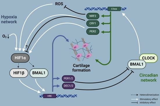
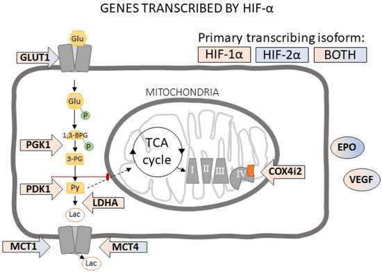
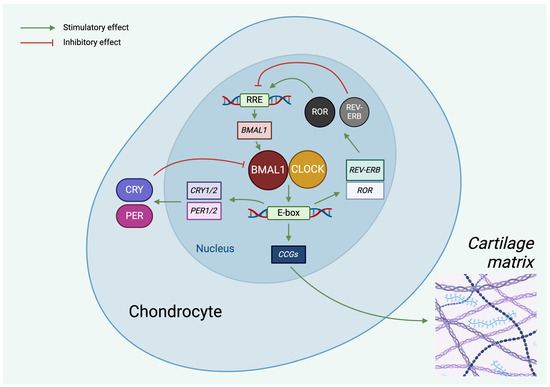
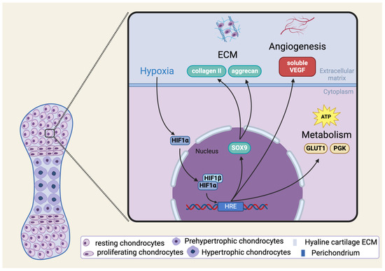
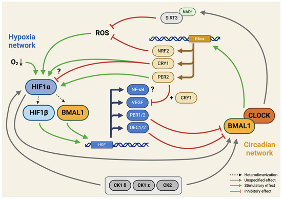
Hypoxia-inducible factor-1 (HIF-1) is a heterodimer transcription factor composed of an alpha and a beta subunit. HIF-1α is a master regulator of cellular response to hypoxia by activating the transcription of genes that facilitate metabolic adaptation to hypoxia. Since chondrocytes in mature articular
[...] Read more.
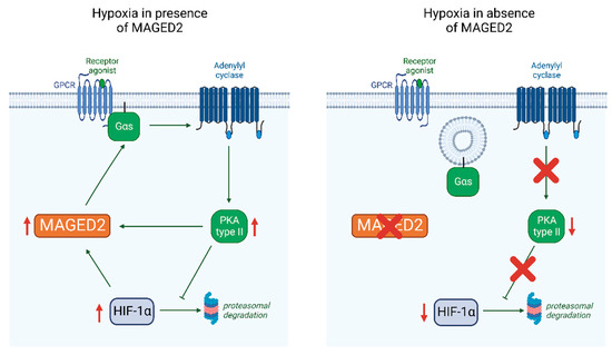
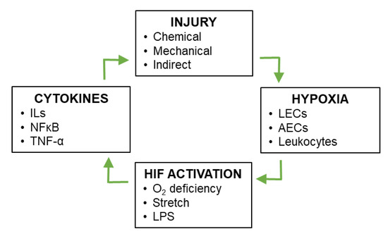
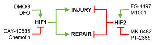
Hypoxia-inducible factor-1 (HIF-1) is a heterodimer transcription factor composed of an alpha and a beta subunit. HIF-1α is a master regulator of cellular response to hypoxia by activating the transcription of genes that facilitate metabolic adaptation to hypoxia. Since chondrocytes in mature articular cartilage reside in a hypoxic environment, HIF-1α plays an important role in chondrogenesis and in the physiological lifecycle of articular cartilage. Accumulating evidence suggests interactions between the HIF pathways and the circadian clock. The circadian clock is an emerging regulator in both developing and mature chondrocytes. However, how circadian rhythm is established during the early steps of cartilage formation and through what signaling pathways it promotes the healthy chondrocyte phenotype is still not entirely known. This narrative review aims to deliver a concise analysis of the existing understanding of the dynamic interplay between HIF-1α and the molecular clock in chondrocytes, in states of both health and disease, while also incorporating creative interpretations. We explore diverse hypotheses regarding the intricate interactions among these pathways and propose relevant therapeutic strategies for cartilage disorders such as osteoarthritis.
Full article
Graphical abstract