


Cells 2024, 13(4), 307; https://doi.org/10.3390/cells13040307 - 07 Feb 2024
Viewed by 742
Abstract
►
Show Figures
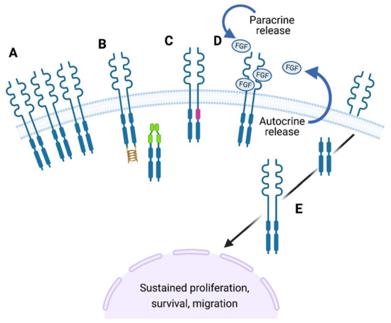
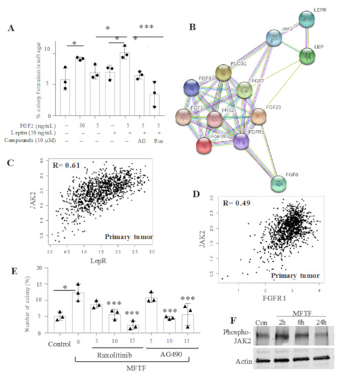
FGF9 is a potent mitogen and survival factor, but FGF9 protein levels are generally low and restricted to a few adult organs. Aberrant expression of FGF9 usually results in cancer. However, the mechanism of FGF9 action has not been fully established. Previous studies
[...] Read more.
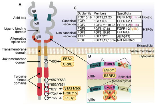
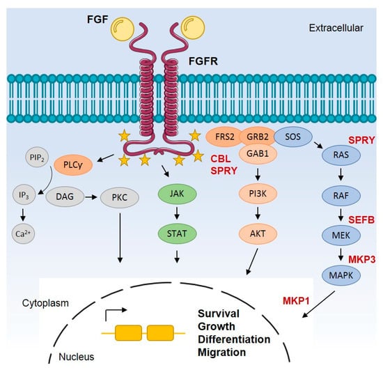
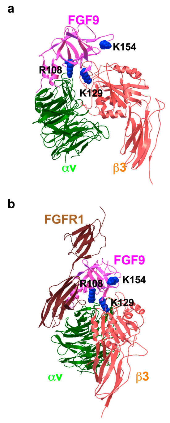
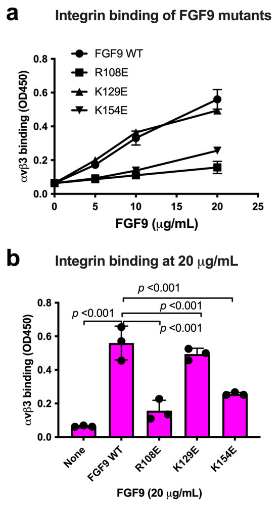
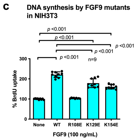
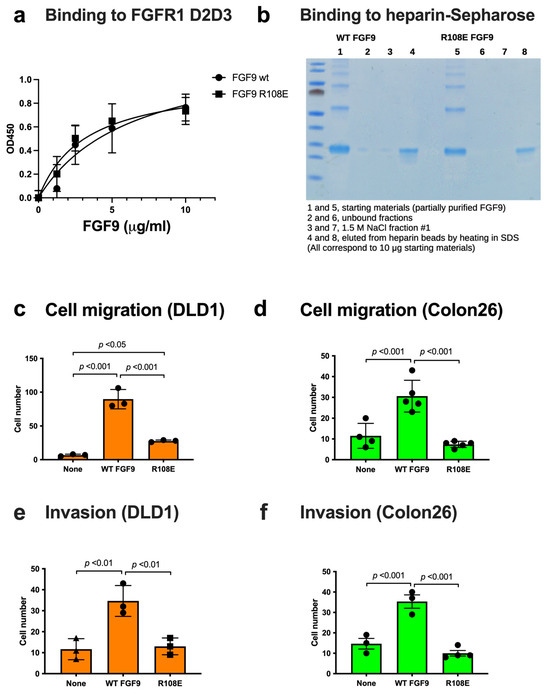
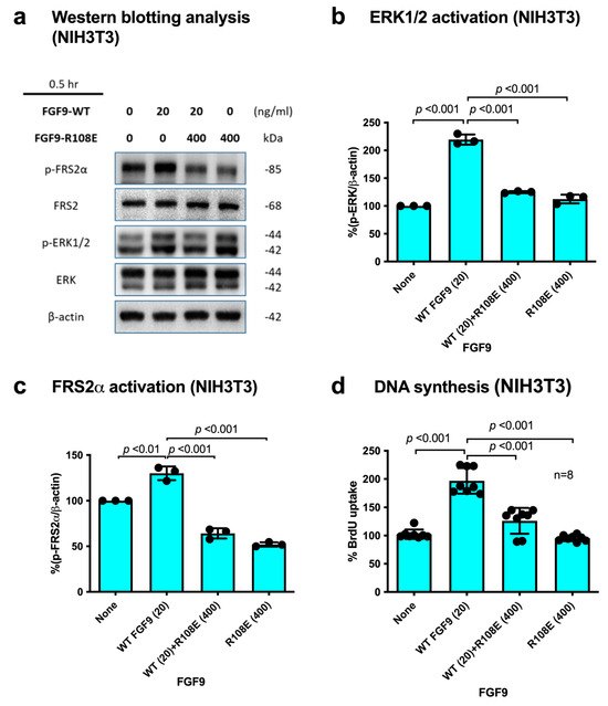
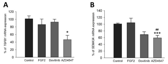
FGF9 is a potent mitogen and survival factor, but FGF9 protein levels are generally low and restricted to a few adult organs. Aberrant expression of FGF9 usually results in cancer. However, the mechanism of FGF9 action has not been fully established. Previous studies showed that FGF1 and FGF2 directly bind to integrin αvβ3, and this interaction is critical for signaling functions (FGF–integrin crosstalk). FGF1 and FGF2 mutants defective in integrin binding were defective in signaling, whereas the mutants still bound to FGFR suppressed angiogenesis and tumor growth, indicating that they act as antagonists. We hypothesize that FGF9 requires direct integrin binding for signaling. Here, we show that docking simulation of the interaction between FGF9 and αvβ3 predicted that FGF9 binds to the classical ligand-binding site of αvβ3. We show that FGF9 bound to integrin αvβ3 and generated FGF9 mutants in the predicted integrin-binding interface. An FGF9 mutant (R108E) was defective in integrin binding, activating FRS2α and ERK1/2, inducing DNA synthesis, cancer cell migration, and invasion in vitro. R108E suppressed DNA synthesis and activation of FRS2α and ERK1/2 induced by WT FGF9 (dominant-negative effect). These findings indicate that FGF9 requires direct integrin binding for signaling and that R108E has potential as an antagonist to FGF9 signaling.
Full article
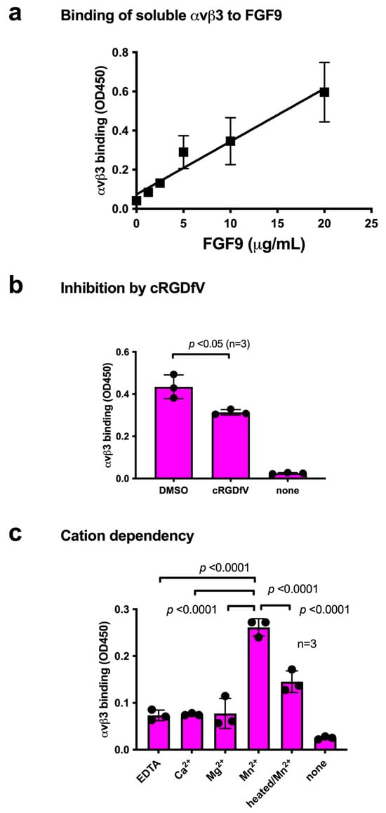
Figure 1