


Cells 2023, 12(21), 2517; https://doi.org/10.3390/cells12212517 - 25 Oct 2023
Cited by 2 | Viewed by 2990
Abstract
►
Show Figures
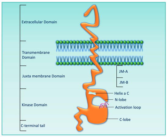
Human epidermal growth factor receptor 3 (HER3) is the only family member of the EGRF/HER family of receptor tyrosine kinases that lacks an active kinase domain (KD), which makes it an obligate binding partner with other receptors for its oncogenic role. When HER3
[...] Read more.
Human epidermal growth factor receptor 3 (HER3) is the only family member of the EGRF/HER family of receptor tyrosine kinases that lacks an active kinase domain (KD), which makes it an obligate binding partner with other receptors for its oncogenic role. When HER3 is activated in a ligand-dependent (NRG1/HRG) or independent manner, it can bind to other receptors (the most potent binding partner is HER2) to regulate many biological functions (growth, survival, nutrient sensing, metabolic regulation, etc.) through the PI3K–AKT–mTOR pathway. HER3 has been found to promote tumorigenesis, tumor growth, and drug resistance in different cancer types, especially breast and non-small cell lung cancer. Given its ubiquitous expression across different solid tumors and role in oncogenesis and drug resistance, there has been a long effort to target HER3. As HER3 cannot be targeted through its KD with small-molecule kinase inhibitors via the conventional method, pharmaceutical companies have used various other approaches, including blocking either the ligand-binding domain or extracellular domain for dimerization with other receptors. The development of treatment options with anti-HER3 monoclonal antibodies, bispecific antibodies, and different combination therapies showed limited clinical efficiency for various reasons. Recent reports showed that the extracellular domain of HER3 is not required for its binding with other receptors, which raises doubt about the efforts and applicability of the development of the HER3-antibodies for treatment. Whereas HER3-directed antibody–drug conjugates showed potentiality for treatment, these drugs are still under clinical trial. The currently understood model for dimerization-induced signaling remains incomplete due to the absence of the crystal structure of HER3 signaling complexes, and many lines of evidence suggest that HER family signaling involves more than the interaction of two members. This review article will significantly expand our knowledge of HER3 signaling and shed light on developing a new generation of drugs that have fewer side effects than the current treatment regimen for these patients.
Full article
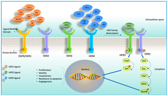
Figure 1
