


Cells 2024, 13(6), 503; https://doi.org/10.3390/cells13060503 - 13 Mar 2024
Viewed by 939
Abstract
►
Show Figures
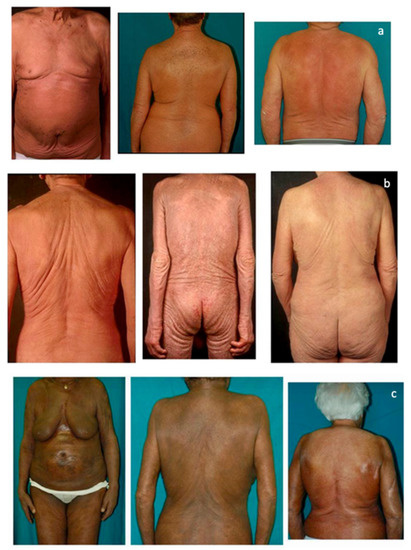
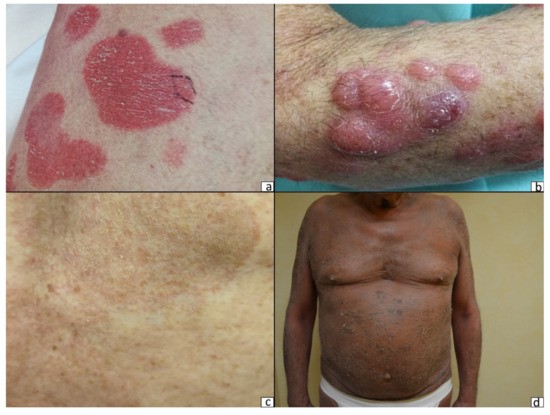
Cutaneous T-cell lymphoma (CTCL) is characterized by the proliferation of malignant T cells in inflamed skin lesions. Mycosis fungoides (MF)—the most common variant of CTCL—often presents with skin lesions around the abdomen and buttocks (“bathing suit” distribution), i.e., in skin areas devoid of
[...] Read more.
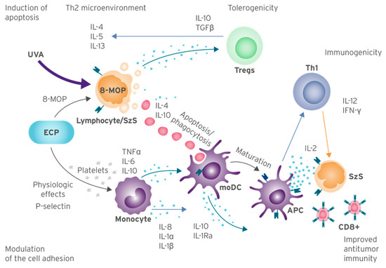
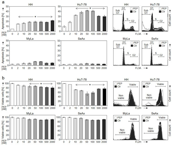
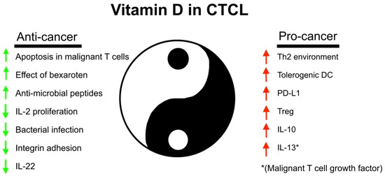
Cutaneous T-cell lymphoma (CTCL) is characterized by the proliferation of malignant T cells in inflamed skin lesions. Mycosis fungoides (MF)—the most common variant of CTCL—often presents with skin lesions around the abdomen and buttocks (“bathing suit” distribution), i.e., in skin areas devoid of sun-induced vitamin D. For decades, sunlight and vitamin D have been connected to CTCL. Thus, vitamin D induces apoptosis and inhibits the expression of cytokines in malignant T cells. Furthermore, CTCL patients often display vitamin D deficiency, whereas phototherapy induces vitamin D and has beneficial effects in CTCL, suggesting that light and vitamin D have beneficial/protective effects in CTCL. Inversely, vitamin D promotes T helper 2 (Th2) cell specific cytokine production, regulatory T cells, tolerogenic dendritic cells, as well as the expression of immune checkpoint molecules, all of which may have disease-promoting effects by stimulating malignant T-cell proliferation and inhibiting anticancer immunity. Studies on vitamin D treatment in CTCL patients showed conflicting results. Some studies found positive effects, others negative effects, while the largest study showed no apparent clinical effect. Taken together, vitamin D may have both pro- and anticancer effects in CTCL. The balance between the opposing effects of vitamin D in CTCL is likely influenced by treatment and may change during the disease course. Therefore, it remains to be discovered whether and how the effect of vitamin D can be tilted toward an anticancer response in CTCL.
Full article
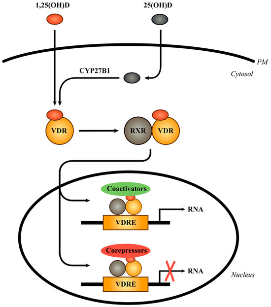
Figure 1