


Cells 2024, 13(6), 476; https://doi.org/10.3390/cells13060476 - 08 Mar 2024
Viewed by 721
Abstract
►
Show Figures
The ubiquitous second messenger 3′,5′-cyclic adenosine monophosphate (cAMP) regulates cardiac excitation-contraction coupling (ECC) by signaling in discrete subcellular microdomains. Phosphodiesterase subfamilies 4B and 4D are critically involved in the regulation of cAMP signaling in mammalian cardiomyocytes. Alterations of PDE4 activity in human hearts
[...] Read more.
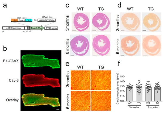
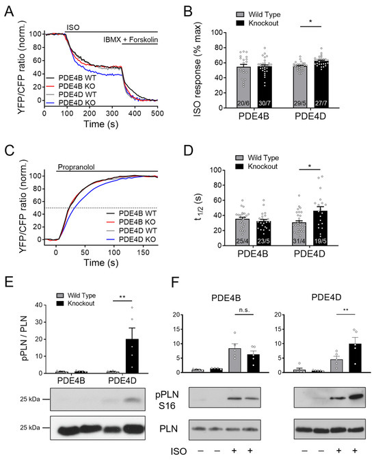
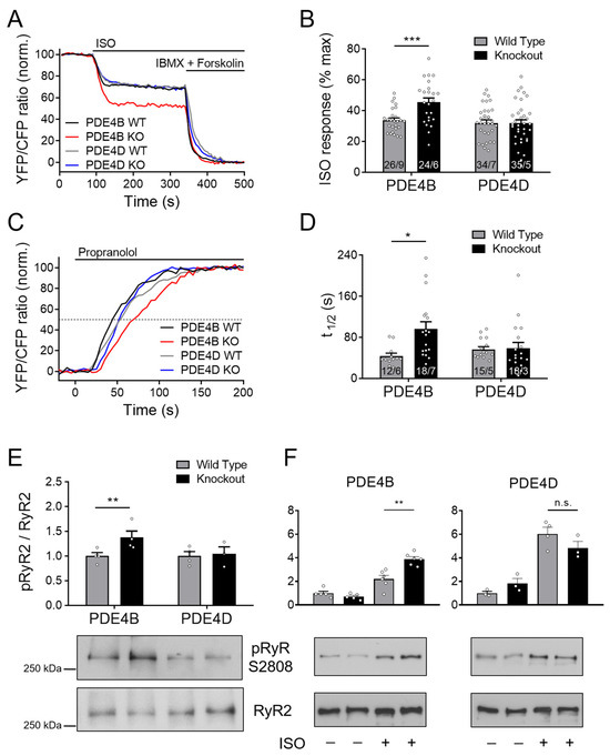
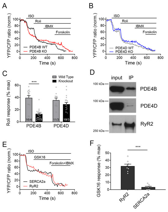
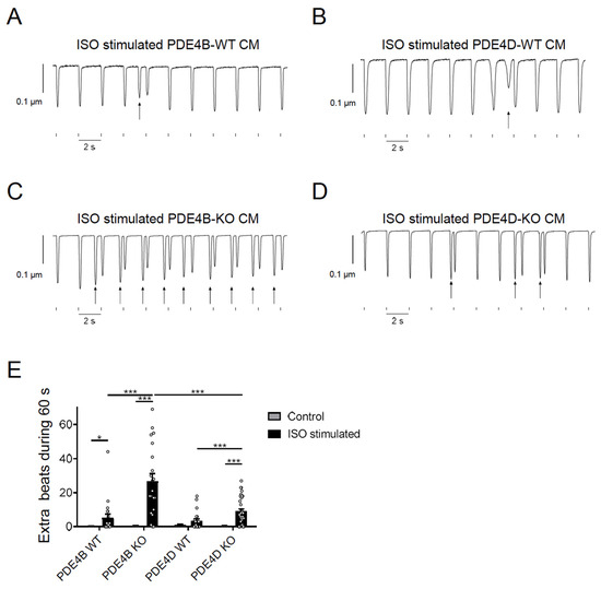
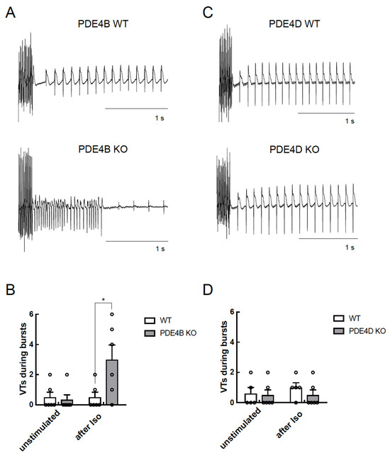
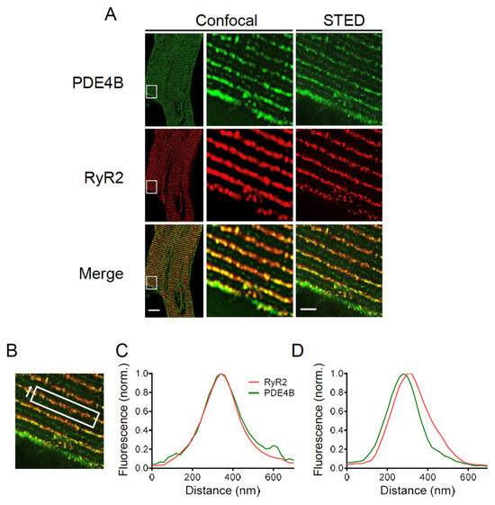
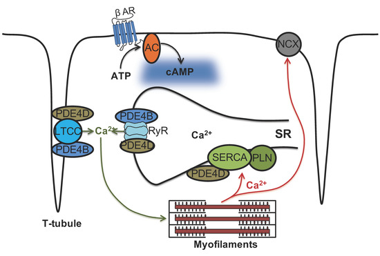
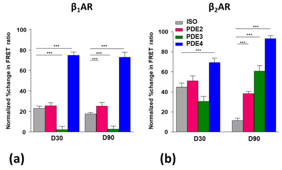
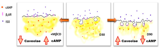
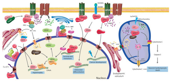
The ubiquitous second messenger 3′,5′-cyclic adenosine monophosphate (cAMP) regulates cardiac excitation-contraction coupling (ECC) by signaling in discrete subcellular microdomains. Phosphodiesterase subfamilies 4B and 4D are critically involved in the regulation of cAMP signaling in mammalian cardiomyocytes. Alterations of PDE4 activity in human hearts has been shown to result in arrhythmias and heart failure. Here, we sought to systematically investigate specific roles of PDE4B and PDE4D in the regulation of cAMP dynamics in three distinct subcellular microdomains, one of them located at the caveolin-rich plasma membrane which harbors the L-type calcium channels (LTCCs), as well as at two sarco/endoplasmic reticulum (SR) microdomains centered around SR Ca2+-ATPase (SERCA2a) and cardiac ryanodine receptor type 2 (RyR2). Transgenic mice expressing Förster Resonance Energy Transfer (FRET)-based cAMP-specific biosensors targeted to caveolin-rich plasma membrane, SERCA2a and RyR2 microdomains were crossed to PDE4B-KO and PDE4D-KO mice. Direct analysis of the specific effects of both PDE4 subfamilies on local cAMP dynamics was performed using FRET imaging. Our data demonstrate that all three microdomains are differentially regulated by these PDE4 subfamilies. Whereas both are involved in cAMP regulation at the caveolin-rich plasma membrane, there are clearly two distinct cAMP microdomains at the SR formed around RyR2 and SERCA2a, which are preferentially controlled by PDE4B and PDE4D, respectively. This correlates with local cAMP-dependent protein kinase (PKA) substrate phosphorylation and arrhythmia susceptibility. Immunoprecipitation assays confirmed that PDE4B is associated with RyR2 along with PDE4D. Stimulated Emission Depletion (STED) microscopy of immunostained cardiomyocytes suggested possible co-localization of PDE4B with both sarcolemmal and RyR2 microdomains. In conclusion, our functional approach could show that both PDE4B and PDE4D can differentially regulate cardiac cAMP microdomains associated with calcium homeostasis. PDE4B controls cAMP dynamics in both caveolin-rich plasma membrane and RyR2 vicinity. Interestingly, PDE4B is the major regulator of the RyR2 microdomain, as opposed to SERCA2a vicinity, which is predominantly under PDE4D control, suggesting a more complex regulatory pattern than previously thought, with multiple PDEs acting at the same location.
Full article
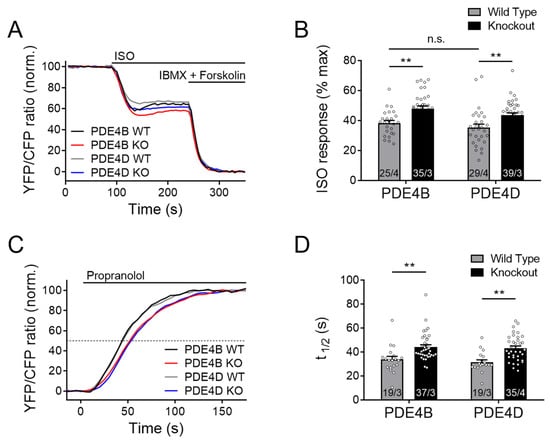
Figure 1


