Molecular Mechanisms to Target Cellular Senescence in Aging and Disease

A topical collection in Cells (ISSN 2073-4409). This collection belongs to the section "Cellular Aging".

Viewed by 28699

Editors


E-Mail Website
Guest Editor
Department of Medicine, University of Udine, Udine, Italy
Interests: heart failure; adult stem cells; cell senescence; mechanotransduction; extracellular vesicles
Special Issues, Collections and Topics in MDPI journals

E-Mail Website
Guest Editor
Advanced Technology Center for Aging Research, Scientific Technological Area, IRCCS INRCA, Ancona, Italy
Interests: biology of aging; biogerontology; experimenetal gerontology; geroscience; cellular senescence
Special Issues, Collections and Topics in MDPI journals

Topical Collection Information

Dear Colleagues,

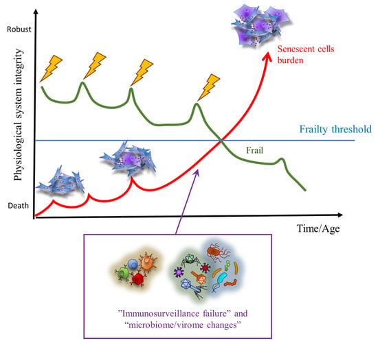
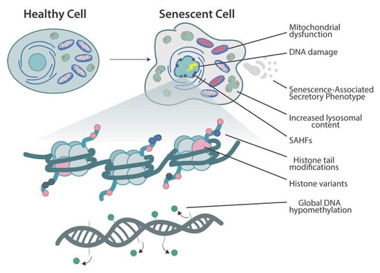
The term cellular senescence indicates a complex cellular response to a variety of stressors that results in the permanent withdrawal of potentially damaged cells from the cell cycle. Further, cellular senescence is associated with a modification of the secretome, promoting extracellular matrix remodeling, recruitment of inflammatory cells, angiogenesis, cell de-differentiation, and induction of cellular senescence in a paracrine fashion. It is conceivable that cellular senescence has been positively selected by evolution both as a barrier to avoid cancerogenesis and to promote tissue repair. However, with aging, senescent cells progressively accumulate in tissues, thus reducing the organism’s capacity to replace lost cells either with normal wear and tear processes or following damage. The immune system plays a special role in these phenomena, as changes in the immune system associated with age may contribute to reduce senescence immunesurveillance (the elimination of senescent cells mediated by the immune system), thus promoting the accumulation of senescent cells in tissues. As a result, the organism becomes more and more vulnerable to minor injuries, while chronic, age-related pathologies develop. Moreover, the higher mortality rate of older people during the current COVID-19 pandemic has raised interest on how senescence impacts our host defense to respiratory viruses. A causal role played by accumulating senescent cells in the pathophysiology of most age-related pathologies is supported by a growing body of literature. Indeed, genetic and drug-based interventions have been tested, aiming at eradicating senescent cells from old tissues with the intent to prevent chronic pathologies and delay aging.

This Topical Collection aims to collect a series of original research and review articles addressing the exciting and emerging field of cellular senescence in aging and age-related pathologies. We would like to put the emphasis on the molecular mechanism that is responsible for the phenomenon and that could be targeted both for diagnostics and therapeutic purposes. Suggested potential topics may be: stem cell senescence in age-related pathologies; mechanisms of proteostasis failure in chronic pathologies; mechanisms of immune surveillance failure in aging and disease; senolytic and rejuvenating therapies; exosomes as therapeutic or diagnostic tools; cell senescence and cancer; cell senescence and infectious diseases, as well as new models and therapeutic tools to target senescence in aging.


Prof. Antonio Paolo Beltrami

Dr. Marco Malavolta
Guest Editors

Manuscript Submission Information

Manuscripts should be submitted online at www.mdpi.com by registering and logging in to this website. Once you are registered, click here to go to the submission form. Manuscripts can be submitted until the deadline. All submissions that pass pre-check are peer-reviewed. Accepted papers will be published continuously in the journal (as soon as accepted) and will be listed together on the collection website. Research articles, review articles as well as short communications are invited. For planned papers, a title and short abstract (about 100 words) can be sent to the Editorial Office for announcement on this website.

Submitted manuscripts should not have been published previously, nor be under consideration for publication elsewhere (except conference proceedings papers). All manuscripts are thoroughly refereed through a single-blind peer-review process. A guide for authors and other relevant information for submission of manuscripts is available on the Instructions for Authors page. Cells is an international peer-reviewed open access semimonthly journal published by MDPI.

Please visit the Instructions for Authors page before submitting a manuscript. The Article Processing Charge (APC) for publication in this open access journal is 2700 CHF (Swiss Francs). Submitted papers should be well formatted and use good English. Authors may use MDPI's English editing service prior to publication or during author revisions.

Keywords

  • cellular senescence
  • proteostasis
  • inflammation
  • inflammaging
  • immunosenescence
  • stem cells
  • exosomes
  • extracellular vesicles
  • senolytics
  • aging

Published Papers (7 papers)

2023

Jump to: 2022, 2021

15 pages, 1911 KiB  
Perspective
Spreading Senescent Cells’ Burden and Emerging Therapeutic Targets for Frailty
by Serena Marcozzi, Giorgia Bigossi, Maria Elisa Giuliani, Giovanni Lai, Robertina Giacconi, Francesco Piacenza and Marco Malavolta
Cells 2023, 12(18), 2287; https://doi.org/10.3390/cells12182287 - 15 Sep 2023
Cited by 1 | Viewed by 911
Abstract
The spreading of senescent cells’ burden holds profound implications for frailty, prompting the exploration of novel therapeutic targets. In this perspective review, we delve into the intricate mechanisms underlying senescent cell spreading, its implications for frailty, and its therapeutic development. We have focused [...] Read more.
The spreading of senescent cells’ burden holds profound implications for frailty, prompting the exploration of novel therapeutic targets. In this perspective review, we delve into the intricate mechanisms underlying senescent cell spreading, its implications for frailty, and its therapeutic development. We have focused our attention on the emerging age-related biological factors, such as microbiome and virome alterations, elucidating their significant contribution to the loss of control over the accumulation rate of senescent cells, particularly affecting key frailty domains, the musculoskeletal system and cerebral functions. We believe that gaining an understanding of these mechanisms could not only aid in elucidating the involvement of cellular senescence in frailty but also offer diverse therapeutic possibilities, potentially advancing the future development of tailored interventions for these highly diverse patients. Full article
Show Figures

Figure 1

14 pages, 3855 KiB  
Article
Hesperidin Ameliorates Sarcopenia through the Regulation of Inflammaging and the AKT/mTOR/FoxO3a Signaling Pathway in 22–26-Month-Old Mice
by Hyun-Ji Oh, Heegu Jin and Boo-Yong Lee
Cells 2023, 12(15), 2015; https://doi.org/10.3390/cells12152015 - 07 Aug 2023
Cited by 3 | Viewed by 1304
Abstract
Faced with a globally aging society, the maintenance of health and quality of life in older people is very important. The age-related loss of muscle mass and strength, known as sarcopenia, severely reduces quality of life and increases the risks of various diseases. [...] Read more.
Faced with a globally aging society, the maintenance of health and quality of life in older people is very important. The age-related loss of muscle mass and strength, known as sarcopenia, severely reduces quality of life and increases the risks of various diseases. In this study, we investigated the inhibitory effect of hesperidin (HES) on inflammaging, with the intention of evaluating its potential use as a treatment for sarcopenia. We studied 22–26-month-old mice, corresponding to humans aged ≥70 years, with aging-related sarcopenia, and young mice aged 3–6 months. The daily administration of HES for 8 weeks resulted in greater muscle mass and strength and increased the fiber size of the old mice. HES also restored the immune homeostasis that had been disrupted by aging, such as the imbalance in M1/M2 macrophage ratio. In addition, we found that HES ameliorated the sarcopenia by regulating AKT/mammalian target of rapamycin/forkhead box 3a signaling through an increase in insulin-like growth factor (IGF)-1 expression in the old mice. Therefore, HES represents a promising candidate inhibitor of sarcopenia in older people, and its effects are achieved through the maintenance of immune homeostasis. Full article
Show Figures

Graphical abstract

2022

Jump to: 2023, 2021

20 pages, 2916 KiB  
Article
Simple Detection of Unstained Live Senescent Cells with Imaging Flow Cytometry
by Marco Malavolta, Robertina Giacconi, Francesco Piacenza, Sergio Strizzi, Maurizio Cardelli, Giorgia Bigossi, Serena Marcozzi, Luca Tiano, Fabio Marcheggiani, Giulia Matacchione, Angelica Giuliani, Fabiola Olivieri, Ilaria Crivellari, Antonio Paolo Beltrami, Alessandro Serra, Marco Demaria and Mauro Provinciali
Cells 2022, 11(16), 2506; https://doi.org/10.3390/cells11162506 - 12 Aug 2022
Cited by 5 | Viewed by 3767
Abstract
Cellular senescence is a hallmark of aging and a promising target for therapeutic approaches. The identification of senescent cells requires multiple biomarkers and complex experimental procedures, resulting in increased variability and reduced sensitivity. Here, we propose a simple and broadly applicable imaging flow [...] Read more.
Cellular senescence is a hallmark of aging and a promising target for therapeutic approaches. The identification of senescent cells requires multiple biomarkers and complex experimental procedures, resulting in increased variability and reduced sensitivity. Here, we propose a simple and broadly applicable imaging flow cytometry (IFC) method. This method is based on measuring autofluorescence and morphological parameters and on applying recent artificial intelligence (AI) and machine learning (ML) tools. We show that the results of this method are superior to those obtained measuring the classical senescence marker, senescence-associated beta-galactosidase (SA-β-Gal). We provide evidence that this method has the potential for diagnostic or prognostic applications as it was able to detect senescence in cardiac pericytes isolated from the hearts of patients affected by end-stage heart failure. We additionally demonstrate that it can be used to quantify senescence “in vivo” and can be used to evaluate the effects of senolytic compounds. We conclude that this method can be used as a simple and fast senescence assay independently of the origin of the cells and the procedure to induce senescence. Full article
Show Figures

Figure 1

16 pages, 1126 KiB  
Review
Epigenetic Regulation of Cellular Senescence
by Jack Crouch, Maria Shvedova, Rex Jeya Rajkumar Samdavid Thanapaul, Vladimir Botchkarev and Daniel Roh
Cells 2022, 11(4), 672; https://doi.org/10.3390/cells11040672 - 15 Feb 2022
Cited by 40 | Viewed by 7748
Abstract
Senescence is a complex cellular stress response that abolishes proliferative capacity and generates a unique secretory pattern that is implicated in organismal aging and age-related disease. How a cell transitions to a senescent state is multifactorial and often requires transcriptional regulation of multiple [...] Read more.
Senescence is a complex cellular stress response that abolishes proliferative capacity and generates a unique secretory pattern that is implicated in organismal aging and age-related disease. How a cell transitions to a senescent state is multifactorial and often requires transcriptional regulation of multiple genes. Epigenetic alterations to DNA and chromatin are powerful regulators of genome architecture and gene expression, and they play a crucial role in mediating the induction and maintenance of senescence. This review will highlight the changes in chromatin, DNA methylation, and histone alterations that establish and maintain cellular senescence, alongside the specific epigenetic regulation of the senescence-associated secretory phenotype (SASP). Full article
Show Figures

Figure 1

2021

Jump to: 2023, 2022

13 pages, 4109 KiB  
Article
A Rare Mutation in LMNB2 Associated with Lipodystrophy Drives Premature Cell Senescence
by Alice-Anaïs Varlet, Camille Desgrouas, Cécile Jebane, Nathalie Bonello-Palot, Patrice Bourgeois, Nicolas Levy, Emmanuèle Helfer, Noémie Dubois, René Valero, Catherine Badens and Sophie Beliard
Cells 2022, 11(1), 50; https://doi.org/10.3390/cells11010050 - 24 Dec 2021
Cited by 4 | Viewed by 2875
Abstract
Many proteins are causative for inherited partial lipodystrophies, including lamins, the essential constituents of the nuclear envelope scaffold called the lamina. By performing high throughput sequencing on a panel of genes involved in lipodystrophies, we identified a heterozygous mutation in LMNB2 gene (c.700C [...] Read more.
Many proteins are causative for inherited partial lipodystrophies, including lamins, the essential constituents of the nuclear envelope scaffold called the lamina. By performing high throughput sequencing on a panel of genes involved in lipodystrophies, we identified a heterozygous mutation in LMNB2 gene (c.700C > T p.(Arg234Trp)) in a female patient presenting early onset type II diabetes, hypertriglyceridemia, and android fat distribution. This mutation is rare in the general population (frequency 0.013% in GnomAD) and was predicted pathogenic by a set of pathogenicity prediction software. Patient-derived fibroblasts showed nuclear shape abnormalities and premature senescence features, which are two typical cellular phenotypes associated with laminopathies. Moreover, we observed an atypical aggregation of lamin B2 in nucleoplasm, which co-distributes with emerin and lamin A/C, along with an abnormal distribution of lamin A/C at the nuclear envelope. Finally, reducing lamin B2 expression level by siRNA targeted toward LMNB2 transcripts resulted in decreased nuclear anomalies and senescence-associated beta-galactosidase, suggesting a role of the mutated protein in the occurrence of the observed cellular phenotype. Altogether, these results suggest that mutations in lamin B2 could produce premature senescence and partial lipodystrophy features as observed with certain mutants of lamin A/C. Full article
Show Figures

Figure 1

21 pages, 3751 KiB  
Article
Dysregulation of Caveolin-1 Phosphorylation and Nuclear Translocation Is Associated with Senescence Onset
by Andreas Goutas, Zozo Outskouni, Ioanna Papathanasiou, Maria Satra, George Koliakos and Varvara Trachana
Cells 2021, 10(11), 2939; https://doi.org/10.3390/cells10112939 - 28 Oct 2021
Cited by 6 | Viewed by 2435
Abstract
We recently reported that the inability of osteoarthritic (OA) chondrocytes to repair oxidative stress (OS) induced DNA damage is linked to Cav-1 overexpression/improper localization. We speculated that the senescent status of OA cells was responsible for this Cav-1 dysregulation. Here, to further investigate [...] Read more.
We recently reported that the inability of osteoarthritic (OA) chondrocytes to repair oxidative stress (OS) induced DNA damage is linked to Cav-1 overexpression/improper localization. We speculated that the senescent status of OA cells was responsible for this Cav-1 dysregulation. Here, to further investigate this hypothesis, we used Wharton Jelly derived mesenchymal stem cells (WJ-MSCs) and investigated Cav-1 function as cells reached replicative senescence or upon stress induced senescence (SIPS). We showed that Cav-1 is upregulated, phosphorylated and translocated to the nucleus in young WJ-MSCs upon acute exogenous OS, and that it returns back to basal/nonphosphorylated levels and exports the nucleus in the recovery phase. However, as cells reach senescence, this regulation is lost. OS did not induce any Cav-1-mediated response, which is concomitant with the inability of older cells to restore DNA damage. Furthermore, downregulation of Cav-1 resulted in persistent OS-induced DNA damage and subsequent onset of senescence. We also report that the establishment of senescence is mediated by autophagy stimulation, since downregulation of autophagy key molecule Atg5, simultaneously with Cav-1 downregulation, was found to inhibit SIPS. Basically, we propose that Cav-1 involvement in DNA damage response can lead to senescence, either because the damage is extensive or because Cav-1 is absent/unable to perform its homeostatic role. Full article
Show Figures

Figure 1

16 pages, 610 KiB  
Review
The Emergence of Senescent Surface Biomarkers as Senotherapeutic Targets
by Martina Rossi and Kotb Abdelmohsen
Cells 2021, 10(7), 1740; https://doi.org/10.3390/cells10071740 - 09 Jul 2021
Cited by 27 | Viewed by 8411
Abstract
Senescence is linked to a wide range of age-associated diseases and physiological declines. Thus, senotherapeutics are emerging to suppress the detrimental effects of senescence either by senomorphics or senolytics. Senomorphics suppress the traits associated with senescence phenotypes, while senolytics aim to clear senescent [...] Read more.
Senescence is linked to a wide range of age-associated diseases and physiological declines. Thus, senotherapeutics are emerging to suppress the detrimental effects of senescence either by senomorphics or senolytics. Senomorphics suppress the traits associated with senescence phenotypes, while senolytics aim to clear senescent cells by suppressing their survival and enhancing the apoptotic pathways. The main goal of these approaches is to suppress the proinflammatory senescence-associated secretory phenotype (SASP) and to promote the immune recognition and elimination of senescent cells. One increasingly attractive approach is the targeting of molecules or proteins specifically present on the surface of senescent cells. These proteins may play roles in the maintenance and survival of senescent cells and hence can be targeted for senolysis. In this review, we summarize the recent knowledge regarding senolysis with a focus on novel surface biomarkers of cellular senescence and discuss their emergence as senotherapeutic targets. Full article
Show Figures

Figure 1

Back to TopTop