


Cells 2023, 12(24), 2816; https://doi.org/10.3390/cells12242816 - 11 Dec 2023
Cited by 1 | Viewed by 1074
Abstract
►
Show Figures
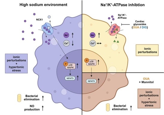
Inflamed and infected tissues can display increased local sodium (Na+) levels, which can have various effects on immune cells. In macrophages, high salt (HS) leads to a Na+/Ca2+-exchanger 1 (NCX1)-dependent increase in intracellular Na+ levels. This
[...] Read more.
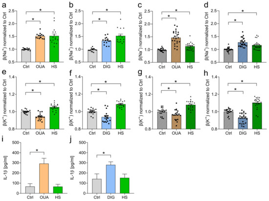
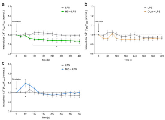
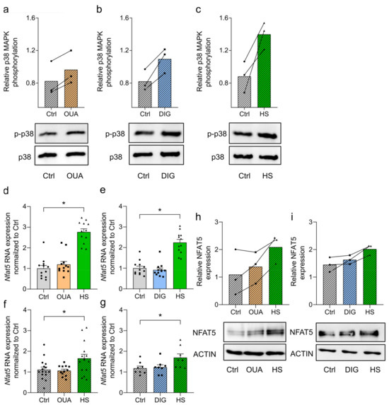
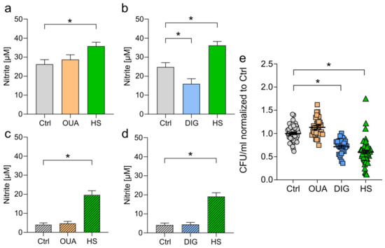
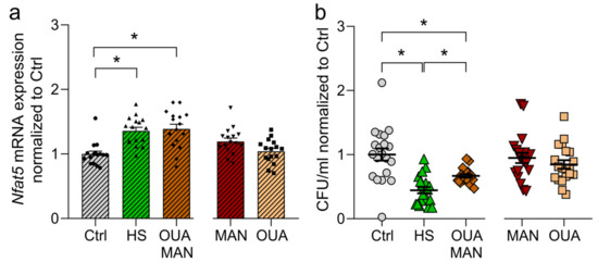
Inflamed and infected tissues can display increased local sodium (Na+) levels, which can have various effects on immune cells. In macrophages, high salt (HS) leads to a Na+/Ca2+-exchanger 1 (NCX1)-dependent increase in intracellular Na+ levels. This results in augmented osmoprotective signaling and enhanced proinflammatory activation, such as enhanced expression of type 2 nitric oxide synthase and antimicrobial function. In this study, the role of elevated intracellular Na+ levels in macrophages was investigated. Therefore, the Na+/K+-ATPase (NKA) was pharmacologically inhibited with two cardiac glycosides (CGs), ouabain (OUA) and digoxin (DIG), to raise intracellular Na+ without increasing extracellular Na+ levels. Exposure to HS conditions and treatment with both inhibitors resulted in intracellular Na+ accumulation and subsequent phosphorylation of p38/MAPK. The CGs had different effects on intracellular Ca2+ and K+ compared to HS stimulation. Moreover, the osmoprotective transcription factor nuclear factor of activated T cells 5 (NFAT5) was not upregulated on RNA and protein levels upon OUA and DIG treatment. Accordingly, OUA and DIG did not boost nitric oxide (NO) production and showed heterogeneous effects toward eliminating intracellular bacteria. While HS environments cause hypertonic stress and ionic perturbations, cardiac glycosides only induce the latter. Cotreatment of macrophages with OUA and non-ionic osmolyte mannitol (MAN) partially mimicked the HS-boosted antimicrobial macrophage activity. These findings suggest that intracellular Na+ accumulation and hypertonic stress are required but not sufficient to mimic boosted macrophage function induced by increased extracellular sodium availability.
Full article
Graphical abstract


