


Cells 2023, 12(21), 2546; https://doi.org/10.3390/cells12212546 - 30 Oct 2023
Viewed by 1376
Abstract
►
Show Figures
Increased medical attention is needed as the prevalence of autism spectrum disorder (ASD) rises. Both cardiovascular disorder (CVD) and hyperlipidemia are closely associated with adult ASD. Shank3 plays a key genetic role in ASD. We hypothesized that Shank3 contributes to CVD development in
[...] Read more.
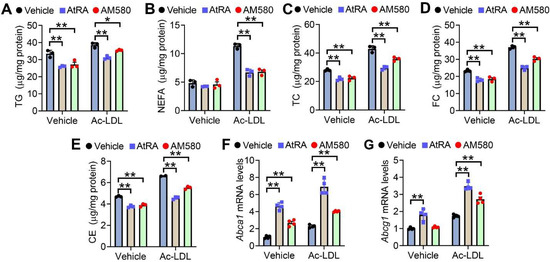
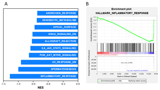
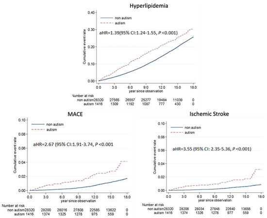
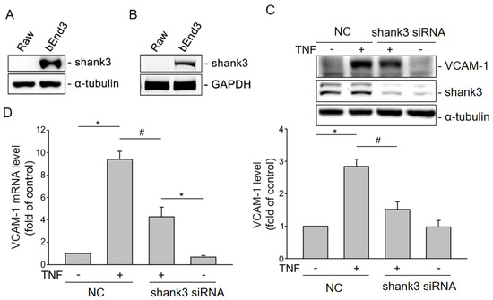
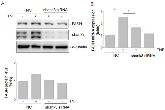
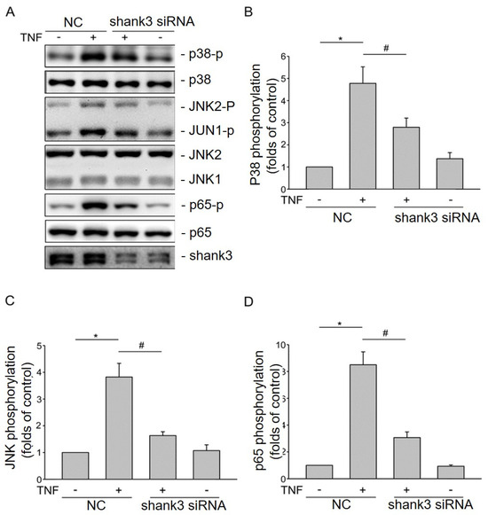
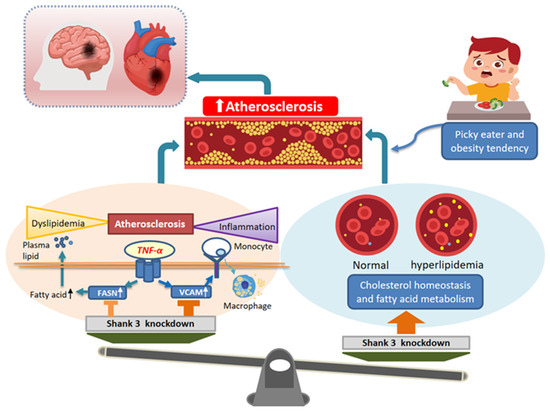
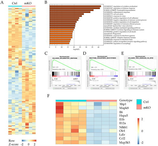
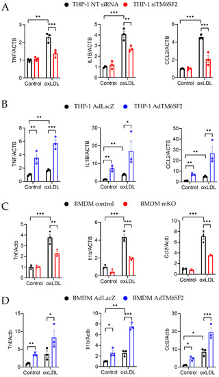
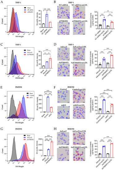
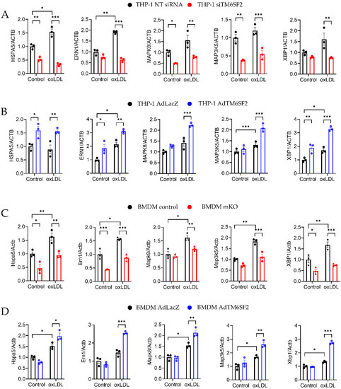
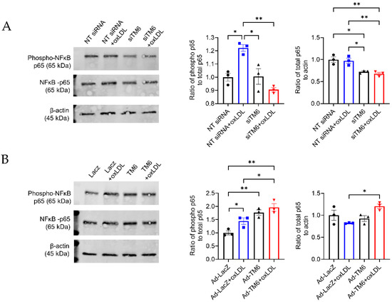
Increased medical attention is needed as the prevalence of autism spectrum disorder (ASD) rises. Both cardiovascular disorder (CVD) and hyperlipidemia are closely associated with adult ASD. Shank3 plays a key genetic role in ASD. We hypothesized that Shank3 contributes to CVD development in young adults with ASD. In this study, we investigated whether Shank3 facilitates the development of atherosclerosis. Using Gene Set Enrichment Analysis software (Version No.: GSEA-4.0.3), we analyzed the data obtained from Shank3 knockout mice (Gene Expression Omnibus database), a human population-based study cohort (from Taiwan’s National Health Insurance Research Database), and a Shank3 knockdown cellular model. Shank3 knockout upregulated the expression of genes of cholesterol homeostasis and fatty acid metabolism but downregulated the expression of genes associated with inflammatory responses. Individuals with autism had higher risks of hyperlipidemia (adjusted hazard ratio (aHR): 1.39; p < 0.001), major adverse cardiac events (aHR: 2.67; p < 0.001), and stroke (aHR: 3.55; p < 0.001) than age- and sex-matched individuals without autism did. Shank3 downregulation suppressed tumor necrosis factor-α-induced fatty acid synthase expression; vascular cell adhesion molecule 1 expression; and downstream signaling pathways involving p38, Jun N-terminal kinase, and nuclear factor-κB. Thus, Shank3 may influence the development of early-onset atherosclerosis and CVD in ASD. Furthermore, regulating Shank3 expression may reduce inflammation-related disorders, such as atherosclerosis, by inhibiting tumor necrosis factor-alpha-mediated inflammatory cascades.
Full article
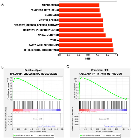
Figure 1
