


Cancers 2024, 16(6), 1112; https://doi.org/10.3390/cancers16061112 - 10 Mar 2024
Viewed by 737
Abstract
►
Show Figures
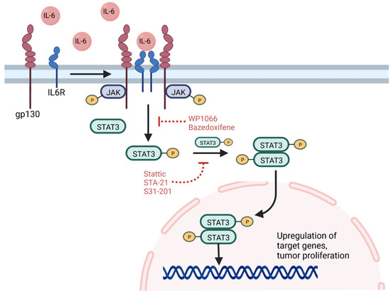
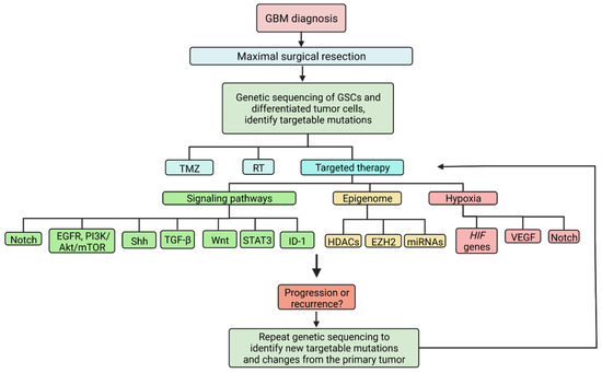
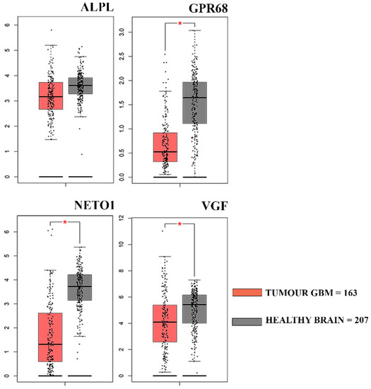
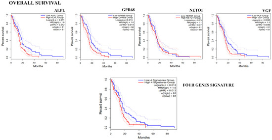
Serpins are serine proteinase inhibitors, with several serpins being overexpressed in cancer cells. Thus, we aim to analyze the single-nucleotide polymorphism (SNP) of Serpinb11 and its association with GBM survival. A cohort of 63 GBM patients recruited from King Abdullah University Hospital in
[...] Read more.
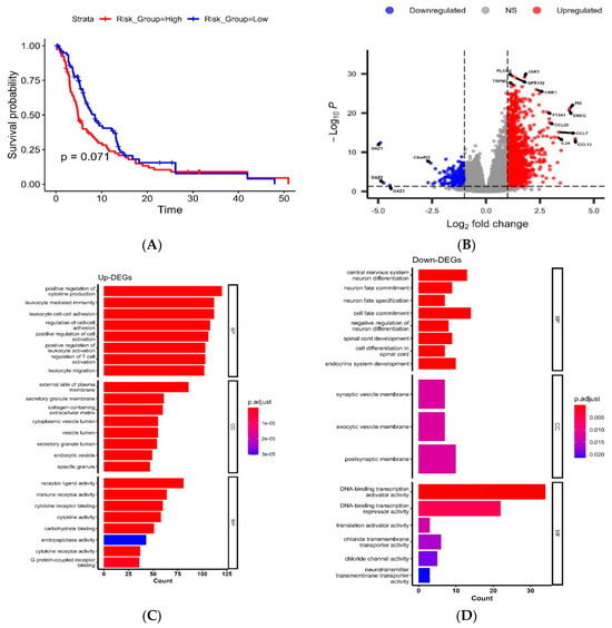
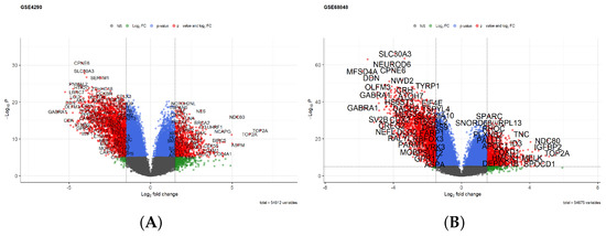
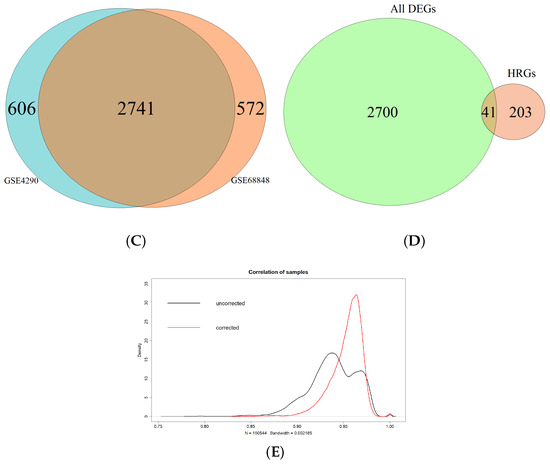
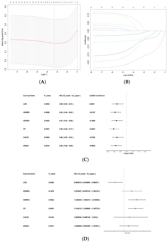
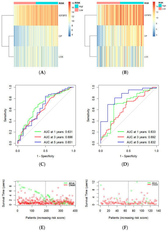
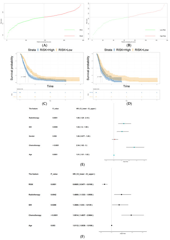
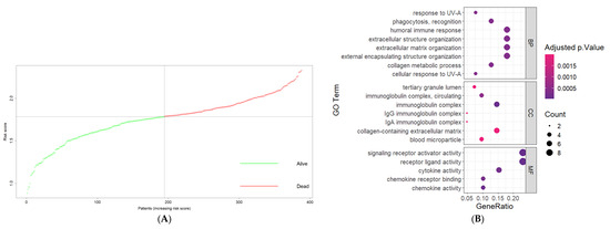
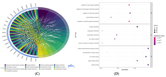
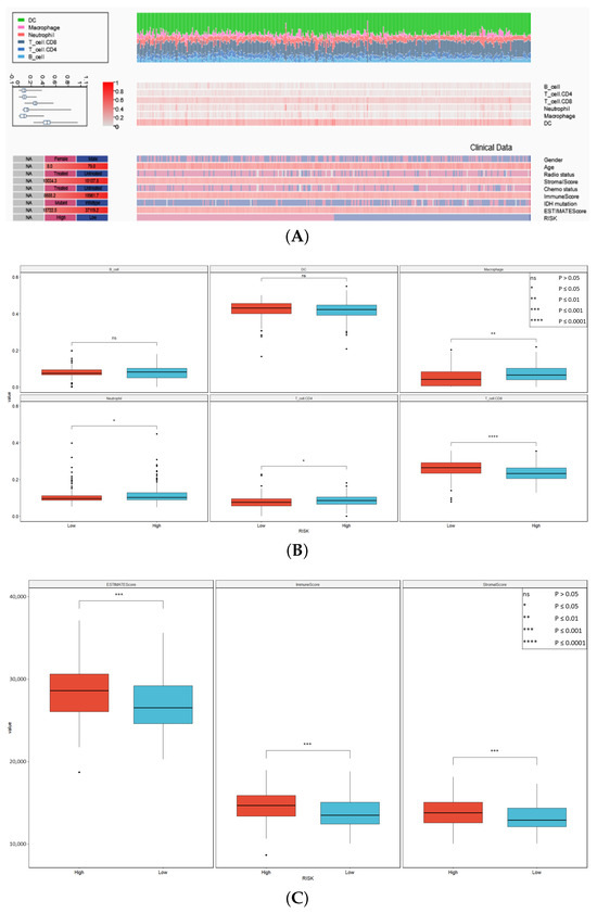
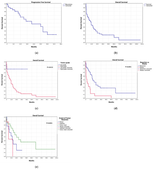
Serpins are serine proteinase inhibitors, with several serpins being overexpressed in cancer cells. Thus, we aim to analyze the single-nucleotide polymorphism (SNP) of Serpinb11 and its association with GBM survival. A cohort of 63 GBM patients recruited from King Abdullah University Hospital in Jordan underwent polymorphism analysis and overall survival (OS) assessments. The Cancer Genome Atlas (GBM) cohort was useful for validation. We constructed a risk score using the principal component analysis for the following Serpin genes: Serpinb3, Serpinb5, Serpinb6, Serpinb11, and Serpinb12, and patients were grouped into high- vs. low-risk groups based on the median cutoff. Univariable Cox models were used to study the survival outcomes. We identified a significant association between rs4940595 and survival. In the TCGA cohort, Serpinb3 alterations showed worse OS. Univariable Cox showed worse PFS outcomes with higher SERPINB5 and SERPINB6 expression. A Serpin B 5-gene risk score showed a trend towards worse PFS in the high-risk group. Upregulated DEGs showed GO enrichment in cytokine regulation and production, positive regulation of leukocyte activation, and the MAPK cascade. The high-risk group showed a significantly higher infiltration of M2 macrophages and activated mast cells. Our findings showed a significant role of the Serpin B family in GBM survival in the Jordanian population.
Full article
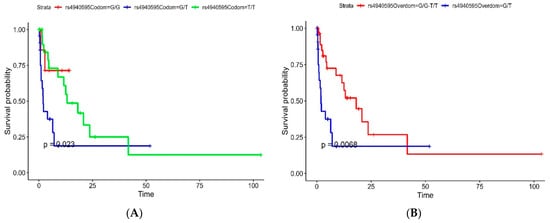
Figure 1


