Deciphering the Crosstalk between Tumor Cells and Their Microenvironment: From Molecular Aspects to Therapeutic Implications

A topical collection in Cancers (ISSN 2072-6694). This collection belongs to the section "Tumor Microenvironment".

Viewed by 21404

Editors


E-Mail Website
Collection Editor
Department of Pharmacological and Biomolecular Sciences, Università degli Studi di Milano, via Balzaretti 9, 20133 Milano, Italy
Interests: cancer cell biology; mechanisms of cancer cell death; hormone receptors in cancer cells; cancer stem cells; metabolic rewiring in cancer cells; drug resistance; tumor microenvironment, anticancer activity of natural compounds; prostate cancer; melanoma
Special Issues, Collections and Topics in MDPI journals

E-Mail Website
Collection Editor
Department of Pharmacological and Biomolecular Sciences, Università degli Studi di Milano, Via Balzaretti 9, 20133 Milano, Italy
Interests: prostate cancer; melanoma; ovarian cancer; cancer biology; mechanisms of cancer cell death; cancer metastasis; cancer metabolism; cancer drug resistance; cancer stem cells; tumor microenvironment; extracellular vesicles
Special Issues, Collections and Topics in MDPI journals

Topical Collection Information

Dear Colleagues,

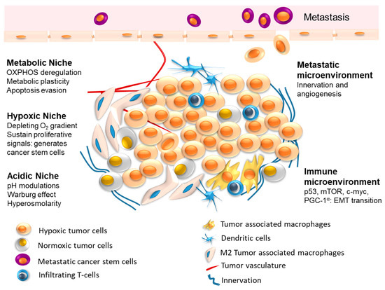
The interplay between tumor cells and their microenvironment (tumor microenvironment, TME) is a well-established hallmark feature of human tumors. TME comprises different cells, such as fibroblasts, immune cells, adipocytes, endothelial cells, as well as bone cells in distant sites of dissemination.

Cancer cells secrete biological factors to educate their neighboring cells toward an aggressive, pro-inflammatory, and pro-tumoral phenotype. In turn, cells of the TME release signals directed toward cancer cells to favor tumor growth, progression, dissemination, and treatment resistance. Interestingly, a cancer metabolic reprogramming is often involved in this pro-tumoral activity of TME cells.

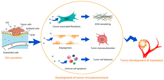
This multifaceted crosstalk is mediated by different biological factors, such as cytokines, chemokines, growth factors, extracellular matrix remodeling enzymes, as well as by extracellular vesicles (EVs). Specifically, EVs serve as transporters of different bioactive cargos, including proteins, nucleic acids, miRNAs, and lipids, from parental to both neighboring and distant target cells to participate in the various steps of malignant tumor development and progression by triggering or suppressing different signaling pathways in recipient cells.

The vicious crosstalk between cancer cells and their neighboring cells in the TME is now considered not only a prognostic/predictive biomarker of tumor growth and progression, but, more importantly, an effective target for novel and valuable therapeutic strategies.

The aim of this Collection is to invite authors to contribute original research as well as review articles highlighting the current concepts of the molecular mechanisms involved in the communication between tumor cells and their microenvironment, paving the way toward more specific and personalized medicine for cancer patients.

Prof. Dr. Patrizia Limonta
Dr. Fabrizio Fontana
Collection Editors

Manuscript Submission Information

Manuscripts should be submitted online at www.mdpi.com by registering and logging in to this website. Once you are registered, click here to go to the submission form. Manuscripts can be submitted until the deadline. All submissions that pass pre-check are peer-reviewed. Accepted papers will be published continuously in the journal (as soon as accepted) and will be listed together on the collection website. Research articles, review articles as well as short communications are invited. For planned papers, a title and short abstract (about 100 words) can be sent to the Editorial Office for announcement on this website.

Submitted manuscripts should not have been published previously, nor be under consideration for publication elsewhere (except conference proceedings papers). All manuscripts are thoroughly refereed through a single-blind peer-review process. A guide for authors and other relevant information for submission of manuscripts is available on the Instructions for Authors page. Cancers is an international peer-reviewed open access semimonthly journal published by MDPI.

Please visit the Instructions for Authors page before submitting a manuscript. The Article Processing Charge (APC) for publication in this open access journal is 2900 CHF (Swiss Francs). Submitted papers should be well formatted and use good English. Authors may use MDPI's English editing service prior to publication or during author revisions.

Keywords

  • cancer cells
  • tumor microenvironment (TME)
  • fibroblasts
  • immune cells
  • adipocytes
  • endothelial cells
  • bone cells
  • biofactors
  • extracellular vesicles (EVs)
  • cytokines
  • chemokines
  • growth factors
  • hormones
  • molecular cargo
  • miRNAs
  • precision medicine

Published Papers (9 papers)

2023

Jump to: 2022, 2021

30 pages, 2487 KiB  
Review
Cancer Bioenergetics and Tumor Microenvironments—Enhancing Chemotherapeutics and Targeting Resistant Niches through Nanosystems
by Aisha Farhana, Abdullah Alsrhani, Yusuf Saleem Khan and Zafar Rasheed
Cancers 2023, 15(15), 3836; https://doi.org/10.3390/cancers15153836 - 28 Jul 2023
Cited by 2 | Viewed by 1569
Abstract
Cancer is an impending bottleneck in the advanced scientific workflow to achieve diagnostic, prognostic, and therapeutic success. Most cancers are refractory to conventional diagnostic and chemotherapeutics due to their limited targetability, specificity, solubility, and side effects. The inherent ability of each cancer to [...] Read more.
Cancer is an impending bottleneck in the advanced scientific workflow to achieve diagnostic, prognostic, and therapeutic success. Most cancers are refractory to conventional diagnostic and chemotherapeutics due to their limited targetability, specificity, solubility, and side effects. The inherent ability of each cancer to evolve through various genetic and epigenetic transformations and metabolic reprogramming underlies therapeutic limitations. Though tumor microenvironments (TMEs) are quite well understood in some cancers, each microenvironment differs from the other in internal perturbations and metabolic skew thereby impeding the development of appropriate diagnostics, drugs, vaccines, and therapies. Cancer associated bioenergetics modulations regulate TME, angiogenesis, immune evasion, generation of resistant niches and tumor progression, and a thorough understanding is crucial to the development of metabolic therapies. However, this remains a missing element in cancer theranostics, necessitating the development of modalities that can be adapted for targetability, diagnostics and therapeutics. In this challenging scenario, nanomaterials are modular platforms for understanding TME and achieving successful theranostics. Several nanoscale particles have been successfully researched in animal models, quite a few have reached clinical trials, and some have achieved clinical success. Nanoparticles exhibit an intrinsic capability to interact with diverse biomolecules and modulate their functions. Furthermore, nanoparticles can be functionalized with receptors, modulators, and drugs to facilitate specific targeting with reduced toxicity. This review discusses the current understanding of different theranostic nanosystems, their synthesis, functionalization, and targetability for therapeutic modulation of bioenergetics, and metabolic reprogramming of the cancer microenvironment. We highlight the potential of nanosystems for enhanced chemotherapeutic success emphasizing the questions that remain unanswered. Full article
Show Figures

Figure 1

17 pages, 5591 KiB  
Article
5-Fluorouracil Suppresses Colon Tumor through Activating the p53-Fas Pathway to Sensitize Myeloid-Derived Suppressor Cells to FasL+ Cytotoxic T Lymphocyte Cytotoxicity
by Yingcui Yang, Mingqing Zhang, Yongdan Zhang, Kebin Liu and Chunwan Lu
Cancers 2023, 15(5), 1563; https://doi.org/10.3390/cancers15051563 - 02 Mar 2023
Cited by 6 | Viewed by 1748
Abstract
Myelosuppression is a major adverse effect of 5-fluorouracil (5-FU) chemotherapy. However, recent findings indicate that 5-FU selectively suppresses myeloid-derived suppressor cells (MDSCs), to enhance antitumor immunity in tumor-bearing mice. 5-FU-mediated myelosuppression may thus have a beneficial effect for cancer patients. The molecular mechanism [...] Read more.
Myelosuppression is a major adverse effect of 5-fluorouracil (5-FU) chemotherapy. However, recent findings indicate that 5-FU selectively suppresses myeloid-derived suppressor cells (MDSCs), to enhance antitumor immunity in tumor-bearing mice. 5-FU-mediated myelosuppression may thus have a beneficial effect for cancer patients. The molecular mechanism underlying 5-FU’s suppression of MDSCs is currently unknown. We aimed at testing the hypothesis that 5-FU suppresses MDSCs through enhancing MDSC sensitivity to Fas-mediated apoptosis. We observed that, although FasL is highly expressed in T cells, Fas is weakly expressed in myeloid cells in human colon carcinoma, indicating that downregulation of Fas is a mechanism underlying myeloid cell survival and accumulation in human colon cancer. 5-FU treatment upregulated expression of both p53 and Fas, and knocking down p53 diminished 5-FU-induced Fas expression in MDSC-like cells, in vitro. 5-FU treatment also increased MDSC-like cell sensitivity to FasL-induced apoptosis in vitro. Furthermore, we determined that 5-FU therapy increased expression of Fas on MDSCs, suppressed MDSC accumulation, and increased CTL tumor infiltration in colon tumor-bearing mice. In human colorectal cancer patients, 5-FU chemotherapy decreased MDSC accumulation and increased CTL level. Our findings determine that 5-FU chemotherapy activates the p53-Fas pathway, to suppress MDSC accumulation, to increase CTL tumor infiltration. Full article
Show Figures

Figure 1

17 pages, 3374 KiB  
Article
The Omega-3 Docosahexaenoyl Ethanolamide Reduces CCL5 Secretion in Triple Negative Breast Cancer Cells Affecting Tumor Progression and Macrophage Recruitment
by Giuseppina Augimeri, Marco Fiorillo, Catia Morelli, Salvatore Panza, Cinzia Giordano, Ines Barone, Stefania Catalano, Diego Sisci, Sebastiano Andò and Daniela Bonofiglio
Cancers 2023, 15(3), 819; https://doi.org/10.3390/cancers15030819 - 29 Jan 2023
Cited by 5 | Viewed by 2007
Abstract
Triple-negative breast cancer (TNBC), an aggressive breast cancer subtype lacking effective targeted therapies, is considered to feature a unique cellular microenvironment with high infiltration of tumor-associated macrophages (TAM), which contribute to worsening breast cancer patient outcomes. Previous studies have shown the antitumoral actions [...] Read more.
Triple-negative breast cancer (TNBC), an aggressive breast cancer subtype lacking effective targeted therapies, is considered to feature a unique cellular microenvironment with high infiltration of tumor-associated macrophages (TAM), which contribute to worsening breast cancer patient outcomes. Previous studies have shown the antitumoral actions of the dietary omega-3 docosahexaenoic acid (DHA) in both tumor epithelial and stromal components of the breast cancer microenvironment. Particularly in breast cancer cells, DHA can be converted into its conjugate with ethanolamine, DHEA, leading to a more effective anti-oncogenic activity of the parent compound in estrogen receptor-positive breast cancer cells. Here, we investigated the ability of DHEA to attenuate the malignant phenotype of MDA-MB-231 and MDA-MB-436 TNBC cell lines, which in turn influenced TAM behaviors. Our findings revealed that DHEA reduced the viability of TNBC cells in a concentration-dependent manner and compromised cell migration and invasion. Interestingly, DHEA inhibited oxygen consumption and extracellular acidification rates, reducing respiration and the glycolytic reserve in both cell lines. In a co-culture system, TNBC cells exposed to DHEA suppressed recruitment of human THP-1 cells, reduced their viability, and the expression of genes associated with TAM phenotype. Interestingly, we unraveled that the effects of DHEA in TNCB cells were mediated by reduced C-C motif chemokine ligand 5 (CCL5) expression and secretion affecting macrophage recruitment. Overall, our data, shedding new light on the antitumoral effects of DHA ethanolamine-conjugated, address this compound as a promising option in the treatment of TNBC patients. Full article
Show Figures

Figure 1

2022

Jump to: 2023, 2021

14 pages, 911 KiB  
Review
Glycocalyx Acts as a Central Player in the Development of Tumor Microenvironment by Extracellular Vesicles for Angiogenesis and Metastasis
by Ye Zeng, Yan Qiu, Wenli Jiang and Bingmei M. Fu
Cancers 2022, 14(21), 5415; https://doi.org/10.3390/cancers14215415 - 03 Nov 2022
Cited by 6 | Viewed by 1969
Abstract
Angiogenesis in tumor growth and progression involves a series of complex changes in the tumor microenvironment. Extracellular vesicles (EVs) are important components of the tumor microenvironment, which can be classified as exosomes, apoptotic vesicles, and matrix vesicles according to their origins and properties. [...] Read more.
Angiogenesis in tumor growth and progression involves a series of complex changes in the tumor microenvironment. Extracellular vesicles (EVs) are important components of the tumor microenvironment, which can be classified as exosomes, apoptotic vesicles, and matrix vesicles according to their origins and properties. The EVs that share many common biological properties are important factors for the microenvironmental modification and play a vital role in tumor growth and progression. For example, vascular endothelial growth factor (VEGF) exosomes, which carry VEGF, participate in the tolerance of anti-angiogenic therapy (AAT). The glycocalyx is a mucopolysaccharide structure consisting of glycoproteins, proteoglycans, and glycosaminoglycans. Both endothelial and tumor cells have glycocalyx at their surfaces. Glycocalyx at both cells mediates the secretion and uptake of EVs. On the other hand, many components carried by EVs can modify the glycocalyx, which finally facilitates the development of the tumor microenvironment. In this short review, we first summarize the role of EVs in the development of the tumor microenvironment. Then we review how the glycocalyx is associated with the tumor microenvironment and how it is modulated by the EVs, and finally, we review the role of the glycocalyx in the synthesis, release, and uptake of EVs that affect tumor microenvironments. This review aims to provide a basis for the mechanistic study of AAT and new clues to address the challenges in AAT tolerance, tumor angiogenesis and metastasis. Full article
Show Figures

Figure 1

17 pages, 1871 KiB  
Article
Actin Retrograde Flow Regulated by the Wiskott–Aldrich Syndrome Protein Drives the Natural Killer Cell Response
by Batel Sabag, Moria Levy, Jessica Kivelevitz, Nataly Dashevsky, Aviad Ben-Shmuel, Abhishek Puthenveetil, Fatima Awwad and Mira Barda-Saad
Cancers 2022, 14(15), 3756; https://doi.org/10.3390/cancers14153756 - 02 Aug 2022
Cited by 3 | Viewed by 1952
Abstract
Understanding the crosstalk between natural killer (NK) cells and the tumor microenvironment (TME) has enhanced the potential of exploiting the interplay between activation and inhibition of NK cells for immunotherapy. This interaction is crucial for understanding how tumor cells escape NK cell immune [...] Read more.
Understanding the crosstalk between natural killer (NK) cells and the tumor microenvironment (TME) has enhanced the potential of exploiting the interplay between activation and inhibition of NK cells for immunotherapy. This interaction is crucial for understanding how tumor cells escape NK cell immune surveillance. NK cell dysfunction is regulated by two molecular mechanisms, downregulated activating receptor ligand expression on the tumor cells, and upregulated inhibitory signals delivered to NK cells. Recent studies demonstrated the role of mechanotransduction in modulating NK cell responses in the TME. The immunological synapse represents a functional interface between the NK cell and its target, regulated by Actin Retrograde Flow (ARF), which drives the adhesion molecules and receptors toward the central zone of the immunological synapse (IS). Here, we further characterize the role of ARF in controlling the immune response of NK cells, using CRISPR/cas9-mediated Wiskott–Aldrich Syndrome protein (WASp) gene silencing of NK cells. We demonstrate that WASp regulates ARF velocity, affecting the conformation and function of the key NK inhibitory regulator, SH2-domain containing protein tyrosine phosphatase-1 (SHP-1), and consequently, the NK cell response. Our results demonstrate the potential of modulating the biophysical and intracellular regulation of NK activation as a promising approach for improving immunotherapy. Full article
Show Figures

Figure 1

13 pages, 1849 KiB  
Article
Melanoma Stem Cells Educate Neutrophils to Support Cancer Progression
by Martina Anselmi, Fabrizio Fontana, Monica Marzagalli, Nicoletta Gagliano, Michele Sommariva and Patrizia Limonta
Cancers 2022, 14(14), 3391; https://doi.org/10.3390/cancers14143391 - 13 Jul 2022
Cited by 15 | Viewed by 2115
Abstract
Background: It is now well-established that cancer stem cells (CSCs) can support melanoma progression by reshaping the tumor immune microenvironment. However, the molecular mechanisms underlying the crosstalk between melanoma SCs and cancer-associated neutrophils have not been elucidated yet. Methods: The aim of the [...] Read more.
Background: It is now well-established that cancer stem cells (CSCs) can support melanoma progression by reshaping the tumor immune microenvironment. However, the molecular mechanisms underlying the crosstalk between melanoma SCs and cancer-associated neutrophils have not been elucidated yet. Methods: The aim of the present study was to unravel the role of melanoma SCs in neutrophil polarization. HL60 neutrophil-like (dHL60) cells were treated with conditioned medium from A375 melanoma SCs (CSC-CM), and their phenotype was investigated. Results: We demonstrated that CSC-CM could specifically activate immune cells by increasing CD66b and CD11b expression. In particular, we revealed that A375 CSCs could release various soluble factors, namely TGF-β, IL-6, and IL-8, able to promote the recruitment of neutrophils and their switch toward an N2 phenotype characterized by the activation of ERK, STAT3, and P38 pathways and the overexpression of CXCR2 and NF-kB. Moreover, after exposure to CSC-CM, dHL60 cells exhibited enhanced ROS production and NET release, without undergoing cell death; increased secretion of MMP-9 and pro-inflammatory cytokines was also observed. Finally, CSC-CM-activated neutrophils endowed A375 cells with stemness traits, stimulating both sphere formation and ABCG2 expression. Conclusion: Collectively, our results suggest that melanoma SCs can prime neutrophils to support cancer progression. Full article
Show Figures

Figure 1

19 pages, 2138 KiB  
Review
Impact of Lipid Metabolism on Antitumor Immune Response
by Nesrine Mabrouk, Baptiste Lecoeur, Ali Bettaieb, Catherine Paul and Frédérique Végran
Cancers 2022, 14(7), 1850; https://doi.org/10.3390/cancers14071850 - 06 Apr 2022
Cited by 18 | Viewed by 3230
Abstract
Over the past decade, metabolic reprogramming has been defined as a hallmark of cancer. More recently, a large number of studies have demonstrated that metabolic reprogramming can modulate the differentiation and functions of immune cells, and thus modify the antitumor response. Increasing evidence [...] Read more.
Over the past decade, metabolic reprogramming has been defined as a hallmark of cancer. More recently, a large number of studies have demonstrated that metabolic reprogramming can modulate the differentiation and functions of immune cells, and thus modify the antitumor response. Increasing evidence suggests that modified energy metabolism could be responsible for the failure of antitumor immunity. Indeed, tumor-infiltrating immune cells play a key role in cancer, and metabolic switching in these cells has been shown to help determine their phenotype: tumor suppressive or immune suppressive. Recent studies in the field of immunometabolism focus on metabolic reprogramming in the tumor microenvironment (TME) by targeting innate and adaptive immune cells and their associated anti- or protumor phenotypes. In this review, we discuss the lipid metabolism of immune cells in the TME as well as the effects of lipids; finally, we expose the link between therapies and lipid metabolism. Full article
Show Figures

Figure 1

2021

Jump to: 2023, 2022

20 pages, 2593 KiB  
Article
Tumor-Infiltrating Lymphocytes and Cancer Markers in Osteosarcoma: Influence on Patient Survival
by José Manuel Casanova, Jani-Sofia Almeida, John David Reith, Luana Madalena Sousa, Ruben Fonseca, Paulo Freitas-Tavares, Manuel Santos-Rosa and Paulo Rodrigues-Santos
Cancers 2021, 13(23), 6075; https://doi.org/10.3390/cancers13236075 - 02 Dec 2021
Cited by 18 | Viewed by 2484
Abstract
Osteosarcoma (OST) is the most common type of high-grade primary bone tumor, which mainly affects young adults. The current standard of care for OST combines surgical resection with chemotherapy. The clinical outcomes and the current options to treat OST patients are unsatisfactory and [...] Read more.
Osteosarcoma (OST) is the most common type of high-grade primary bone tumor, which mainly affects young adults. The current standard of care for OST combines surgical resection with chemotherapy. The clinical outcomes and the current options to treat OST patients are unsatisfactory and novel treatment strategies are needed. The crosstalk between tumor cells and immune cells is essential to the OST microenvironment. Despite the efforts that have been made to address the importance of immune-related factors in OST, there is still a lot to understand. The purpose of the current study was to evaluate the tumor-infiltrating lymphocytes (TIL), the expression of proteins involved in tumor biology, and their impact on the clinical outcome of OST patients. We studied 93 samples of OST patients using immunohistochemistry and histomorphometry. We looked for the infiltration of CD3+, CD4+, CD8+, TIA1+ and CD20+ cells and for the expression of CD44 standard (CD44s) and variant 6 (CD44v6), CD95/Fas, Fas-L, p53 and p-glycoprotein. All the parameters were analyzed for the influence on the occurrence of death and metastasis, plus patient overall survival (OS) and progression-free survival (PFS). The effect of sex, age, tumor location (distal femur or proximal tibia) and the combination with neoadjuvant chemotherapy was also assessed. Our results suggest that the presence of tumor-infiltrating CD4+ cells provides protection to OST patients, and that CD8+ cells have a significant impact on the patient’s overall survival (OS) and progression-free survival (PFS), which is more evident in male patients. In addition, a strong association between tumor-infiltrating CD4+ cells and the presence of CD44s expression in tumor samples was observed. Analysis of TIL and tumor markers related to tumor biology could be useful to stratify patients and monitor the response to therapy, as well as to assist with the development of immunotherapy strategies to improve the effects of cytotoxic TIL to eradicate the tumor cells. Full article
Show Figures

Figure 1

14 pages, 2838 KiB  
Article
Response to FEC Chemotherapy and Oncolytic HSV-1 Is Associated with Macrophage Polarization and Increased Expression of S100A8/A9 in Triple Negative Breast Cancer
by Alyssa Vito, Nader El-Sayes, Omar Salem, Yonghong Wan and Karen L. Mossman
Cancers 2021, 13(21), 5590; https://doi.org/10.3390/cancers13215590 - 08 Nov 2021
Cited by 2 | Viewed by 2399
Abstract
The era of immunotherapy has seen an insurgence of novel therapies driving oncologic research and the clinical management of the disease. We have previously reported that a combination of chemotherapy (FEC) and oncolytic virotherapy (oHSV-1) can be used to sensitize otherwise non-responsive tumors [...] Read more.
The era of immunotherapy has seen an insurgence of novel therapies driving oncologic research and the clinical management of the disease. We have previously reported that a combination of chemotherapy (FEC) and oncolytic virotherapy (oHSV-1) can be used to sensitize otherwise non-responsive tumors to immune checkpoint blockade and that tumor-infiltrating B cells are required for the efficacy of our therapeutic regimen in a murine model of triple-negative breast cancer. In the studies herein, we have performed gene expression profiling using microarray analyses and have investigated the differential gene expression between tumors treated with FEC + oHSV-1 versus untreated tumors. In this work, we uncovered a therapeutically driven switch of the myeloid phenotype and a gene signature driving increased tumor cell killing. Full article
Show Figures

Figure 1

Planned Papers

The below list represents only planned manuscripts. Some of these manuscripts have not been received by the Editorial Office yet. Papers submitted to MDPI journals are subject to peer-review.

Title: The metastatic niche formation: a dialogue between lung tumor cells and microenvironment
Authors: Francesca Pontis1, Orazio Fortunato1,*, Luca Rozand, Gabriella Sozzi and Giulia Bertolini1
Affiliation: Tumor Genomics Unit, Department of Research, Fondazione IRCCS Istituto Nazionale dei Tumori, Via Venezian 1, 20133 Milan, Italy
Abstract: Abstract: Lung cancer is the most deadly cancer in the world. Approximately 30% of diagnosed patients have an advanced lung cancer disease. Understanding the mechanisms that regulated the metastasis formation from primary lung cancer is an important unmet clinical need. One of the first steps during the establishment of lung cancer metastases includes the formation of the pre-metastatic niche in the distant organs that may occur at the first phase of the cancer development. The pre-metastatic niche is established by the cross-talk among primary tumor secreted factors and stromal components at the distant site. Mechanisms of primary tumors escape and seeding of distant organs rely on specific properties of tumor cells and they are also tightly regulated by the interactions with stromal cells at the metastatic niche that finally dictate the success of metastasis establishment. Here, we summarize mechanisms underlying the pre-metastatic niche formation starting from how lung primary tumor cells modulate the distant site of metastasis through the release of several factors focusing on Extracellular Vesicles. Then, we describe the complexity of Circulating Tumor Cells (CTCs) that represent the seed of metastasis and how stromal cells can help their metastatic dissemination. Finally, we evaluate the contribution of stromal and immune cells at the distant site in the control and/or support of metastatic niche formation. Overall, we present an overview of the step of metastatic cascade of lung cancer focusing on the interactions, physical or mediated by soluble factors, between tumor cells and stroma cells subtending metastasis development. Keywords: extracellular vesicles; Circulating Tumor Cells; lung cancer

Back to TopTop