


Cancers 2023, 15(18), 4529; https://doi.org/10.3390/cancers15184529 - 12 Sep 2023
Cited by 1 | Viewed by 1792
Abstract
►
Show Figures
Autophagy plays a complex role in breast cancer cell survival, metastasis, and chemotherapeutic resistance. Dipeptidyl peptidase (DPP)-4, a therapeutic target for type 2 diabetes mellitus, is also involved in autophagic flux. The potential influence of DPP-4 suppression on cancer biology remains unknown. Here,
[...] Read more.
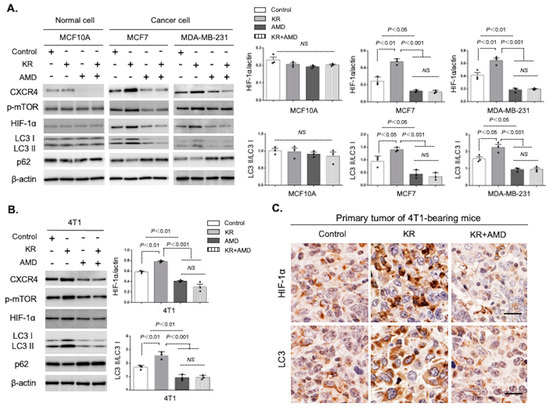
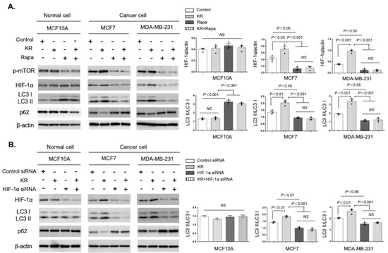
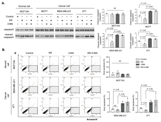
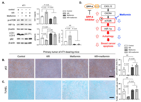
Autophagy plays a complex role in breast cancer cell survival, metastasis, and chemotherapeutic resistance. Dipeptidyl peptidase (DPP)-4, a therapeutic target for type 2 diabetes mellitus, is also involved in autophagic flux. The potential influence of DPP-4 suppression on cancer biology remains unknown. Here, we report that DPP-4 deficiency promotes breast cancer cell survival via the induction of autophagy by the C-X-C motif chemokine 12 (CXCL12)/C-X-C receptor 4 (CXCR4)/mammalian target of rapamycin (mTOR)/hypoxia inducible factor (HIF)-1α axis. DPP-4 knockdown and DPP-4 inhibitor KR62436 (KR) treatment both increased the levels of LC3II and HIF-1α in cultured human breast and mouse mammary cancer cells. The KR-induced autophagic phenotype in cancer cells was inhibited by treatment with the CXCR4 inhibitor AMD3100 and rapamycin. HIF-1α knockdown also suppressed breast cancer autophagy induced by KR. The autophagy inhibitor 3-methyladenine significantly blocked the KR-mediated suppression of cleaved caspase-3 levels and apoptosis in breast cancer cell lines. Finally, we found that the metformin-induced apoptosis of DPP-4-deficient 4T1 mammary cancer cells was associated with the suppression of autophagy. Our findings identify a novel role for DPP-4 inhibition in the promotion of breast cancer survival by inducing CXCL12/CXCR4/mTOR/HIF-1α axis-dependent autophagy. Metformin is a potential drug that counteracts the breast cancer cell survival system.
Full article
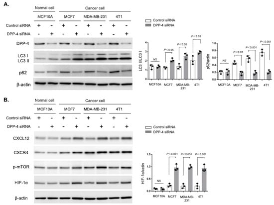
Figure 1