miRNAs: New Insights in Tumor Biology
Share This Topical Collection
Editor
 Dr. Francesca Orso
Dr. Francesca Orso
E-Mail
Website
Collection Editor
Department of Translational Medicine (DIMET), University of Eastern Piedmont, 28100 Novara, Italy
Interests: miRNAs; tumor progression; metastasis; melanoma; breast cancer; miRNA therapy
Topical Collection Information
Dear Colleagues,
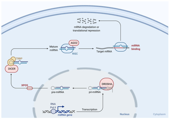
MicroRNAs (miRNAs) are short non-coding RNAs involved in the initiation and progression of cancer. In fact, comparisons of expression profiles for normal tissues versus human cancers have revealed significant differences in miRNA expression. Moreover, miRNAs regulate different steps of cancer progression. However, the precise mechanisms through which they act need to be further characterized. Therefore, studies investigating the molecular mechanism underlying their actions and dysregulation are urgent. This Topical Collection invites original papers and reviews covering a wide range of topics related to miRNAs, such as miRNA-activated functional networks, miRNA expression regulation, miRNA therapeutics, miRNA function as biomarkers, miRNA role in cancer drug resistance in various kinds of cancers, as well as other related topics not listed here.
Dr. Francesca Orso
Collection Editor
Manuscript Submission Information
Manuscripts should be submitted online at www.mdpi.com by registering and logging in to this website. Once you are registered, click here to go to the submission form. Manuscripts can be submitted until the deadline. All submissions that pass pre-check are peer-reviewed. Accepted papers will be published continuously in the journal (as soon as accepted) and will be listed together on the collection website. Research articles, review articles as well as short communications are invited. For planned papers, a title and short abstract (about 100 words) can be sent to the Editorial Office for announcement on this website.
Submitted manuscripts should not have been published previously, nor be under consideration for publication elsewhere (except conference proceedings papers). All manuscripts are thoroughly refereed through a single-blind peer-review process. A guide for authors and other relevant information for submission of manuscripts is available on the Instructions for Authors page. Cancers is an international peer-reviewed open access semimonthly journal published by MDPI.
Please visit the Instructions for Authors page before submitting a manuscript.
The Article Processing Charge (APC) for publication in this open access journal is 2900 CHF (Swiss Francs).
Submitted papers should be well formatted and use good English. Authors may use MDPI's
English editing service prior to publication or during author revisions.
Keywords
- miRNAs
- tumor progression
- miRNA expression regulation
- miRNA therapeutics
- biomarkers
Published Papers (22 papers)
Open AccessArticle
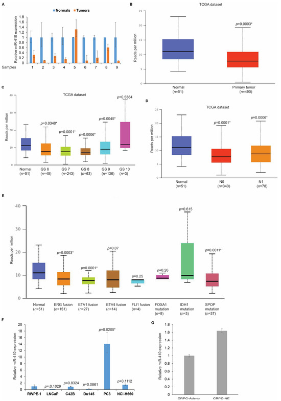
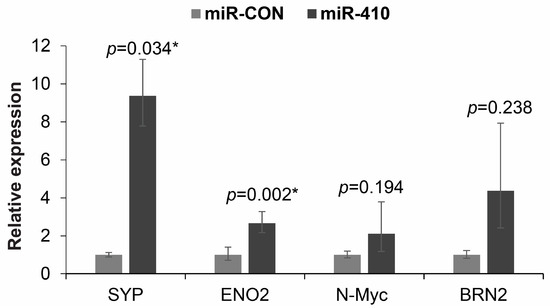
miR-410 Is a Key Regulator of Epithelial-to-Mesenchymal Transition with Biphasic Role in Prostate Cancer
by
Diana M. Asante, Amritha Sreekumar, Sandip Nathani, Tae Jin Lee, Ashok Sharma, Nikhil Patel, Matthew N. Simmons and Sharanjot Saini
Viewed by 900
Abstract
The molecular basis of prostate cancer (PCa) progression from the primary disease to metastatic castration-resistant prostate cancer (CRPC) followed by therapy-induced neuroendocrine prostate cancer is not fully understood. In this study, we elucidate the role of miR-410, a little-studied microRNA located on chromosome
[...] Read more.
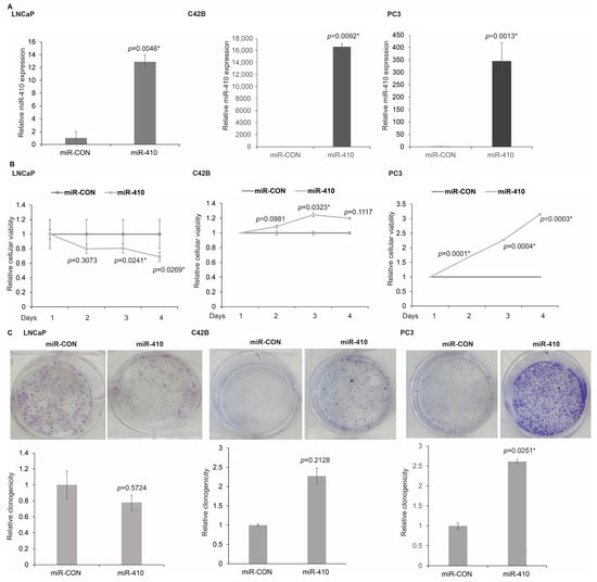
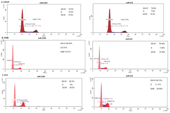
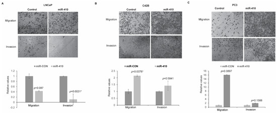
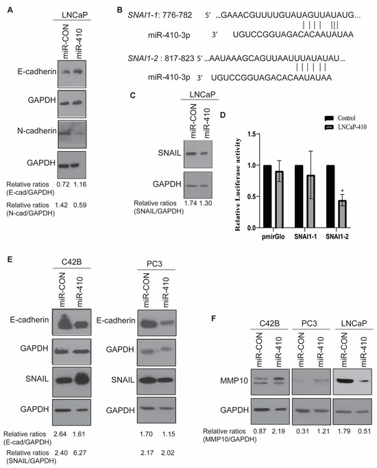
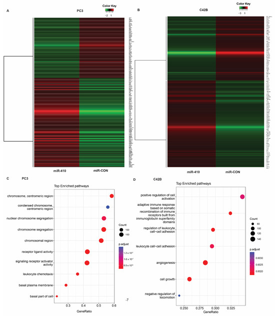
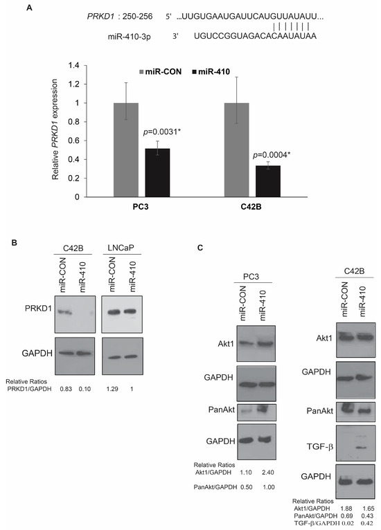
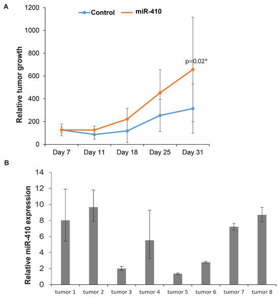
The molecular basis of prostate cancer (PCa) progression from the primary disease to metastatic castration-resistant prostate cancer (CRPC) followed by therapy-induced neuroendocrine prostate cancer is not fully understood. In this study, we elucidate the role of miR-410, a little-studied microRNA located on chromosome 14q32.31 within the DLK1-DIO3 cluster, in PCa. miR-410 expression analyses in primary and metastatic PCa tissues and cell lines show that its levels are decreased in initial stages and increased in advanced PCa. Functional studies were performed in a series of PCa cell lines. In LNCaP cells, miR-410 overexpression led to decreases in cellular viability, proliferation, invasiveness, and migration. On the other hand, miR-410 overexpression in PC3 and C42B cells led to increased viability, proliferation, and invasiveness. Our data suggest that miR-410 represses epithelial-to-mesenchymal transition (EMT) in LNCaP cells by directly repressing SNAIL. However, it promotes EMT and upregulates PI3K/Akt signaling in PC3 and C42B cells. In vivo studies with PC3 xenografts support an oncogenic role of miR-410. These data suggest that miR-410 acts as a tumor suppressor in the initial stages of PCa and play an oncogenic role in advanced PCa. Our findings have important implications in understanding the molecular basis of PCa progression with potential translational implications.
Full article
►▼
Show Figures
Open AccessReview
Clinical Significance of microRNAs in Hematologic Malignancies and Hematopoietic Stem Cell Transplantation
by
Aneta Sevcikova, Ivana Fridrichova, Nataliia Nikolaieva, Lenka Kalinkova, Radoslav Omelka, Monika Martiniakova and Sona Ciernikova
Cited by 5 | Viewed by 2127
Abstract
Hematologic malignancies are a group of neoplastic conditions that can develop from any stage of the hematopoiesis cascade. Small non-coding microRNAs (miRNAs) play a crucial role in the post-transcriptional regulation of gene expression. Mounting evidence highlights the role of miRNAs in malignant hematopoiesis
[...] Read more.
Hematologic malignancies are a group of neoplastic conditions that can develop from any stage of the hematopoiesis cascade. Small non-coding microRNAs (miRNAs) play a crucial role in the post-transcriptional regulation of gene expression. Mounting evidence highlights the role of miRNAs in malignant hematopoiesis via the regulation of oncogenes and tumor suppressors involved in proliferation, differentiation, and cell death. In this review, we provide current knowledge about dysregulated miRNA expression in the pathogenesis of hematological malignancies. We summarize data about the clinical utility of aberrant miRNA expression profiles in hematologic cancer patients and their associations with diagnosis, prognosis, and the monitoring of treatment response. Moreover, we will discuss the emerging role of miRNAs in hematopoietic stem cell transplantation (HSCT), and severe post-HSCT complications, such as graft-versus-host disease (GvHD). The therapeutical potential of the miRNA-based approach in hemato-oncology will be outlined, including studies with specific antagomiRs, mimetics, and circular RNAs (circRNAs). Since hematologic malignancies represent a full spectrum of disorders with different treatment paradigms and prognoses, the potential use of miRNAs as novel diagnostic and prognostic biomarkers might lead to improvements, resulting in a more accurate diagnosis and better patient outcomes.
Full article
►▼
Show Figures
Open AccessArticle
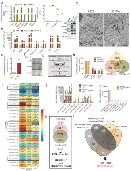
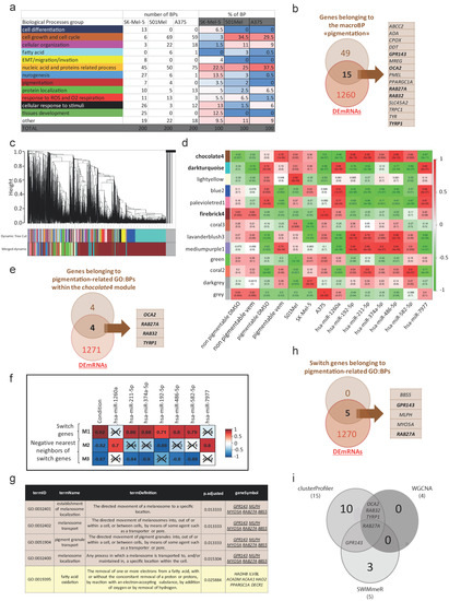
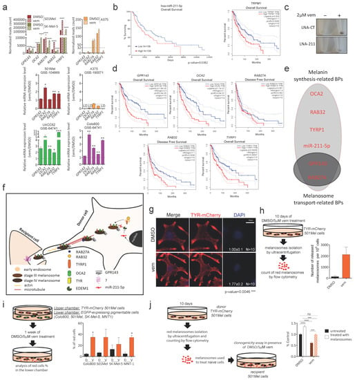
A Network of MicroRNAs and mRNAs Involved in Melanosome Maturation and Trafficking Defines the Lower Response of Pigmentable Melanoma Cells to Targeted Therapy
by
Marianna Vitiello, Alberto Mercatanti, Maurizio Salvatore Podda, Caterina Baldanzi, Antonella Prantera, Samanta Sarti, Milena Rizzo, Alessandra Salvetti, Federica Conte, Giulia Fiscon, Paola Paci and Laura Poliseno
Cited by 1 | Viewed by 1877
Abstract
Background: The ability to increase their degree of pigmentation is an adaptive response that confers pigmentable melanoma cells higher resistance to BRAF inhibitors (BRAFi) compared to non-pigmentable melanoma cells. Methods: Here, we compared the miRNome and the transcriptome profile of pigmentable 501Mel and
[...] Read more.
Background: The ability to increase their degree of pigmentation is an adaptive response that confers pigmentable melanoma cells higher resistance to BRAF inhibitors (BRAFi) compared to non-pigmentable melanoma cells. Methods: Here, we compared the miRNome and the transcriptome profile of pigmentable 501Mel and SK-Mel-5 melanoma cells vs. non-pigmentable A375 melanoma cells, following treatment with the BRAFi vemurafenib (vem). In depth bioinformatic analyses (clusterProfiler, WGCNA and SWIMmeR) allowed us to identify the miRNAs, mRNAs and biological processes (BPs) that specifically characterize the response of pigmentable melanoma cells to the drug. Such BPs were studied using appropriate assays in vitro and in vivo (xenograft in zebrafish embryos). Results: Upon vem treatment, miR-192-5p, miR-211-5p, miR-374a-5p, miR-486-5p, miR-582-5p, miR-1260a and miR-7977, as well as
GPR143,
OCA2,
RAB27A,
RAB32 and
TYRP1 mRNAs, are differentially expressed only in pigmentable cells. These miRNAs and mRNAs belong to BPs related to pigmentation, specifically melanosome maturation and trafficking. In fact, an increase in the number of intracellular melanosomes—due to increased maturation and/or trafficking—confers resistance to vem. Conclusion: We demonstrated that the ability of pigmentable cells to increase the number of intracellular melanosomes fully accounts for their higher resistance to vem compared to non-pigmentable cells. In addition, we identified a network of miRNAs and mRNAs that are involved in melanosome maturation and/or trafficking. Finally, we provide the rationale for testing BRAFi in combination with inhibitors of these biological processes, so that pigmentable melanoma cells can be turned into more sensitive non-pigmentable cells.
Full article
►▼
Show Figures
Open AccessArticle
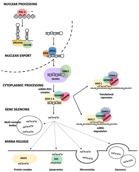
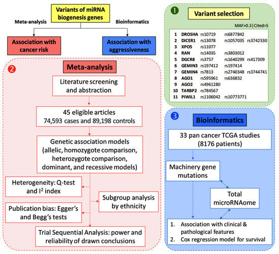
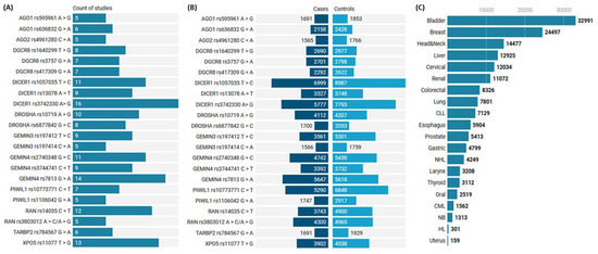
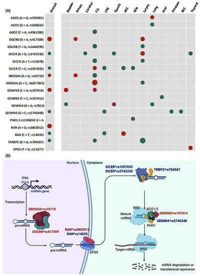
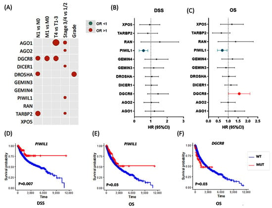
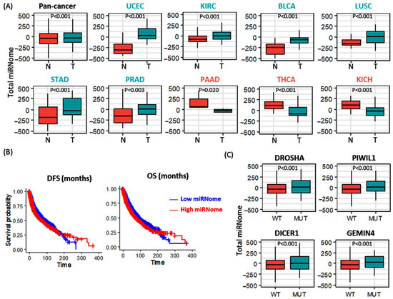
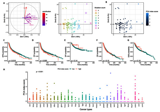
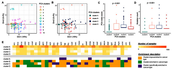
Pan-Cancer Study on Variants of Canonical miRNA Biogenesis Pathway Components: A Pooled Analysis
by
Rami M. Elshazli, Eman A. Toraih, Mohammad H. Hussein, Emmanuelle M. Ruiz, Emad Kandil and Manal S. Fawzy
Cited by 7 | Viewed by 1882
Abstract
Single nucleotide polymorphisms in genes involved in microRNA processing/maturation and release may deregulate the microRNAome expression levels. We aimed to assess the relationship between miRNA machinery genetic variants and human cancer risk using integrative bioinformatics analyses to identify the role of these genes
[...] Read more.
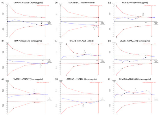
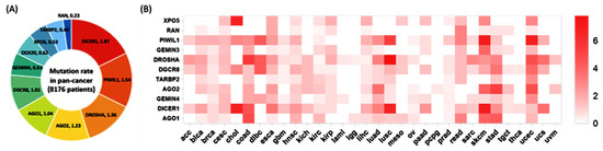
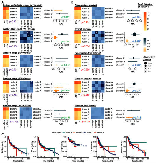
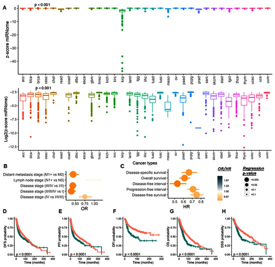
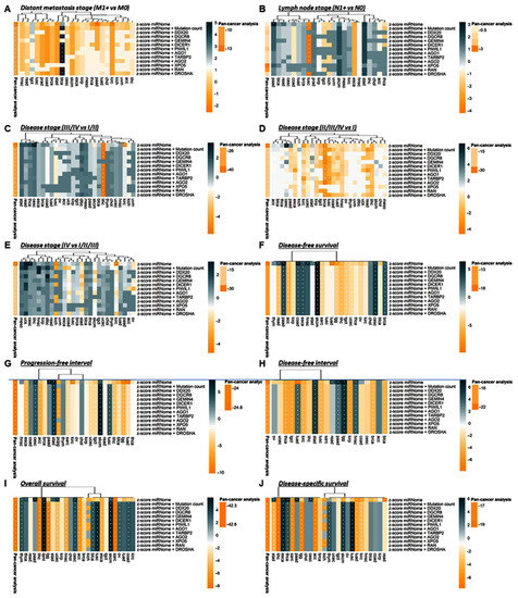
Single nucleotide polymorphisms in genes involved in microRNA processing/maturation and release may deregulate the microRNAome expression levels. We aimed to assess the relationship between miRNA machinery genetic variants and human cancer risk using integrative bioinformatics analyses to identify the role of these genes in cancer aggressiveness. Mutations of 8176 pan-cancer samples were retrieved from 33 studies in “TCGA” database, and a Cox regression model for survival was performed. Next, 22 computationally identified variants within 11 genes were selected based on their high citation rate and MAF. Relevant articles through March 2020 were included. Pooled estimates under the five genetic association models were calculated. Publication bias and heterogeneity between articles were evaluated. Trial Sequential Analysis (TSA) was applied to assess the power and reliability of the draw conclusions. TCGA patients with different cancer types revealed significant alterations in miRNA machinery genes, with mutation frequency ranging from 0.6–13% of samples. RAN was associated with LN metastasis, while TARBP2 and PIWIL1 gene mutations exhibited better overall survival. In the meta-analysis, 45 articles (74,593 cases and 89,198 controls) met the eligibility criteria. Pooled analysis revealed an increased cancer risk with DROSHArs10719*G, RANrs3803012*G, DGCR8rs417309*A, and GEMIN3rs197414*A. In contrast, both DICER1rs1057035*T and GEMIN4rs2743048*G conferred protection against developing cancer. TSA showed the cumulative evidence is inadequate, and the addition of further primary studies is necessary. This study suggests a potential role of miRNA biogenesis genes in cancer development/prognosis. Further functional studies may reveal biological explanations for the differential risks of the machinery variants in different cancer types.
Full article
►▼
Show Figures
Open AccessReview
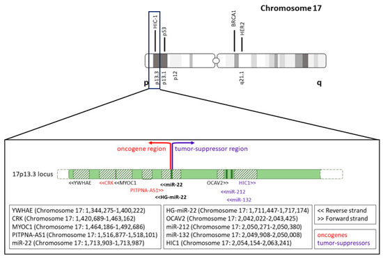
An Immunocompetent Environment Unravels the Proto-Oncogenic Role of miR-22
by
Maria Laura Centomo, Marianna Vitiello, Laura Poliseno and Pier Paolo Pandolfi
Cited by 4 | Viewed by 1756
Abstract
MiR-22 was first identified as a proto-oncogenic microRNA (miRNA) due to its ability to post-transcriptionally suppress the expression of the potent PTEN (Phosphatase And Tensin Homolog) tumor suppressor gene. miR-22 tumorigenic role in cancer was subsequently supported by its ability to positively trigger
[...] Read more.
MiR-22 was first identified as a proto-oncogenic microRNA (miRNA) due to its ability to post-transcriptionally suppress the expression of the potent PTEN (Phosphatase And Tensin Homolog) tumor suppressor gene. miR-22 tumorigenic role in cancer was subsequently supported by its ability to positively trigger lipogenesis, anabolic metabolism, and epithelial-mesenchymal transition (EMT) towards the metastatic spread. However, during the following years, the picture was complicated by the identification of targets that support a tumor-suppressive role in certain tissues or cell types. Indeed, many papers have been published where in vitro cellular assays and in vivo immunodeficient or immunosuppressed xenograft models are used. However, here we show that all the studies performed
in vivo, in immunocompetent transgenic and knock-out animal models, unanimously support a proto-oncogenic role for miR-22. Since miR-22 is actively secreted from and readily exchanged between normal and tumoral cells, a functional immune dimension at play could well represent the divider that allows reconciling these contradictory findings. In addition to a critical review of this vast literature, here we provide further proof of the oncogenic role of miR-22 through the analysis of its genomic locus
vis a vis the genetic landscape of human cancer.
Full article
►▼
Show Figures
Open AccessArticle
Identifying Tissue- and Cohort-Specific RNA Regulatory Modules in Cancer Cells Using Multitask Learning
by
Milad Mokhtaridoost, Philipp G. Maass and Mehmet Gönen
Viewed by 1716
Abstract
MicroRNA (miRNA) alterations significantly impact the formation and progression of human cancers. miRNAs interact with messenger RNAs (mRNAs) to facilitate degradation or translational repression. Thus, identifying miRNA–mRNA regulatory modules in cohorts of primary tumor tissues are fundamental for understanding the biology of tumor
[...] Read more.
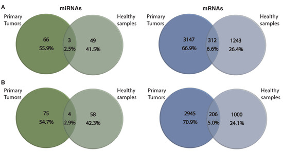
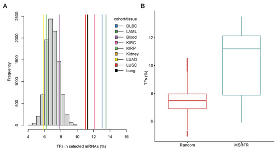
MicroRNA (miRNA) alterations significantly impact the formation and progression of human cancers. miRNAs interact with messenger RNAs (mRNAs) to facilitate degradation or translational repression. Thus, identifying miRNA–mRNA regulatory modules in cohorts of primary tumor tissues are fundamental for understanding the biology of tumor heterogeneity and precise diagnosis and treatment. We established a multitask learning sparse regularized factor regression (MSRFR) method to determine key tissue- and cohort-specific miRNA–mRNA regulatory modules from expression profiles of tumors. MSRFR simultaneously models the sparse relationship between miRNAs and mRNAs and extracts tissue- and cohort-specific miRNA–mRNA regulatory modules separately. We tested the model’s ability to determine cohort-specific regulatory modules of multiple cancer cohorts from the same tissue and their underlying tissue-specific regulatory modules by extracting similarities between cancer cohorts (i.e., blood, kidney, and lung). We also detected tissue-specific and cohort-specific signatures in the corresponding regulatory modules by comparing our findings from various other tissues. We show that MSRFR effectively determines cancer-related miRNAs in cohort-specific regulatory modules, distinguishes tissue- and cohort-specific regulatory modules from each other, and extracts tissue-specific information from different cohorts of disease-related tissue. Our findings indicate that the MSRFR model can support current efforts in precision medicine to define tumor-specific miRNA–mRNA signatures.
Full article
►▼
Show Figures
Open AccessArticle
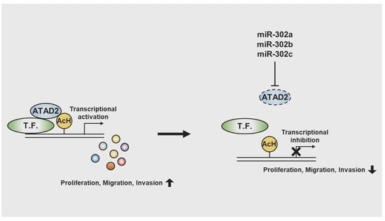
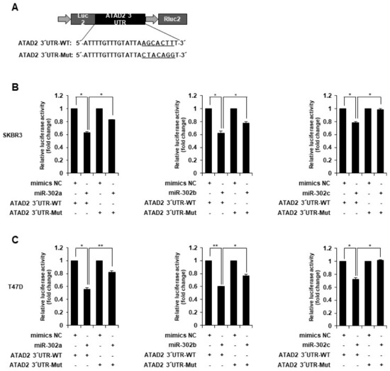
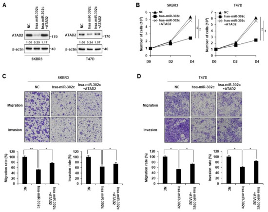
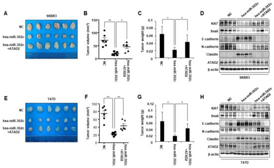
miR-302 Suppresses the Proliferation, Migration, and Invasion of Breast Cancer Cells by Downregulating ATAD2
by
Yo Sep Hwang, Eun Sun Park, Byung Moo Oh, Tae Gi Uhm, Suk Ran Yoon, Jong-Lyul Park, Hee Jun Cho and Hee Gu Lee
Cited by 3 | Viewed by 1778
Abstract
Breast cancer is the most common malignant tumor in women. The ATPase family AAA domain-containing protein 2 (ATAD2) contains an ATPase domain and a bromodomain, and is abnormally expressed in various human cancers, including breast cancer. However, the molecular mechanisms underlying the regulation
[...] Read more.
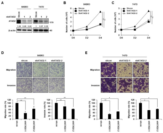
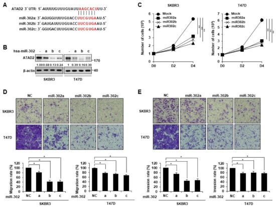
Breast cancer is the most common malignant tumor in women. The ATPase family AAA domain-containing protein 2 (ATAD2) contains an ATPase domain and a bromodomain, and is abnormally expressed in various human cancers, including breast cancer. However, the molecular mechanisms underlying the regulation of ATAD2 expression in breast cancer remain unclear. This study aimed to investigate the expression and function of ATAD2 in breast cancer. We found that ATAD2 was highly expressed in human breast cancer tissues and cell lines. ATAD2 depletion via RNA interference inhibited the proliferation, migration, and invasive ability of the SKBR3 and T47D breast cancer cell lines. Furthermore, Western blot analysis and luciferase assay results revealed that ATAD2 is a putative target of miR-302. Transfection with miR-302 mimics markedly reduced cell migration and invasion. These inhibitory effects of miR-302 were restored by ATAD2 overexpression. Moreover, miR-302 overexpression in SKBR3 and T47D cells suppressed tumor growth in the xenograft mouse model. However, ATAD2 overexpression rescued the decreased tumor growth seen after miR-302 overexpression. Our findings indicate that miR-302 plays a prominent role in inhibiting the cancer cell behavior associated with tumor progression by targeting ATAD2, and could thus be a valuable target for breast cancer therapy.
Full article
►▼
Show Figures
Open AccessReview
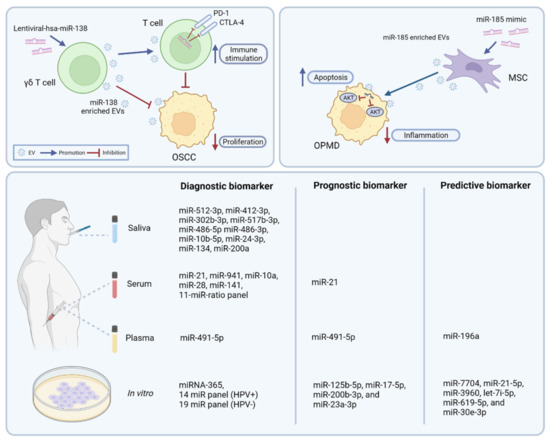
More than a Bubble: Extracellular Vesicle microRNAs in Head and Neck Squamous Cell Carcinoma
by
Wittaya Panvongsa, D. Michiel Pegtel and Jens Voortman
Cited by 13 | Viewed by 3059
Abstract
MicroRNAs (miRNAs) are a class of small non-coding RNA molecules that play a pivotal regulatory role in a broad variety of biological processes. Dysregulation of miRNAs is associated with several human diseases, particularly cancer. Extracellular vesicles (EVs) are crucial components in intercellular communication.
[...] Read more.
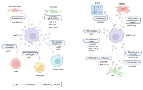
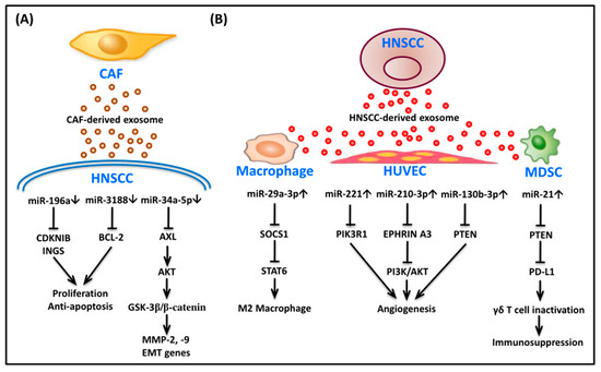
MicroRNAs (miRNAs) are a class of small non-coding RNA molecules that play a pivotal regulatory role in a broad variety of biological processes. Dysregulation of miRNAs is associated with several human diseases, particularly cancer. Extracellular vesicles (EVs) are crucial components in intercellular communication. As part of the cargo of EVs, miRNAs are involved in EV-mediated cell-to-cell interactions, including promotion or suppression of tumor development. The knowledge on the molecular mechanisms and clinical importance of EV-miRNAs in head and neck squamous cell carcinoma (HNSCC) has rapidly grown over the past years. In the present review, the current understanding regarding the effect of EV-miRNAs on HNSCC tumorigenesis is summarized, which includes effects on tumor proliferation, angiogenesis, invasion and metastasis, the tumor microenvironment, immune modulation, and treatment resistance. EV-miRNA-based biomarkers in liquid biopsies such as blood and saliva may open up new possibilities for employing EV-miRNAs for screening and early diagnostics as well as disease monitoring. Future perspectives include the promise of EV-miRNAs as a novel therapeutic target.
Full article
►▼
Show Figures
Open AccessArticle
Multiomics Topic Modeling for Breast Cancer Classification
by
Filippo Valle, Matteo Osella and Michele Caselle
Cited by 4 | Viewed by 2507
Abstract
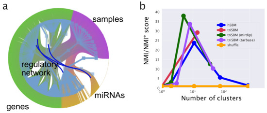
The integration of transcriptional data with other layers of information, such as the post-transcriptional regulation mediated by microRNAs, can be crucial to identify the driver genes and the subtypes of complex and heterogeneous diseases such as cancer. This paper presents an approach based
[...] Read more.
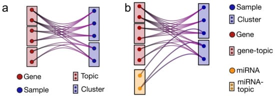
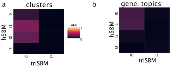
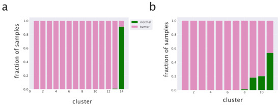
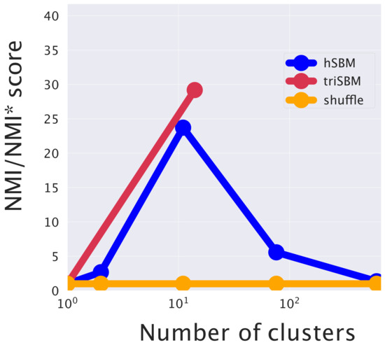
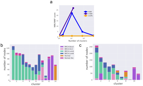
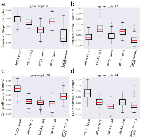
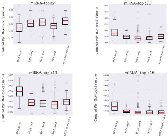
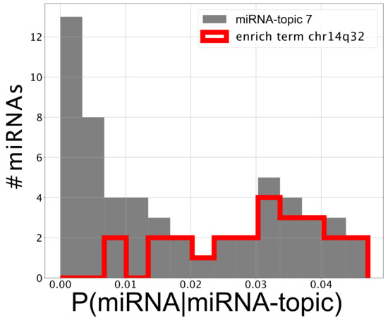
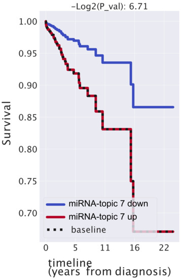
The integration of transcriptional data with other layers of information, such as the post-transcriptional regulation mediated by microRNAs, can be crucial to identify the driver genes and the subtypes of complex and heterogeneous diseases such as cancer. This paper presents an approach based on topic modeling to accomplish this integration task. More specifically, we show how an algorithm based on a hierarchical version of stochastic block modeling can be naturally extended to integrate any combination of ’omics data. We test this approach on breast cancer samples from the TCGA database, integrating data on messenger RNA, microRNAs, and copy number variations. We show that the inclusion of the microRNA layer significantly improves the accuracy of subtype classification. Moreover, some of the hidden structures or “topics” that the algorithm extracts actually correspond to genes and microRNAs involved in breast cancer development and are associated to the survival probability.
Full article
►▼
Show Figures
Open AccessReview
MicroRNA Profile Alterations in Parathyroid Carcinoma: Latest Updates and Perspectives
by
Marta Wielogórska, Beata Podgórska, Magdalena Niemira, Małgorzata Szelachowska, Adam Krętowski and Katarzyna Siewko
Cited by 3 | Viewed by 1936
Abstract
Parathyroid tumors are a genetically heterogenous group with a significant variability in clinical features. Due to a lack of specific signs and symptoms and uncertain histopathological criteria, parathyroid carcinomas (PCs) are challenging to diagnose, both before and after surgery. There is a great
[...] Read more.
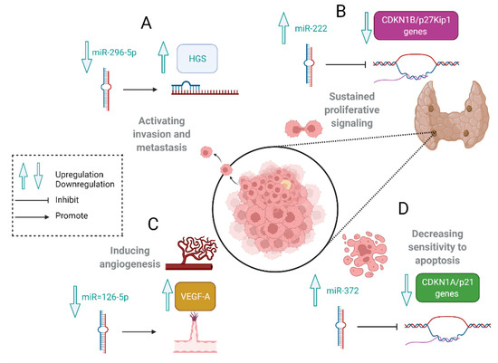
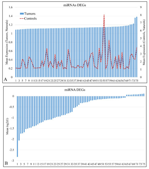
Parathyroid tumors are a genetically heterogenous group with a significant variability in clinical features. Due to a lack of specific signs and symptoms and uncertain histopathological criteria, parathyroid carcinomas (PCs) are challenging to diagnose, both before and after surgery. There is a great interest in searching for accurate molecular biomarkers for early detection, disease monitoring, and clinical management. Due to improvements in molecular pathology, the latest studies have reported that PC tumorigenesis is strongly linked to the epigenetic regulation of gene expression. MicroRNA (miRNA) profiling may serve as a helpful adjunct in distinguishing parathyroid adenoma (PAd) from PC and provide further insight into regulatory pathways involved in PTH release and parathyroid tumorigenesis. So far, only a few studies have attempted to show the miRNA signature for PC, and very few overlaps could be found between these relatively similar studies. A global miRNA downregulation was detected in PC compared with normal glands among differentially expressed miRNAs. This review summarizes changes in miRNA expression in PC and discusses the future research directions in this area.
Full article
►▼
Show Figures
Open AccessReview
MicroRNAs: Emerging Regulators of Metastatic Bone Disease in Breast Cancer
by
Marie-Therese Haider, Daniel J. Smit and Hanna Taipaleenmäki
Cited by 10 | Viewed by 3128
Abstract
Bone metastasis is a frequent complication in patients with advanced breast cancer. Once in the bone, cancer cells disrupt the tightly regulated cellular balance within the bone microenvironment, leading to excessive bone destruction and further tumor growth. Physiological and pathological interactions in the
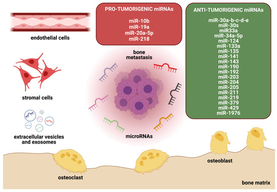
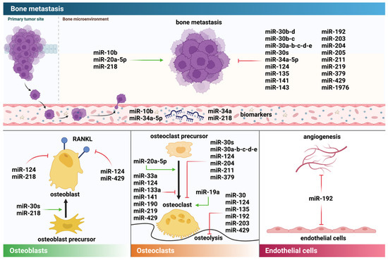
[...] Read more.
Bone metastasis is a frequent complication in patients with advanced breast cancer. Once in the bone, cancer cells disrupt the tightly regulated cellular balance within the bone microenvironment, leading to excessive bone destruction and further tumor growth. Physiological and pathological interactions in the bone marrow are mediated by cell–cell contacts and secreted molecules that include soluble proteins as well as RNA molecules. MicroRNAs (miRNAs) are short non-coding RNAs that post-transcriptionally interfere with their target messenger RNA (mRNA) and subsequently reduce protein abundance. Since their discovery, miRNAs have been identified as critical regulators of physiological and pathological processes, including breast cancer and associated metastatic bone disease. Depending on their targets, miRNAs can exhibit pro-tumorigenic or anti-tumorigenic functions and serve as diagnostic and prognostic biomarkers. These properties have encouraged pre-clinical and clinical development programs to investigate miRNAs as biomarkers and therapeutic targets in various diseases, including metastatic cancers. In this review, we discuss the role of miRNAs in metastatic bone disease with a focus on breast cancer and the bone microenvironment and elaborate on their potential use for diagnostic and therapeutic purposes in metastatic bone disease and beyond.
Full article
►▼
Show Figures
Open AccessArticle
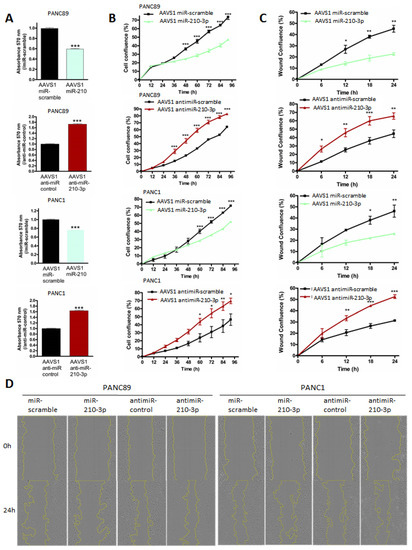
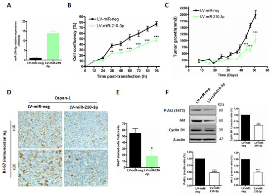
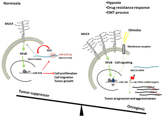
Antagonistic Roles of the Tumor Suppressor miR-210-3p and Oncomucin MUC4 Forming a Negative Feedback Loop in Pancreatic Adenocarcinoma
by
Nihad Boukrout, Mouloud Souidi, Fatima Lahdaoui, Belinda Duchêne, Bernadette Neve, Lucie Coppin, Emmanuelle Leteurtre, Jérôme Torrisani, Isabelle Van Seuningen and Nicolas Jonckheere
Cited by 2 | Viewed by 2627
Abstract
Background: Pancreatic adenocarcinoma (PDAC) is a deadly cancer with an extremely poor prognosis. MUC4 membrane-bound mucin is neoexpressed in early pancreatic neoplastic lesions and is associated with PDAC progression and chemoresistance. In cancers, microRNAs (miRNAs, small noncoding RNAs) are crucial regulators of carcinogenesis,
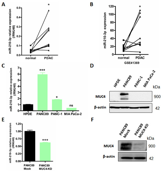
[...] Read more.
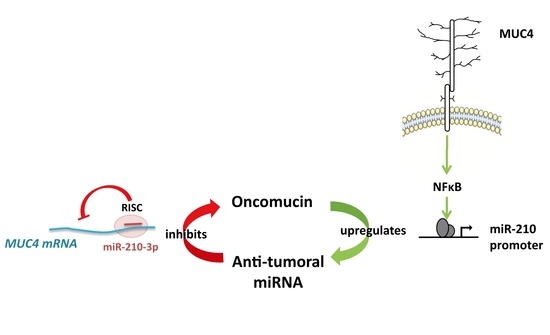
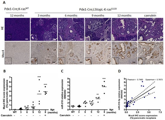
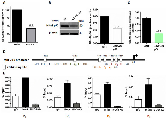
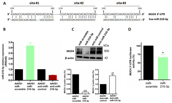
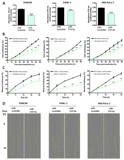
Background: Pancreatic adenocarcinoma (PDAC) is a deadly cancer with an extremely poor prognosis. MUC4 membrane-bound mucin is neoexpressed in early pancreatic neoplastic lesions and is associated with PDAC progression and chemoresistance. In cancers, microRNAs (miRNAs, small noncoding RNAs) are crucial regulators of carcinogenesis, chemotherapy response and even metastatic processes. In this study, we aimed at identifying and characterizing miRNAs activated downstream of MUC4-associated signaling in pancreatic adenocarcinoma. MiRnome analysis comparing MUC4-KD versus Mock cancer cells showed that MUC4 inhibition impaired miR-210-3p expression. Therefore, we aimed to better understand the miR-210-3p biological roles. Methods: miR-210-3p expression level was analyzed by RT-qPCR in PDAC-derived cell lines (PANC89 Mock and MUC4-KD, PANC-1 and MiaPACA-2), as well as in mice and patients tissues. The MUC4-miR-210-3p regulation was investigated using luciferase reporter construct and chromatin immunoprecipitation experiments. Stable cell lines expressing miR-210-3p or anti-miR-210-3p were established using CRISPR/Cas9 technology or lentiviral transduction. We evaluated the biological activity of miR-210-3p in vitro by measuring cell proliferation and migration and in vivo using a model of subcutaneous xenograft. Results: miR-210-3p expression is correlated with MUC4 expression in PDAC-derived cells and human samples, and in pancreatic PanIN lesions of Pdx1-Cre; LstopL-KrasG12D mice. MUC4 enhances miR-210-3p expression levels via alteration of the NF-κB signaling pathway. Chromatin immunoprecipitation experiments showed p50 NF-κB subunit binding on miR-210-3p promoter regions. We established a reciprocal regulation since miR-210-3p repressed MUC4 expression via its 3′-UTR. MiR-210-3p transient transfection of PANC89, PANC-1 and MiaPACA-2 cells led to a decrease in cell proliferation and migration. These biological effects were validated in cells overexpressing or knocked-down for miR-210-3p. Finally, we showed that miR-210-3p inhibits pancreatic tumor growth and proliferation in vivo. Conclusion: We identified a MUC4-miR-210-3p negative feedback loop in early-onset PDAC, but also revealed new functions of miR-210-3p in both in vitro and in vivo proliferation and migration of pancreatic cancer cells, suggesting a complex balance between MUC4 pro-oncogenic roles and miR-210-3p anti-tumoral effects.
Full article
►▼
Show Figures
Open AccessReview
miRNAs in the Regulation of Cancer Immune Response: Effect of miRNAs on Cancer Immunotherapy
by
Faheem Hyder Pottoo, Ashif Iqubal, Mohammad Kashif Iqubal, Mohammed Salahuddin, Jawad Ur Rahman, Noora AlHajri and Mustafa Shehadeh
Cited by 6 | Viewed by 2652
Abstract
In the last few decades, carcinogenesis has been extensively explored and substantial research has identified immunogenic involvement in various types of cancers. As a result, immune checkpoint blockers and other immune-based therapies were developed as novel immunotherapeutic strategies. However, despite being a promising
[...] Read more.
In the last few decades, carcinogenesis has been extensively explored and substantial research has identified immunogenic involvement in various types of cancers. As a result, immune checkpoint blockers and other immune-based therapies were developed as novel immunotherapeutic strategies. However, despite being a promising therapeutic option, immunotherapy has significant constraints such as a high cost of treatment, unpredictable toxicity, and clinical outcomes. miRNAs are non-coding, small RNAs actively involved in modulating the immune system’s multiple signalling pathways by binding to the 3′-UTR of target genes. miRNAs possess a unique advantage in modulating multiple targets of either the same or different signalling pathways. Therefore, miRNA follows a ‘one drug multiple target’ hypothesis. Attempts are made to explore the therapeutic promise of miRNAs in cancer so that it can be transported from bench to bedside for successful immunotherapeutic results. Therefore, in the current manuscript, we discussed, in detail, the mechanism and role of miRNAs in different types of cancers relating to the immune system, its diagnostic and therapeutic aspect, the effect on immune escape, immune-checkpoint molecules, and the tumour microenvironment. We have also discussed the existing limitations, clinical success and the prospective use of miRNAs in cancer.
Full article
►▼
Show Figures
Open AccessReview
MicroRNAs: Their Role in Metabolism, Tumor Microenvironment, and Therapeutic Implications in Head and Neck Squamous Cell Carcinoma
by
Shine-Gwo Shiah, Sung-Tau Chou and Jang-Yang Chang
Cited by 11 | Viewed by 2529
Abstract
MicroRNAs (miRNAs) are endogenous small non-coding RNA molecules that negatively regulate gene expression by binding to target mRNAs. Deregulated miRNAs can act as either oncogenic miRNAs or tumor suppressor miRNAs in controlling proliferation, differentiation, apoptosis, metastasis, epithelial–mesenchymal transition, and immune responses, which are
[...] Read more.
MicroRNAs (miRNAs) are endogenous small non-coding RNA molecules that negatively regulate gene expression by binding to target mRNAs. Deregulated miRNAs can act as either oncogenic miRNAs or tumor suppressor miRNAs in controlling proliferation, differentiation, apoptosis, metastasis, epithelial–mesenchymal transition, and immune responses, which are all involved in the carcinogenesis process of HNSCC. Recent findings have shown that metabolic reprogramming is an important hallmark of cancer, which is necessary for malignant transformation and tumor development. Some reprogrammed metabolisms are believed to be required for HNSCC against an unfavorable tumor microenvironment (TME). The TME is composed of various cell types embedded in the altered extracellular matrix, among which exosomes, secreted by cancer cells, are one of the most important factors. Tumor-derived exosomes reshape the tumor microenvironment and play a crucial role in cell-to-cell communication during HNSCC development. Exosomes encapsulate many biomolecules, including miRNAs, circulate in body fluids, and can transmit intercellular regulatory messages to nearby and distant sites, which indicates that exosomal miRNAs have the potential to become non-invasive biomarkers. This review aims to clarify the functions of diverse miRNAs in HNSCC metabolic reprogramming and tumor-derived exosomes. In addition, it also emphasizes the potential role of miRNA as a biomarker in the diagnosis, prognosis, and treatment of HNSCC cancer.
Full article
►▼
Show Figures
Open AccessArticle
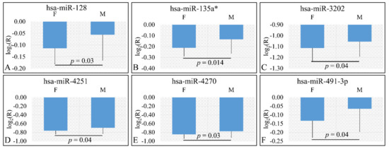
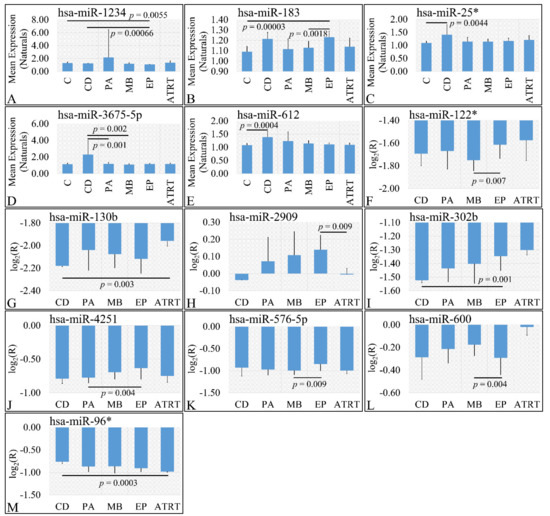
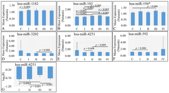
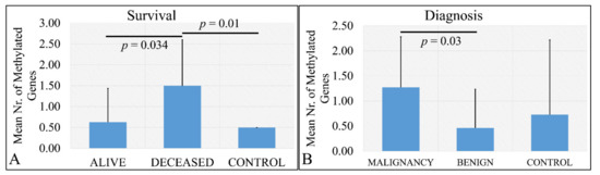
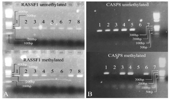
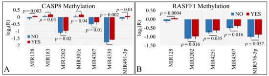
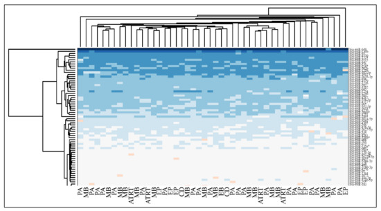
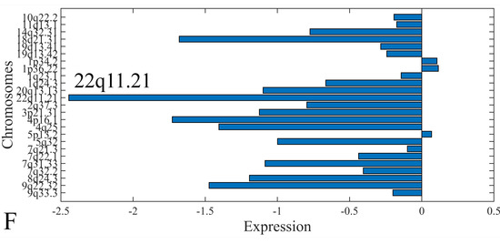
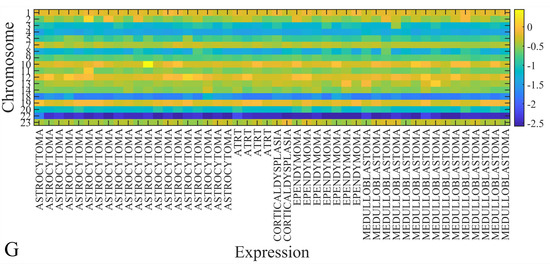
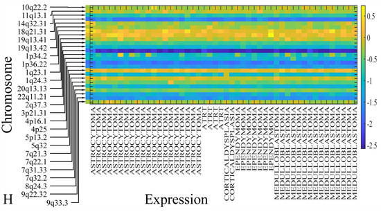
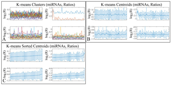
Differential and Common Signatures of miRNA Expression and Methylation in Childhood Central Nervous System Malignancies: An Experimental and Computational Approach
by
George I. Lambrou, Myrto Poulou, Krinio Giannikou, Marios Themistocleous, Apostolos Zaravinos and Maria Braoudaki
Viewed by 2274
Abstract
Epigenetic modifications are considered of utmost significance for tumor ontogenesis and progression. Especially, it has been found that miRNA expression, as well as DNA methylation plays a significant role in central nervous system tumors during childhood. A total of 49 resected brain tumors
[...] Read more.
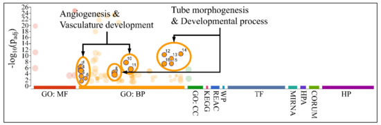
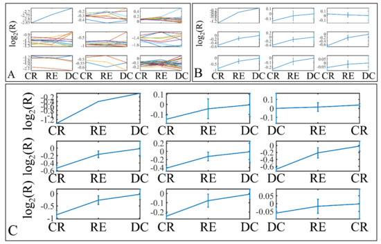
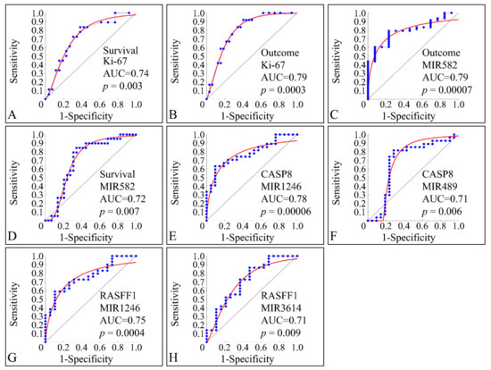
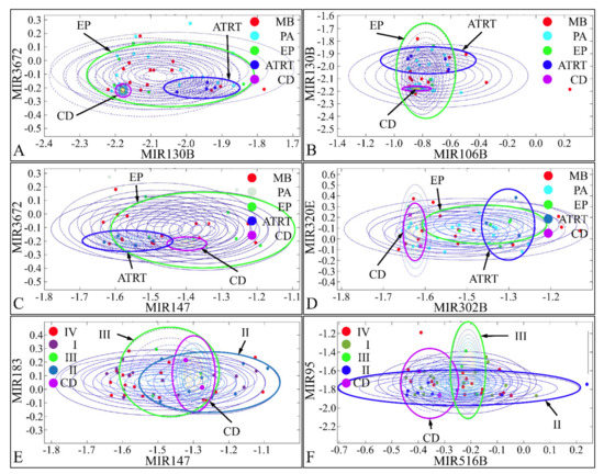
Epigenetic modifications are considered of utmost significance for tumor ontogenesis and progression. Especially, it has been found that miRNA expression, as well as DNA methylation plays a significant role in central nervous system tumors during childhood. A total of 49 resected brain tumors from children were used for further analysis. DNA methylation was identified with methylation-specific MLPA and, in particular, for the tumor suppressor genes CASP8, RASSF1, MGMT, MSH6, GATA5, ATM1, TP53, and CADM1. miRNAs were identified with microarray screening, as well as selected samples, were tested for their mRNA expression levels. CASP8, RASSF1 were the most frequently methylated genes in all tumor samples. Simultaneous methylation of genes manifested significant results with respect to tumor staging, tumor type, and the differentiation of tumor and control samples. There was no significant dependence observed with the methylation of one gene promoter, rather with the simultaneous presence of all detected methylated genes’ promoters. miRNA expression was found to be correlated to gene methylation. Epigenetic regulation appears to be of major importance in tumor progression and pathophysiology, making it an imperative field of study.
Full article
►▼
Show Figures
Open AccessReview
Functional and Clinical Significance of Dysregulated microRNAs in Liver Cancer
by
Po-Shuan Huang, Chia-Jung Liao, Ya-Hui Huang, Chau-Ting Yeh, Cheng-Yi Chen, Hui-Chi Tang, Cheng-Chih Chang and Kwang-Huei Lin
Cited by 8 | Viewed by 2555
Abstract
Liver cancer is the leading cause of cancer-related mortality in the world. This mainly reflects the lack of early diagnosis tools and effective treatment methods. MicroRNAs (miRNAs) are a class of non-transcribed RNAs, some of which play important regulatory roles in liver cancer.
[...] Read more.
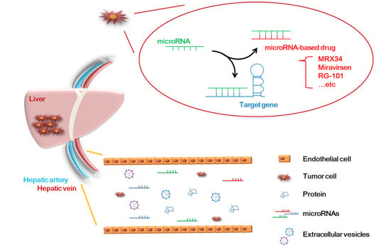
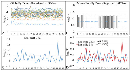
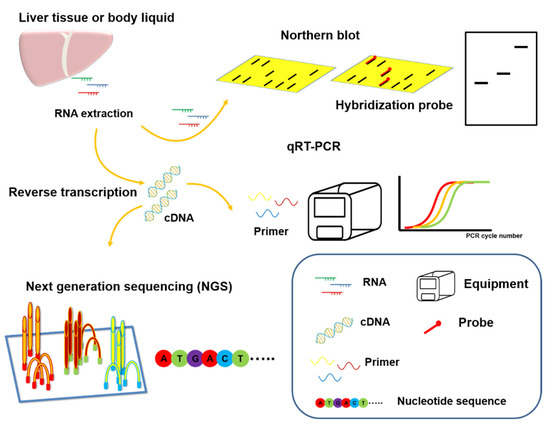
Liver cancer is the leading cause of cancer-related mortality in the world. This mainly reflects the lack of early diagnosis tools and effective treatment methods. MicroRNAs (miRNAs) are a class of non-transcribed RNAs, some of which play important regulatory roles in liver cancer. Here, we discuss microRNAs with key impacts on liver cancer, such as miR-122, miR-21, miR-214, and miR-199. These microRNAs participate in various physiological regulatory pathways of liver cancer cells, and their modulation can have non-negligible effects in the treatment of liver cancer. We discuss whether these microRNAs can be used for better clinical diagnosis and/or drug development. With the advent of novel technologies, fast, inexpensive, and non-invasive RNA-based biomarker research has become a new mainstream approach. However, the clinical application of microRNA-based markers has been limited by the high sequence similarity among them and the potential for off-target problems. Therefore, researchers particularly value microRNAs that are specific to or have special functions in liver cancer. These include miR-122, which is specifically expressed in the liver, and miR-34, which is necessary for the replication of the hepatitis C virus in liver cancer. Clinical treatment drugs have been developed based on miR-34 and miR-122 (MRX34 and Miravirsen, respectively), but their side effects have not yet been overcome. Future research is needed to address these weaknesses and establish a feasible microRNA-based treatment strategy for liver cancer.
Full article
►▼
Show Figures
Open AccessReview
MicroRNAs in Molecular Classification and Pathogenesis of Breast Tumors
by
Vinitha Richard, Matthew G. Davey, Heidi Annuk, Nicola Miller, Róisín M. Dwyer, Aoife Lowery and Michael J. Kerin
Cited by 21 | Viewed by 2755
Abstract
The current clinical practice of breast tumor classification relies on the routine immunohistochemistry-based expression analysis of hormone receptors, which is inadequate in addressing breast tumor heterogeneity and drug resistance. MicroRNA expression profiling in tumor tissue and in the circulation is an efficient alternative
[...] Read more.
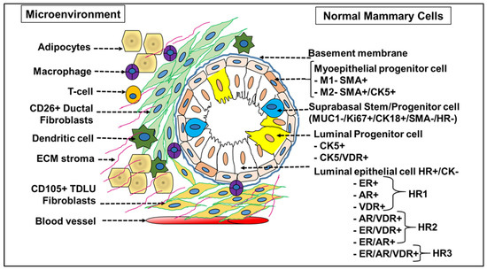
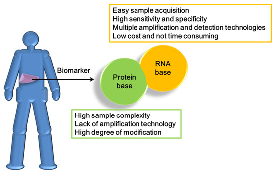
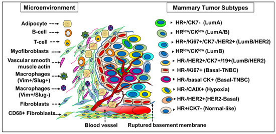
The current clinical practice of breast tumor classification relies on the routine immunohistochemistry-based expression analysis of hormone receptors, which is inadequate in addressing breast tumor heterogeneity and drug resistance. MicroRNA expression profiling in tumor tissue and in the circulation is an efficient alternative to intrinsic molecular subtyping that enables precise molecular classification of breast tumor variants, the prediction of tumor progression, risk stratification and also identifies critical regulators of the tumor microenvironment. This review integrates data from protein, gene and miRNA expression studies to elaborate on a unique miRNA-based 10-subtype taxonomy, which we propose as the current gold standard to allow appropriate classification and separation of breast cancer into a targetable strategy for therapy.
Full article
►▼
Show Figures
Open AccessArticle
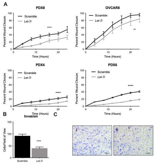
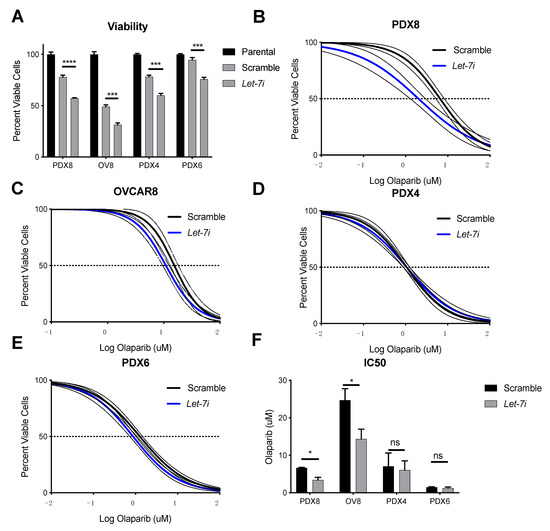
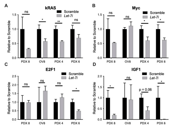
Let-7i Reduces Aggressive Phenotype and Induces BRCAness in Ovarian Cancer Cells
by
Evgeny Chirshev, Tise Suzuki, Hanmin Wang, Anthony Nguyen, Nozomi Hojo, Linda Sanderman, Saied Mirshahidi, Yevgeniya J. Ioffe and Juli J. Unternaehrer
Cited by 6 | Viewed by 2299
Abstract
High-grade serous carcinoma of the ovary is a deadly gynecological cancer with poor long-term survival. Dysregulation of microRNAs has been shown to contribute to the formation of cancer stem cells (CSCs), an important part of oncogenesis and tumor progression. The
let-7 family of
[...] Read more.
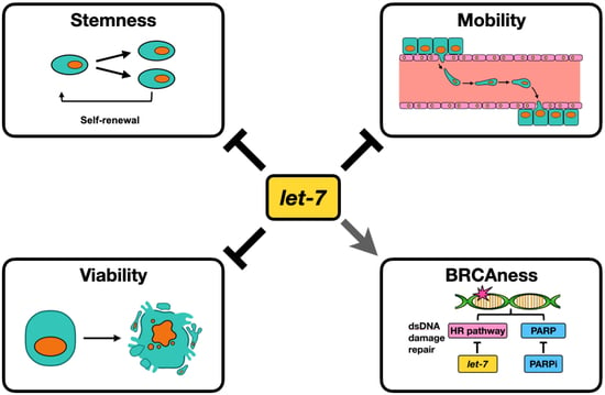
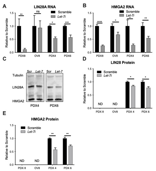
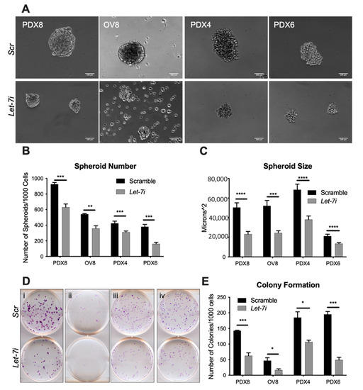
High-grade serous carcinoma of the ovary is a deadly gynecological cancer with poor long-term survival. Dysregulation of microRNAs has been shown to contribute to the formation of cancer stem cells (CSCs), an important part of oncogenesis and tumor progression. The
let-7 family of microRNAs has previously been shown to regulate stemness and has tumor suppressive actions in a variety of cancers, including ovarian. Here, we demonstrate tumor suppressor actions of
let-7i: repression of cancer cell stemness, inhibition of migration and invasion, and promotion of apoptosis, features important for cancer progression, relapse, and metastasis.
Let-7i over-expression results in increased sensitivity to the PARP inhibitor olaparib in samples without BRCA mutations, consistent with induction of
BRCAness phenotype. We also show that
let-7i inhibits the expression of several factors involved in the homologous recombination repair (HRR) pathway, providing potential mechanisms by which the
BRCAness phenotype could be induced. These actions of
let-7i add to the rationale for use of this miRNA as a treatment for ovarian cancer patients, including those without mutations in the HRR pathway.
Full article
►▼
Show Figures
Open AccessArticle
Human Prostate Tissue MicroRNAs and Their Predicted Target Pathways Linked to Prostate Cancer Risk Factors
by
Max Enwald, Terho Lehtimäki, Pashupati P. Mishra, Nina Mononen, Teemu J. Murtola and Emma Raitoharju
Cited by 2 | Viewed by 1888
Abstract
MicroRNAs are important in prostate cancer development, progression and metastasis. The aim of this study was to test microRNA expression profile in prostate tissue obtained from prostate cancer patients for associations with various prostate cancer related factors and to pinpoint the predicted target
[...] Read more.
MicroRNAs are important in prostate cancer development, progression and metastasis. The aim of this study was to test microRNA expression profile in prostate tissue obtained from prostate cancer patients for associations with various prostate cancer related factors and to pinpoint the predicted target pathways for these microRNAs. Prostate tissue samples were obtained at prostatectomy from patients participating in a trial evaluating impact of pre-operative atorvastatin on serum prostate specific antigen (PSA) and Ki-67 expression in prostate tissue. Prostate tissue microRNA expression profiles were analyzed using OpenArray
® MicroRNA Panel. Pathway enrichment analyses were conducted for predicted target genes of microRNAs that correlated significantly with studied factors. Eight microRNAs correlated significantly with studied factors of patients after Bonferroni multiple testing correction. MiR-485-3p correlated with serum HDL-cholesterol levels. In atorvastatin-treated subjects, miR-34c-5p correlated with a change in serum PSA and miR-138-3p with a change in total cholesterol. In the placebo arm, both miR-576-3p and miR-550-3p correlated with HDL-cholesterol and miR-627 with PSA. In pathway analysis, these eight microRNAs related significantly to several pathways relevant to prostate cancer. This study brings new evidence from the expression of prostate tissue microRNAs and related pathways that may link risk factors to prostate cancer and pinpoint new therapeutic possibilities.
Full article
Open AccessReview
Epitranscriptomics: A New Layer of microRNA Regulation in Cancer
by
Veronica De Paolis, Elisa Lorefice, Elisa Orecchini, Claudia Carissimi, Ilaria Laudadio and Valerio Fulci
Cited by 17 | Viewed by 4017
Abstract
MicroRNAs are pervasive regulators of gene expression at the post-transcriptional level in metazoan, playing key roles in several physiological and pathological processes. Accordingly, these small non-coding RNAs are also involved in cancer development and progression. Furthermore, miRNAs represent valuable diagnostic and prognostic biomarkers
[...] Read more.
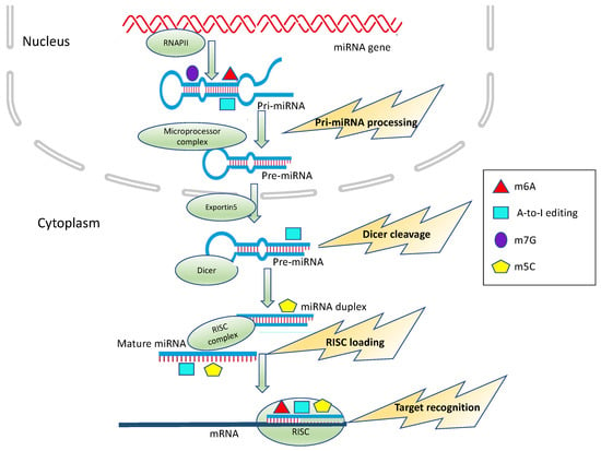
MicroRNAs are pervasive regulators of gene expression at the post-transcriptional level in metazoan, playing key roles in several physiological and pathological processes. Accordingly, these small non-coding RNAs are also involved in cancer development and progression. Furthermore, miRNAs represent valuable diagnostic and prognostic biomarkers in malignancies. In the last twenty years, the role of RNA modifications in fine-tuning gene expressions at several levels has been unraveled. All RNA species may undergo post-transcriptional modifications, collectively referred to as epitranscriptomic modifications, which, in many instances, affect RNA molecule properties. miRNAs are not an exception, in this respect, and they have been shown to undergo several post-transcriptional modifications. In this review, we will summarize the recent findings concerning miRNA epitranscriptomic modifications, focusing on their potential role in cancer development and progression.
Full article
►▼
Show Figures
Open AccessFeature PaperReview
Mutual Correlation between Non-Coding RNA and S-Adenosylmethionine in Human Cancer: Roles and Therapeutic Opportunities
by
Laura Mosca, Francesca Vitiello, Luigi Borzacchiello, Alessandra Coppola, Roberta Veglia Tranchese, Martina Pagano, Michele Caraglia, Giovanna Cacciapuoti and Marina Porcelli
Cited by 9 | Viewed by 2077
Abstract
Epigenetics includes modifications in DNA methylation, histone and chromatin structure, and expression of non-coding RNAs (ncRNAs), especially microRNAs (miRNAs) and long non-coding RNAs (lncRNAs). Knowledge of the relationships between S-adenosylmethionine (AdoMet or SAM), the universal methyl donor for all epigenetic methylation reactions and
[...] Read more.
Epigenetics includes modifications in DNA methylation, histone and chromatin structure, and expression of non-coding RNAs (ncRNAs), especially microRNAs (miRNAs) and long non-coding RNAs (lncRNAs). Knowledge of the relationships between S-adenosylmethionine (AdoMet or SAM), the universal methyl donor for all epigenetic methylation reactions and miRNAs or lncRNAs in human cancer may provide helpful insights for the development of new end more effective anticancer therapeutic approaches. In recent literature, a complex network of mutual interconnections between AdoMet and miRNAs or lncRNAs has been reported and discussed. Indeed, ncRNAs expression may be regulated by epigenetic mechanisms such as DNA and RNA methylation and histone modifications. On the other hand, miRNAs or lncRNAs may influence the epigenetic apparatus by modulating the expression of its enzymatic components at the post-transcriptional level. Understanding epigenetic mechanisms, such as dysregulation of miRNAs/lncRNAs and DNA methylation, has become of central importance in modern research. This review summarizes the recent findings on the mechanisms by which AdoMet and miRNA/lncRNA exert their bioactivity, providing new insights to develop innovative and more efficient anticancer strategies based on the interactions between these epigenetic modulators.
Full article
►▼
Show Figures
Open AccessReview
The Role of MicroRNAs in Lung Cancer Metabolism
by
Mohamed Iman Hidayat Nor Azizi, Iekhsan Othman and Rakesh Naidu
Cited by 15 | Viewed by 3314
Abstract
MicroRNAs (miRNAs) are short-strand non-coding RNAs that are responsible for post-transcriptional regulation of many biological processes. Their differential expression is important in supporting tumorigenesis by causing dysregulation in normal biological functions including cell proliferation, apoptosis, metastasis and invasion and cellular metabolism. Cellular metabolic
[...] Read more.
MicroRNAs (miRNAs) are short-strand non-coding RNAs that are responsible for post-transcriptional regulation of many biological processes. Their differential expression is important in supporting tumorigenesis by causing dysregulation in normal biological functions including cell proliferation, apoptosis, metastasis and invasion and cellular metabolism. Cellular metabolic processes are a tightly regulated mechanism. However, cancer cells have adapted features to circumvent these regulations, recognizing metabolic reprogramming as an important hallmark of cancer. The miRNA expression profile may differ between localized lung cancers, advanced lung cancers and solid tumors, which lead to a varying extent of metabolic deregulation. Emerging evidence has shown the relationship between the differential expression of miRNAs with lung cancer metabolic reprogramming in perpetuating tumorigenesis. This review provides an insight into the role of different miRNAs in lung cancer metabolic reprogramming by targeting key enzymes, transporter proteins or regulatory components alongside metabolic signaling pathways. These discussions would allow a deeper understanding of the importance of miRNAs in tumor progression therefore providing new avenues for diagnostic, therapeutic and disease management applications.
Full article
►▼
Show Figures


