


Brain Sci. 2023, 13(7), 1080; https://doi.org/10.3390/brainsci13071080 - 17 Jul 2023
Viewed by 1184
Abstract
►
Show Figures
(1) Introduction: Symptom spectrum can be of great diversity and heterogeneity in mitochondrial encephalomyopathy, lactic acidosis, and stroke-like episodes (MELAS) patients in clinical practice. Here, we report a case of MELAS presenting asymptomatic refractory hypotension with m.3243 A>G mutation. (2) Case representation: A
[...] Read more.
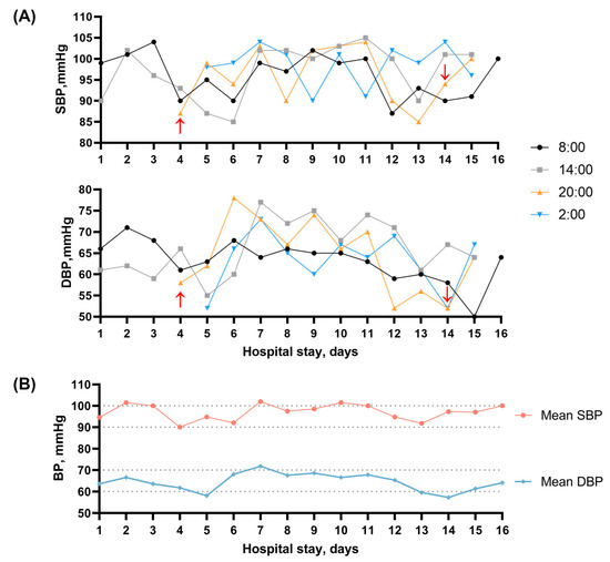
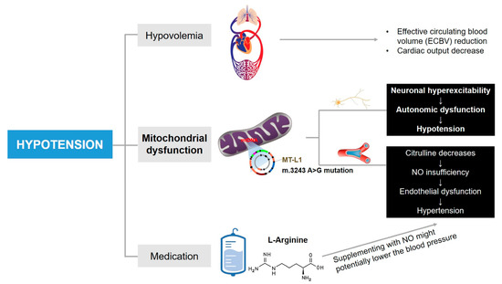
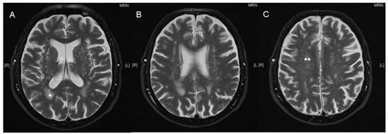
(1) Introduction: Symptom spectrum can be of great diversity and heterogeneity in mitochondrial encephalomyopathy, lactic acidosis, and stroke-like episodes (MELAS) patients in clinical practice. Here, we report a case of MELAS presenting asymptomatic refractory hypotension with m.3243 A>G mutation. (2) Case representation: A 51-year-old male patient presented with a headache, vertigo, and difficulty in expression and understanding. The magnetic resonance imaging of the brain revealed an acute stroke-like lesion involving the left temporoparietal lobe. A definitive diagnosis of MELAS was given after the genetic test identified the chrM-3243 A>G mutation. The patient suffered recurrent stroke-like episodes in the 1-year follow-up. Notably, refractory hypotension was observed during hospitalizations, and no significant improvement in blood pressure was found after continuous use of vasopressor drugs and fluid infusion therapy. (3) Conclusions: We report a case of refractory hypotension which was unresponsive to fluid infusion therapy found in a patient with MELAS. Our case suggests that comprehensive management should be paid attention to during treatment. A further study on the pathological mechanism of the multisystem symptoms in MELAS would be beneficial to the treatment of patients.
Full article
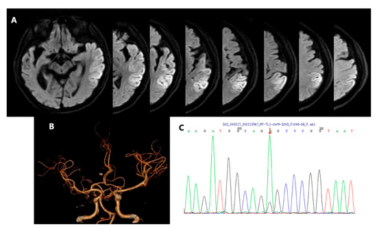
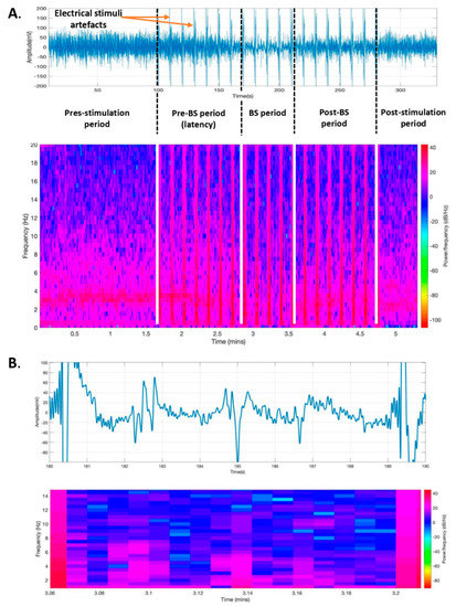
Figure 1

