Nonmotor Symptoms in Parkinson's Disease (PD)
Share This Topical Collection
Editor
Topical Collection Information
Dear Colleagues,
Parkinson’s Disease (PD) is classically defined as a movement disorder, but PD is increasingly being acknowledged as a multidimensional disease involving progressive motor and nonmotor symptoms that negatively impact quality of life. Mild cognitive impairment, depression, anxiety, and disordered sleep are common nonmotor symptoms in PD, and frequently dominate prodromal disease stages. This Topical Collection of Brain Sciences will examine the relationships between nonmotor symptoms in PD and their impact on quality of life. This Topical Collection will also publish original research reporting novel therapeutic interventions for ameliorating nonmotor symptoms, with specific interest given to the neuroprotective potential of interventions at the prodromal stages of PD. We welcome original research and review articles that provide evidence to increase our current understanding of (and interventions for) nonmotor symptoms in PD and provide a direction for future research in this domain. We also encourage submissions from established and early-career researchers from a range of disciplines to ensure this collection adopts a multidisciplinary approach to understanding nonmotor symptoms in PD.
Dr. Andrea Loftus
Dr. Blake Lawrence
Collection Editors
Manuscript Submission Information
Manuscripts should be submitted online at www.mdpi.com by registering and logging in to this website. Once you are registered, click here to go to the submission form. Manuscripts can be submitted until the deadline. All submissions that pass pre-check are peer-reviewed. Accepted papers will be published continuously in the journal (as soon as accepted) and will be listed together on the collection website. Research articles, review articles as well as short communications are invited. For planned papers, a title and short abstract (about 100 words) can be sent to the Editorial Office for announcement on this website.
Submitted manuscripts should not have been published previously, nor be under consideration for publication elsewhere (except conference proceedings papers). All manuscripts are thoroughly refereed through a single-blind peer-review process. A guide for authors and other relevant information for submission of manuscripts is available on the Instructions for Authors page. Brain Sciences is an international peer-reviewed open access monthly journal published by MDPI.
Please visit the Instructions for Authors page before submitting a manuscript.
The Article Processing Charge (APC) for publication in this open access journal is 2200 CHF (Swiss Francs).
Submitted papers should be well formatted and use good English. Authors may use MDPI's
English editing service prior to publication or during author revisions.
Keywords
- Parkinson’s Disease
- nonmotor symptoms
- mild cognitive impairment
- depression
- anxiety
- sleep disorders
- quality of life
Published Papers (2 papers)
2023
Open AccessArticle
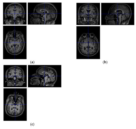
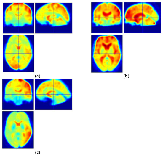
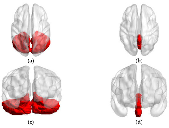
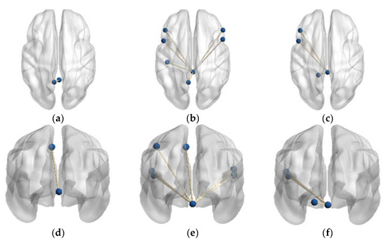
Differences and Changes in Cerebellar Functional Connectivity of Parkinson’s Patients with Visual Hallucinations
by
Liangcheng Qu, Chuan Liu, Yiting Cao, Jingping Shi, Kuiying Yin and Weiguo Liu
Viewed by 959
Abstract
Recent studies have discovered that functional connections are impaired in patients with Parkinson’s disease (PD) accompanied by hallucinations (PD-H), even at the preclinical stage. The cerebellum has been implicated in playing a role in cognitive processes. However, the functional connectivity (FC) between the
[...] Read more.
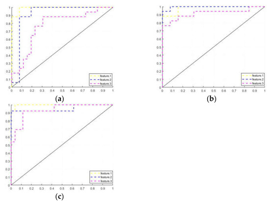
Recent studies have discovered that functional connections are impaired in patients with Parkinson’s disease (PD) accompanied by hallucinations (PD-H), even at the preclinical stage. The cerebellum has been implicated in playing a role in cognitive processes. However, the functional connectivity (FC) between the cognitive sub-regions of the cerebellum in PD patients with hallucinations needs further clarification. Resting-state functional magnetic resonance imaging (rs-fMRI) data were collected from three groups (17 PD-H patients, 13 patients with Parkinson’s disease not accompanied by hallucinations (PD-NH), and 26 healthy controls (HC)). The data were collected in this study to investigate the impact of cerebellar FC changes on cognitive performance. Additionally, we define cerebellar FC as a training feature for classifying all subjects using Support Vector Machines (SVMs). We found that in the PD-H patients, there was an increase in FC within the left side of the precuneus (PCUN) compared to the HC. Additionally, there was an increase in FC within the bilateral opercular part of the inferior frontal gyrus (IFGoprec) and triangular part of the inferior frontal gyrus (IFCtriang), as well as the left side of the postcentral gyrus (PoCG), inferior parietal lobe (IPL), and PCUN compared to the PD-NH patients. In the machine learning training results, cerebellar FC has also been proven to be an effective biomarker feature, achieving a recognition rate of over 90% for PD-H. These findings indicate that the cortico-cerebellar FC in PD-H and PD-NH patients was significantly disrupted, with different patterns of distribution. The proposed pipeline offers a promising, low-cost alternative for diagnosing preclinical PD-H and may also be beneficial for other degenerative brain disorders.
Full article
►▼
Show Figures
Open AccessArticle
Detecting Subtle Cognitive Impairment in Patients with Parkinson’s Disease and Normal Cognition: A Novel Cognitive Control Challenge Task (C3T)
by
Karmen Resnik Robida, Vida Ana Politakis, Aleš Oblak, Anka Slana Ozimič, Helena Burger, Zvezdan Pirtošek and Jurij Bon
Viewed by 1384
Abstract
Patients with Parkinson’s disease (PD) often show early deficits in cognitive control, with primary difficulties in flexibility and relatively intact stable representations. The aim of our study was to assess executive function using an ecologically valid approach that combines measures of stability and
[...] Read more.
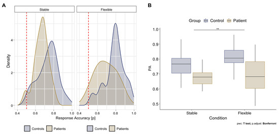
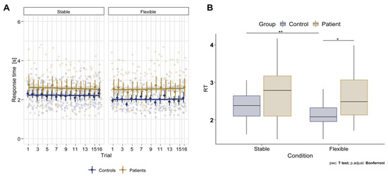
Patients with Parkinson’s disease (PD) often show early deficits in cognitive control, with primary difficulties in flexibility and relatively intact stable representations. The aim of our study was to assess executive function using an ecologically valid approach that combines measures of stability and flexibility. Fourteen patients without cognitive deficits and sixteen comparable control subjects completed a standardized neuropsychological test battery and a newly developed cognitive control challenge task (C3T). We found that the accuracy of C3T performance decreased with age in healthy participants and remained impaired in PD patients regardless of age. In addition, PD patients showed significantly lower overall performance for cognitive control tasks than healthy controls, even when they scored in the normal range on standardized neuropsychological tests. PD Patients responded significantly faster than healthy control subjects regarding flexible cognitive control tasks due to their impulsivity. Correlations showed that the C3T task targets multiple cognitive systems, including working memory, inhibition, and task switching, providing a reliable measure of complex cognitive control. C3T could be a valuable tool for characterizing cognitive deficits associated with PD and appears to be a more sensitive measure than standardized neuropsychological tests. A different assessment approach could potentially detect early signs of the disease and identify opportunities for early intervention with neuroprotective therapies.
Full article
►▼
Show Figures


