


Biomolecules 2023, 13(10), 1445; https://doi.org/10.3390/biom13101445 - 26 Sep 2023
Viewed by 1247
Abstract
►
Show Figures
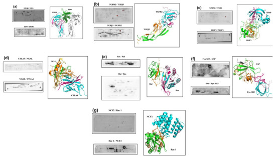
Dynein motors facilitate the majority of minus-end-directed transport events on microtubules. The dynein adaptor Bicaudal D2 (BicD2) recruits the dynein machinery to several cellular cargo for transport, including Nup358, which facilitates a nuclear positioning pathway that is essential for the differentiation of distinct
[...] Read more.
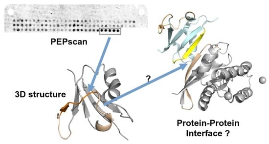
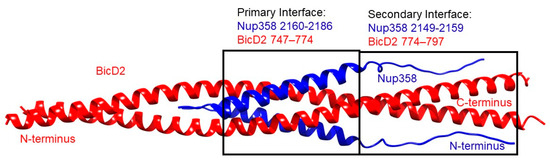
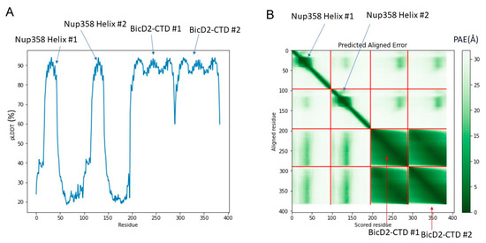
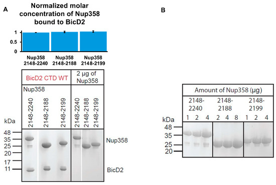
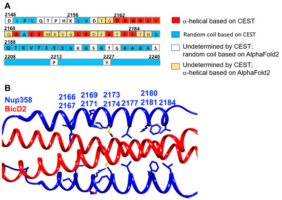
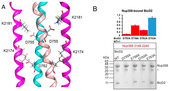
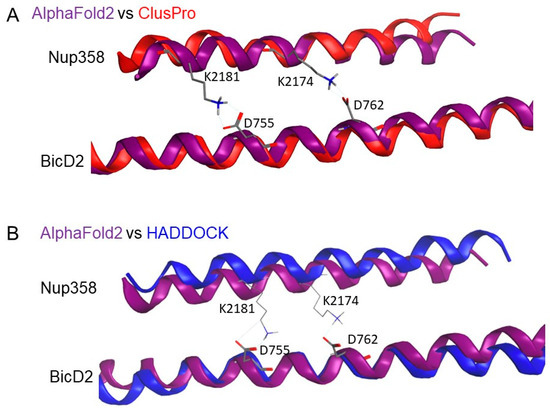
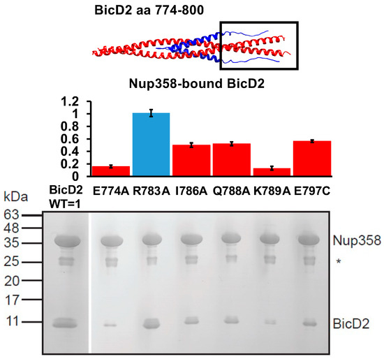
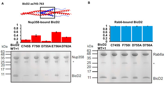
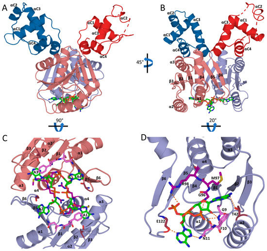
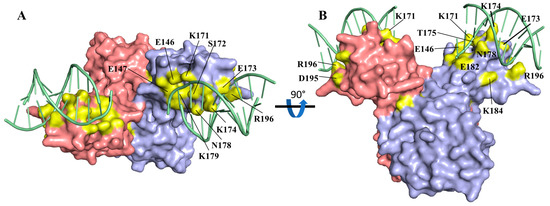
Dynein motors facilitate the majority of minus-end-directed transport events on microtubules. The dynein adaptor Bicaudal D2 (BicD2) recruits the dynein machinery to several cellular cargo for transport, including Nup358, which facilitates a nuclear positioning pathway that is essential for the differentiation of distinct brain progenitor cells. Previously, we showed that Nup358 forms a “cargo recognition α-helix” upon binding to BicD2; however, the specifics of the BicD2-Nup358 interface are still not well understood. Here, we used AlphaFold2, complemented by two additional docking programs (HADDOCK and ClusPro) as well as mutagenesis, to show that the Nup358 cargo-recognition α-helix binds to BicD2 between residues 747 and 774 in an anti-parallel manner, forming a helical bundle. We identified two intermolecular salt bridges that are important to stabilize the interface. In addition, we uncovered a secondary interface mediated by an intrinsically disordered region of Nup358 that is directly N-terminal to the cargo-recognition α-helix and binds to BicD2 between residues 774 and 800. This is the same BicD2 domain that binds to the competing cargo adapter Rab6, which is important for the transport of Golgi-derived and secretory vesicles. Our results establish a structural basis for cargo recognition and selection by the dynein adapter BicD2, which facilitates transport pathways that are important for brain development.
Full article
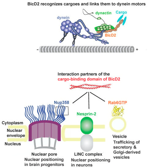
Figure 1

