Feature Papers in Biochemistry
Share This Topical Collection
Editor
 Dr. Eugene A. Permyakov
Dr. Eugene A. Permyakov
E-Mail
Website
Collection Editor
Institute for Biological Instrumentation, Russian Academy of Sciences, 142290 Pushchino, Russia
Interests: protein physics; luminescence (fluorescence, phosphorescence) spectroscopy of proteins; scanning and titration calorimetry of proteins; metal binding proteins; calcium binding proteins
Special Issues, Collections and Topics in MDPI journals
Topical Collection Information
Dear Colleagues,
This Topical Collection, “Feature Papers in Biochemistry”, aims to collect high-quality research articles, review articles, and communications on all aspects of biochemistry. It is dedicated to recent advances in the research area of biochemistry, and comprises a selection of exclusive papers from the Editorial Board Members (EBMs) of the Biochemistry Section, as well as invited papers from relevant experts. We also welcome senior experts in the field to make contributions to this Topical Collection. Kindly note that all invited papers will be published online free of charge once accepted. We aim to represent our Section as an attractive open-access publishing platform for biochemistry research.
Dr. Eugene A. Permyakov
Collection Editor
Manuscript Submission Information
Manuscripts should be submitted online at www.mdpi.com by registering and logging in to this website. Once you are registered, click here to go to the submission form. Manuscripts can be submitted until the deadline. All submissions that pass pre-check are peer-reviewed. Accepted papers will be published continuously in the journal (as soon as accepted) and will be listed together on the collection website. Research articles, review articles as well as short communications are invited. For planned papers, a title and short abstract (about 100 words) can be sent to the Editorial Office for announcement on this website.
Submitted manuscripts should not have been published previously, nor be under consideration for publication elsewhere (except conference proceedings papers). All manuscripts are thoroughly refereed through a single-blind peer-review process. A guide for authors and other relevant information for submission of manuscripts is available on the Instructions for Authors page. Biomolecules is an international peer-reviewed open access monthly journal published by MDPI.
Please visit the Instructions for Authors page before submitting a manuscript.
The Article Processing Charge (APC) for publication in this open access journal is 2700 CHF (Swiss Francs).
Submitted papers should be well formatted and use good English. Authors may use MDPI's
English editing service prior to publication or during author revisions.
Published Papers (32 papers)
Open AccessArticle
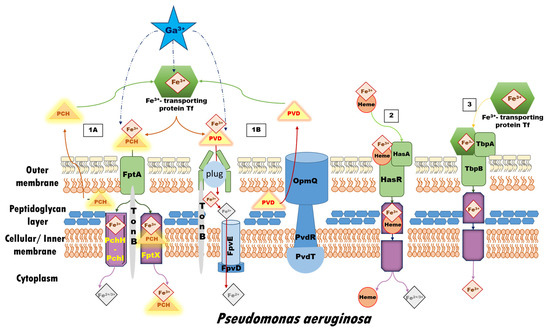
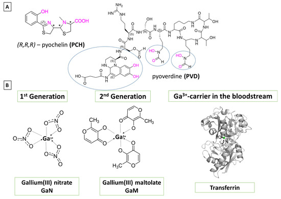
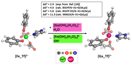
In Silico Analysis of the Ga3+/Fe3+ Competition for Binding the Iron-Scavenging Siderophores of P. aeruginosa—Implementation of Three Gallium-Based Complexes in the “Trojan Horse” Antibacterial Strategy
by
Nikoleta Kircheva, Stefan Dobrev, Vladislava Petkova, Lyubima Yocheva, Silvia Angelova and Todor Dudev
Viewed by 324
Abstract
The emergence of multidrug-resistant (MDR) microorganisms combined with the ever-draining antibiotic pipeline poses a disturbing and immensely growing public health challenge that requires a multidisciplinary approach and the application of novel therapies aimed at unconventional targets and/or applying innovative drug formulations. Hence, bacterial
[...] Read more.
The emergence of multidrug-resistant (MDR) microorganisms combined with the ever-draining antibiotic pipeline poses a disturbing and immensely growing public health challenge that requires a multidisciplinary approach and the application of novel therapies aimed at unconventional targets and/or applying innovative drug formulations. Hence, bacterial iron acquisition systems and bacterial Fe
2+/3+-containing enzymes have been identified as a plausible target of great potential. The intriguing “Trojan horse” approach deprives microorganisms from the essential iron. Recently, gallium’s potential in medicine as an iron mimicry species has attracted vast attention. Different Ga
3+ formulations exhibit diverse effects upon entering the cell and thus supposedly have multiple targets. The aim of the current study is to specifically distinguish characteristics of great significance in regard to the initial gallium-based complex, allowing the alien cation to effectively compete with the native ferric ion for binding the siderophores pyochelin and pyoverdine secreted by the bacterium
P. aeruginosa. Therefore, three gallium-based formulations were taken into consideration: the first-generation gallium nitrate, Ga(NO
3)
3, metabolized to Ga
3+-hydrated forms, the second-generation gallium maltolate (tris(3-hydroxy-2-methyl-4-pyronato)gallium), and the experimentally proven Ga carrier in the bloodstream—the protein transferrin. We employed a reliable in silico approach based on DFT computations in order to understand the underlying biochemical processes that govern the Ga
3+/Fe
3+ rivalry for binding the two bacterial siderophores.
Full article
►▼
Show Figures
Open AccessEditor’s ChoiceArticle
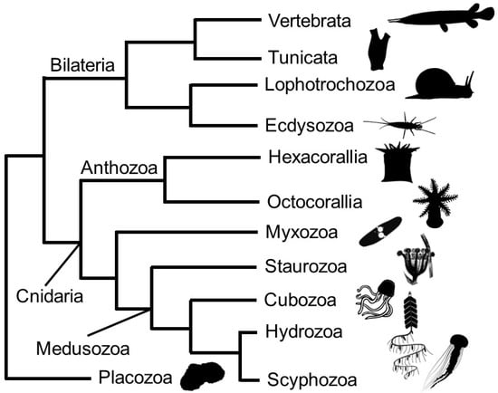
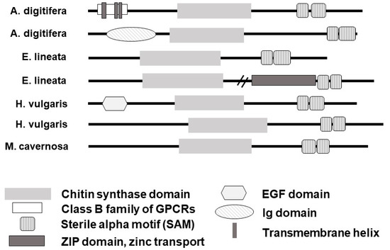
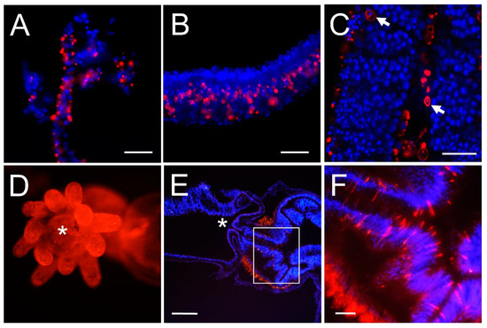
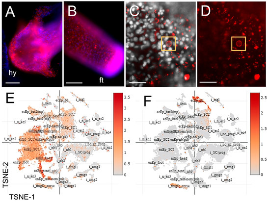
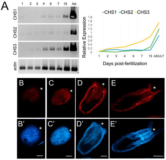
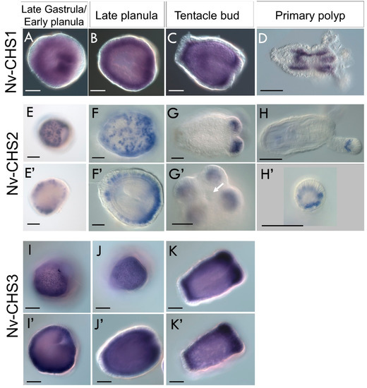
Unexpected Distribution of Chitin and Chitin Synthase across Soft-Bodied Cnidarians
by
Lauren E. Vandepas, Michael G. Tassia, Kenneth M. Halanych and Chris T. Amemiya
Cited by 2 | Viewed by 1988
Abstract
Cnidarians are commonly recognized as sea jellies, corals, or complex colonies such as the Portuguese man-of-war. While some cnidarians possess rigid internal calcareous skeletons (e.g., corals), many are soft-bodied. Intriguingly, genes coding for the chitin-biosynthetic enzyme,
chitin synthase (CHS)
, were recently identified
[...] Read more.
Cnidarians are commonly recognized as sea jellies, corals, or complex colonies such as the Portuguese man-of-war. While some cnidarians possess rigid internal calcareous skeletons (e.g., corals), many are soft-bodied. Intriguingly, genes coding for the chitin-biosynthetic enzyme,
chitin synthase (CHS)
, were recently identified in the model anemone
Nematostella vectensis, a species lacking hard structures. Here we report the prevalence and diversity of CHS across Cnidaria and show that cnidarian
chitin synthase genes display diverse protein domain organizations. We found that
CHS is expressed in cnidarian species and/or developmental stages with no reported chitinous or rigid morphological structures. Chitin affinity histochemistry indicates that chitin is present in soft tissues of some scyphozoan and hydrozoan medusae. To further elucidate the biology of chitin in cnidarian soft tissues, we focused on
CHS expression in
N. vectensis. Spatial expression data show that three
CHS orthologs are differentially expressed in
Nematostella embryos and larvae during development, suggesting that chitin has an integral role in the biology of this species. Understanding how a non-bilaterian lineage such as Cnidaria employs chitin may provide new insight into hitherto unknown functions of polysaccharides in animals, as well as their role in the evolution of biological novelty.
Full article
►▼
Show Figures
Open AccessFeature PaperReview
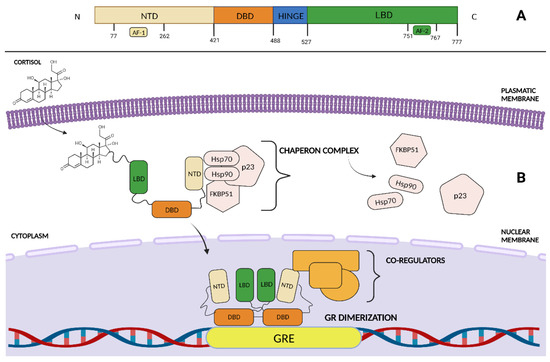
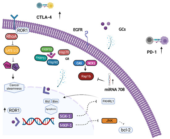
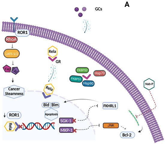
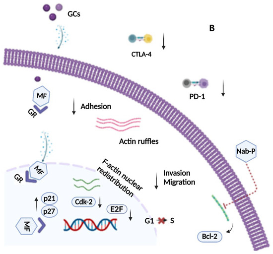
Glucocorticoid Receptor and Ovarian Cancer: From Biology to Therapeutic Intervention
by
Roberto Buonaiuto, Giuseppe Neola, Sabrina Chiara Cecere, Aldo Caltavituro, Amedeo Cefaliello, Erica Pietroluongo, Pietro De Placido, Mario Giuliano, Grazia Arpino and Carmine De Angelis
Viewed by 2010
Abstract
Ovarian cancer (OC) is the leading cause of death from gynecological malignancies worldwide. Fortunately, recent advances in OC biology and the discovery of novel therapeutic targets have led to the development of novel therapeutic agents that may improve the outcome of OC patients.
[...] Read more.
Ovarian cancer (OC) is the leading cause of death from gynecological malignancies worldwide. Fortunately, recent advances in OC biology and the discovery of novel therapeutic targets have led to the development of novel therapeutic agents that may improve the outcome of OC patients. The glucocorticoid receptor (GR) is a ligand-dependent transcriptional factor known for its role in body stress reactions, energy homeostasis and immune regulation. Notably, evidence suggests that GR may play a relevant role in tumor progression and may affect treatment response. In cell culture models, administration of low levels of glucocorticoids (GCs) suppresses OC growth and metastasis. Conversely, high GR expression has been associated with poor prognostic features and long-term outcomes in patients with OC. Moreover, both preclinical and clinical data have shown that GR activation impairs the effectiveness of chemotherapy by inducing the apoptotic pathways and cell differentiation. In this narrative review, we summarize data related to the function and role of GR in OC. To this aim, we reorganized the controversial and fragmented data regarding GR activity in OC and herein describe its potential use as a prognostic and predictive biomarker. Moreover, we explored the interplay between GR and BRCA expression and reviewed the latest therapeutic strategies such as non-selective GR antagonists and selective GR modulators to enhance chemotherapy sensitivity, and to finally provide new treatment options in OC patients.
Full article
►▼
Show Figures
Open AccessFeature PaperArticle
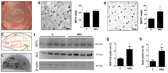
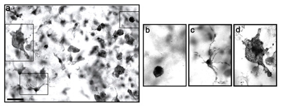
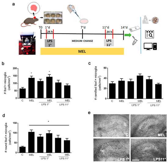
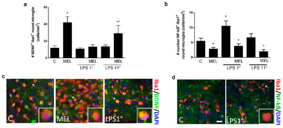
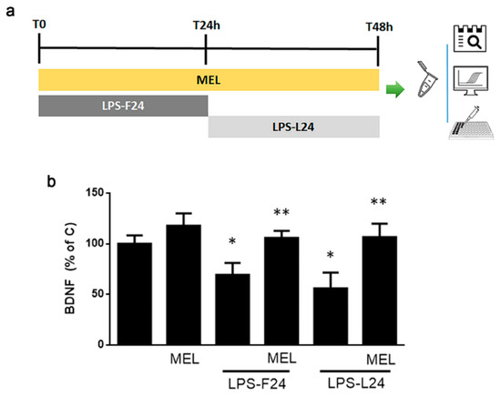
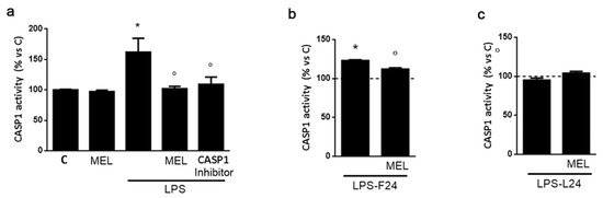
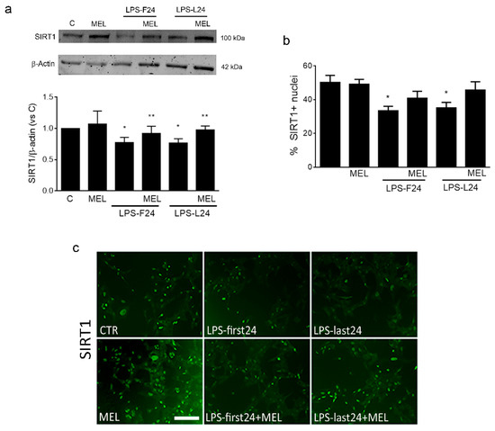
Melatonin Activates Anti-Inflammatory Features in Microglia in a Multicellular Context: Evidence from Organotypic Brain Slices and HMC3 Cells
by
Sara Merlo, Grazia Ilaria Caruso, Dhwani Sunil Korde, Alla Khodorovska, Christian Humpel and Maria Angela Sortino
Cited by 2 | Viewed by 1926
Abstract
Melatonin (MEL) is a neurohormone endowed with neuroprotective activity, exerted both directly on neuronal cells and indirectly through modulation of responsive glial cells. In particular, MEL’s effects on microglia are receptor-mediated and in part dependent on SIRT1 activation. In the present study, we
[...] Read more.
Melatonin (MEL) is a neurohormone endowed with neuroprotective activity, exerted both directly on neuronal cells and indirectly through modulation of responsive glial cells. In particular, MEL’s effects on microglia are receptor-mediated and in part dependent on SIRT1 activation. In the present study, we exploited the highly preserved cytoarchitecture of organotypic brain cultures (OC) to explore the effects of MEL on hippocampal microglia in a 3D context as compared to a single cell type context represented by the human HMC3 cell line. We first evaluated the expression of MEL receptor MT1 and SIRT1 and then investigated MEL action against an inflammatory stimulation with LPS: OCs were cultured for a total of 2 weeks and during this time exposed to 0.1 μg/mL of LPS for 24 h either on day 1 (LPS 1°) or on day 11 (LPS 11°). MEL was added immediately after plating and kept for the entire experiment. Under these conditions, both MEL and LPS induced amoeboid microglia. However, the same round phenotype matched different polarization features. LPS increased the number of nuclear-NF-kB+ round cells and MEL alone or in combination with LPS increased BDNF+ round microglia. In addition, MEL contrasted LPS effects on NF-kB expression. Data from HMC3 microglia confirmed MEL’s anti-inflammatory effects against LPS in terms of CASP1 induction and BDNF release, identifying SIRT1 as a mediator. However, no effects were evident for MEL alone on HMC3 microglia. Overall, our results point to the importance of the multicellular context for full MEL activity, especially in a preventive view, and support the use of OCs as a favorable model to explore inflammatory responses.
Full article
►▼
Show Figures
Open AccessArticle
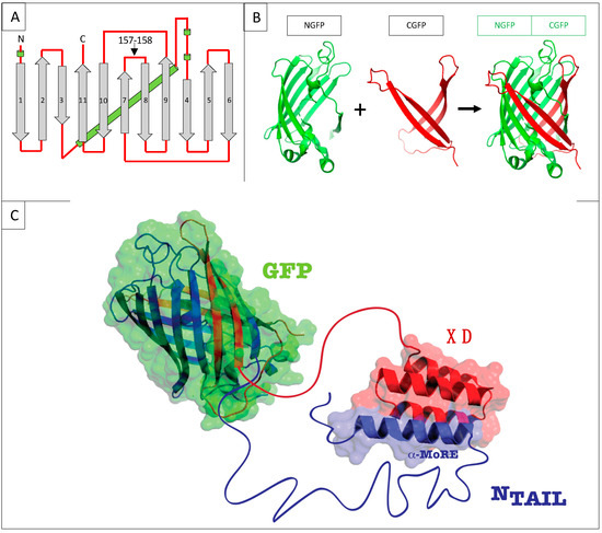
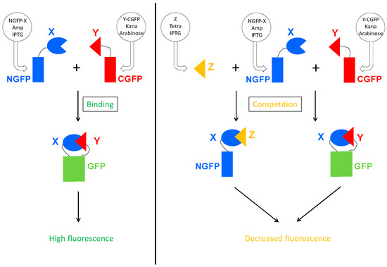
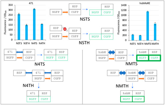
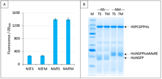
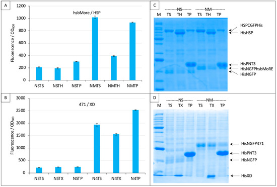
In Vivo Protein–Protein Binding Competition Assay Based on Split-GFP Reassembly: Proof of Concept
by
Christophe Bignon and Sonia Longhi
Cited by 1 | Viewed by 2091
Abstract
The split-green fluorescent protein (GFP) reassembly assay is a well-established approach to study protein–protein interactions (PPIs). In this assay, when two interacting proteins X and Y, respectively fused to residues 1–157 and to residues 158–237 of GFP, are co-expressed in
E. coli,
[...] Read more.
The split-green fluorescent protein (GFP) reassembly assay is a well-established approach to study protein–protein interactions (PPIs). In this assay, when two interacting proteins X and Y, respectively fused to residues 1–157 and to residues 158–237 of GFP, are co-expressed in
E. coli, the two GFP halves are brought to sufficient proximity to reassociate and fold to recreate the functional GFP. At constant protein expression level, the intensity of fluorescence produced by the bacteria is proportional to the binding affinity of X to Y. We hypothesized that adding a third partner (Z) endowed with an affinity for either X or Y would lead to an in vivo competition assay. We report here the different steps of the set-up of this competition assay, and define the experimental conditions required to obtained reliable results. Results show that this competition assay is a potentially interesting tool for screening libraries of binding inhibitors, Z being either a protein or a chemical reagent.
Full article
►▼
Show Figures
Open AccessArticle
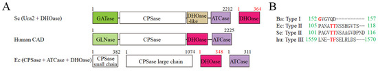
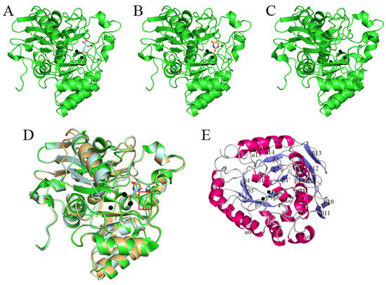
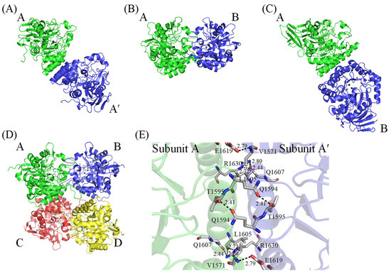
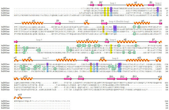
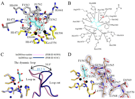
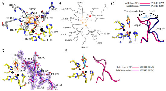
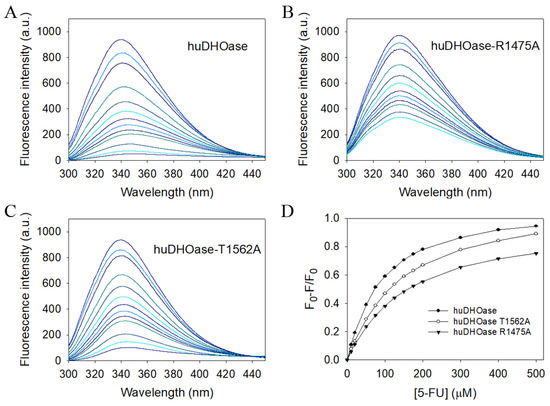
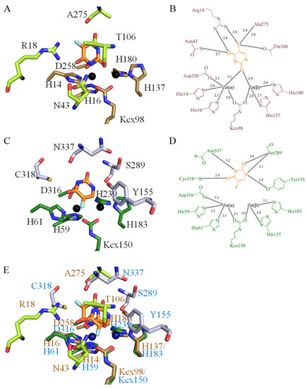
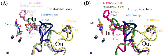
Complexed Crystal Structure of the Dihydroorotase Domain of Human CAD Protein with the Anticancer Drug 5-Fluorouracil
by
En-Shyh Lin, Yen-Hua Huang, Po-Chun Yang, Wei-Feng Peng and Cheng-Yang Huang
Cited by 4 | Viewed by 1756
Abstract
Dihydroorotase (DHOase) is the third enzyme in the pathway used for the biosynthesis of pyrimidine nucleotides. In mammals, DHOase is active in a trifunctional enzyme, CAD, which also carries out the activities of carbamoyl phosphate synthetase and aspartate transcarbamoylase. Prior to this study,
[...] Read more.
Dihydroorotase (DHOase) is the third enzyme in the pathway used for the biosynthesis of pyrimidine nucleotides. In mammals, DHOase is active in a trifunctional enzyme, CAD, which also carries out the activities of carbamoyl phosphate synthetase and aspartate transcarbamoylase. Prior to this study, it was unknown whether the FDA-approved clinical drug 5-fluorouracil (5-FU), which is used as an anticancer therapy, could bind to the DHOase domain of human CAD (huDHOase). Here, we identified huDHOase as a new 5-FU binding protein, thereby extending the 5-FU interactome to this human enzyme. In order to investigate where 5-FU binds to huDHOase, we solved the complexed crystal structure at 1.97 Å (PDB ID 8GVZ). The structure of huDHOase complexed with malate was also determined for the sake of comparison (PDB ID 8GW0). These two nonsubstrate ligands were bound at the active site of huDHOase. It was previously established that the substrate
N-carbamoyl-L-aspartate is either bound to or moves away from the active site, but it is the loop that is extended towards (loop-in mode) or moved away (loop-out mode) from the active site. DHOase also binds to nonsubstrate ligands via the loop-out mode. In contrast to the
Escherichia coli DHOase model, our complexed structures revealed that huDHOase binds to either 5-FU or malate via the loop-in mode. We further characterized the binding of 5-FU to huDHOase using site-directed mutagenesis and the fluorescence quenching method. Considering the loop-in mode, the dynamic loop in huDHOase should be a suitable drug-targeting site for further designing inhibitors and clinical chemotherapies to suppress pyrimidine biosynthesis in cancer cell lines.
Full article
►▼
Show Figures
Open AccessArticle
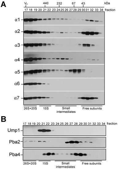
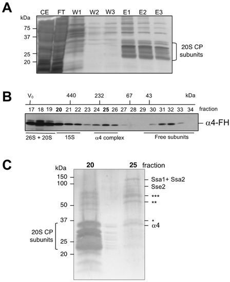
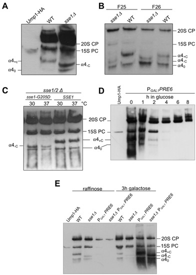
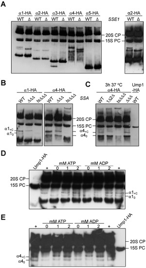
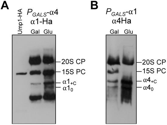
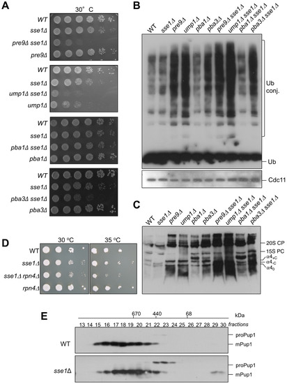
Hsp70 and Hsp110 Chaperones Promote Early Steps of Proteasome Assembly
by
Ana C. Matias, Joao Matos, R. Jürgen Dohmen and Paula C. Ramos
Cited by 2 | Viewed by 1422
Abstract
Whereas assembly of the 20S proteasome core particle (CP) in prokaryotes apparently occurs spontaneously, the efficiency of this process in eukaryotes relies on the dedicated assembly chaperones Ump1, Pba1-Pba2, and Pba3-Pba4. For mammals, it was reported that CP assembly initiates with formation of
[...] Read more.
Whereas assembly of the 20S proteasome core particle (CP) in prokaryotes apparently occurs spontaneously, the efficiency of this process in eukaryotes relies on the dedicated assembly chaperones Ump1, Pba1-Pba2, and Pba3-Pba4. For mammals, it was reported that CP assembly initiates with formation of a complete α-ring that functions as a template for β subunit incorporation. By contrast, we were not able to detect a ring composed only of a complete set of α subunits in
S. cerevisiae. Instead, we found that the CP subunits α1, α2, and α4 each form independent small complexes. Purification of such complexes containing α4 revealed the presence of chaperones of the Hsp70/Ssa and Hsp110/Sse families. Consistently, certain small complexes containing α1, α2, and α4 were not formed in strains lacking these chaperones. Deletion of the
SSE1 gene in combination with deletions of
PRE9 (α3),
PBA3, or
UMP1 genes resulted in severe synthetic growth defects, high levels of ubiquitin-conjugates, and an accumulation of distinct small complexes with α subunits. Our study shows that Hsp70 and Hsp110 chaperones cooperate to promote the folding of individual α subunits and/or their assembly with other CP subunits, Ump1, and Pba1-Pba4 in subsequent steps.
Full article
►▼
Show Figures
Open AccessReview
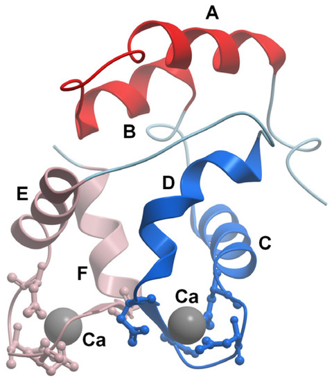
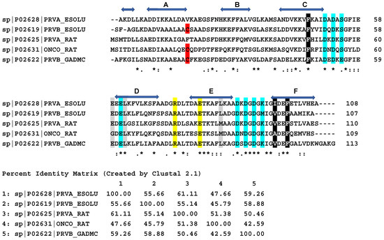
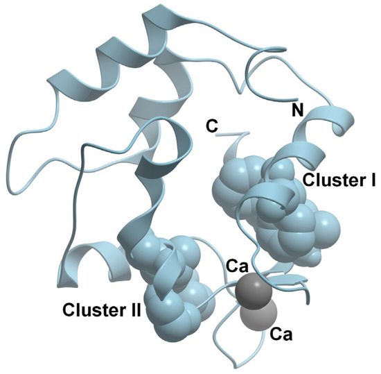
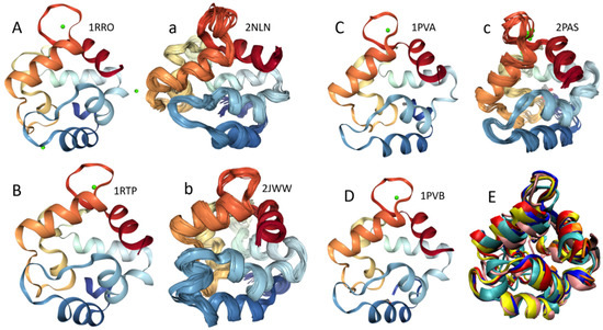
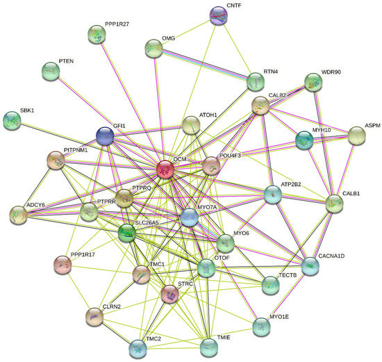
What Is Parvalbumin for?
by
Eugene A. Permyakov and Vladimir N. Uversky
Cited by 13 | Viewed by 4854
Abstract
Parvalbumin (PA) is a small, acidic, mostly cytosolic Ca
2+-binding protein of the EF-hand superfamily. Structural and physical properties of PA are well studied but recently two highly conserved structural motifs consisting of three amino acids each (clusters I and II), which
[...] Read more.
Parvalbumin (PA) is a small, acidic, mostly cytosolic Ca
2+-binding protein of the EF-hand superfamily. Structural and physical properties of PA are well studied but recently two highly conserved structural motifs consisting of three amino acids each (clusters I and II), which contribute to the hydrophobic core of the EF-hand domains, have been revealed. Despite several decades of studies, physiological functions of PA are still poorly known. Since no target proteins have been revealed for PA so far, it is believed that PA acts as a slow calcium buffer. Numerous experiments on various muscle systems have shown that PA accelerates the relaxation of fast skeletal muscles. It has been found that oxidation of PA by reactive oxygen species (ROS) is conformation-dependent and one more physiological function of PA in fast muscles could be a protection of these cells from ROS. PA is thought to regulate calcium-dependent metabolic and electric processes within the population of gamma-aminobutyric acid (GABA) neurons. Genetic elimination of PA results in changes in GABAergic synaptic transmission. Mammalian oncomodulin (OM), the β isoform of PA, is expressed mostly in cochlear outer hair cells and in vestibular hair cells. OM knockout mice lose their hearing after 3–4 months. It was suggested that, in sensory cells, OM maintains auditory function, most likely affecting outer hair cells’ motility mechanisms.
Full article
►▼
Show Figures
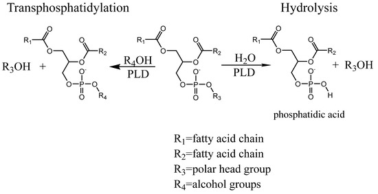
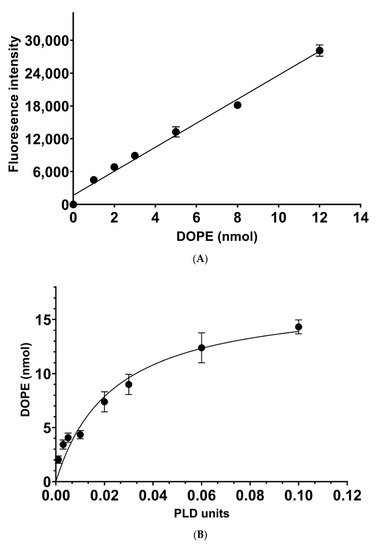
Open AccessFeature PaperArticle
A Novel High-Throughput Assay Reveals That the Temperature Induced Increases in Transphosphatidylation of Phospholipase D Are Dependent on the Alcohol Acceptor Concentration
by
Hengzhang Yang and Rüdiger Woscholski
Cited by 1 | Viewed by 1835
Abstract
Phospholipase D reacts with alcohols or water, transphosphatidylating or hydrolysing lipids such as phosphatidylcholine, generating phosphatidylalcohols or phosphatidic acid, respectively. The enzyme has been employed in many applications making use of the transphosphatidylation reaction and the enzyme’s tolerance for organic solvents in order
[...] Read more.
Phospholipase D reacts with alcohols or water, transphosphatidylating or hydrolysing lipids such as phosphatidylcholine, generating phosphatidylalcohols or phosphatidic acid, respectively. The enzyme has been employed in many applications making use of the transphosphatidylation reaction and the enzyme’s tolerance for organic solvents in order to synthesize natural and artificial phospholipids. Yet, its catalytic properties with respect to the transphosphatidylation reaction are not well understood. Here, we introduce a novel high-throughput assay, making use of 96-well plates, that employs Fluorescamine for the detection of transphosphatidylated amino alcohols. This assay allowed to monitor the
KM and
VMax at different temperatures, revealing that the former will be elevated by the temperature, while the latter is increased by a combination of both temperature and alcohol acceptor concentration being elevated, suggesting that increase in temperature may open up a new binding site for the alcohol acceptor.
Full article
►▼
Show Figures
Open AccessArticle
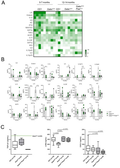
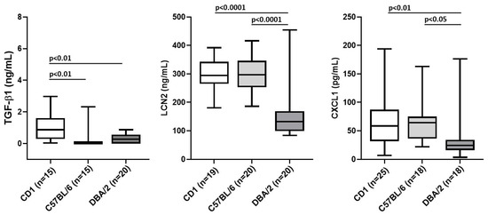
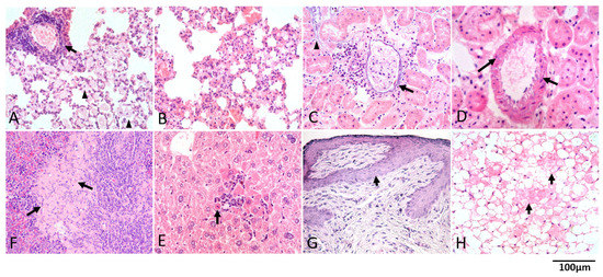
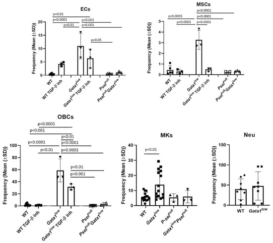
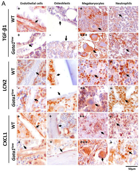
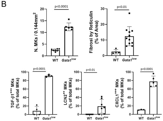
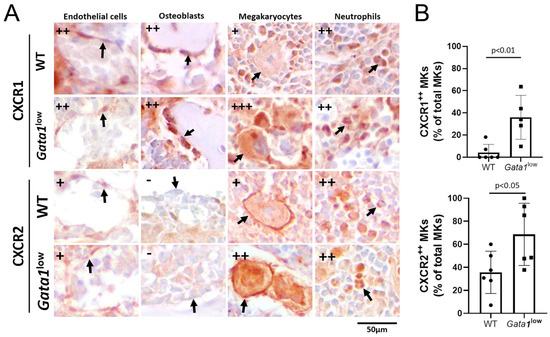
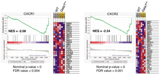
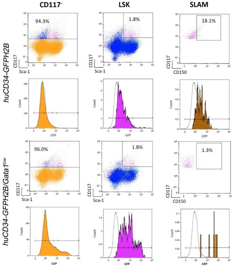
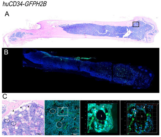
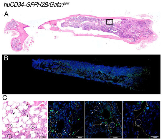
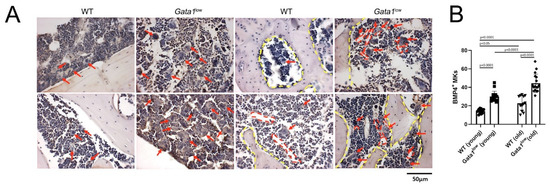
Resident Self-Tissue of Proinflammatory Cytokines Rather Than Their Systemic Levels Correlates with Development of Myelofibrosis in Gata1low Mice
by
Maria Zingariello, Paola Verachi, Francesca Gobbo, Fabrizio Martelli, Mario Falchi, Maria Mazzarini, Mauro Valeri, Giuseppe Sarli, Christian Marinaccio, Johanna Melo-Cardenas, John D. Crispino and Anna Rita Migliaccio
Cited by 5 | Viewed by 3340
Abstract
Serum levels of inflammatory cytokines are currently investigated as prognosis markers in myelofibrosis, the most severe Philadelphia-negative myeloproliferative neoplasm. We tested this hypothesis in the
Gata1low model of myelofibrosis.
Gata1low mice, and age-matched wild-type littermates, were analyzed before and after disease
[...] Read more.
Serum levels of inflammatory cytokines are currently investigated as prognosis markers in myelofibrosis, the most severe Philadelphia-negative myeloproliferative neoplasm. We tested this hypothesis in the
Gata1low model of myelofibrosis.
Gata1low mice, and age-matched wild-type littermates, were analyzed before and after disease onset. We assessed cytokine serum levels by Luminex-bead-assay and ELISA, frequency and cytokine content of stromal cells by flow cytometry, and immunohistochemistry and bone marrow (BM) localization of GFP-tagged hematopoietic stem cells (HSC) by confocal microscopy. Differences in serum levels of 32 inflammatory-cytokines between prefibrotic and fibrotic
Gata1low mice and their wild-type littermates were modest. However, BM from fibrotic
Gata1low mice contained higher levels of lipocalin-2, CXCL1, and TGF-β1 than wild-type BM. Although frequencies of endothelial cells, mesenchymal cells, osteoblasts, and megakaryocytes were higher than normal in
Gata1low BM, the cells which expressed these cytokines the most were malignant megakaryocytes. This increased bioavailability of proinflammatory cytokines was associated with altered HSC localization:
Gata1low HSC were localized in the femur diaphysis in areas surrounded by microvessels, neo-bones, and megakaryocytes, while wild-type HSC were localized in the femur epiphysis around adipocytes. In conclusion, bioavailability of inflammatory cytokines in BM, rather than blood levels, possibly by reshaping the HSC niche, correlates with myelofibrosis in
Gata1low mice.
Full article
►▼
Show Figures
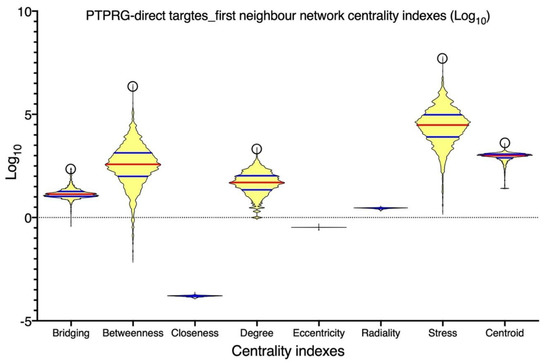
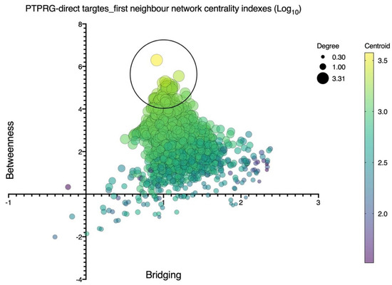
Open AccessReview
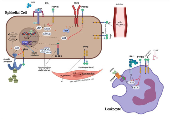
A Comprehensive Review of Receptor-Type Tyrosine-Protein Phosphatase Gamma (PTPRG) Role in Health and Non-Neoplastic Disease
by
Christian Boni, Carlo Laudanna and Claudio Sorio
Cited by 10 | Viewed by 2837
Abstract
Protein tyrosine phosphatase receptor gamma (PTPRG) is known to interact with and regulate several tyrosine kinases, exerting a tumor suppressor role in several type of cancers. Its wide expression in human tissues compared to the other component of group 5 of receptor phosphatases,
[...] Read more.
Protein tyrosine phosphatase receptor gamma (PTPRG) is known to interact with and regulate several tyrosine kinases, exerting a tumor suppressor role in several type of cancers. Its wide expression in human tissues compared to the other component of group 5 of receptor phosphatases, PTPRZ expressed as a chondroitin sulfate proteoglycan in the central nervous system, has raised interest in its role as a possible regulatory switch of cell signaling processes. Indeed, a carbonic anhydrase-like domain (CAH) and a fibronectin type III domain are present in the N-terminal portion and were found to be associated with its role as [HCO
3−] sensor in vascular and renal tissues and a possible interaction domain for cell adhesion, respectively. Studies on PTPRG ligands revealed the contactins family (CNTN) as possible interactors. Furthermore, the correlation of PTPRG phosphatase with inflammatory processes in different normal tissues, including cancer, and the increasing amount of its soluble form (sPTPRG) in plasma, suggest a possible role as inflammatory marker. PTPRG has important roles in human diseases; for example, neuropsychiatric and behavioral disorders and various types of cancer such as colon, ovary, lung, breast, central nervous system, and inflammatory disorders. In this review, we sum up our knowledge regarding the latest discoveries in order to appreciate PTPRG function in the various tissues and diseases, along with an interactome map of its relationship with a group of validated molecular interactors.
Full article
►▼
Show Figures
Open AccessArticle
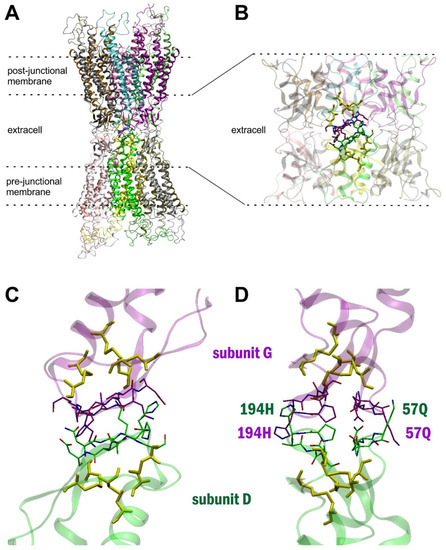
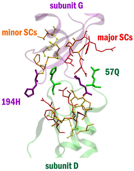
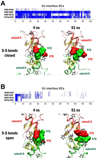
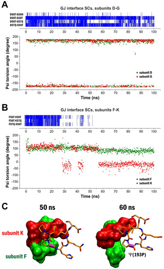
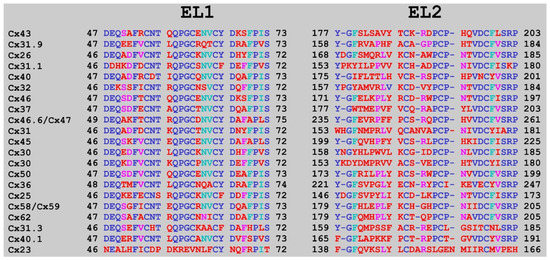
Connexons Coupling to Gap Junction Channel: Potential Role for Extracellular Protein Stabilization Centers
by
László Héja, Ágnes Simon, Zsolt Szabó and Julianna Kardos
Cited by 6 | Viewed by 1778
Abstract
Connexin (Cx) proteins establish intercellular gap junction channels (Cx GJCs) through coupling of two apposed hexameric Cx hemichannels (Cx HCs, connexons). Pre- and post-GJ interfaces consist of extracellular EL1 and EL2 loops, each with three conserved cysteines. Previously, we reported that known peptide
[...] Read more.
Connexin (Cx) proteins establish intercellular gap junction channels (Cx GJCs) through coupling of two apposed hexameric Cx hemichannels (Cx HCs, connexons). Pre- and post-GJ interfaces consist of extracellular EL1 and EL2 loops, each with three conserved cysteines. Previously, we reported that known peptide inhibitors, mimicking a variety of Cx43 sequences, appear non-selective when binding to homomeric Cx43 vs. Cx36 GJC homology model subtypes. In pursuit of finding potentially Cx subtype-specific inhibitors of connexon-connexon coupling, we aimed at to understand better how the GJ interface is formed. Here we report on the discovery of Cx GJC subtype-specific protein stabilization centers (SCs) featuring GJ interface architecture. First, the Cx43 GJC homology model, embedded in two opposed membrane bilayers, has been devised. Next, we endorsed the fluctuation dynamics of SCs of the interface domain of Cx43 GJC by applying standard molecular dynamics under open and closed cystine disulfide bond (
CS-S
C) preconditions. The simulations confirmed the major role of the unique trans-GJ SC pattern comprising conserved (55
N, 56
T) and non-conserved (57Q) residues of the apposed EL1 loops in the stabilization of the GJC complex. Importantly, clusters of SC patterns residing close to the GJ interface domain appear to orient the interface formation via the numerous SCs between EL1 and EL2. These include central
54CS-S
198C or
61CS-S
192C contacts with residues 53R, 54
C, 55
N, 197
D, 199
F or 64
V, 191
P, respectively. In addition, we revealed that GJC interface formation is favoured when the psi dihedral angle of the nearby 193
P residue is stable around 180° and the interface SCs disappear when this angle moves to the 0° to −45° range. The potential of the association of non-conserved residues with SC motifs in connexon-connexon coupling makes the development of Cx subtype-specific inhibitors viable.
Full article
►▼
Show Figures
Open AccessArticle
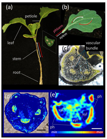
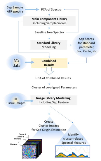
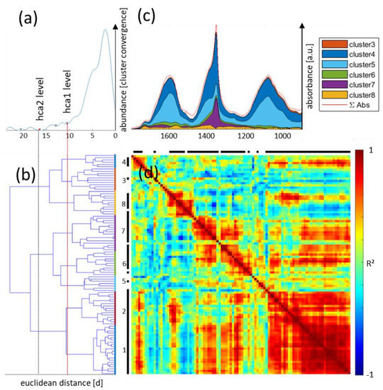
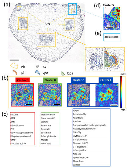
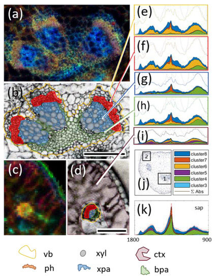
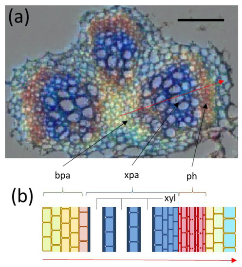
Probing the Metabolic Landscape of Plant Vascular Bundles by Infrared Fingerprint Analysis, Imaging and Mass Spectrometry
by
André Guendel, Alexander Hilo, Hardy Rolletschek and Ljudmilla Borisjuk
Cited by 3 | Viewed by 2047
Abstract
Fingerprint analysis is a common technique in forensic and criminal investigations. Similar techniques exist in the field of infrared spectroscopy to identify biomolecules according to their characteristic spectral fingerprint features. These unique markers are located in a wavenumber range from 1800 to 600
[...] Read more.
Fingerprint analysis is a common technique in forensic and criminal investigations. Similar techniques exist in the field of infrared spectroscopy to identify biomolecules according to their characteristic spectral fingerprint features. These unique markers are located in a wavenumber range from 1800 to 600 cm
−1 in the mid infrared region. Here, a novel bioanalytical concept of correlating these spectral features with corresponding mass spectrometry datasets to unravel metabolic clusters within complex plant tissues was applied. As proof of concept, vascular bundles of oilseed rape (
Brassica napus) were investigated, one of the most important and widely cultivated temperate zone oilseed crops. The link between mass spectrometry data and spectral data identified features that co-aligned within both datasets. Regions of origin were then detected by searching for these features in hyperspectral images of plant tissues. This approach, based on co-alignment and co-localization, finally enabled the detection of eight distinct metabolic clusters, reflecting functional and structural arrangements within the vascular bundle. The proposed analytical concept may assist future synergistic research approaches and may lead to biotechnological innovations with regard to crop yield and sustainability.
Full article
►▼
Show Figures
Open AccessReview
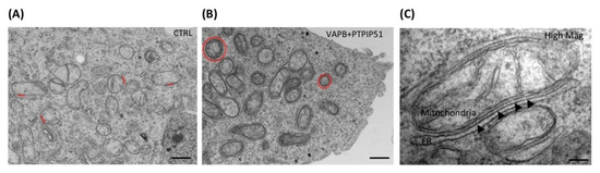
Mitochondria-Endoplasmic Reticulum Crosstalk in Parkinson’s Disease: The Role of Brain Renin Angiotensin System Components
by
Tuladhar Sunanda, Bipul Ray, Arehally M. Mahalakshmi, Abid Bhat, Luay Rashan, Wiramon Rungratanawanich, Byoung-Joon Song, Musthafa Mohamed Essa, Meena Kishore Sakharkar and Saravana Babu Chidambaram
Cited by 18 | Viewed by 3318
Abstract
The past few decades have seen an increased emphasis on the involvement of the mitochondrial-associated membrane (MAM) in various neurodegenerative diseases, particularly in Parkinson’s disease (PD) and Alzheimer’s disease (AD). In PD, alterations in mitochondria, endoplasmic reticulum (ER), and MAM functions affect the
[...] Read more.
The past few decades have seen an increased emphasis on the involvement of the mitochondrial-associated membrane (MAM) in various neurodegenerative diseases, particularly in Parkinson’s disease (PD) and Alzheimer’s disease (AD). In PD, alterations in mitochondria, endoplasmic reticulum (ER), and MAM functions affect the secretion and metabolism of proteins, causing an imbalance in calcium homeostasis and oxidative stress. These changes lead to alterations in the translocation of the MAM components, such as IP3R, VDAC, and MFN1 and 2, and consequently disrupt calcium homeostasis and cause misfolded proteins with impaired autophagy, distorted mitochondrial dynamics, and cell death. Various reports indicate the detrimental involvement of the brain renin–angiotensin system (RAS) in oxidative stress, neuroinflammation, and apoptosis in various neurodegenerative diseases. In this review, we attempted to update the reports (using various search engines, such as PubMed, SCOPUS, Elsevier, and Springer Nature) demonstrating the pathogenic interactions between the various proteins present in mitochondria, ER, and MAM with respect to Parkinson’s disease. We also made an attempt to speculate the possible involvement of RAS and its components, i.e., AT1 and AT2 receptors, angiotensinogen, in this crosstalk and PD pathology. The review also collates and provides updated information on the role of MAM in calcium signaling, oxidative stress, neuroinflammation, and apoptosis in PD.
Full article
►▼
Show Figures
Open AccessReview
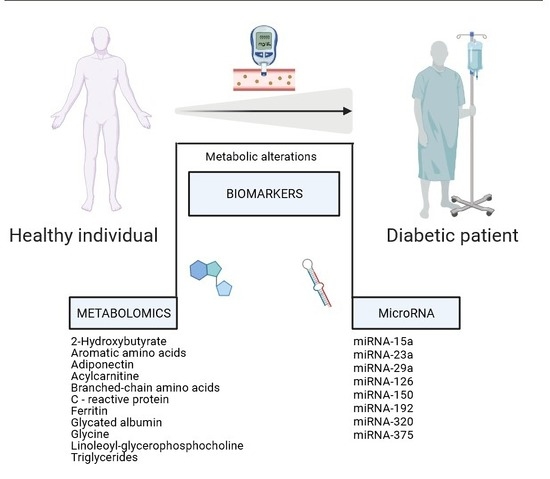
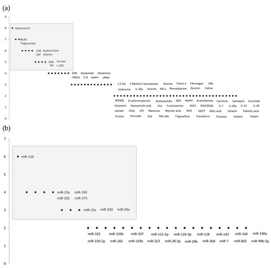
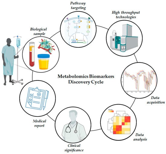
Metabolic Dysfunction Biomarkers as Predictors of Early Diabetes
by
Carla Luís, Pilar Baylina, Raquel Soares and Rúben Fernandes
Cited by 5 | Viewed by 2759
Abstract
During the pathophysiological course of type 2 diabetes (T2D), several metabolic imbalances occur. There is increasing evidence that metabolic dysfunction far precedes clinical manifestations. Thus, knowing and understanding metabolic imbalances is crucial to unraveling new strategies and molecules (biomarkers) for the early-stage prediction
[...] Read more.
During the pathophysiological course of type 2 diabetes (T2D), several metabolic imbalances occur. There is increasing evidence that metabolic dysfunction far precedes clinical manifestations. Thus, knowing and understanding metabolic imbalances is crucial to unraveling new strategies and molecules (biomarkers) for the early-stage prediction of the disease’s non-clinical phase. Lifestyle interventions must be made with considerable involvement of clinicians, and it should be considered that not all patients will respond in the same manner. Individuals with a high risk of diabetic progression will present compensatory metabolic mechanisms, translated into metabolic biomarkers that will therefore show potential predictive value to differentiate between progressors/non-progressors in T2D. Specific novel biomarkers are being proposed to entrap prediabetes and target progressors to achieve better outcomes. This study provides a review of the latest relevant biomarkers in prediabetes. A search for articles published between 2011 and 2021 was conducted; duplicates were removed, and inclusion criteria were applied. From the 29 studies considered, a survey of the most cited (relevant) biomarkers was conducted and further discussed in the two main identified fields: metabolomics, and miRNA studies.
Full article
►▼
Show Figures
Open AccessReview
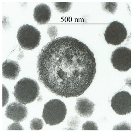
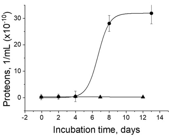
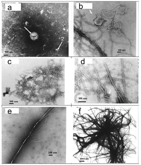
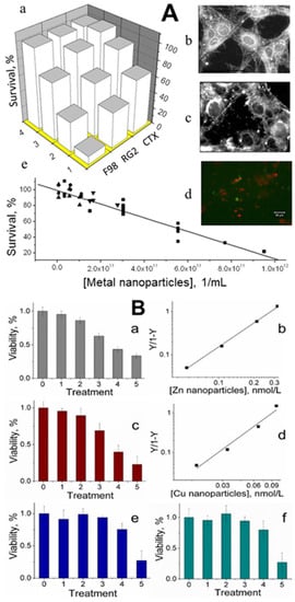
The Role of Endogenous Metal Nanoparticles in Biological Systems
by
Vitaly Vodyanoy
Cited by 2 | Viewed by 2012
Abstract
The blood and tissues of vertebrate animals and mammals contain small endogenous metal nanoparticles. These nanoparticles were observed to be composed of individual atoms of iron, copper, zinc, silver, gold, platinum, and other metals. Metal nanoparticles can bind proteins and produce proteinaceous particles
[...] Read more.
The blood and tissues of vertebrate animals and mammals contain small endogenous metal nanoparticles. These nanoparticles were observed to be composed of individual atoms of iron, copper, zinc, silver, gold, platinum, and other metals. Metal nanoparticles can bind proteins and produce proteinaceous particles called proteons. A small fraction of the entire pool of nanoparticles is usually linked with proteins to form proteons. These endogenous metal nanoparticles, along with engineered zinc and copper nanoparticles at subnanomolar levels, were shown to be lethal to cultured cancer cells. These nanoparticles appear to be elemental crystalline metal nanoparticles. It was discovered that zinc nanoparticles produce no odor response but increase the odor reaction if mixed with an odorant. Some other metal nanoparticles, including copper, silver, gold, and platinum nanoparticles, do not affect the responses to odorants. The sources of metal nanoparticles in animal blood and tissues may include dietary plants and gut microorganisms. The solid physiological and biochemical properties of metal nanoparticles reflect their importance in cell homeostasis and disease.
Full article
►▼
Show Figures
Open AccessReview
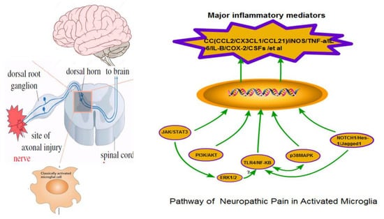
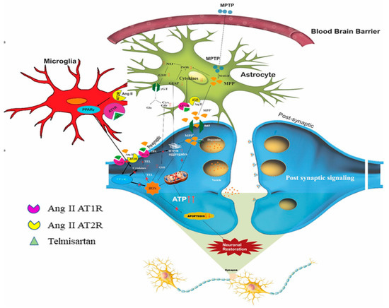
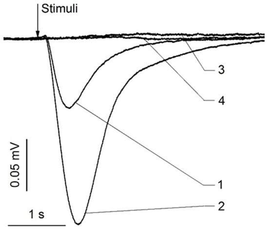
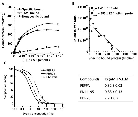
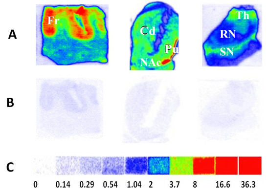
Neuropathic Pain: Biomolecular Intervention and Imaging via Targeting Microglia Activation
by
Aijun Ji and Jinbin Xu
Cited by 18 | Viewed by 3971
Abstract
Many diseases, including cancer, can lead to neuropathic pain (NP). NP is one of the accompanying symptoms of suffering in many conditions and the life quality of NP patient is seriously affected. Due to complex causes, the effects of clinical treatments have been
[...] Read more.
Many diseases, including cancer, can lead to neuropathic pain (NP). NP is one of the accompanying symptoms of suffering in many conditions and the life quality of NP patient is seriously affected. Due to complex causes, the effects of clinical treatments have been very unsatisfactory. Many experts have found that neuron-microglia interaction plays an essential role in NP occurrence and development. Therefore, the activation of microglia, related inflammatory mediators and molecular and cellular signaling pathways have become the focus of NP research. With the help of modern functional imaging technology, advanced pre-and clinical studies have been carried out and NP interventions have been attempted by using the different pharmaceuticals and the extracted active components of various traditional herbal medicines. In this communication, we review the mechanism of microglia on NP formation and treatment and molecular imaging technology’s role in the clinical diagnosis and evaluation of NP therapies.
Full article
►▼
Show Figures
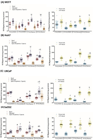
Open AccessArticle
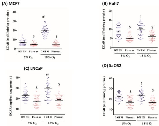
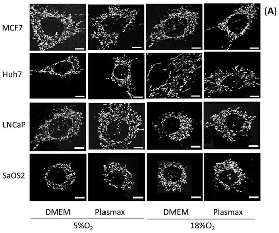
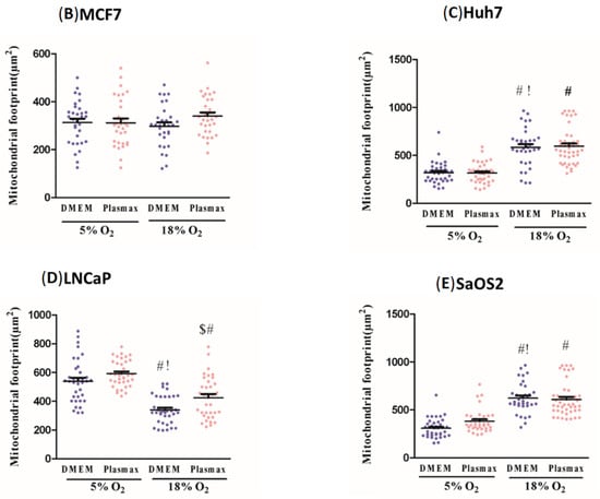
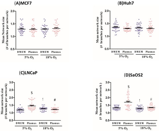
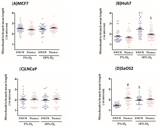
The Effect of Oxygen and Micronutrient Composition of Cell Growth Media on Cancer Cell Bioenergetics and Mitochondrial Networks
by
Fereshteh Moradi, Christopher Moffatt and Jeffrey A. Stuart
Cited by 11 | Viewed by 2877
Abstract
Cancer cell culture is routinely performed under superphysiologic O
2 levels and in media such as Dulbecco’s Modified Eagle Medium (DMEM) with nutrient composition dissimilar to mammalian extracellular fluid. Recently developed cell culture media (e.g., Plasmax, Human Plasma-Like Medium (HPLM)), which are modeled
[...] Read more.
Cancer cell culture is routinely performed under superphysiologic O
2 levels and in media such as Dulbecco’s Modified Eagle Medium (DMEM) with nutrient composition dissimilar to mammalian extracellular fluid. Recently developed cell culture media (e.g., Plasmax, Human Plasma-Like Medium (HPLM)), which are modeled on the metabolite composition of human blood plasma, have been shown to shift key cellular activities in several cancer cell lines. Similar effects have been reported with respect to O
2 levels in cell culture. Given these observations, we investigated how media composition and O
2 levels affect cellular energy metabolism and mitochondria network structure in MCF7, SaOS2, LNCaP, and Huh7 cells. Cells were cultured in physiologic (5%) or standard (18%) O
2 levels, and in physiologic (Plasmax) or standard cell culture media (DMEM). We show that both O
2 levels and media composition significantly affect mitochondrial abundance and network structure, concomitantly with changes in cellular bioenergetics. Extracellular acidification rate (ECAR), a proxy for glycolytic activity, was generally higher in cells cultured in DMEM while oxygen consumption rates (OCR) were lower. This effect of media on energy metabolism is an important consideration for the study of cancer drugs that target aspects of energy metabolism, including lactate dehydrogenase activity.
Full article
►▼
Show Figures
Open AccessReview
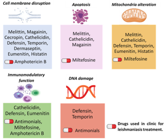
Activity of Anti-Microbial Peptides (AMPs) against Leishmania and Other Parasites: An Overview
by
Rima El-Dirany, Hawraa Shahrour, Zeinab Dirany, Fadi Abdel-Sater, Gustavo Gonzalez-Gaitano, Klaus Brandenburg, Guillermo Martinez de Tejada and Paul A. Nguewa
Cited by 18 | Viewed by 4178
Abstract
Anti-microbial peptides (AMPs), small biologically active molecules, produced by different organisms through their innate immune system, have become a considerable subject of interest in the request of novel therapeutics. Most of these peptides are cationic-amphipathic, exhibiting two main mechanisms of action, direct lysis
[...] Read more.
Anti-microbial peptides (AMPs), small biologically active molecules, produced by different organisms through their innate immune system, have become a considerable subject of interest in the request of novel therapeutics. Most of these peptides are cationic-amphipathic, exhibiting two main mechanisms of action, direct lysis and by modulating the immunity. The most commonly reported activity of AMPs is their anti-bacterial effects, although other effects, such as anti-fungal, anti-viral, and anti-parasitic, as well as anti-tumor mechanisms of action have also been described. Their anti-parasitic effect against leishmaniasis has been studied. Leishmaniasis is a neglected tropical disease. Currently among parasitic diseases, it is the second most threating illness after malaria. Clinical treatments, mainly antimonial derivatives, are related to drug resistance and some undesirable effects. Therefore, the development of new therapeutic agents has become a priority, and AMPs constitute a promising alternative. In this work, we describe the principal families of AMPs (melittin, cecropin, cathelicidin, defensin, magainin, temporin, dermaseptin, eumenitin, and histatin) exhibiting a potential anti-leishmanial activity, as well as their effectiveness against other microorganisms.
Full article
►▼
Show Figures
Open AccessArticle
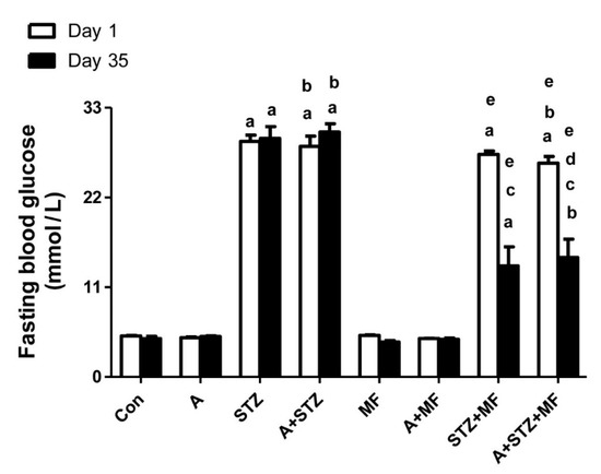
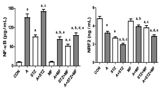
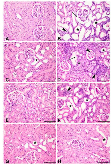
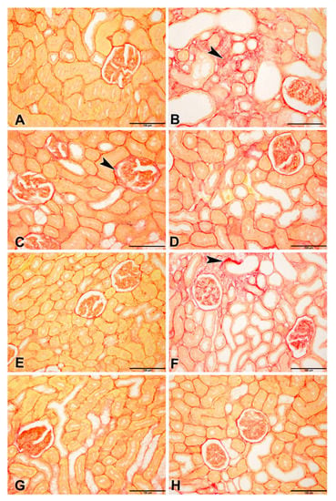
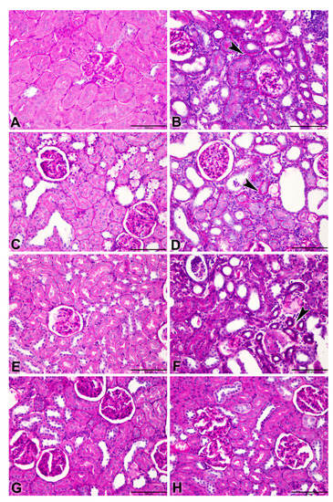
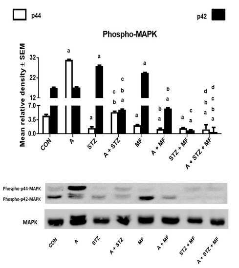
The Effect of Metformin in Diabetic and Non-Diabetic Rats with Experimentally-Induced Chronic Kidney Disease
by
Mohammed Al Za’abi, Badreldin H. Ali, Yousuf Al Suleimani, Sirin A. Adham, Haytham Ali, Priyadarsini Manoj, Mohammed Ashique and Abderrahim Nemmar
Cited by 15 | Viewed by 3509
Abstract
This work aimed to investigate whether treatment with the antidiabetic drug metformin would affect adenine-induced chronic kidney disease (CKD) in non-diabetic rats and rats with streptozotocin (STZ)-induced diabetes. Rats were randomly divided into eight groups, and given either normal feed, or feed mixed
[...] Read more.
This work aimed to investigate whether treatment with the antidiabetic drug metformin would affect adenine-induced chronic kidney disease (CKD) in non-diabetic rats and rats with streptozotocin (STZ)-induced diabetes. Rats were randomly divided into eight groups, and given either normal feed, or feed mixed with adenine (0.25%
w/w, for five weeks) to induce CKD. Some of these groups were also simultaneously treated orally with metformin (200 mg/kg/day). Rats given adenine showed the typical signs of CKD that included detrimental changes in several physiological and traditional and novel biochemical biomarkers in plasma urine and kidney homogenates such as albumin/creatinine ratio, N-acetyl-beta-D-glucosaminidase, neutrophil gelatinase-associated lipocalin, 8-isoprostane, adiponectin, cystatin C, as well as plasma urea, creatinine, uric acid, indoxyl sulfate, calcium, and phosphorus. Several indices of inflammation and oxidative stress, and renal nuclear factor-κB and nuclear factor erythroid 2-related factor 2 levels were also measured. Histopathologically, adenine caused renal tubular necrosis and fibrosis. The activation of the intracellular mitogen-activated protein kinase signaling pathway was inhibited in the groups that received metformin and STZ together, with or without adenine induced-CKD. Induction of diabetes worsened most of the actions induced by adenine. Metformin significantly ameliorated the renal actions induced by adenine and STZ when these were given singly, and more so when given together. The results suggest that metformin can be a useful drug in attenuating the progression of CKD in both diabetic and non-diabetic rats.
Full article
►▼
Show Figures
Open AccessEditor’s ChoiceReview
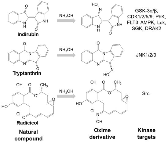
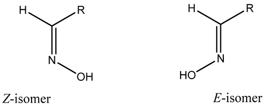
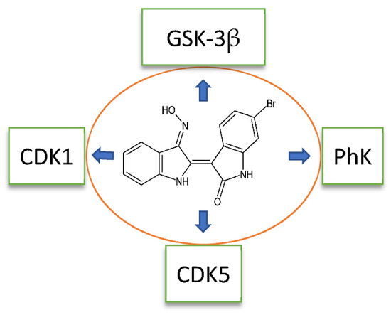
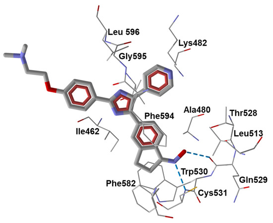
Oximes: Novel Therapeutics with Anticancer and Anti-Inflammatory Potential
by
Igor A. Schepetkin, Mark B. Plotnikov, Andrei I. Khlebnikov, Tatiana M. Plotnikova and Mark T. Quinn
Cited by 35 | Viewed by 5126
Abstract
Oximes have been studied for decades because of their significant roles as acetylcholinesterase reactivators. Over the last twenty years, a large number of oximes have been reported with useful pharmaceutical properties, including compounds with antibacterial, anticancer, anti-arthritis, and anti-stroke activities. Many oximes are
[...] Read more.
Oximes have been studied for decades because of their significant roles as acetylcholinesterase reactivators. Over the last twenty years, a large number of oximes have been reported with useful pharmaceutical properties, including compounds with antibacterial, anticancer, anti-arthritis, and anti-stroke activities. Many oximes are kinase inhibitors and have been shown to inhibit over 40 different kinases, including AMP-activated protein kinase (AMPK), phosphatidylinositol 3-kinase (PI3K), cyclin-dependent kinase (CDK), serine/threonine kinases glycogen synthase kinase 3 α/β (GSK-3α/β), Aurora A, B-Raf, Chk1, death-associated protein-kinase-related 2 (DRAK2), phosphorylase kinase (PhK), serum and glucocorticoid-regulated kinase (SGK), Janus tyrosine kinase (JAK), and multiple receptor and non-receptor tyrosine kinases. Some oximes are inhibitors of lipoxygenase 5, human neutrophil elastase, and proteinase 3. The oxime group contains two H-bond acceptors (nitrogen and oxygen atoms) and one H-bond donor (OH group), versus only one H-bond acceptor present in carbonyl groups. This feature, together with the high polarity of oxime groups, may lead to a significantly different mode of interaction with receptor binding sites compared to corresponding carbonyl compounds, despite small changes in the total size and shape of the compound. In addition, oximes can generate nitric oxide. This review is focused on oximes as kinase inhibitors with anticancer and anti-inflammatory activities. Oximes with non-kinase targets or mechanisms of anti-inflammatory activity are also discussed.
Full article
►▼
Show Figures
Open AccessArticle
Immunohistochemical Analysis of the Expression of Adhesion Proteins: TNS1, TNS2 and TNS3 in Correlation with Clinicopathological Parameters in Gastric Cancer
by
Marcin Nizioł, Justyna Zińczuk, Konrad Zaręba, Katarzyna Guzińska-Ustymowicz and Anna Pryczynicz
Cited by 9 | Viewed by 2088
Abstract
Tensins belong to the group of adhesion proteins that are involved in cell adhesion and migration, actin cytoskeleton maintenance and intercellular communication. TNS1, TNS2 and TNS3 proteins expression was evaluated in 90 patients with gastric cancer by immunohistochemistry method. TNS1 was more frequently
[...] Read more.
Tensins belong to the group of adhesion proteins that are involved in cell adhesion and migration, actin cytoskeleton maintenance and intercellular communication. TNS1, TNS2 and TNS3 proteins expression was evaluated in 90 patients with gastric cancer by immunohistochemistry method. TNS1 was more frequently present in non-differentiated tumors compared to poorly and moderately differentiated tumors (
p = 0.016). TNS1 was also more often observed in metastatic tumors compared to those without distant metastases (
p = 0.001). TNS2 was more common in moderately differentiated tumors than in poorly or non-differentiated ones (
p = 0.041). TNS2 expression was also more frequently present in tumors with peritumoral inflammation (
p = 0.041) and with concomitant
H. pylori infection (
p = 0.023). In contrast, TNS3 protein was more prevalent in moderately than in poorly and non-differentiated tumors (
p = 0.023). No significant relationship was found between tensins’ expression and the overall survival rate of patients. TNS1 protein expression is associated with a poor-prognosis type of GC. Higher expression of TNS2 is accompanied by peritumoral inflammation and
H. pylori infection, which favor the development of GC of a better prognosis, similarly to higher TNS3 protein expression.
Full article
►▼
Show Figures
Open AccessReview
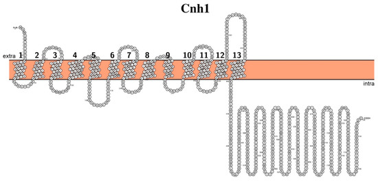
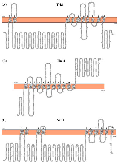
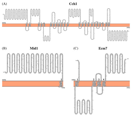
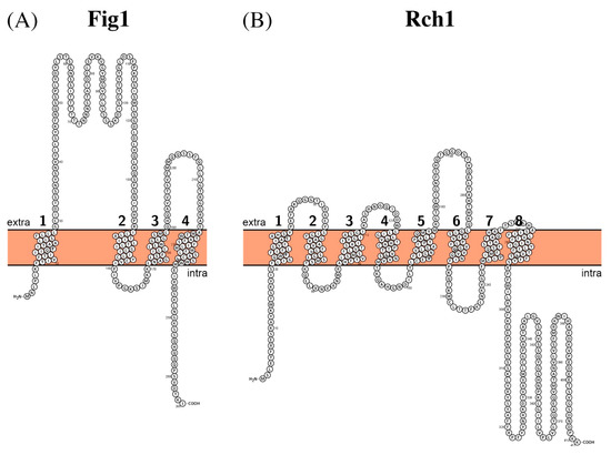
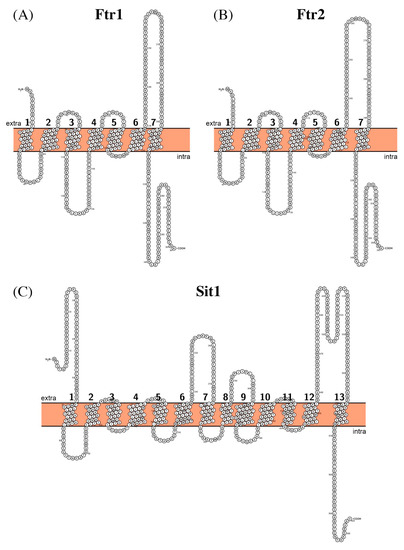
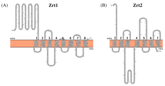
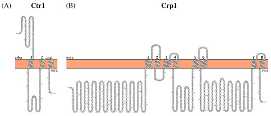
Cation Transporters of Candida albicans—New Targets to Fight Candidiasis?
by
Marina Volkova, Anastasia Atamas, Alexey Tsarenko, Andrey Rogachev and Albert Guskov
Cited by 7 | Viewed by 3134
Abstract
Candidiasis is the wide-spread fungal infection caused by numerous strains of yeast, with the prevalence of
Candida albicans. The current treatment of candidiasis is becoming rather ineffective and costly owing to the emergence of resistant strains; hence, the exploration of new possible
[...] Read more.
Candidiasis is the wide-spread fungal infection caused by numerous strains of yeast, with the prevalence of
Candida albicans. The current treatment of candidiasis is becoming rather ineffective and costly owing to the emergence of resistant strains; hence, the exploration of new possible drug targets is necessary. The most promising route is the development of novel antibiotics targeting this pathogen. In this review, we summarize such candidates found in
C. albicans and those involved in the transport of (metal) cations, as the latter are essential for numerous processes within the cell; hence, disruption of their fluxes can be fatal for
C. albicans.
Full article
►▼
Show Figures
Open AccessReview
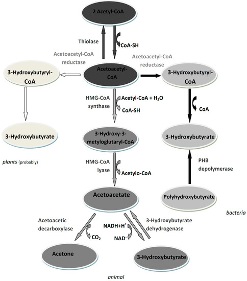
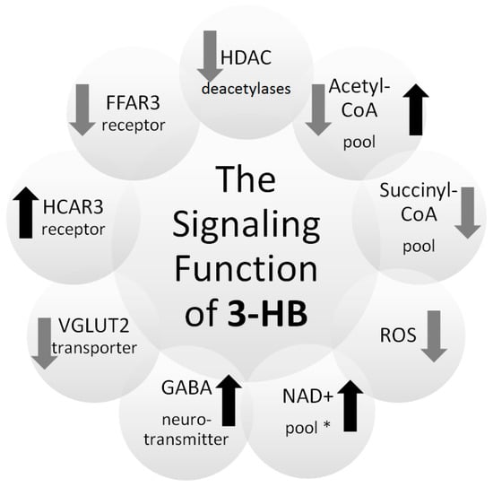
3-Hydroxybutyrate as a Metabolite and a Signal Molecule Regulating Processes of Living Organisms
by
Justyna Mierziak, Marta Burgberger and Wioleta Wojtasik
Cited by 73 | Viewed by 8785
Abstract
3-hydroxybutyrate (3-HB) as a very important metabolite occurs in animals, bacteria and plants. It is well known that in animals, 3-HB is formed as a product of the normal metabolism of fatty acid oxidation and can therefore be used as an energy source
[...] Read more.
3-hydroxybutyrate (3-HB) as a very important metabolite occurs in animals, bacteria and plants. It is well known that in animals, 3-HB is formed as a product of the normal metabolism of fatty acid oxidation and can therefore be used as an energy source in the absence of sufficient blood glucose. In microorganisms, 3-HB mainly serves as a substrate for the synthesis of polyhydroxybutyrate, which is a reserve material. Recent studies show that in plants, 3-HB acts as a regulatory molecule that most likely influences the expression of genes involved in DNA methylation, thereby altering DNA methylation levels. Additionally, in animals, 3-HB is not only an intermediate metabolite, but also an important regulatory molecule that can influence gene expression, lipid metabolism, neuronal function, and overall metabolic rate. Some of these effects are the direct effects of 3-HB itself, while others are indirect effects, regulated by the metabolites into which 3-HB is converted. One of the most important regulatory functions of 3-HB is the inhibition of the activity of histone deacetylases and thus the epigenetic regulation of many genes. Due to the number of functions of this compound, it also shows promising therapeutic properties.
Full article
►▼
Show Figures
Open AccessFeature PaperReview
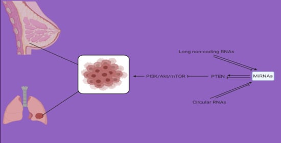
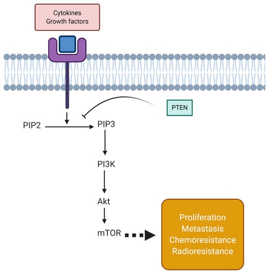
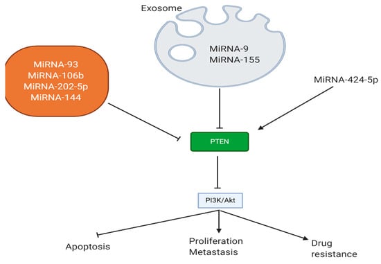
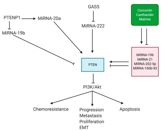
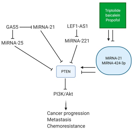
Small in Size, but Large in Action: microRNAs as Potential Modulators of PTEN in Breast and Lung Cancers
by
Asal Jalal Abadi, Ali Zarrabi, Mohammad Hossein Gholami, Sepideh Mirzaei, Farid Hashemi, Amirhossein Zabolian, Maliheh Entezari, Kiavash Hushmandi, Milad Ashrafizadeh, Haroon Khan and Alan Prem Kumar
Cited by 48 | Viewed by 4845
Abstract
MicroRNAs (miRNAs) are well-known regulators of biological mechanisms with a small size of 19–24 nucleotides and a single-stranded structure. miRNA dysregulation occurs in cancer progression. miRNAs can function as tumor-suppressing or tumor-promoting factors in cancer via regulating molecular pathways. Breast and lung cancers
[...] Read more.
MicroRNAs (miRNAs) are well-known regulators of biological mechanisms with a small size of 19–24 nucleotides and a single-stranded structure. miRNA dysregulation occurs in cancer progression. miRNAs can function as tumor-suppressing or tumor-promoting factors in cancer via regulating molecular pathways. Breast and lung cancers are two malignant thoracic tumors in which the abnormal expression of miRNAs plays a significant role in their development. Phosphatase and tensin homolog (PTEN) is a tumor-suppressor factor that is capable of suppressing the growth, viability, and metastasis of cancer cells via downregulating phosphatidylinositol 3-kinase (PI3K)/protein kinase B (Akt) signaling. PTEN downregulation occurs in lung and breast cancers to promote PI3K/Akt expression, leading to uncontrolled proliferation, metastasis, and their resistance to chemotherapy and radiotherapy. miRNAs as upstream mediators of PTEN can dually induce/inhibit PTEN signaling in affecting the malignant behavior of lung and breast cancer cells. Furthermore, long non-coding RNAs and circular RNAs can regulate the miRNA/PTEN axis in lung and breast cancer cells. It seems that anti-tumor compounds such as baicalein, propofol, and curcumin can induce PTEN upregulation by affecting miRNAs in suppressing breast and lung cancer progression. These topics are discussed in the current review with a focus on molecular pathways.
Full article
►▼
Show Figures
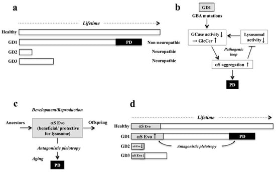
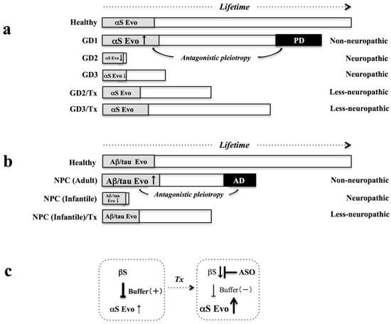
Open AccessPerspective
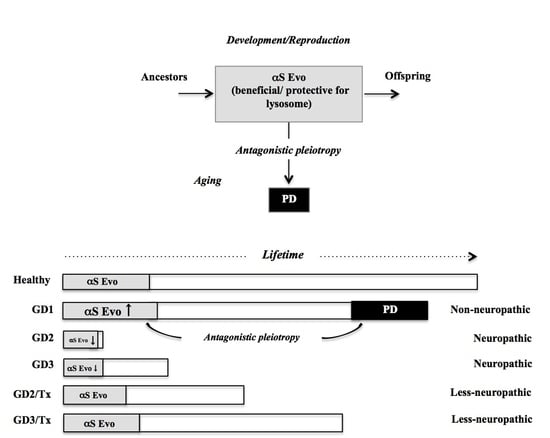
Therapeutic Potential of αS Evolvability for Neuropathic Gaucher Disease
by
Jianshe Wei, Yoshiki Takamatsu, Ryoko Wada, Masayo Fujita, Gilbert Ho, Eliezer Masliah and Makoto Hashimoto
Cited by 4 | Viewed by 2790
Abstract
Gaucher disease (GD), the most common lysosomal storage disorder (LSD), is caused by autosomal recessive mutations of the glucocerebrosidase gene,
GBA1. In the majority of cases, GD has a non-neuropathic chronic form with adult onset (GD1), while other cases are more acute
[...] Read more.
Gaucher disease (GD), the most common lysosomal storage disorder (LSD), is caused by autosomal recessive mutations of the glucocerebrosidase gene,
GBA1. In the majority of cases, GD has a non-neuropathic chronic form with adult onset (GD1), while other cases are more acute and severer neuropathic forms with early onset (GD2/3). Currently, no radical therapies are established for GD2/3. Notably, GD1, but not GD2/3, is associated with increased risk of Parkinson’s disease (PD), the elucidation of which might provide a clue for novel therapeutic strategies. In this context, the objective of the present study is to discuss that the evolvability of α-synuclein (αS) might be differentially involved in GD subtypes. Hypothetically, aging-associated PD features with accumulation of αS, and the autophagy-lysosomal dysfunction might be an antagonistic pleiotropy phenomenon derived from αS evolvability in the development in GD1, without which neuropathies like GD2/3 might be manifested due to the autophagy-lysosomal dysfunction. Supposing that the increased severity of GD2/3 might be attributed to the decreased activity of αS evolvability, suppressing the expression of β-synuclein (βS), a potential buffer against αS evolvability, might be therapeutically efficient. Of interest, a similar view might be applicable to Niemann-Pick type C (NPC), another LSD, given that the adult type of NPC, which is comorbid with Alzheimer’s disease, exhibits milder medical symptoms compared with those of infantile NPC. Thus, it is predicted that the evolvability of amyloid β and tau, might be beneficial for the adult type of NPC. Collectively, a better understanding of amyloidogenic evolvability in the pathogenesis of LSD may inform rational therapy development.
Full article
►▼
Show Figures
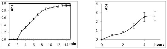
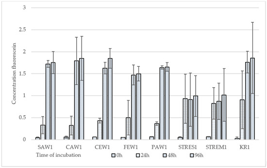
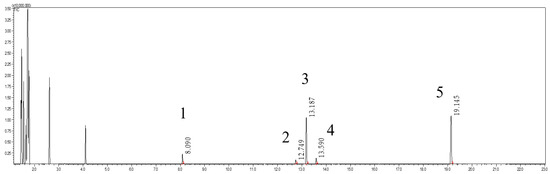
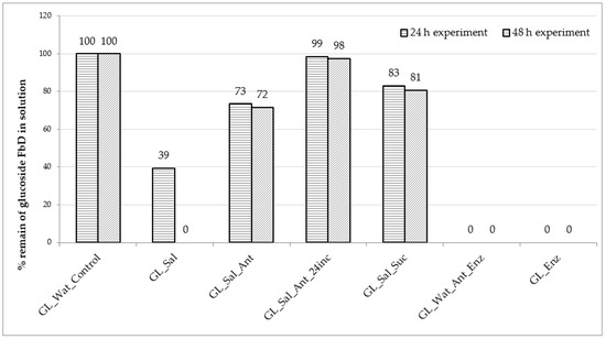
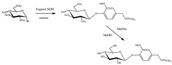
Open AccessArticle
Human Saliva-Mediated Hydrolysis of Eugenyl-β-D-Glucoside and Fluorescein-di-β-D-Glucoside in In Vivo and In Vitro Models
by
Mariusz Dziadas, Adam Junka and Henryk Jeleń
Cited by 3 | Viewed by 2216
Abstract
Eugenyl-β-D-glucopyranoside, also referred to as Citrusin C, is a natural glucoside found among others in cloves, basil and cinnamon plants. Eugenol in a form of free aglycone is used in perfumeries, flavourings, essential oils and in medicinal products. Synthetic Citrusin C was incubated
[...] Read more.
Eugenyl-β-D-glucopyranoside, also referred to as Citrusin C, is a natural glucoside found among others in cloves, basil and cinnamon plants. Eugenol in a form of free aglycone is used in perfumeries, flavourings, essential oils and in medicinal products. Synthetic Citrusin C was incubated with human saliva in several in vitro models together with substrate-specific enzyme and antibiotics (clindamycin, ciprofloxacin, amoxicillin trihydrate and potassium clavulanate). Citrusin C was detected using liquid chromatography with tandem mass spectrometry (LC-MS/MS). Citrusin C was completely degraded only when incubated with substrate-specific
A. niger glucosidase E.C 3.2.1.21 (control sample) and when incubated with human saliva (tested sample). The addition of antibiotics to the above-described experimental setting, stopped Citrusin C degradation, indicating microbiologic origin of hydrolysis observed. Our results demonstrate that Citrusin C is subjected to complete degradation by salivary/oral cavity microorganisms. Extrapolation of our results allows to state that in the human oral cavity, virtually all β-D-glucosides would follow this type of hydrolysis. Additionally, a new method was developed for an in vivo rapid test of glucosidase activity in the human mouth on the tongue using fluorescein-di-β-D-glucoside as substrate. The results presented in this study serve as a proof of concept for the hypothesis that microbial hydrolysis path of β-D-glucosides begins immediately in the human mouth and releases the aglycone directly into the gastrointestinal tract.
Full article
►▼
Show Figures
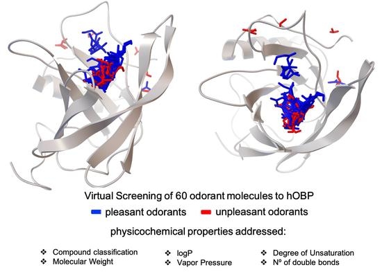
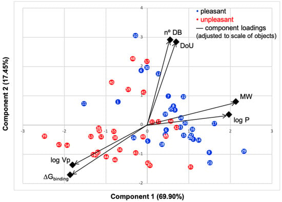
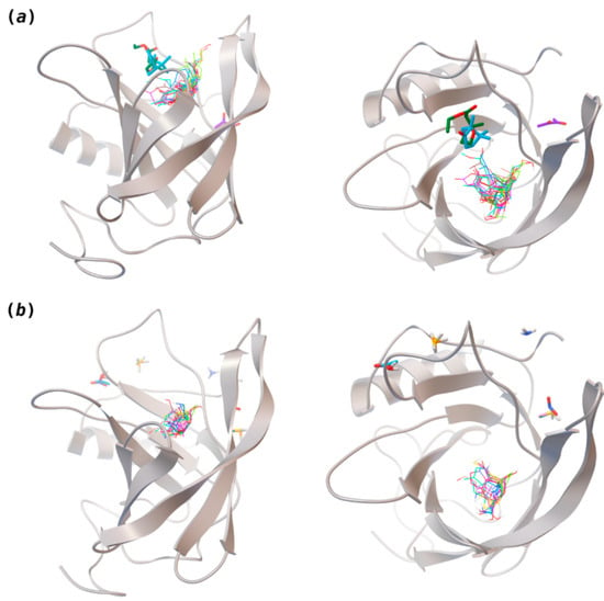
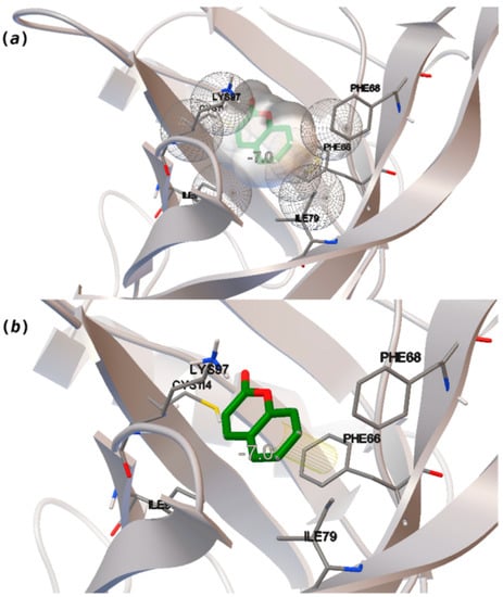
Open AccessArticle
The Structural Properties of Odorants Modulate Their Association to Human Odorant Binding Protein
by
Tarsila G. Castro, Carla Silva, Teresa Matamá and Artur Cavaco-Paulo
Cited by 5 | Viewed by 2594
Abstract
The binding of known odorant molecules to the human odorant-binding protein (hOBP) was evaluated
in silico. Docking experiments elucidate the preferable binding site and binding affinity of odorant molecules to hOBP. The physicochemical properties molecular weight (MW), vapor pressure (Vp), hydrophobicity level (logP),
[...] Read more.
The binding of known odorant molecules to the human odorant-binding protein (hOBP) was evaluated
in silico. Docking experiments elucidate the preferable binding site and binding affinity of odorant molecules to hOBP. The physicochemical properties molecular weight (MW), vapor pressure (Vp), hydrophobicity level (logP), number of double bonds (NºDB), degree of unsaturation (DoU) and the chemical classification, were selected for the study of odorant modulation. Here, these properties were analyzed concerning 30 pleasant and 30 unpleasant odorants, chosen to represent a wide variety of compounds and to determine their influence on the binding energy to hOBP. Our findings indicate that MW, logP and Vp are the most important odorant variables, directly correlated to odorant-binding energies (
G
binding) towards hOBP. Understanding how the odorants behave when complexed with the OBP in human olfaction opens new possibilities for the development of future biotechnological applications, including sensory devices, medical diagnosis, among others.
Full article
►▼
Show Figures
Open AccessArticle
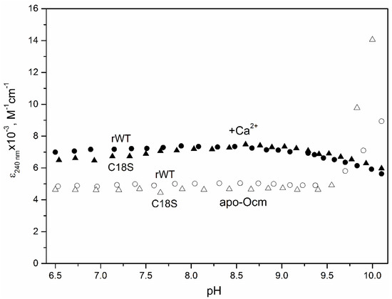
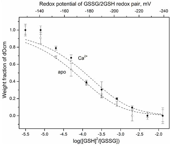
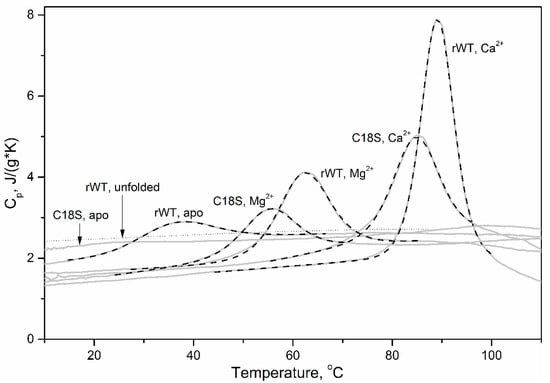
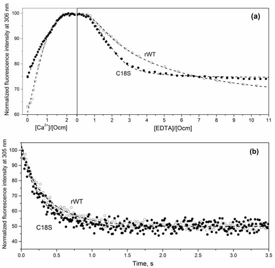
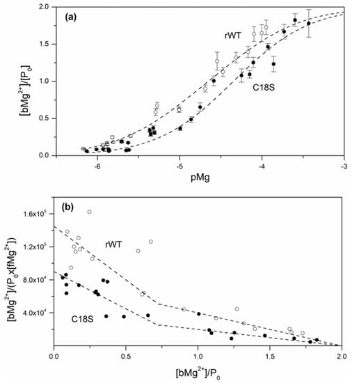
The Highly Conservative Cysteine of Oncomodulin as a Feasible Redox Sensor
by
Alisa A. Vologzhannikova, Polina A. Khorn, Marina P. Shevelyova, Alexei S. Kazakov, Victor I. Emelyanenko, Eugene A. Permyakov and Sergei E. Permyakov
Cited by 3 | Viewed by 2001
Abstract
Oncomodulin (Ocm), or parvalbumin β, is an 11–12 kDa Ca
2+-binding protein found inside and outside of vertebrate cells, which regulates numerous processes via poorly understood mechanisms. Ocm consists of two active Ca
2+-specific domains of the EF-hand type (“helix-loop-helix” motif),
[...] Read more.
Oncomodulin (Ocm), or parvalbumin β, is an 11–12 kDa Ca
2+-binding protein found inside and outside of vertebrate cells, which regulates numerous processes via poorly understood mechanisms. Ocm consists of two active Ca
2+-specific domains of the EF-hand type (“helix-loop-helix” motif), covered by an EF-hand domain with inactive EF-hand loop, which contains a highly conservative cysteine with unknown function. In this study, we have explored peculiarities of the microenvironment of the conservative Cys18 of recombinant rat Ocm (rWT Ocm), redox properties of this residue, and structural/functional sensitivity of rWT Ocm to the homologous C18S substitution. We have found that
pKa of the Cys18 thiol lays beyond the physiological pH range. The measurement of redox dependence of rWT Ocm thiol–disulfide equilibrium (glutathione redox pair) showed that redox potential of Cys18 for the metal-free and Ca
2+-loaded protein is of −168 mV and −176 mV, respectively. Therefore, the conservative thiol of rWT Ocm is prone to disulfide dimerization under physiological redox conditions. The C18S substitution drastically reduces α-helices content of the metal-free and Mg
2+-bound Ocm, increases solvent accessibility of its hydrophobic residues, eliminates the cooperative thermal transition in the apo-protein, suppresses Ca
2+/Mg
2+ affinity of the EF site, and accelerates Ca
2+ dissociation from Ocm. The distinct structural and functional consequences of the minor structural modification of Cys18 indicate its possible redox sensory function. Since some other EF-hand proteins also contain a conservative redox-sensitive cysteine located in an inactive EF-hand loop, it is reasonable to suggest that in the course of evolution, some of the EF-hands attained redox sensitivity at the expense of the loss of their Ca
2+ affinity.
Full article
►▼
Show Figures
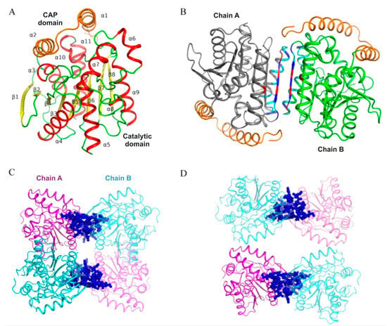
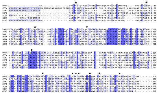
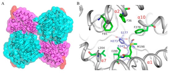
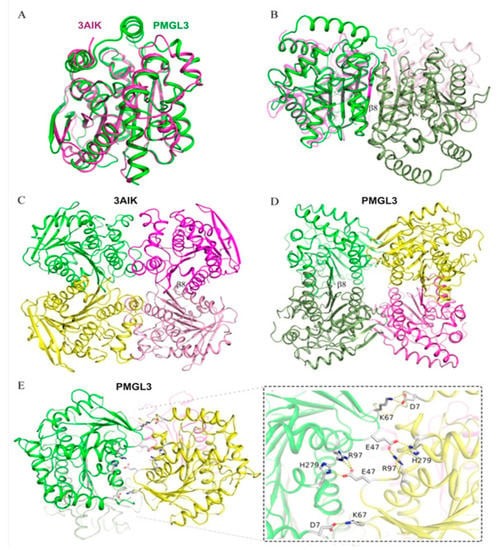
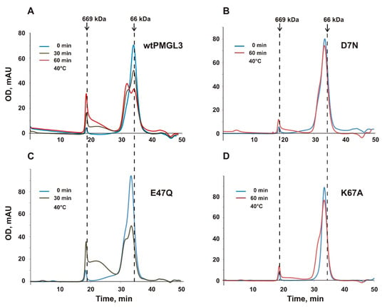
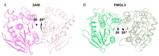
Open AccessFeature PaperArticle
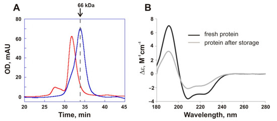
Structural and Biochemical Characterization of a Cold-Active PMGL3 Esterase with Unusual Oligomeric Structure
by
Konstantin M. Boyko, Mariya V. Kryukova, Lada E. Petrovskaya, Elena A. Kryukova, Alena Y. Nikolaeva, Dmitry A. Korzhenevsky, Galina Yu. Lomakina, Ksenia A. Novototskaya-Vlasova, Elizaveta M. Rivkina, Dmitry A. Dolgikh, Mikhail P. Kirpichnikov and Vladimir O. Popov
Cited by 6 | Viewed by 2308
Abstract
The gene coding for a novel cold-active esterase PMGL3 was previously obtained from a Siberian permafrost metagenomic DNA library and expressed in
Escherichia coli. We elucidated the 3D structure of the enzyme which belongs to the hormone-sensitive lipase (HSL) family. Similar to
[...] Read more.
The gene coding for a novel cold-active esterase PMGL3 was previously obtained from a Siberian permafrost metagenomic DNA library and expressed in
Escherichia coli. We elucidated the 3D structure of the enzyme which belongs to the hormone-sensitive lipase (HSL) family. Similar to other bacterial HSLs, PMGL3 shares a canonical α/β hydrolase fold and is presumably a dimer in solution but, in addition to the dimer, it forms a tetrameric structure in a crystal and upon prolonged incubation at 4 °C. Detailed analysis demonstrated that the crystal tetramer of PMGL3 has a unique architecture compared to other known tetramers of the bacterial HSLs. To study the role of the specific residues comprising the tetramerization interface of PMGL3, several mutant variants were constructed. Size exclusion chromatography (SEC) analysis of D7N, E47Q, and K67A mutants demonstrated that they still contained a portion of tetrameric form after heat treatment, although its amount was significantly lower in D7N and K67A compared to the wild type. Moreover, the D7N and K67A mutants demonstrated a 40 and 60% increase in the half-life at 40 °C in comparison with the wild type protein.
Km values of these mutants were similar to that of the wt PMGL3. However, the catalytic constants of the E47Q and K67A mutants were reduced by ~40%.
Full article
►▼
Show Figures
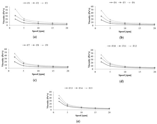
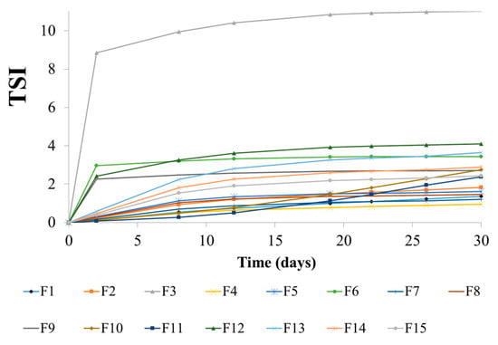
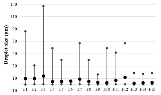
Open AccessArticle
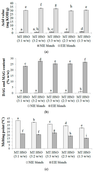
Enzymatically Modified Fats Applied in Emulsions Stabilized by Polysaccharides
by
Magdalena Woźniak, Małgorzata Kowalska, Serge Tavernier and Anna Żbikowska
Cited by 10 | Viewed by 2040
Abstract
The subject of the study was emulsions based on enzymatically modified fats and stabilized with polysaccharides (xanthan gum and scleroglucan). Emulsion oil phases (blends of mutton tallow and hemp seed oil in a ratio of 3:1, 3:2, 3:3, 2:3 and 1:3) were characterized
[...] Read more.
The subject of the study was emulsions based on enzymatically modified fats and stabilized with polysaccharides (xanthan gum and scleroglucan). Emulsion oil phases (blends of mutton tallow and hemp seed oil in a ratio of 3:1, 3:2, 3:3, 2:3 and 1:3) were characterized in the terms of acid value, melting point and mono- and diacylglycerols content before and after the modification. Emulsions containing modified fat blends and various amount (0.6, 0.8 and 1.0%
w/w) of polysaccharides were investigated in the terms of their color, rheological properties, microstructure, droplet size and stability. The obtained results confirmed that enzymatic modification allowed to produce new fats, which can successfully be applied as an emulsion oil phases equipped with a sufficient amount of emulsifiers. The use of a variable amount of texture modifier in the proposed formulations did not show clear differences in the stability of the systems. Therefore, it does not seem justified to use greater amounts of a modifier (above 0.6%
w/w) in this type of emulsions. The proposed formulations could be of interest to the cosmetics, food or pharmaceutical industry.
Full article
►▼
Show Figures
Open AccessReview
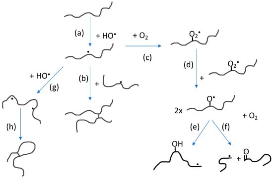
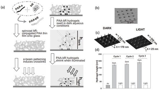
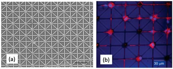
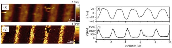
Micro- to Nanoscale Bio-Hybrid Hydrogels Engineered by Ionizing Radiation
by
Clelia Dispenza, Daniela Giacomazza and Mats Jonsson
Cited by 10 | Viewed by 2883
Abstract
Bio-hybrid hydrogels consist of a water-swollen hydrophilic polymer network encapsulating or conjugating single biomolecules, or larger and more complex biological constructs like whole cells. By modulating at least one dimension of the hydrogel system at the micro- or nanoscale, the activity of the
[...] Read more.
Bio-hybrid hydrogels consist of a water-swollen hydrophilic polymer network encapsulating or conjugating single biomolecules, or larger and more complex biological constructs like whole cells. By modulating at least one dimension of the hydrogel system at the micro- or nanoscale, the activity of the biological component can be extremely upgraded with clear advantages for the development of therapeutic or diagnostic micro- and nano-devices. Gamma or e-beam irradiation of polymers allow a good control of the chemistry at the micro-/nanoscale with minimal recourse to toxic reactants and solvents. Another potential advantage is to obtain simultaneous sterilization when the absorbed doses are within the sterilization dose range. This short review will highlight opportunities and challenges of the radiation technologies to produce bio-hybrid nanogels as delivery devices of therapeutic biomolecules to the target cells, tissues, and organs, and to create hydrogel patterns at the nano-length and micro-length scales on surfaces.
Full article
►▼
Show Figures
Planned Papers
The below list represents only planned manuscripts. Some of these
manuscripts have not been received by the Editorial Office yet. Papers
submitted to MDPI journals are subject to peer-review.
Title: Novel perspectives for microRNAs in prediabetes state and its complications
Authors: Adriana Georgescu, et al.
Affiliation: Institute of Cellular Biology and Pathology ‘Nicolae Simionescu’ of Romanian Academy, 8 B.P Hasdeu Street, Bucharest, Romania
Title: Evaluation of a new PLD high-throughput assay
Authors: Rudiger Woscholski; et al.
Affiliation: Institute of Chemical Biology, Department of Chemistry, Imperial College, Molecular Science Research Hub, White City Campus, London W12 0BZ, UK
Title: The role of endogenous metal nanoparticles in biological systems
Authors: Vitaly Vodyanoy; et al.
Affiliation: Department of Anatomy, Physiology and Pharmacology, 207 Greene Hall, Auburn University, Auburn, ALÂ 36849Â, USA
Title: Adhesion and motile molecular machineries of mycoplasmas from the pneumoniae cluster
Authors: Oscar Quijada Pích; et al.
Affiliation: Universidad Autónoma Barcelona, Instituto de Biología Fundamental, Campus UAB (08193) Bellaterra, Barcelona, Spain
Title: New molecules with potential use in clinical to treat infections caused by non-tuberculous mycobacteria
Authors: Jesús Navas; et al.
Affiliation: Grupo BIOMEDAGE, Facultad de Medicina, Universidad de Cantabria, Herrera Oria 2, 39011 Santander, Spain.
Title: Cardiovascular disease as a consequence or a cause of cancer: potential role of extracellular vesicles
Authors: Adriana Georgescu; et al.
Affiliation: Institute of Cellular Biology and Pathology ‘Nicolae Simionescu’ of Romanian Academy, 8 B.P Hasdeu Street, Bucharest, Romania
Title: Metabolic dysfunction biomarkers as predictors for early Diabetes. A systematic review
Authors: Carla Luis; Pilar Baylin; Raquel Soares; Ruben Fernandes
Affiliation: i3S - Institute of Research and Innovation, University of Porto; Faculty of Medicine, University of Porto, Polytechnic Institute of Porto


