


Biomolecules 2024, 14(4), 474; https://doi.org/10.3390/biom14040474 - 12 Apr 2024
Viewed by 351
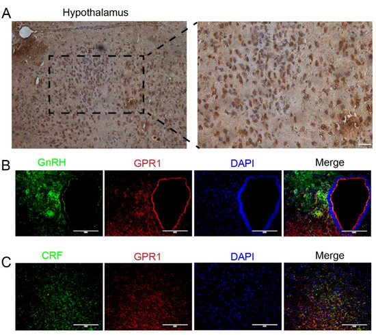
Abstract
►
Show Figures
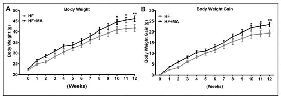
Vitamin D (vit D) and fish oil (FO) both offer unique health benefits, however, their combined effects have not been evaluated in obesity and nonalcoholic fatty liver disease (NAFLD). Hence, we hypothesized that vit D and FO supplementation would have additive effects in
[...] Read more.
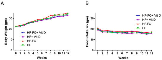
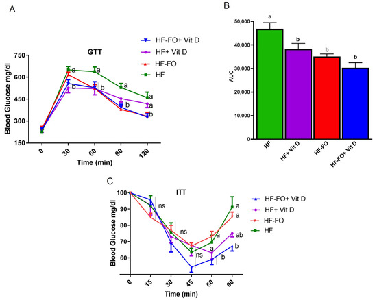
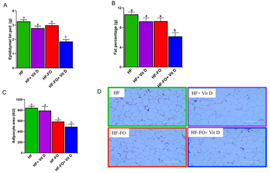
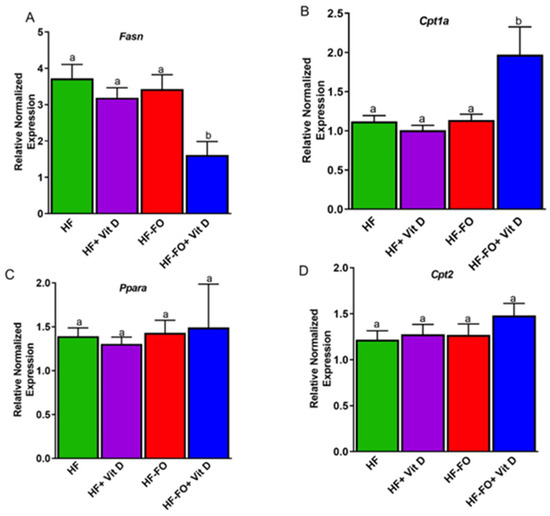
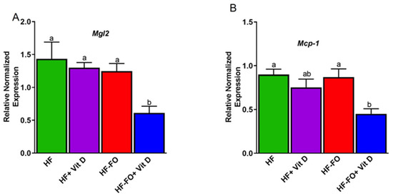
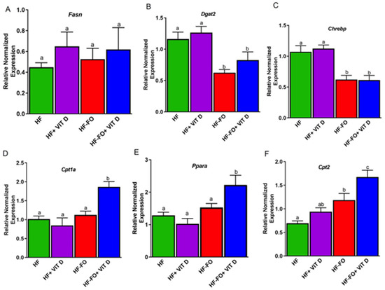
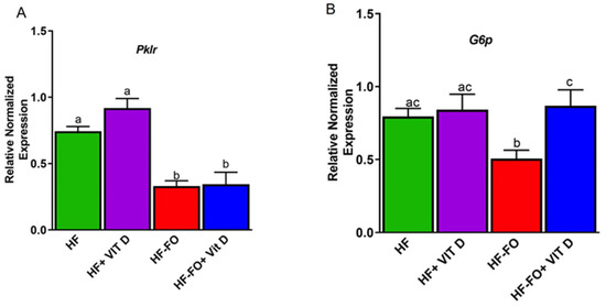
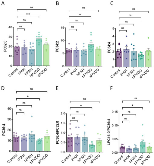
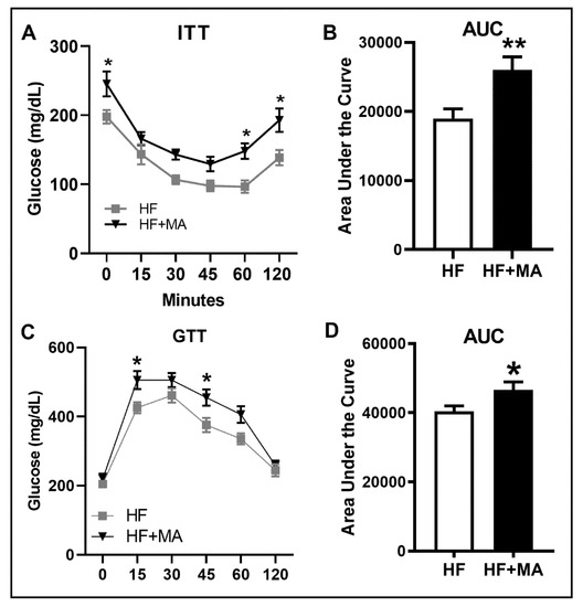
Vitamin D (vit D) and fish oil (FO) both offer unique health benefits, however, their combined effects have not been evaluated in obesity and nonalcoholic fatty liver disease (NAFLD). Hence, we hypothesized that vit D and FO supplementation would have additive effects in reducing obesity-associated inflammation and NAFLD. Male C57BL6 mice were split into four groups and fed a high fat (HF) diet supplemented with a low (HF; +200 IU vit D) or high dose of vitamin D (HF + D; +1000 IU vit D); combination of vit D and FO (HF-FO; +1000 IU vit D); or only FO (HF-FO; +200 IU vit D) for 12 weeks. We measured body weight, food intake, glucose tolerance, and harvested epididymal fat pad and liver for gene expression analyses. Adiposity was reduced in groups supplemented with both FO and vit D. Glucose clearance was higher in FO-supplemented groups compared to mice fed HF. In adipose tissue, markers of fatty acid synthesis and oxidation were comparable in groups that received vit D and FO individually in comparison to HF. However, the vit D and FO group had significantly lower fatty acid synthesis and higher oxidation compared to the other groups. Vit D and FO also significantly improved fatty acid oxidation, despite similar fatty acid synthesis among the four groups in liver. Even though we did not find additive effects of vit D and FO, our data provide evidence that FO reduces markers of obesity in the presence of adequate levels of vit D.
Full article
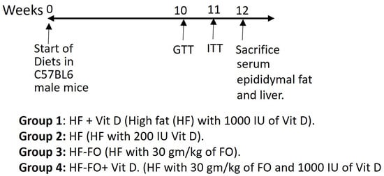
Figure 1


