


Biomolecules 2023, 13(2), 256; https://doi.org/10.3390/biom13020256 - 30 Jan 2023
Cited by 3 | Viewed by 1636
Abstract
►
Show Figures
Thorough study of composition and fluorescence properties of a commercial reagent of active equine NAD-dependent alcohol dehydrogenase expressed and purified from E. coli has been carried out. Several experimental methods: spectral- and time-resolved two-photon excited fluorescence, sodium dodecyl sulfate–polyacrylamide gel electrophoresis, fast protein
[...] Read more.
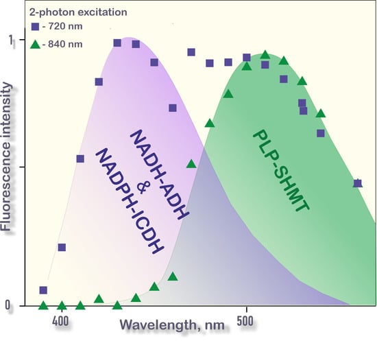
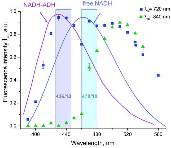
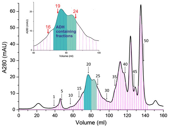
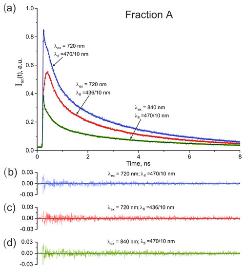
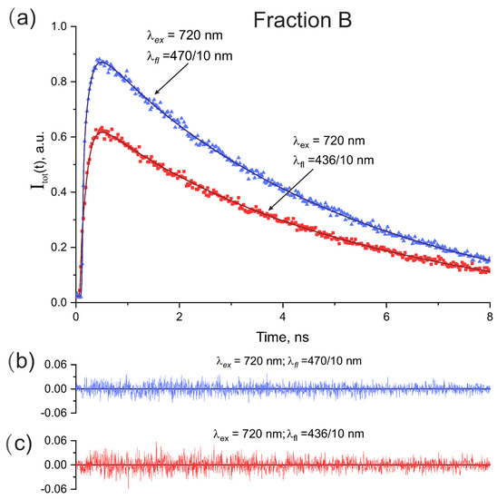
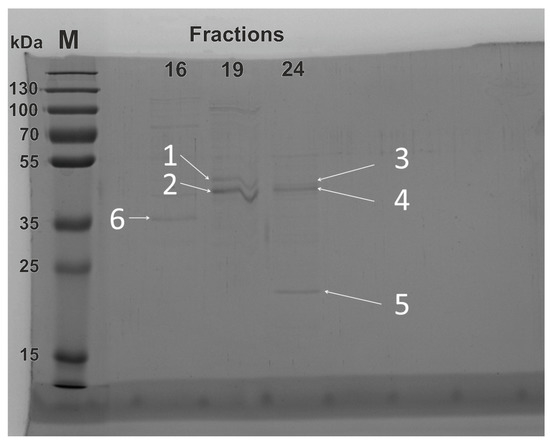
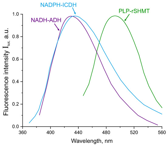
Thorough study of composition and fluorescence properties of a commercial reagent of active equine NAD-dependent alcohol dehydrogenase expressed and purified from E. coli has been carried out. Several experimental methods: spectral- and time-resolved two-photon excited fluorescence, sodium dodecyl sulfate–polyacrylamide gel electrophoresis, fast protein liquid chromatography, and mass spectrometry were used for analysis. The reagent under study was found to contain also a number of natural fluorophores: free NAD(P)H, NADH-alcohol dehydrogenase, NADPH-isocitrate dehydrogenase, and pyridoxal 5-phosphate—serine hydroxymethyltransferase complexes. The results obtained demonstrated the potential and limitations of popular optical methods as FLIM for separation of fluorescence signals from free and protein-bound forms of NADH, NADPH, and FAD that are essential coenzymes in redox reactions in all living cells. In particular, NADH-alcohol dehydrogenase and NADPH-isocitrate dehydrogenase complexes could not be optically separated in our experimental conditions although fast protein liquid chromatography and mass spectrometry analysis undoubtedly indicated the presence of both enzymes in the molecular sample used. Also, the results of fluorescence, fast protein liquid chromatography, and mass spectrometry analysis revealed a significant contribution of the enzyme-bound coenzyme pyridoxal 5-phosphate to the fluorescence signal that could be separated from enzyme-bound NADH by using bandpass filters, but could effectively mask contribution from enzyme-bound FAD because the fluorescence spectra of the species practically overlapped. It was shown that enzyme-bound pyridoxal 5-phosphate fluorescence can be separated from enzyme-bound NAD(P)H and FAD through analysis of short fluorescence decay times of about tens of picoseconds. However, this analysis was found to be effective only at relatively high number of peak photon counts in recorded fluorescence signals. The results obtained in this study can be used for interpretation of fluorescence signals from a mixture of enzyme-bound fluorophores and should be taken into consideration when determining the intracellular NADH/FAD ratio using FLIM.
Full article
Graphical abstract
