


Biology 2024, 13(3), 146; https://doi.org/10.3390/biology13030146 - 26 Feb 2024
Viewed by 1243
Abstract
►
Show Figures
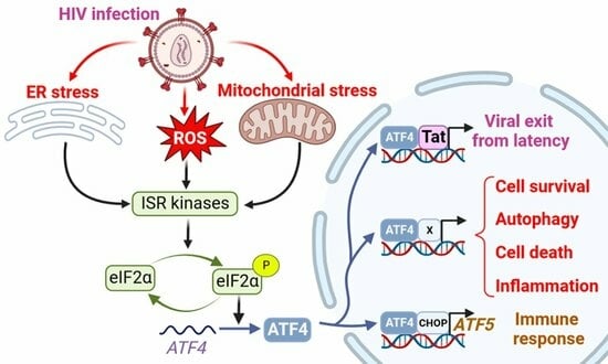
Cellular integrated stress response (ISR), the mitochondrial unfolded protein response (UPRmt), and IFN signaling are associated with viral infections. Activating transcription factor 4 (ATF4) plays a pivotal role in these pathways and controls the expression of many genes involved in redox processes, amino
[...] Read more.
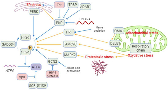
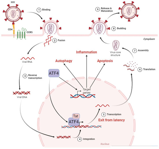
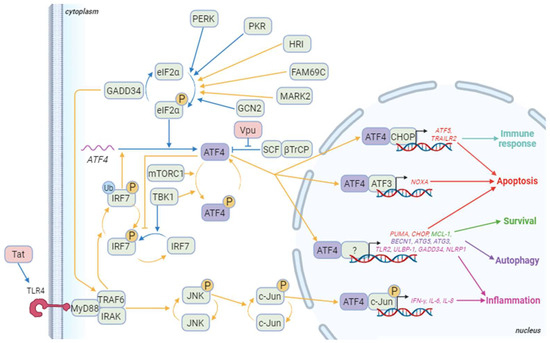
Cellular integrated stress response (ISR), the mitochondrial unfolded protein response (UPRmt), and IFN signaling are associated with viral infections. Activating transcription factor 4 (ATF4) plays a pivotal role in these pathways and controls the expression of many genes involved in redox processes, amino acid metabolism, protein misfolding, autophagy, and apoptosis. The precise role of ATF4 during viral infection is unclear and depends on cell hosts, viral agents, and models. Furthermore, ATF4 signaling can be hijacked by pathogens to favor viral infection and replication. In this review, we summarize the ATF4-mediated signaling pathways in response to viral infections, focusing on human immunodeficiency virus 1 (HIV-1). We examine the consequences of ATF4 activation for HIV-1 replication and reactivation. The role of ATF4 in autophagy and apoptosis is explored as in the context of HIV-1 infection programmed cell deaths contribute to the depletion of CD4 T cells. Furthermore, ATF4 can also participate in the establishment of innate and adaptive immunity that is essential for the host to control viral infections. We finally discuss the putative role of the ATF4 paralogue, named ATF5, in HIV-1 infection. This review underlines the role of ATF4 at the crossroads of multiple processes reflecting host–pathogen interactions.
Full article
Graphical abstract
