Anatomy and Regenerative Medicine: From Methods to Applications

A special issue of Applied Biosciences (ISSN 2813-0464).

Deadline for manuscript submissions: 30 September 2024 | Viewed by 6566

Special Issue Editors


E-Mail Website
Guest Editor
Department of Biomedicine, Neurosciences and Advanced Diagnostic (BiND), Human Anatomy Section, University of Palermo, via del Vespro 129, 90127 Palermo, Italy
Interests: tissue engineering; human anatomy; stem-cell therapies

E-Mail Website
Guest Editor
Department of Theoretical and Applied Sciences, eCampus University, 22060 Novedrate, CO, Italy
Interests: artificial organs; bioengineering; regenerative medicine; tissue engineering; biomaterials

E-Mail Website
Guest Editor
Department of Biomedicine, Neurosciences and Advanced Diagnostic (BiND), Human Anatomy Section, University of Palermo, via del Vespro 129, 90127 Palermo, Italy
Interests: human anatomy; biomedical engineering; 3D cell cultures

Special Issue Information

Dear Colleagues,

Anatomy and regenerative medicine (RM) are two disciplines that are strongly interconnected. The promising field of regenerative medicine may be defined as the process of replacing or "regenerating" human cells, tissues or organs to restore or establish normal functions. Beginning from the basics provided by human anatomy remains the best approach. Years of studies of and insights into the composition of the human body offer a solid starting ground on which to develop new therapeutic paths. Macro-anatomy data contribute to the replacement/healing of entire organs, and the field of nanotechnology uses micro-anatomy discoveries.

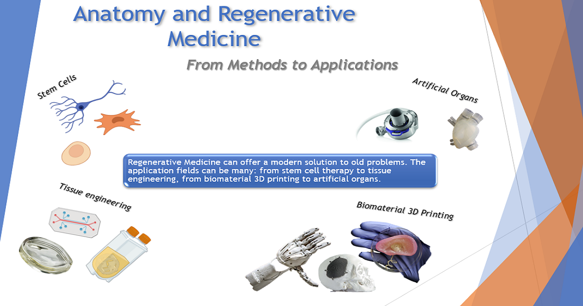
RM can offer a modern solution to existing long-term problems. There is an extensive number of application fields: from stem-cell therapy to tissue engineering; from biomaterial 3D printing to artificial organs.

The achievements of these applications are often the result of collaboration with scientists outside of the clinical area (bioengineers, materials engineers, biologists).

This Special Issue of Applied Biosciences, "Anatomy and Regenerative Medicine: From Methods to Applications", is committed to all new discoveries and applications of RM. Research papers that emphasize the shift from anatomical data to practical applications will be particularly appreciated.

Dr. Alessandro Pitruzzella
Dr. Alberto Fucarino
Prof. Dr. Fabio Bucchieri
Guest Editors

Manuscript Submission Information

Manuscripts should be submitted online at www.mdpi.com by registering and logging in to this website. Once you are registered, click here to go to the submission form. Manuscripts can be submitted until the deadline. All submissions that pass pre-check are peer-reviewed. Accepted papers will be published continuously in the journal (as soon as accepted) and will be listed together on the special issue website. Research articles, review articles as well as short communications are invited. For planned papers, a title and short abstract (about 100 words) can be sent to the Editorial Office for announcement on this website.

Submitted manuscripts should not have been published previously, nor be under consideration for publication elsewhere (except conference proceedings papers). All manuscripts are thoroughly refereed through a single-blind peer-review process. A guide for authors and other relevant information for submission of manuscripts is available on the Instructions for Authors page. Applied Biosciences is an international peer-reviewed open access quarterly journal published by MDPI.

Please visit the Instructions for Authors page before submitting a manuscript. The Article Processing Charge (APC) for publication in this open access journal is 1000 CHF (Swiss Francs). Submitted papers should be well formatted and use good English. Authors may use MDPI's English editing service prior to publication or during author revisions.

Keywords

  • human anatomy
  • regenerative medicine
  • nanotechnology
  • bioengineering
  • stem cell therapies
  • tissue engineering
  • artificial organs
  • biomaterials

Published Papers (4 papers)

Order results
Result details
Select all
Export citation of selected articles as:

Research

Jump to: Review

21 pages, 5459 KiB  
Article
Methods for Testing Meniscal Repair Using a 3D-Printed Meniscus
by Andrew Nelson, Steven Voinier, Jeremy Tran, Kristin H. Gilchrist, Melvin Helgeson, Vincent B. Ho and George J. Klarmann
Appl. Biosci. 2024, 3(1), 102-122; https://doi.org/10.3390/applbiosci3010007 - 06 Feb 2024
Viewed by 1101
Abstract
Torn and damaged menisci resulting from trauma are very common knee injuries, which can cause pain and mobility limitations and lead to osteoarthritis. Meniscal injuries can require surgery to repair the tissue damage and restore mobility. Here we describe the biomechanical testing of [...] Read more.
Torn and damaged menisci resulting from trauma are very common knee injuries, which can cause pain and mobility limitations and lead to osteoarthritis. Meniscal injuries can require surgery to repair the tissue damage and restore mobility. Here we describe the biomechanical testing of a 3D-printed meniscus to illustrate methods to determine if it has the strength and durability to effectively repair meniscal tears and restore knee biomechanics. This work was designed to demonstrate the steps needed to test novel meniscus repair devices prior to moving toward animal testing. The first testing step determined the ability of the 3D-printed meniscus to withstand surgical fixation by measuring the suture pull-out force. We show that vertical 2/0 silk or Fiberwire sutures need an average of 1.4 or 1.8 N, respectively, to pull through the meniscus, while horizontal sutures need only 0.7 and 1.2 N, respectively. The next step measured the compressive strength of normal, damaged, and repaired porcine meniscus tissue. Here, we show that meniscectomy decreased the stiffness of meniscus tissue from 26.7 ± 0.85 N to 7.43 ± 0.81 N at 25% strain. Menisci repaired with the 3D-printed tissue restored 66% of the measured force at 25% strain. The final step measured the contact pressures and areas in an ex vivo porcine knee before and after meniscal repair was made with the 3D-printed meniscus tissue. The example 3D-printed meniscus was successfully sutured into the porcine knee joint but failed to restore normal knee contact pressures. This work demonstrates the need for an iterative biomechanical testing process of biomaterial development, 3D-printing optimization, and knee kinematics to develop a durable and functional meniscus repair device. In summary, the methods described here serve as a guide for the functional evaluation of novel meniscus repair devices. Full article
(This article belongs to the Special Issue Anatomy and Regenerative Medicine: From Methods to Applications)
Show Figures

Figure 1

29 pages, 13940 KiB  
Article
Computational and Experimental Investigation of the Combined Effect of Various 3D Scaffolds and Bioreactor Stimulation on Human Cells’ Feedback
by Foteini K. Kozaniti, Aikaterini E. Manara, Vassilis Kostopoulos, Panagiotis Mallis, Efstathios Michalopoulos, Demosthenes Polyzos, Despina D. Deligianni and Diana V. Portan
Appl. Biosci. 2023, 2(2), 249-277; https://doi.org/10.3390/applbiosci2020018 - 01 Jun 2023
Cited by 3 | Viewed by 1339
Abstract
Computational methods were combined with an experimental setup in order to investigate the response of human umbilical cord stem cells to 3D electrospun and printed scaffolds, when dynamically stimulated in a bioreactor. Key parameters associated to bioreactor working conditions were computationally investigated using [...] Read more.
Computational methods were combined with an experimental setup in order to investigate the response of human umbilical cord stem cells to 3D electrospun and printed scaffolds, when dynamically stimulated in a bioreactor. Key parameters associated to bioreactor working conditions were computationally investigated using Comsol software to use the output for the planned experimental setup. Based on the theoretical observations, the influence of the inlet velocity, cell number, and exposure time in the bioreactor were analyzed and the in vitro parameters were adjusted accordingly. MSCs were seeded in different numbers in the 3D porous scaffolds and stimulated in the bioreactor (0.5 and 2 h duration, 3 and 6 mm/s inlet velocity). Polycaprolactone 3D electrospun, and polyurethane and polylactic acid 3D-printed scaffolds were fabricated and fibronectin-coated. The computational study predicted initial events in the process of cells deposition and attachment. Total protein, osteopontin, and osteocalcin levels in cells deposited in scaffolds were investigated; SEM and confocal imaging confirmed the biomarker analysis. MSCs proliferated well in PCL. Polyurethane enabled extremely rapid proliferation followed by differentiation, while PLA induced a moderate proliferation and parallel mineralization. The scaffolds stiffness has been found as the key enabling parameter decisive for cells feedback. Full article
(This article belongs to the Special Issue Anatomy and Regenerative Medicine: From Methods to Applications)
Show Figures

Figure 1

Review

Jump to: Research

22 pages, 1494 KiB  
Review
Nanobiotechnology in Bone Tissue Engineering Applications: Recent Advances and Future Perspectives
by Neelam Iqbal, Tejal Pant, Nanda Rohra, Abhishek Goyal, Merin Lawrence, Anomitra Dey and Payal Ganguly
Appl. Biosci. 2023, 2(4), 617-638; https://doi.org/10.3390/applbiosci2040039 - 15 Nov 2023
Cited by 1 | Viewed by 1003
Abstract
Bone regeneration and repair are complex processes with the potential of added complications, like delayed repair, fracture non-union, and post-surgical infections. These conditions remain a challenge globally, pressurizing the economy and patients suffering from these conditions. Applications of nanotechnology (NBT) in the field [...] Read more.
Bone regeneration and repair are complex processes with the potential of added complications, like delayed repair, fracture non-union, and post-surgical infections. These conditions remain a challenge globally, pressurizing the economy and patients suffering from these conditions. Applications of nanotechnology (NBT) in the field of medicine have provided a medium for several approaches to support these global challenges. Tissue engineering is one such field that has been on the rise in the last three decades through the utilization of NBT for addressing the challenges related to bone regeneration. First, NBT enables the formation of scaffolds at the nanoscale needed for bone tissue engineering (BTE) using natural and synthetic polymers, as well as with minerals and metals. Then, it aids the development of the nano-formulation strategized to deliver antimicrobial drugs and/or growth factors through various ways to enhance bone repair through the scaffold. Third, NBT facilitates the use of specialized nanoparticles to image and track cellular events in vitro as well as in vivo. This review is an effort to bring together the current knowledge in the field of BTE and present the scope of ever-evolving NBT, a contribution towards precision medicine. Full article
(This article belongs to the Special Issue Anatomy and Regenerative Medicine: From Methods to Applications)
Show Figures

Figure 1

16 pages, 1913 KiB  
Review
How Far Are We from Research That Is Independent of the Use of Animal Models? A Comparative Analysis between Animal and 3D/On-a-Chip Models for the Study of Respiratory Diseases
by Stefano Burgio, Olga Maria Manna, Giorgia Intili, Francesco Cappello and Fabio Bucchieri
Appl. Biosci. 2023, 2(2), 157-172; https://doi.org/10.3390/applbiosci2020012 - 02 Apr 2023
Viewed by 1846
Abstract
Over the last ten years, with the progress of in vitro culture methods, it has been possible to build increasingly reliable models to effectively mimic in vivo ones. The translational methodological approach that combined biotechnology and biomedical engineering has produced remarkable results, such [...] Read more.
Over the last ten years, with the progress of in vitro culture methods, it has been possible to build increasingly reliable models to effectively mimic in vivo ones. The translational methodological approach that combined biotechnology and biomedical engineering has produced remarkable results, such as the development of ex vivo 3D culture models, the construction of on-a-chip organoids, and the construction of complex systems capable of bypassing the static nature of the two-dimensional cultural models that have been typical of in vitro studies conducted to date. However, nowadays, there is still reluctance to completely abandon the animal model as an essential reference or as an integrated step for the validation of a model or a proposed study. This is due to the partially correct conviction of the impossibility of reproducing, in vitro or ex vivo, the complexity of pathological models or the spatial communication between different cytotypes, as well as, more generally, the lack of systems capable of mimicking the dynamism of a complex in vivo system. In this study, we will compare different methodological approaches in the study of the three most common types of respiratory diseases: chronic obstructive pulmonary disease (COPD), asthma, and lung carcinomas. The purpose of this comparative study is to evaluate the most current methodological approaches to understand how far research is from being independent from animal models. Animal studies are generally considered necessary, but are still questioned because of the ethics and the cost–benefit ratio involved. Full article
(This article belongs to the Special Issue Anatomy and Regenerative Medicine: From Methods to Applications)
Show Figures

Figure 1

Back to TopTop