Clinical Pathology in Animals

A topical collection in Animals (ISSN 2076-2615). This collection belongs to the section "Veterinary Clinical Studies".

Viewed by 58980

Editors


E-Mail Website
Guest Editor
School of Biosciences and Veterinary Medicine, University of Camerino, 62032 Camerino, Italy
Interests: clinical pathology; hematology; onco-hematology; transfusion medicine
Special Issues, Collections and Topics in MDPI journals

E-Mail Website
Guest Editor
Department of Veterinary Medicine, University of Milan, Lodi, Italy
Interests: veterinary clinical pathology; hematology; feline infectious peritonitis

Topical Collection Information

Dear Colleagues,

The theme of this Special Issue of Animals is Clinical Pathology in Animals.

The scope of this Special Issue is to provide a source for the publication of articles, reviews, case reports, and case studies covering all aspects of the clinical laboratory diagnosis of animal diseases. Particularly, we encourage improving the knowledge and comparative aspects between different mammalian species, including humans, and breeds with regard of clinical chemistry, cytopathology, hematopathology, hemoparasites, coagulation, and coagulative disorders, bone marrow and hematopoietic neoplasia, biomarkers of disease, hormonal analysis, endocrine disease, infectious diseases of blood and bone marrow, inflammation and inflammatory mediators, genetic blood and biochemical disorders, as well as assay validations and method comparisons. This Special Issue should encourage good contact between clinicians, pathologists, and biochemical experts to support daily work, scientific research, increase knowledge, and to facilitate coordination of clinical research in all species.

Constant work by experts in the many areas of interest in Veterinary Clinical Pathology is important so that knowledge can be extended both in terms of early and accurate laboratory diagnoses of animal diseases and in terms of new comparative aspects with human diseases. 

Dr. Arianna Miglio
Dr. Angelica Stranieri
Guest Editors

Manuscript Submission Information

Manuscripts should be submitted online at www.mdpi.com by registering and logging in to this website. Once you are registered, click here to go to the submission form. Manuscripts can be submitted until the deadline. All submissions that pass pre-check are peer-reviewed. Accepted papers will be published continuously in the journal (as soon as accepted) and will be listed together on the collection website. Research articles, review articles as well as short communications are invited. For planned papers, a title and short abstract (about 100 words) can be sent to the Editorial Office for announcement on this website.

Submitted manuscripts should not have been published previously, nor be under consideration for publication elsewhere (except conference proceedings papers). All manuscripts are thoroughly refereed through a single-blind peer-review process. A guide for authors and other relevant information for submission of manuscripts is available on the Instructions for Authors page. Animals is an international peer-reviewed open access semimonthly journal published by MDPI.

Please visit the Instructions for Authors page before submitting a manuscript. The Article Processing Charge (APC) for publication in this open access journal is 2400 CHF (Swiss Francs). Submitted papers should be well formatted and use good English. Authors may use MDPI's English editing service prior to publication or during author revisions.

Keywords

  • clinical chemistry
  • cytopathology
  • hemoparasites
  • bone marrow
  • hemostasis
  • immunology
  • immunophenotyping
  • hematopoietic neoplasia
  • endocrine disease
  • inflammatory mediators
  • biomarkers

Published Papers (21 papers)

2023

Jump to: 2022, 2021, 2020

10 pages, 287 KiB  
Article
Establishment of Reference Intervals of Hematological Parameters and Evaluation of Sex and Age Effect in the Miranda Donkey
by Grasiene Silva, Felisbina Queiroga, Madalena Ferreira, Daniela Andrade and Ana C. Silvestre-Ferreira
Animals 2023, 13(14), 2331; https://doi.org/10.3390/ani13142331 - 17 Jul 2023
Cited by 1 | Viewed by 1427
Abstract
The Miranda donkey is an autochthonous Portuguese breed that is considered endangered. Several studies have been carried out on this breed, but to the authors’ best knowledge, no studies have been conducted on their clinical pathology. The aims of this study were to [...] Read more.
The Miranda donkey is an autochthonous Portuguese breed that is considered endangered. Several studies have been carried out on this breed, but to the authors’ best knowledge, no studies have been conducted on their clinical pathology. The aims of this study were to determine the hematological reference intervals (RIs) in healthy Miranda donkeys and to estimate the influence of age and sex. Blood samples from 75 clinically healthy animals were analyzed for 22 hematological parameters on the IDEXX ProCyte Dx, an automated hematology analyzer previously validated for the species. The RIs were estimated following the ASVCP guidelines with the Reference Value Advisor software. Regarding sex, no significant differences were found between groups. Regarding age, significant statistical differences (p < 0.05) were observed for red blood cells, red cell distribution width, white blood cells, lymphocytes, monocytes, platelets, plateletcrit (higher mean in young animals), mean corpuscular volume, mean corpuscular haemoglobin, neutrophils and eosinophils (higher mean in adults). The RIs described here can be used to evaluate and monitor the health status of animals and herds, as well as to guide diagnoses or select fit and healthy animals for reproduction, contributing to the preservation of the breed. Full article
17 pages, 1774 KiB  
Article
Simplified Light’s Criteria and Acute Phase Proteins Reflect Aetiology of Feline Body Cavity Effusions Better than the Traditional Classification Scheme
by Katarina Hazuchova, Susanne Held, Isabell Klemm and Natali Bauer
Animals 2023, 13(12), 1918; https://doi.org/10.3390/ani13121918 - 08 Jun 2023
Cited by 1 | Viewed by 1256
Abstract
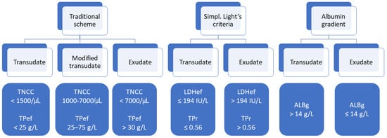
The traditional veterinary classification (TVC) of effusions based on cell count and total protein (TP) does not adequately reflect the aetiology. Light’s criteria (LC) (activity of lactate dehydrogenase [LDH] in the effusion [LDHef], effusion/serum LDH ratio [LDHr], effusion/serum TP ratio [TPr]), serum–effusion albumin [...] Read more.
The traditional veterinary classification (TVC) of effusions based on cell count and total protein (TP) does not adequately reflect the aetiology. Light’s criteria (LC) (activity of lactate dehydrogenase [LDH] in the effusion [LDHef], effusion/serum LDH ratio [LDHr], effusion/serum TP ratio [TPr]), serum–effusion albumin gradient (ALBg), acute phase proteins (APPs) [serum amyloid A (SAA), α1-acid glycoprotein (AGP), haptoglobin] might aid classification. The aim was to evaluate the utility of these parameters except LDHr in differentiating exudates from transudates. Sixty-five cats with effusions (33 peritoneal, 31 pleural, 1 pericardial), with 18 transudates and 47 exudates based on aetiological classification (AC), were included. The sensitivity, specificity and accuracy of several parameters to identify exudates (based on AC) was assessed. APPs were compared between exudates and transudates based on AC and TVC, with receiver operating characteristics analysis identifying the best APP to recognise exudates. Simplified LC (LDHef, TPr) had an accuracy of 79% and TVC of 48%. ALBg had the highest sensitivity (98%) and LDHef the highest specificity (83%) in identifying exudates in cats. All APPs but effusion SAA could differentiate exudates from transudates based on AC (effusion AGP had the largest area under the curve 0.79) but not TVC. All parameters were better than TVC in identifying exudates. The conformity of APPs with AC but not TVC favours the use of AC to classify effusions. Full article
Show Figures

Figure 1

11 pages, 1487 KiB  
Article
Ocular and Superficial Body Thermographic Findings in Sled Dogs before and after Competition
by Giuseppe Spinella, Andrea Galimberti, Giorgia Casagrande, Sergio Maffi, Vincenzo Musella and Simona Valentini
Animals 2023, 13(5), 854; https://doi.org/10.3390/ani13050854 - 26 Feb 2023
Cited by 1 | Viewed by 2049
Abstract
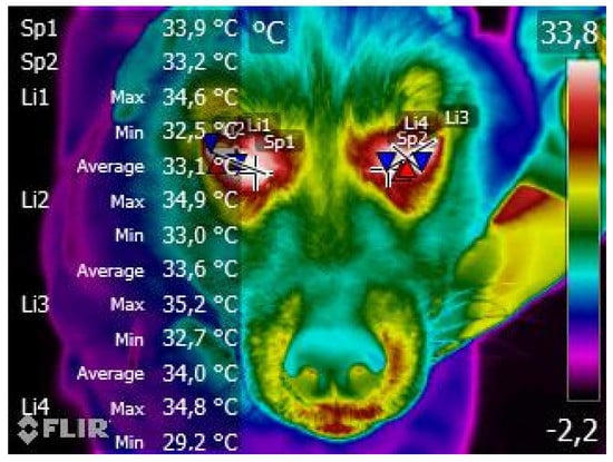
Competitions involving sled dogs are rapidly growing and body temperature assessment could represent a prompt and non-invasive method of screening for potential pathological conditions during or after activity. The aim of this clinical study was to evaluate if thermography is able to monitor [...] Read more.
Competitions involving sled dogs are rapidly growing and body temperature assessment could represent a prompt and non-invasive method of screening for potential pathological conditions during or after activity. The aim of this clinical study was to evaluate if thermography is able to monitor the pre- and post-competition ocular and superficial body temperature variations during a sled dog competition. It subsequently compared the data relating to the ocular temperatures in different race types: mid-distance (30 km) and sprint (≤16 km). Results showed a statistically significant increase in post-competition ocular temperature of both eyes, regardless of the length of the race. The relative increase in the temperatures of the other body surfaces was lower than the expected values, probably due to the influence of environmental and subjective factors such as the type of coat of the Siberian Husky or subcutaneous fat. Infrared thermography has therefore proved to be useful method in sled dog competition conditions for screening superficial temperature variations, as the investigation is normally conducted in an external environment and often in demanding work conditions. Full article
Show Figures

Figure 1

2022

Jump to: 2023, 2021, 2020

12 pages, 1306 KiB  
Article
Neutrophil Gelatinase-Associated Lipocalin in Synovial Fluid from Horses with and without Septic Arthritis
by Stine Jacobsen, Camilla Drejer Mortensen, Elisabeth Alkærsig Høj, Anne Mette Vinther, Lise Charlotte Berg, Ditte Marie Top Adler, Denis Verwilghen and Gaby van Galen
Animals 2023, 13(1), 29; https://doi.org/10.3390/ani13010029 - 21 Dec 2022
Cited by 1 | Viewed by 1260
Abstract
Neutrophil gelatinase-associated lipocalin (NGAL) has been suggested to be a highly sensitive and specific marker of joint infection in humans. The aim of the study was to investigate NGAL concentrations in synovial fluid (SF) from horses with septic synovitis, horses without septic synovitis, [...] Read more.
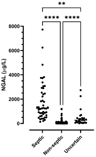
Neutrophil gelatinase-associated lipocalin (NGAL) has been suggested to be a highly sensitive and specific marker of joint infection in humans. The aim of the study was to investigate NGAL concentrations in synovial fluid (SF) from horses with septic synovitis, horses without septic synovitis, and horses with uncertain status. NGAL was measured in 177 admission samples obtained from 152 horses. From a subset of horses (n = 35), additional samples obtained sequentially over the course of treatment were available. Concentrations of NGAL were significantly higher in septic synovitis (n = 47 samples) than in samples classified as non-septic (n = 103) or samples with uncertain status (n = 27), with median NGAL concentrations in the three groups being 1236, 16.8, and 266.4 µg/L, respectively. NGAL discriminated nearly perfectly between septic and non-septic (area under the receiver operating characteristic curve 0.98, 95% confidence interval 0.95–1.00). The optimal cut-off value for maximal sensitivity (87.2%) and specificity (75.0%) to discriminate septic samples from those with uncertain status was 444.6 µg/L, with an area under the receiver operating characteristic curve of 0.85 (95% confidence interval 0.74–0.93). Concentrations declined over time in horses undergoing treatment. NGAL is a novel biomarker that seems to have great potential for identifying septic synovitis and for monitoring the response to treatment of synovial infection in horses. Full article
Show Figures

Figure 1

9 pages, 284 KiB  
Article
Evaluation of Eosinopenia as a SIRS Biomarker in Critically Ill Horses
by María Martín-Cuervo, Luis Alfonso Gracia-Calvo, Beatriz Macías-García, Luis Javier Ezquerra and Rafael Barrera
Animals 2022, 12(24), 3547; https://doi.org/10.3390/ani12243547 - 15 Dec 2022
Cited by 1 | Viewed by 1125
Abstract
Systemic inflammatory response syndrome (SIRS) is a very common finding in critically ill patients. To accurately identify patients with SIRS and those who need intensive care, several markers have been evaluated, including cortisol, WBC or lactate. It is widely known that a stress [...] Read more.
Systemic inflammatory response syndrome (SIRS) is a very common finding in critically ill patients. To accurately identify patients with SIRS and those who need intensive care, several markers have been evaluated, including cortisol, WBC or lactate. It is widely known that a stress leukogram includes eosinopenia as one of its main markers (neutrophilia, eosinopenia, lymphopenia and mild monocytes). It is known that cortisol concentration in plasma is the main stress biomarker and is strongly correlated with the severity of disease in horses. However, it is not possible to measure this parameter routinely in clinical conditions. Hence, in this study it was hypothesized that the eosinophil count could be a reliable parameter to identify critically ill horses. Horses included in this study were divided into three groups: Group A (sick horses received at the Emergency Unit which did not fulfil the criteria for SIRS), Group B (horses that meet two or more criteria for inclusion in the definition of SIRS) and a control group of healthy horses. In this study, horses with SIRS showed lower eosinophil counts than healthy horses. Moreover, non-surviving horses exhibited lower eosinophil counts than survivors. Eosinopenia could be used to identify horses with SIRS and can be useful as a prognostic marker. Full article
12 pages, 593 KiB  
Article
Prognostic Role of Red Cell Distribution Width and Other Routine Clinico-Pathological Parameters in Dogs with Acute Pancreatitis
by Carlo Guglielmini, Paolo Emidio Crisi, Antonio Maria Tardo, Roberta Di Maggio, Barbara Contiero, Andrea Boari, Federico Fracassi and Arianna Miglio
Animals 2022, 12(24), 3483; https://doi.org/10.3390/ani12243483 - 09 Dec 2022
Cited by 5 | Viewed by 1925
Abstract
This study aimed to assess the prognostic value of red cell distribution width (RDW) RDW-to-calcium ratio (RDW/Ca), neutrophils-to-lymphocytes ratio (N/L), platelets-to-lymphocytes ratio (P/L) and other easy to obtain and inexpensive hematological and biochemical parameters in dogs with acute pancreatitis. This is a multicenter, [...] Read more.
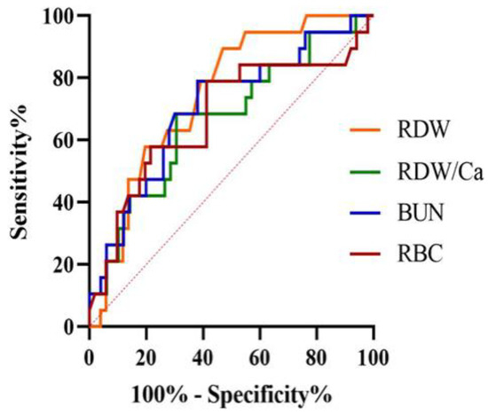
This study aimed to assess the prognostic value of red cell distribution width (RDW) RDW-to-calcium ratio (RDW/Ca), neutrophils-to-lymphocytes ratio (N/L), platelets-to-lymphocytes ratio (P/L) and other easy to obtain and inexpensive hematological and biochemical parameters in dogs with acute pancreatitis. This is a multicenter, retrospective cohort study including 70 client-owned dogs. The accuracy of clinical and laboratory variables to predict short-term death (i.e., dead by 14 days) was tested by calculating the area under the receiver-operating characteristic curve (AUC). Independent predictors of death were identified using the multivariable Cox proportional hazards regression model. The survival rate was 72.9% (51 dogs) and 19 dogs died within 14 days of admission from AP. RDW and blood urea nitrogen (BUN) had good accuracy to predict short-term dead with AUC of 0.74 and 0.70 at the cut-off of >12.7% and >42 mg/dL, respectively. According to the multivariable model, RDW (hazard ratio and 95% confidence interval [HR, 95% CI] = 5.08, 95% CI = 1.14–22.67; p = 0.03), BUN (HR = 1.00, 95% CI = 1.00–1.01; p < 0.01) and bilirubin (HR = 2.46, 95% CI = 1.38–4.39; p < 0.01) were independent predictors of death. The results indicate that RDW, BUN and bilirubin are useful predictors of short-term death in dogs with acute pancreatitis. Full article
Show Figures

Figure 1

13 pages, 876 KiB  
Article
Evaluation of a Comprehensive Profile of Salivary Analytes for the Diagnosis of the Equine Gastric Ulcer Syndrome
by Alberto Muñoz-Prieto, José J. Cerón, Camila P. Rubio, María Dolores Contreras-Aguilar, Luis Pardo-Marín, Ignacio Ayala-de la Peña, María Martín-Cuervo, Ida-Marie Holm Henriksen, Julián J. Arense-Gonzalo, Fernando Tecles and Sanni Hansen
Animals 2022, 12(23), 3261; https://doi.org/10.3390/ani12233261 - 23 Nov 2022
Cited by 4 | Viewed by 2430
Abstract
In this report, the measurement of salivary biomarkers as an aid for diagnosis of equine gastric ulcer syndrome (EGUS) was studied. A comprehensive panel of 23 salivary analytes was measured in the saliva of horses affected by EGUS and compared to healthy animals [...] Read more.
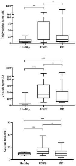
In this report, the measurement of salivary biomarkers as an aid for diagnosis of equine gastric ulcer syndrome (EGUS) was studied. A comprehensive panel of 23 salivary analytes was measured in the saliva of horses affected by EGUS and compared to healthy animals and horses with other diseases clinically similar to EGUS but with a negative diagnosis at gastroscopic examination. A total of 147 horses were included in the study and divided into heathy population (n = 12), the EGUS group (n = 110), and the group of horses with other diseases (n = 25). From the 23 analytes studied, 17 showed increased values in EGUS horses when compared to healthy ones, and uric acid, triglycerides, and calcium were significantly increased in horses with EGUS compared to the group of other diseases. The receiver operating characteristic curve analyses showed a modest but significant discriminatory power of those three analytes to identify EGUS from other diseases with similar symptoms. The discriminatory power enhanced when the results of the three analytes were combined. In conclusion, the results showed that selected salivary analytes could have potential use as biomarkers in horses with EGUS. Full article
Show Figures

Figure 1

12 pages, 1197 KiB  
Article
Evaluation of the Effect of Sampling Time on Biomarkers of Stress, Immune System, Redox Status and Other Biochemistry Analytes in Saliva of Finishing Pigs
by Alba Ortín-Bustillo, María D. Contreras-Aguilar, Camila P. Rubio, María Botia, José J. Cerón, Marina López-Arjona, Silvia Martínez-Subiela, Damián Escribano and Fernando Tecles
Animals 2022, 12(16), 2127; https://doi.org/10.3390/ani12162127 - 19 Aug 2022
Cited by 4 | Viewed by 1519
Abstract
This study aims to evaluate the possible variations due to the sampling time in the day in 26 analytes of pigs’ saliva, related to stress, the immune system, redox status and other biomarkers related to metabolism and selected tissues and organs, in order [...] Read more.
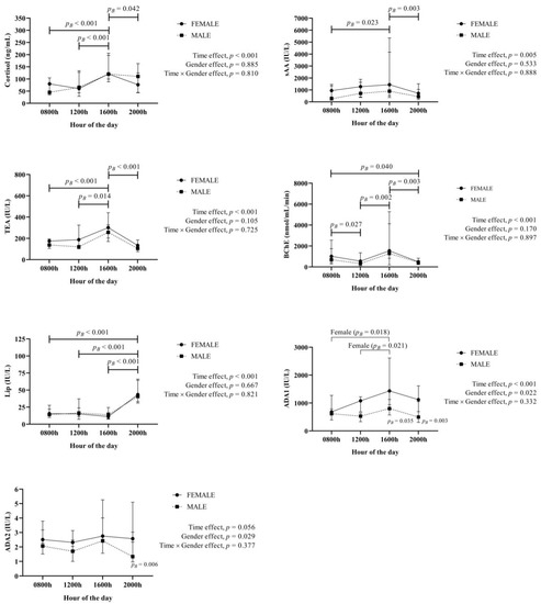
This study aims to evaluate the possible variations due to the sampling time in the day in 26 analytes of pigs’ saliva, related to stress, the immune system, redox status and other biomarkers related to metabolism and selected tissues and organs, in order to know the possible effects of the hour of the day in their interpretation. These analytes were measured in saliva obtained from a population of 40 clinically healthy pigs from 8 a.m. to 8 p.m., every 4 h in the same day. In our experimental conditions, daily variations were observed in cortisol, salivary α-amylase, total esterase activity, butyrylcholinesterase, lipase, adenosine deaminase isoenzyme 1, uric acid, superoxide dismutase, alanine aminotransferase, aspartate aminotransferase, alkaline phosphatase, creatine kinase, lactate dehydrogenase, lactate and triglycerides. These changes appeared in both sexes, except for adenosine deaminase isoenzyme 1 and superoxide dismutase which only showed differences in females. In conclusion, this report indicates that, in the experimental conditions of this trial, the time of the day and sex can influence the values obtained in various salivary analytes in pigs. These variations should thus be taken into consideration for an adequate interpretation of these analytes when used for the evaluation of health and welfare in this species. Full article
Show Figures

Figure 1

15 pages, 1123 KiB  
Article
Development of Colorimetric Reverse Transcription Loop-Mediated Isothermal Amplification Assay for Detecting Feline Coronavirus
by Witsanu Rapichai, Wichayet Saejung, Kotchaporn Khumtong, Chaiwat Boonkaewwan, Supansa Tuanthap, Peter A. Lieberzeit, Kiattawee Choowongkomon and Jatuporn Rattanasrisomporn
Animals 2022, 12(16), 2075; https://doi.org/10.3390/ani12162075 - 14 Aug 2022
Cited by 7 | Viewed by 2790
Abstract
Feline infectious peritonitis (FIP) is a worldwide fatal disease caused by a mutant feline coronavirus (FCoV). Simple and efficient molecular detection methods are needed. Here, sensitive, specific, rapid, and reliable colorimetric reverse transcription loop-mediated isothermal amplification (RT-LAMP) was developed to detect the ORF1a/1b [...] Read more.
Feline infectious peritonitis (FIP) is a worldwide fatal disease caused by a mutant feline coronavirus (FCoV). Simple and efficient molecular detection methods are needed. Here, sensitive, specific, rapid, and reliable colorimetric reverse transcription loop-mediated isothermal amplification (RT-LAMP) was developed to detect the ORF1a/1b gene of FCoV from cats with suspected FIP using neutral red as an indicator. Novel LAMP primers were specifically designed based on the gene of interest. The isothermal assay could visually detect FCoV at 58 °C for 50 min. The RT-LAMP assay was highly specific and had no cross-reactivity with other related feline viruses. The detection limit of FCoV detection by RT-LAMP was 20 fg/µL. A blind clinical test (n = 81) of the developed RT-LAMP procedure was in good agreement with the conventional PCR method. In the light of its performance specificity, sensitivity, and easy visualization, this neutral-red-based RT-LAMP approach would be a fruitful alternative molecular diagnostic tool for veterinary inspection of FCoV when combined with nucleotide sequencing or specific PCR to affirm the highly virulent FIP-associated FCoV. Full article
Show Figures

Figure 1

14 pages, 300 KiB  
Article
Changes in a Comprehensive Profile of Saliva Analytes in Fattening Pigs during a Complete Productive Cycle: A Longitudinal Study
by Alba Ortín-Bustillo, Damián Escribano, Marina López-Arjona, María Botia, Pablo Fuentes, Silvia Martínez-Miró, Camila P. Rubio, Edgar García-Manzanilla, Lorena Franco-Martínez, Luis Pardo-Marín, José J. Cerón, Pol Llonch and Fernando Tecles
Animals 2022, 12(14), 1865; https://doi.org/10.3390/ani12141865 - 21 Jul 2022
Cited by 9 | Viewed by 1577
Abstract
A comprehensive panel of 29 salivary analytes was measured in fattening pigs to evaluate its possible changes along their productive cycle. The identification of those changes would allow a better interpretation of the results according to the productive phase of the animal. Saliva [...] Read more.
A comprehensive panel of 29 salivary analytes was measured in fattening pigs to evaluate its possible changes along their productive cycle. The identification of those changes would allow a better interpretation of the results according to the productive phase of the animal. Saliva samples were obtained from 49 Large-White pigs (24 females, 25 males) in suckling phase, at the beginning and the end of the nursery phase, and at the beginning and the end of the growing phase. Several analytes changed according to the phase of the productive cycle, with most of the analytes showing higher values at lactation and at the beginning of nursery. Additionally, differences were seen due to sex. When possible relations between performance parameters and analytes were evaluated, significant positive but weak relationships were found between weight at birth and salivary γ-glutamyl transferase, and between back-fat thickness and salivary lactate dehydrogenase. In conclusion, differences in the values of salivary analytes can be found in fattening pigs depending on the productive phase and sex of the animals. Full article
14 pages, 301 KiB  
Article
Changes in Proteins in Saliva and Serum in Equine Gastric Ulcer Syndrome Using a Proteomic Approach
by Alberto Muñoz-Prieto, Maria Dolores Contreras-Aguilar, Jose Joaquín Cerón, Ignacio Ayala, Maria Martin-Cuervo, Juan Carlos Gonzalez-Sanchez, Stine Jacobsen, Josipa Kuleš, Anđelo Beletić, Ivana Rubić, Vladimir Mrljak, Fernando Tecles and Sanni Hansen
Animals 2022, 12(9), 1169; https://doi.org/10.3390/ani12091169 - 02 May 2022
Cited by 12 | Viewed by 9763
Abstract
Changes in the salivary proteome in 12 horses with the two diseases included in equine gastric ulcer syndrome (EGUS), equine glandular gastric disease (EGGD) (n = 6) and equine squamous gastric disease (ESGD) (n = 6), were evaluated using a high-resolution [...] Read more.
Changes in the salivary proteome in 12 horses with the two diseases included in equine gastric ulcer syndrome (EGUS), equine glandular gastric disease (EGGD) (n = 6) and equine squamous gastric disease (ESGD) (n = 6), were evaluated using a high-resolution LC-MS/MS analysis of TMT-labelled peptides and compared to 10 healthy control horses. Serum was also analysed for comparative purposes. The comparison between the horses with EGGD and controls showed significant changes in 10 salivary proteins, whereas 36 salivary proteins were differently abundant between ESGD and control groups. The most upregulated proteins in the case of EGGD were related to immune activation whereas, in horses with ESGD, the most significantly changed proteins were associated with squamous cell regulation and growth. Compared to serum, saliva showed a higher number of proteins with significant changes and a different pattern of changes. The proteins identified in our study, in addition to providing new information about the pathophysiological mechanisms in these diseases, could have the potential to be novel biomarkers for the diagnosis or monitoring of EGGD and ESGD. Full article

2021

Jump to: 2023, 2022, 2020

13 pages, 22526 KiB  
Communication
Natural Bovine Coronavirus Infection in a Calf Persistently Infected with Bovine Viral Diarrhea Virus: Viral Shedding, Immunological Features and S Gene Variations
by Annamaria Pratelli, Maria Stella Lucente, Marco Cordisco, Stefano Ciccarelli, Roberta Di Fonte, Alessio Sposato, Viviana Mari, Paolo Capozza, Francesco Pellegrini, Grazia Carelli, Amalia Azzariti and Canio Buonavoglia
Animals 2021, 11(12), 3350; https://doi.org/10.3390/ani11123350 - 23 Nov 2021
Cited by 4 | Viewed by 2085
Abstract
The evolution of a bovine coronavirus (BCoV) natural infection in a calf persistently infected with bovine viral diarrhea virus (BVDV) was described. The infected calf developed intermittent nasal discharge, diarrhea and hyperthermia. The total number of leukocytes/mL and the absolute differential number of [...] Read more.
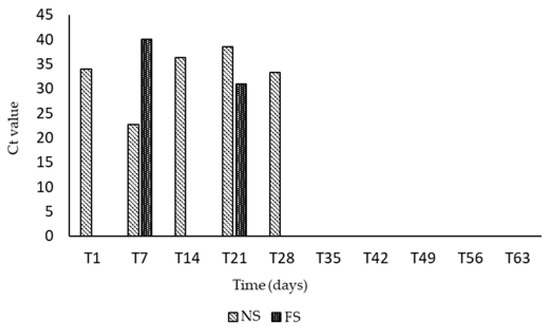
The evolution of a bovine coronavirus (BCoV) natural infection in a calf persistently infected with bovine viral diarrhea virus (BVDV) was described. The infected calf developed intermittent nasal discharge, diarrhea and hyperthermia. The total number of leukocytes/mL and the absolute differential number of neutrophils and lymphocytes resulted within the normal range, but monocytes increased at T28 (time 28 post-infection). Flow-cytometry analysis evidenced that the CD8+ subpopulation increased at T7 and between T28 and T35. BCoV shedding in nasal discharges and feces was detected up to three weeks post infection and high antibody titers persisted up to T56. The RNA BCoV load increased until T14, contrary to what was observed in a previous study where the fecal excretion of BCoV was significantly lower in the co-infected (BCoV/BVDV) calves than in the calves infected with BCoV only. We can suppose that BVDV may have modulated the BCoV infection exacerbating the long viral excretion, as well as favoring the onset of mutations in the genome of BCoV detected in fecal samples at T21. An extensive study was performed to verify if the selective pressure in the S gene could be a natural mode of variation of BCoV, providing data for the identification of new epidemic strains, genotypes or recombinant betacoronaviruses. Full article
Show Figures

Figure 1

16 pages, 350 KiB  
Review
Current Diagnostic Methods for Assessing Transfer of Passive Immunity in Calves and Possible Improvements: A Literature Review
by Rayanne Soalheiro de Souza, Lucas Braga Costa dos Santos, Isabela Oliveira Melo, Daiane Maria Cerqueira, Juliana Vieira Dumas, Fabiola de Oliveira Paes Leme, Tiago Facury Moreira, Rodrigo Melo Meneses, Antônio Ultimo de Carvalho and Elias Jorge Facury-Filho
Animals 2021, 11(10), 2963; https://doi.org/10.3390/ani11102963 - 14 Oct 2021
Cited by 10 | Viewed by 2964
Abstract
Several direct or indirect methods can be used to assess immunoglobulin G (IgG) concentrations in calves, which evaluates the transfer of passive immunity (TPI). Radial immunodiffusion (RID) is the gold standard method to measure serum IgG in bovines. Previous studies have shown that [...] Read more.
Several direct or indirect methods can be used to assess immunoglobulin G (IgG) concentrations in calves, which evaluates the transfer of passive immunity (TPI). Radial immunodiffusion (RID) is the gold standard method to measure serum IgG in bovines. Previous studies have shown that colostrum provides several molecules in addition to immunoglobulins, which play an important role in the passive immunity of the calf. However, no studies have yet determined the level of interference of these components in the immunity, health and survival of calves. In this sense, the objective of this study is to review the methods of evaluation available for the laboratory and field diagnosis of TPI in calves and discuss the main aspects of each technique. Several methods available for TPI evaluation in calves may provide insights into the various components of colostrum involved in passive immunity. Full article
7 pages, 867 KiB  
Communication
Analytical Validation of Two Point-of-Care Assays for Serum Amyloid A Measurements in Cats
by Damián Escribano, Alba Ortín Bustillo, Luis Pardo Marín, Andrea Navarro Rabasco, Pablo Ruiz Herrera, Jose J. Cerón and Asta Tvarijonaviciute
Animals 2021, 11(9), 2518; https://doi.org/10.3390/ani11092518 - 27 Aug 2021
Cited by 3 | Viewed by 2452
Abstract
Serum Amyloid A (SAA) is one of the most sensitive tests to detect inflammation in cats. In this study, two point-of-care assays for SAA measurements in cats (FUJI DRI-CHEM IMMUNO AU CARTRIDGE vf-SAA (method A), and CUBE-VET analyser (Method B), were analytically evaluated. [...] Read more.
Serum Amyloid A (SAA) is one of the most sensitive tests to detect inflammation in cats. In this study, two point-of-care assays for SAA measurements in cats (FUJI DRI-CHEM IMMUNO AU CARTRIDGE vf-SAA (method A), and CUBE-VET analyser (Method B), were analytically evaluated. Regarding the imprecision precision only the method A showed intra-assay and inter-assay CV < 10% at all concentrations. Both assays showed linearity with r close to 1 and the recovery were in the range of 81–112% for assay A and 85–125% for assay B and the limit of detection were 3.75 and 0.5 mg/dL for method A and B, respectively. A previously validated method for SAA quantification SAATIA; LZ-SAA (method C) was used as gold-standard to evaluate the accuracy of the assays. Significant correlations (p < 0.0001) were found between assays A and C (r = 0.94) and B and C (r = 0.91). In addition, an overlap performance test was made using serum samples from cats with non-inflammatory and cats with inflammatory. Both assays showed higher median SAA concentrations in cats with inflammatory diseases than in cats without inflammatory diseases (p < 0.0001). In conclusion, this manuscript provides data about the possible application of two point-of-care assays for the measurement of SAA concentration in cats. Full article
Show Figures

Figure 1

14 pages, 1521 KiB  
Article
Altered Proteomic Profile of Adipose Tissue-Derived Mesenchymal Stem Cell Exosomes from Cats with Severe Chronic Gingivostomatitis
by Antonio J. Villatoro, María del Carmen Martín-Astorga, Cristina Alcoholado, Casimiro Cárdenas, Fernando Fariñas, José Becerra and Rick Visser
Animals 2021, 11(8), 2466; https://doi.org/10.3390/ani11082466 - 23 Aug 2021
Cited by 5 | Viewed by 3507
Abstract
Feline chronic gingivostomatitis (FCGS) is a pathology with a complicated therapeutic approach and with a prevalence between 0.7 and 12%. Although the etiology of the disease is diverse, feline calicivirus infection is known to be a predisposing factor. To date, the available treatment [...] Read more.
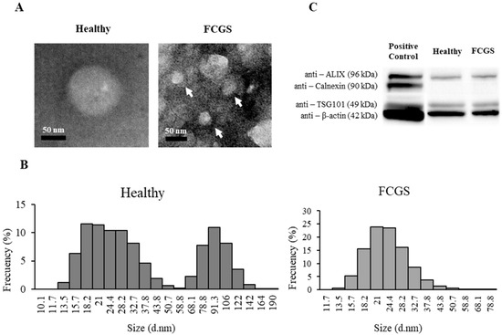
Feline chronic gingivostomatitis (FCGS) is a pathology with a complicated therapeutic approach and with a prevalence between 0.7 and 12%. Although the etiology of the disease is diverse, feline calicivirus infection is known to be a predisposing factor. To date, the available treatment helps in controlling the disease, but cannot always provide a cure, which leads to a high percentage of refractory animals. Mesenchymal stem cells (MSCs) play a pivotal role in the homeostasis and reparation of different tissues and have the ability to modulate the immune system responses. This ability is, in part, due to the capacity of exosomes to play a part in intercellular cell communication. However, the precise role of MSC-derived exosomes and their alterations in immunocompromised pathologies remains unknown, especially in veterinary patients. The goal of this work was to analyze the proteomic profile of feline adipose tissue-derived MSCs (fAd-MSCs) from calicivirus-positive FCGS patients, and to detect possible modifications of the exosomal cargo, to gain better knowledge of the disease’s etiopathogenesis. Using high-resolution mass spectrometry and functional enrichment analysis with Gene Ontology, exosomes isolated from the fAd-MSCs of five healthy cats and five calicivirus-positive FCGS patients, were pooled and compared. The results showed that the fAd-MSCs from cats suffering from FCGS not only had a higher exosome production, but also their exosomes showed significant alterations in their proteomic profile. Eight proteins were exclusively found in the exosomes from the FCGS group, and five proteins could only be found in the exosomes from the healthy cats. When comparing the exosomal cargo between the two groups, significant upregulation of 17 and downregulation of 13 proteins were detected in the FCGS group compared to the control group. These findings shed light on new perspectives on the roles of MSCs and their relation to this disease, which may help in identifying new therapeutic targets and selecting specific biomarkers. Full article
Show Figures

Figure 1

15 pages, 2682 KiB  
Article
Calcinosis in Alpaca Crias (Vicugna pacos) Due to Vitamin D Intoxication—Clinical, Laboratory and Pathological Findings with a Focus on Kidney Function
by Matthias Gerhard Wagener, Carina Helmer, Patricia Kammeyer, Sven Kleinschmidt, Teresa Maria Punsmann, Johanna Maria Meilwes, Cornelia Schwennen, Alexandra von Altrock, Mirja Wilkens, Barbara Schwert, Nicole von Keyserlingk-Eberius and Martin Ganter
Animals 2021, 11(8), 2332; https://doi.org/10.3390/ani11082332 - 07 Aug 2021
Cited by 4 | Viewed by 3681
Abstract
Alpacas kept in Central Europe are often deficient in vitamin D3, which is supplemented orally or by injection by the owners or veterinarians. Vitamin D3 can be specified in two different units (IU and µg), which differ by a factor [...] Read more.
Alpacas kept in Central Europe are often deficient in vitamin D3, which is supplemented orally or by injection by the owners or veterinarians. Vitamin D3 can be specified in two different units (IU and µg), which differ by a factor of 40. By mixing up these units, an overdosage can be induced. In this study, three alpaca crias were examined after vitamin D3 intoxication, with particular reference to kidney function. All three animals developed non-specific clinical alterations 1–2 weeks after a vitamin D3 overdose of approximately 40 times. Plasma of the animals revealed several alterations. The main findings were severe azotemia, hypercalcemia and hyperphosphatemia, 15 days after treatment. Kidney function analysis (endogenous creatinine clearance) in two of the crias revealed severe glomerular damage. All crias died despite intensive treatment within 23 days after vitamin D3 treatment. Necropsy revealed calcification in different organs, mainly the kidneys, lungs and liver. Since nine other crias in the same group were treated with comparable doses of vitamin D3 and no clinical signs were observed in these animals, it is concluded that individual animals show different levels of sensitivity to vitamin D3. Full article
Show Figures

Figure 1

11 pages, 2696 KiB  
Case Report
Successful Insulin Glargine Treatment in Two Pet Guinea Pigs with Suspected Type 1 Diabetes Mellitus
by Theresa Kreilmeier-Berger, Florian K. Zeugswetter, Klaas-Ole Blohm, Ilse Schwendenwein, Elisabeth Baszler, Bernadette Ploderer, Iwan Anton Burgener and Frank Künzel
Animals 2021, 11(4), 1025; https://doi.org/10.3390/ani11041025 - 05 Apr 2021
Cited by 3 | Viewed by 3724
Abstract
Scientific information on spontaneous type I diabetes mellitus (DM) and treatment modalities in guinea pigs is scarce. As most diabetic guinea pigs are overweight and respond to dietary changes, a disorder resembling type II-DM in humans seems to be most prevalent in this [...] Read more.
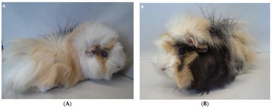
Scientific information on spontaneous type I diabetes mellitus (DM) and treatment modalities in guinea pigs is scarce. As most diabetic guinea pigs are overweight and respond to dietary changes, a disorder resembling type II-DM in humans seems to be most prevalent in this species. In the present report, a nine-month-old female intact guinea pig (GP1) was presented because of a cataract and polyphagia. The physical examinations in GP1 and its littermate, GP2, were unremarkable. Laboratory tests revealed hyperglycemia, hyperlipidemia, elevated fructosamine concentrations, and glucosuria in GP1 and GP2. Not responding to dietary changes, an insulin-dependent diabetes mellitus was suspected in both animals. Treatment with 0.5 IU of glargine insulin (Lantus®) per guinea pig subcutaneously (s.c.) once daily was initiated in both animals. Monitoring included repeated clinical evaluations and the measurement of plasma glucose and fructosamine concentrations. Capillary glucose concentration was measured using a glucometer, and glucosuria was monitored by dipstick. Blood glucose concentrations decreased quickly in both GPs, and glucosuria resolved. Including several dose adjustments, DM remained controlled for over 1.5 years. Bilateral cataracts and lens-induced uveitis in GP1 were medically managed with only slight progression. This is the first report of guinea pigs with insulin-dependent diabetes mellitus that were successfully treated with long-acting basal insulin glargine. Full article
Show Figures

Figure 1

13 pages, 1227 KiB  
Article
Clinical Value of CT-Guided Fine Needle Aspiration and Tissue-Core Biopsy of Thoracic Masses in the Dog and Cat
by Massimo Vignoli, Roberto Tamburro, Andrea Felici, Francesca Del Signore, Annalisa Dettori, Morena Di Tommaso, Angela Ghiraldelli, Rossella Terragni, Francesco Simeoni, Ilaria Falerno and Arianna Miglio
Animals 2021, 11(3), 883; https://doi.org/10.3390/ani11030883 - 19 Mar 2021
Cited by 2 | Viewed by 3289
Abstract
Diagnosis of thoracic lesions on the basis of history and physical examination is often challenging. Diagnostic imaging is therefore of paramount importance in this field. Radiology has traditionally been considered the diagnostic procedure of choice for these diseases. Nevertheless, it is often not [...] Read more.
Diagnosis of thoracic lesions on the basis of history and physical examination is often challenging. Diagnostic imaging is therefore of paramount importance in this field. Radiology has traditionally been considered the diagnostic procedure of choice for these diseases. Nevertheless, it is often not possible to differentiate inflammatory/infectious lesions from neoplastic diseases. A correct cytological and histopathologic diagnosis is therefore needed for an accurate diagnosis and subsequent prognostic and therapeutic approach. In human medicine, Computed Tomography (CT) and CT-guided biopsy are used in the presence of lesions which are not adequately diagnosed with other procedures. In the present study, thoracic lesions from 52 dogs and 10 cats of different sex, breed and size underwent both CT-guided fine-needle aspiration (FNAB) and tissue-core biopsy (TCB). Clinical examination, hematobiochemical analysis and chest radiography were performed on all animals. In this study, 59 of 62 histopathological samples were diagnostic (95.2%). Cytology was diagnostic in 43 of 62 samples (69.4%). General sensitivity, accuracy and PPV for FNAB and TCB were 67.7%, 67.7% and 100% and 96.7%, 95.2% and 98.3%, respectively. Combining the two techniques, the overall mean accuracy for diagnosis was 98.4%. Nineteen of 62 cases showed complications (30.6%). Mild pneumothorax was seen in 16 cases, whereas mild hemorrhage occurred in three cases. No major complications were encountered. CT-guided FNAB cytology can be considered a useful and reliable technique, especially for small lesions or lesions located close to vital organs and therefore dangerous to biopsy in other way. Full article
Show Figures

Figure 1

10 pages, 712 KiB  
Article
Changes in Saliva Analytes in Dairy Cows during Peripartum: A Pilot Study
by María D. Contreras-Aguilar, Pedro J. Vallejo-Mateo, Elsa Lamy, Damián Escribano, Jose J. Cerón, Fernando Tecles and Camila P. Rubio
Animals 2021, 11(3), 749; https://doi.org/10.3390/ani11030749 - 09 Mar 2021
Cited by 4 | Viewed by 2047
Abstract
This pilot study aimed to study the possible changes in a profile of 26 salivary analytes in thirteen healthy dairy cows along their peripartum period. Analytes associated with the stress (salivary cortisol, salivary alpha-amylase, butyrylcholinesterase, and total esterase), inflammation (adenosine deaminase), oxidative status [...] Read more.
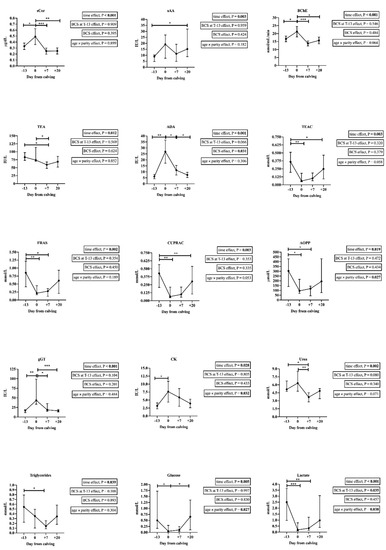
This pilot study aimed to study the possible changes in a profile of 26 salivary analytes in thirteen healthy dairy cows along their peripartum period. Analytes associated with the stress (salivary cortisol, salivary alpha-amylase, butyrylcholinesterase, and total esterase), inflammation (adenosine deaminase), oxidative status (total antioxidant capacity and the advanced oxidation protein products), and general metabolism (creatine kinase, γ-glutamyl transferase, urea, triglycerides, glucose, and lactate) varied along the sampling times. A positive correlation between the white blood cells counts, and the lipase, Trolox equivalent antioxidant capacity, advanced oxidation protein products, and lactate levels in saliva were observed at the delivery. A linear association between selected salivary analytes at different sampling times and the milk yield after calving was observed. In conclusion, in our experimental conditions, it was observed that the peripartum period in dairy cows can induce changes in salivary analytes. Some of them were associated with inflammatory status and the capacity of milk production after calving. Full article
Show Figures

Figure 1

12 pages, 917 KiB  
Article
Detection of Serum Allergen-Specific IgE in Atopic Dogs Tested in Northern Italy: Preliminary Study
by Morena Di Tommaso, Alessia Luciani, Paolo Emidio Crisi, Marica Beschi, Paolo Rosi, Francesca Rocconi and Arianna Miglio
Animals 2021, 11(2), 358; https://doi.org/10.3390/ani11020358 - 01 Feb 2021
Cited by 7 | Viewed by 2738
Abstract
Canine atopic dermatitis (CAD) is a pruritic allergic skin disease associated with IgE-mediated hypersensitivity. IgE is detected using Serum Allergen-Specific IgE test (SAT) in order to identify allergens. The present study aims to identify the environmental allergens in atopic dogs living in Northern [...] Read more.
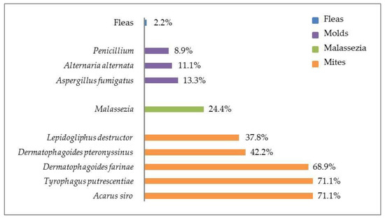
Canine atopic dermatitis (CAD) is a pruritic allergic skin disease associated with IgE-mediated hypersensitivity. IgE is detected using Serum Allergen-Specific IgE test (SAT) in order to identify allergens. The present study aims to identify the environmental allergens in atopic dogs living in Northern Italy using SAT. The screening SAT (sSAT), using a monoclonal antibody cocktail-based ELISA to identify indoor and outdoor allergens, was performed. In all positive samples, an anti-IgE monoclonal antibody ELISA test was performed to extend panel of allergens. Out of 117 selected dogs, 69 were included in the study; 71% were positive and 29% were negative to sSAT. Among the 49 positive sSAT, 53% were positive for both indoor and outdoor, 38.8% only for indoor, and 8.2% only for outdoor allergens. This is the first study on the frequency of allergens involved in CAD in Italy using SAT. IgE hypersensitivity in atopic dogs of Northern Italy is usually associated with indoor allergens, primarily house dust mites. Among the outdoor allergens, an important role was played by Rumex acetosa. Polysensitization also commonly occurs. Therefore, since the numerous factors affect the IgE positivity in CAD, specific panels for geographical areas should be considered and re-evaluated at time intervals. Full article
Show Figures

Figure 1

2020

Jump to: 2023, 2022, 2021

9 pages, 420 KiB  
Article
Incidence of Bacteremia Consequent to Different Endoscopic Procedures in Dogs: A Preliminary Study
by Alba Gaspardo, Maria Chiara Sabetti, Renato Giulio Zanoni, Benedetto Morandi, Giorgia Galiazzo, Domenico Mion and Marco Pietra
Animals 2020, 10(12), 2265; https://doi.org/10.3390/ani10122265 - 01 Dec 2020
Viewed by 1698
Abstract
Endoscopic procedures are widely used in veterinary medicine, and their role in producing transient bacteremia is debatable. The growing issue of antibiotic resistance requires the correct use of antibiotics, avoiding their administration when not strictly necessary. Studies highlighting post-endoscopy bacteremia in veterinary medicine [...] Read more.
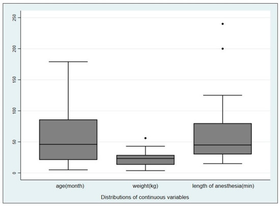
Endoscopic procedures are widely used in veterinary medicine, and their role in producing transient bacteremia is debatable. The growing issue of antibiotic resistance requires the correct use of antibiotics, avoiding their administration when not strictly necessary. Studies highlighting post-endoscopy bacteremia in veterinary medicine are extremely rare and often involve very few animals. This study describes the results from 74 owned dogs, brought to the Veterinary Teaching Hospital of the Department of Veterinary Medical Science of the University of Bologna, for the purpose of undergoing an endoscopic procedure. Two blood samples were taken from each dog, one before and one after the procedure, in order to assess the incidence of bacteremia linked to endoscopic procedures. Eight dogs were tested positive at the second blood culture with an Incidence Risk (IR) of 10.8%. No statistical differences were found by comparing positive and negative blood cultures with respect to sex, age, weight and anesthesia duration. In addition, no difference was found between airway and digestive tract procedures. The present findings showed that the probability of developing bacteremia after an endoscopic procedure was quite low, and additional studies confirming this are certainly recommended as well as the evaluation of categories of patients potentially considered at risk. Full article
Show Figures

Figure 1

Back to TopTop