Article

Lymphoscintigraphy versus Indocyanine Green Lymphography—Which Should Be the Gold Standard for Lymphedema Imaging?

Department of Plastic Surgery, Cleveland Clinic Foundation, 9500 Euclid Avenue, Desk A60, Cleveland, OH 44195, USA
*
Author to whom correspondence should be addressed.
Lymphatics 2023, 1(1), 25-33; https://doi.org/10.3390/lymphatics1010004
Submission received: 7 March 2023 / Revised: 19 April 2023 / Accepted: 4 May 2023 / Published: 5 May 2023

Abstract

:
Early detection and treatment can slow the progression of lymphedema. To diagnose lymphedema in the subclinical phase, a sensitive imaging modality is required. Radioisotope-based lymphoscintigraphy (LSG) has been the “gold standard” for a century. Indocyanine green lymphography (ICGL) is being used at our institute for diagnosing and grading all lymphedema patients. In this study, ICGL disease detection rate was compared to that of LSG. Chart review of all patients who presented for lymphedema consult between February 2020 and April 2022 was conducted. Patients who underwent both LSG and ICG for extremity edema in symptomatic/asymptomatic limbs were included. A total of 50 limbs in 23 patients met the inclusion criteria. Of those, 37 were symptomatic and 13 were asymptomatic. LSG detected lymphatic dysfunction in 26/37(70%) of the symptomatic limbs while ICG detected the same in 37/37(100%) limbs (p < 0.01). In the asymptomatic group, LSG detected the disease in 1/13(8%) limbs while ICG detected lymphatic dysfunction in 8/13 (62%) limbs (p < 0.01). LSG missed symptomatic limbs 30% of the time, whereas ICG did not miss any symptomatic limbs (p < 0.01). LSG missed asymptomatic disease 54% of the time (p < 0.01) compared to ICG. In conclusion, ICG lymphography was determined to have a higher lymphatic dysfunction detection rate compared to LSG.

1. Introduction

Historically, lymphedema diagnosis relied heavily on clinical history and physical examination. However, with new insights into the intricacies of lymphatic physiology and the recognition of the subclinical stages of the disease, there has been a notable shift in this approach [1]. In cases where clinical signs and symptoms are inconclusive or absent, a reliable imaging modality is an indispensable tool for accurate diagnosis. Consequently, imaging has gained widespread acceptance as a necessary method for achieving a reliable lymphedema diagnosis [2,3].
Lymphoscintigraphy (LSG), a radioisotope-based study, has been the conventional gold standard for nearly a century [4] by supplanting traditional bipedal lymphography as the preferred method for determining lymphedema due to the technical difficulty of cannulating lymphatic veins and the morbidity of the oil-based contrast agent used. However, due to LSG’s poor spatial and temporal resolution, as well as ionizing radiation exposure for both patients and clinicians, its application has been limited [5]. In attempt to improve LSG’s spatial and temporal resolution, magnetic resonance lymphangiography (MRL), with or without the use of gadolinium-based contrast agent, is a widely described technique in the recent literature [6,7] as a valid support to diagnose lymphedema and to map lymphatic vessels. MRL assesses the small subdermal lymphatic channels and venules, as nuclear lymphoscintigraphy is not able to discern singular lymphatic channels, venules and ICGL has a limited penetration depth of about 2 cm [8]. As a result, indocyanine green lymphography (ICGL), a fluorophore-based imaging modality, has gained widespread popularity, particularly among lymphedema surgeons [4,9,10]. ICG lymphography and lymphoscintigraphy provide distinct perspectives on the lymphatic system when utilized individually [4,9].
The dynamic utility of ICG lymphography is currently being used to effectively diagnose the disease, plan treatment and tracking treatment outcomes. However, despite its growing use, there are limited data available that directly compare these two imaging techniques [10,11,12]. The advantages of ICG lymphography compared with lymphoscintigraphy are the absence of radiation exposure, the short time required for the test, usefulness for lymphedema clinical stratification, and the real-time visualization of lymph flow. In contrast, the camera position can be freely changed in ICG lymphography, enabling acquisition in all directions. Therefore, the whole affected limb can be examined, and this may be the cause of the greater accuracy of ICG lymphography for early diagnosis of lymphedema. However, ICG lymphography has the disadvantage that only regions at a depth up to 2 cm from the skin can be observed, whereas lymphatic function can be observed in all layers using LSG. Therefore, more detailed evaluation of lymphatic function and identification of the location of lymph vessels are possible using ICG lymphography and lymphoscintigraphy in combination.
We present the results of our study comparing LSG and ICGL in patients with lymphedema. Our findings shed light on the relative strengths and limitations of these two diagnostic imaging modalities and may aid healthcare professionals in making informed decisions about which diagnostic technique to use.

2. Results

In total, 416 patients were evaluated for extremity lymphedema using ICG lymphography, of which 23 had LSG, meeting the inclusion criteria. In the selected cohort, 5 were males and 18 were females with an average body mass index (BMI) of 26 ± 5.47 kg/m2. The mean age was 49 ± 18.55 years. The mean lymphedema duration was 11 ± 11.38 years. Lower extremities were more commonly involved than upper extremities (Table 1).
Lymphedema was diagnosed as primary in 15 (65%) patients and secondary in 8 (35%) patients. Secondary lymphedema was acquired due to breast cancer in two cases (8.7%), urogynecological malignancies in five cases (21.7%), and in two cases (8.6%) it was acquired from other causes (e.g., prostate cancer, melanoma, filariasis, and cosmetic procedures).
In the 23 patients, 50 limbs were examined by both LSG and ICGL. A total of 37 limbs were symptomatic, and the remaining 13 limbs were asymptomatic at the time of testing. LSG detected lymphatic dysfunction in 26/37 (70%) of the symptomatic limbs while ICGL confirmed lymphatic abnormality in 37/37 (100%) limbs (p < 0.01). In the asymptomatic group, LSG detected the disease in 1/13 (8%) limbs, while ICGL detected lymphatic dysfunction in 8/13 (62%) limbs (p < 0.01). Hence, LSG missed symptomatic limbs 30% of the time, whereas ICGL did not miss any symptomatic limbs (p < 0.01). LSG also missed asymptomatic disease 54% of the time (p < 0.01) compared to ICGL (Table 2).
In the 11 symptomatic limbs (37 minus 26) which had normal LSG but abnormal ICGL, 9/11 (82%) limbs were of patients with primary lymphedema. The remaining two symptomatic limbs were of patients with secondary lymphedema (18%). The abnormalities noted in the ICG scans of the symptomatic limbs that were missed by LSG were delayed transit (six patients), abnormally oriented/collateral channels (two patients), and dermal backflow (three patients). Out of the seven asymptomatic limbs that were diagnosed with disease by ICGL, but exhibited normal LSG, four (57%) were present in patients with primary lymphedema, while the remaining three (43%) were observed in patients with secondary lymphedema. The most common findings on ICGL for these seven asymptomatic limbs were delayed transit (six patients) and abnormally oriented/collateral channels (Figure 1). A case is presented herein, in which LSG yielded a false negative result, which was later detected by indocyanine green lymphography (ICGL).
A 64-year-old male presented with left lower extremity lymphedema secondary to bladder cancer with metastasis to intrapelvic lymph nodes. The patient received chemotherapy and underwent a left inguinal lymph node dissection seven years prior. Lymphedema had developed in the left lower extremity three years ago and had gradually worsened despite treatment with lymph massage and elastic stockings. Physical examination revealed pitting edema of 2+ at the leg and 1+ at the thigh of the left lower extremity. Preoperative lymphoscintigraphy showed dermal radiotracer uptake extending to the knee and visualization of the main lymphatic ducts of the left lower extremity. Prompt visualization of the main lymphatic channels was observed in the right lower extremity. In contrast, on indocyanine green (ICG) lymphography, the left lower extremity exhibited stage III dermal backflow, with the right lower extremity showing lymph flow disturbance. Based on the ICG findings, the patient underwent lymphaticovenous anastomosis (LVA).

3. Discussion

Lymphatic damage is followed by a period of compensation by the lymphatic system. Exhaustion of these compensatory mechanisms by disease progression manifests as symptoms—edema, pain, tightness, and sensory changes [1]. Diagnosis of the disease during the compensating phase or Stage 0 (International Society of Lymphography—ISL) requires a sensitive imaging modality [1]. Therapeutic intervention initiated during this subclinical phase can prevent abnormal changes from setting in the lymphatic system and prevent disease progression. The challenge is to identify the lymphatic function abnormality during this asymptomatic stage of the disease.
Both nuclear lymphoscintigraphy and ICG lymphography can reveal aberrant structure and function in the form of slow or no transit, collateral flow pathways, and dermal backflow in established disease. A standardized test interpretation is required to reduce missed diagnoses and estimate the true disease burden to allow timely intervention.
Across the world, variable-sized technetium-99m-labeled (Tc99m) lymphatic-specific tracers are utilized therapeutically. Typically, they are 99mTc-nanocolloidal human serum albumin (5–80 nm diameter) utilized in Europe, 99mTc-sulphur colloid (filtered to a diameter below 100–200 nm) in the United States, and 99mTc-antimonium-trisulfide (5–30 nm) in Canada and Australia [13]. Small-sized tracers are chosen for functional imaging of the lymphatic system to ensure quick absorption from the injection site into lymphatic channels which can be useful in clinical scenarios such as sentinel lymph node biopsies. The larger, 99mTc-sulphur colloid can take 18–20 h to reach its intended target [14]. Particles such as this (>100 nm) become trapped in the interstitial compartment for extended periods prohibiting time-effective radioisotopic examination [15]. Several other variables unrelated to the lymphatic system of the extremity influence the uptake of the radioactive isotope. Important among these considerations are the depth of injection of the labeled colloid, the unintended administration of the colloid into a small vein, and post-injection muscular exercise. Gloviczki et al. demonstrated that muscular exercise increased the measured levels of radioactivity by as much as tenfold [16]. In light of this, quantitative investigations are of little use unless muscular exercise is standardized. Most importantly, lymphoscintigraphic protocols are variegated between institutions based on radiotracer administration, dosage, injection site, and timing of imaging acquisition. Despite numerous attempts towards improving objective interpretation by measuring lymphatic function, no universal consensus has been obtained to date [2,3,13,17]. On the other hand, ICG is a tricarbocyanine dye that gives off light after being excited by 806 nm near-infrared light. Indocyanine green dissolves easily in water and sticks to β-lipoproteins, especially albumin. Indocyanine green builds up in the lymphatic pathways and lymph nodes because lymph is high in proteins. Because of this, ICGL has many logistical advantages over LSG. Indocyanine green lymphography does not expose patients to radiation, has a shorter test duration and allows for real-time imaging of lymph flow due to faster transit. In addition, the ICGL image resolution is higher and allows a 3D spatial orientation when compared to LSG planar images [4,9,18]. Furthermore, ICG lymphography can be used as a perioperative lymph mapping tool for lymphaticovenous anastomosis [18,19,20]. In this study, we compared the diagnostic ability of lymphoscintigraphy and ICGL to identify symptomatic and asymptomatic extremity lymphedema. Indocyanine green lymphography was determined to have a significantly higher subclinical disease detection rate (62% versus 8%; p < 0.01) compared to LSG. This is in accordance with reports on ICGL detection of subclinical disease in cancer-related lymphedema patients [21,22]. In a prospective longitudinal cohort study involving patients with breast cancer-related lymphedema resulting from axillary lymph node dissection and regional nodal radiotherapy, Aldrich and colleagues [23] conducted serial follow-ups using indocyanine green lymphangiography (ICGL) over a period of eight months. The results showed that ICGL was able to detect dermal backflow patterns prior to the clinical manifestation of lymphedema in 83% of the patients.
Furthermore, LSG also missed 30% of symptomatic limbs, none of which were missed by ICGL. Interestingly, the majority of limbs where LSG failed to detect disease were those of primary lymphedema patients. This could be because there were no localized lymphatic system obstructions in these patients but a more diffuse lymphatic hypoplasia/pump function, which could be more easily displayed using a real-time flow study such as ICGL.
In patients suspected of primary lymphedema, we performed ICGL in all four limbs as these patients tend to have a global disruption in lymphatic function. However, the same was not performed for LSG at the referring facility, where only the symptomatic limb was tested, or in some cases, the contralateral limb. Hence, very few upper limbs could be included in this study, as they did not have LSG scans performed, especially in the unaffected limbs.
It is important to note that all ICGLs were performed and interpreted by the same examiner using a standardized technique [24]. This examiner was not blinded to the clinical exam. On the other hand, LSG was conducted at different centers, where we know there is a lack of uniformity in injected sites, injectable composition and volume, study period, activity during the study period, and measures used for study interpretation [2,3,13,17].
Studies have demonstrated that the interobserver reliability of LSG interpretation is low for early-stage lymphedema, which may contribute to the failure to detect subclinical disease [25]. It is worth mentioning that the study interpretation alone may not entirely account for the missed subclinical disease, as LSG may simply be inferior in detecting early or subclinical lymphedema due to its inability to accurately visualize superficial lymphatics. In our study, the 11 clinically diagnosed limbs that failed to be detected by LSG could be related to mild disease severity or a relatively short study period, or a high threshold used for interpreting the result as abnormal. To improve the interpretation and diagnostic accuracy of lymphoscintigraphy, the Genoa Protocol [15] suggests adding a SPECT scan. However, this requires special equipment and significantly increases the investigation costs.
Our results are consistent with those of the similar work published by other researchers [10], showing superiority of ICGL over LSG in detecting lymphedema in asymptomatic limbs. On the contrary, Akita et al. discovered ICGL to have a higher sensitivity (97%) than specificity in diagnosing primary and secondary lymphedema [26].
The demonstrated higher sensitivity and known safety, logistic ease, time, and cost-effectiveness of ICG lymphography suggest that it has the potential to become the standard for lymphedema evaluation. However, the lack of uniformity in lymphoscintigraphy (LSG) technique and measures for interpretation may affect its diagnostic value and could have biased our results. Additionally, the limitations of ICG lymphography, such as the lack of depth penetration of the dye, should also be considered. While ICG lymphography has shown promise as a lymphedema evaluation tool, further research is necessary to determine its true potential as a gold standard. Standardization of LSG technique and interpretation measures, as well as comparative studies with other imaging modalities such as MR lymphangiography could clarify the role of ICG lymphography as the gold standard for lymphedema assessment.

4. Methods

4.1. Patient Population and Study Design

The study was approved by the institutional review board and conducted in accordance with the Declaration of Helsinki. Given the retrospective nature of this study, the need for informed consents was waived. Consent was only acquired for any photographs used for publication. A retrospective chart review was performed on all patients with extremity lymphedema who presented to Cleveland Clinic Multidisciplinary Lymphedema Center (CCMLC) for evaluation between February 2020 and April 2022. All patients who were referred to our facility underwent indocyanine green lymphography (ICGL) to confirm the diagnosis and grade the baseline disease severity. We included patients who had completed ICGL and had previously undergone lymphoscintigraphy (LSG) and compared the imaging modalities for limbs evaluated by both LSG and ICGL. Patients who had undergone surgical interventions on the affected limbs between imaging studies or had a time gap of over two years between studies were excluded. LSG reports were reviewed by the authors for completeness. The patient demographics, limbs tested, presence or absence of lymphedema symptoms and disease etiology were analyzed. The results of LSG and ICGL interpreted as normal or abnormal were compared. Asymptomatic patients were considered in the absence of the following signs and symptoms: extremity swelling, heaviness or tightness, restricted range of motion, hardening and thickening of the skin (fibrosis). For symptomatic limbs, the International Society of Lymphology (ISL) classification was utilized [27].

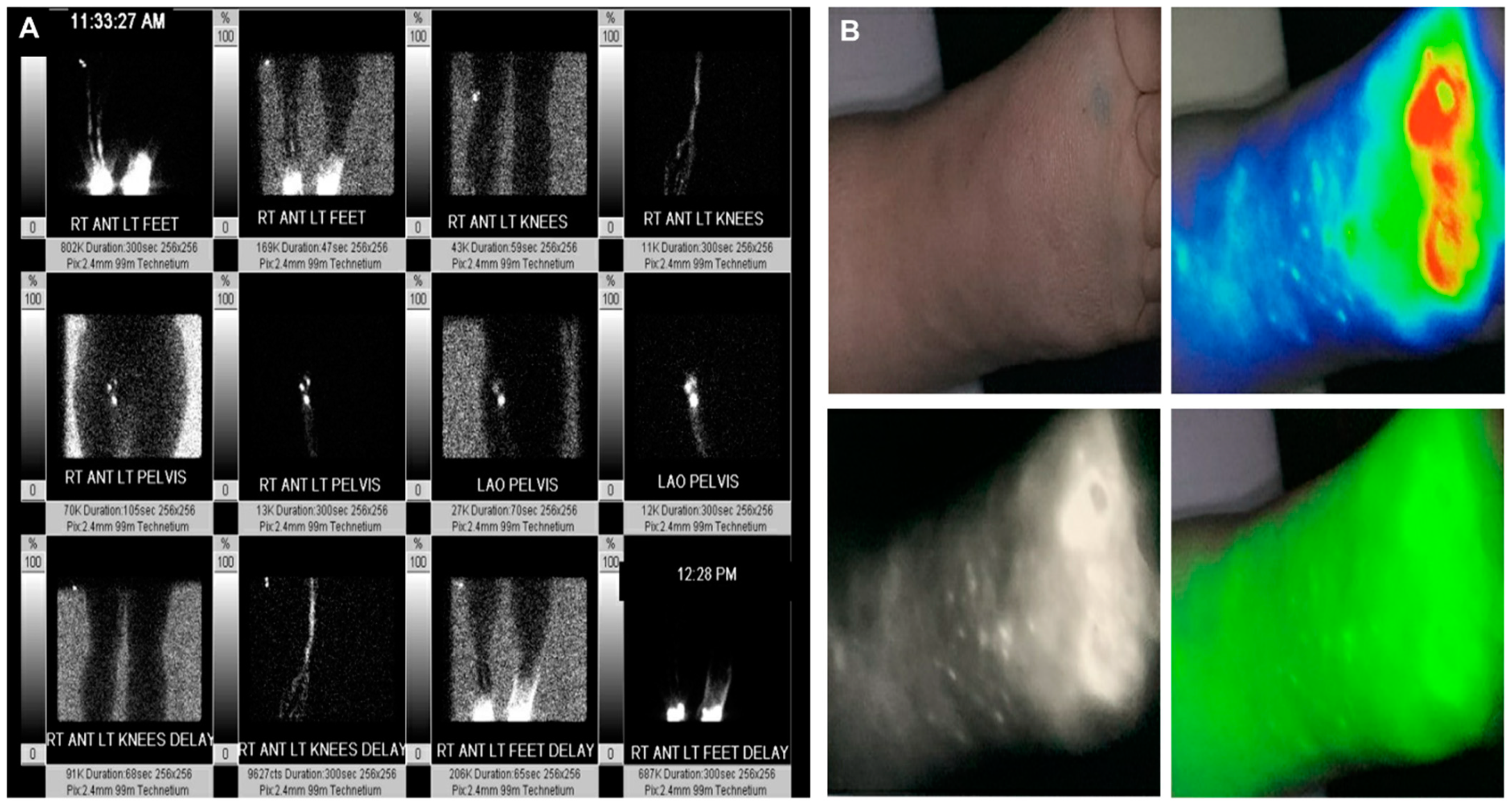
4.2. Indocyanine Green Lymphography

ICGL was performed using our standard technique, as described previously [24]. A total of 0.1 mL of 0.25% ICG (Diagnostic Green LLC, Farmington Hills, MI, USA) was injected into three intradermal sites on two interdigital web spaces, the wrist and medial malleolus of each arm and leg (total amount injected—200 µg). The injected sites were massaged by the examiner for one minute. Lymphatic imaging was carried out using Quest Spectrum HD (Olympus, Netherlands). Each patient received two scans: an immediate scan, and a delayed scan, with a duration of three minutes each. The first scan was performed immediately after the injection and a massage of the fluorophore, allowing the evaluation of lymphatic pump velocity, normal/aberrant anatomy, collateralization, flow distance and flow patterns. The second scan, which was conducted 6 h after the initial scan, enabled visualization of lymphographic patterns for estimating disease severity. In patients with a history consistent with secondary lymphedema, ICGL was performed on both the affected limb and the contralateral limb for comparison. The contralateral asymptomatic limbs were scanned to detect subclinical lymphatic dysfunction. In patients with a history consistent with primary lymphedema, ICGL was performed on all four extremities, irrespective of symptoms distribution. The study was interpreted as abnormal when one or more of the following were seen. In the immediate scans: if ICG did not reach the groin/axilla in 3 min from one or more of the injected sites [28]; in delayed scans, results were interpreted as abnormal when (1) ICG did not reach the groin/axilla from one or more of the injected sites, (2) abnormally oriented linear channels were present, (3) linear channels were absent, or (4) there was presence of dermal backflow.

4.3. Lymphoscintigraphy

Prior to the referred patient presenting to our facility, other institutions previously performed lymphoscintigraphy. Lymphoscintigraphy is typically performed with subcutaneous injections of a small amount (approximately 1.0 mCi) of technetium-99m-labelled filtered sulfur colloid administered in bilateral upper or lower extremities, depending on symptomatic limb. LSG reports were brought by the patient and studied for visualization of regional lymph nodes, lymph stagnation points, dermal backflow patterns, absence of tracer migration, drainage delays, presence of collateral lymphatic vessels, and lymphatic pathway interruptions. Information was extracted from the data available in the LSG reports, which were read by the referring hospital radiological department. The data were analyzed for injected colloid type, sites of injection, duration of study, study interpretation as normal or abnormal.

4.4. Data Analysis

Categorical variables were described using frequencies and percentage, and the p-values were obtained from either Pearson Chi-square or Fisher’s exact test as appropriate. Continuous variables with normal distributions were described using means and standard deviations, and p-values were obtained from Student’s t-tests. A significance level of 0.05 was assumed for all statistical tests. The analyses were performed using JMP SAS Software (version 16.2; Cary, NC, USA).

Author Contributions

Conceptualization, W.F.C.; methodology, B.A.F.; formal analysis, B.A.F. and J.D.L.; investigation, B.A.F., J.D.L., M.A.-M., S.P. and W.F.C.; data curation, J.D.L. and M.A.-M.; writing—original draft preparation, B.A.F.; writing—review and editing, J.D.L., M.A.-M., S.P. and W.F.C.; visualization, W.F.C.; supervision, W.F.C. All authors have read and agreed to the published version of the manuscript.

Funding

This research received no external funding.

Institutional Review Board Statement

The study was conducted in accordance with the Declaration of Helsinki, and approved by the Institutional Review Board (IRB) of The Cleveland Clinic Foundation (IRB 22658; approved January 2020).

Informed Consent Statement

Informed consent was obtained from all subjects involved in the study.

Data Availability Statement

Not applicable.

Conflicts of Interest

The authors declare no conflict of interest.

References

  1. Executive, C. The Diagnosis and Treatment of Peripheral Lymphedema: 2016 Consensus Document of the International Society of Lymphology. Lymphology 2016, 49, 170–184. [Google Scholar]
  2. Partsch, H. Assessment of abnormal lymph drainage for the diagnosis of lymphedema by isotopic lymphangiography and by indirect lymphography. Clin. Dermatol. 1995, 13, 445–450. [Google Scholar] [CrossRef]
  3. Weissleder, H.; Weissleder, R. Lymphedema: Evaluation of qualitative and quantitative lymphoscintigraphy in 238 patients. Radiology 1988, 167, 729–735. [Google Scholar] [CrossRef] [PubMed]
  4. Ogata, F.; Azuma, R.; Kikuchi, M.; Koshima, I.; Morimoto, Y. Novel lymphography using indocyanine green dye for near-infrared fluorescence labeling. Ann. Plast. Surg. 2007, 58, 652–655. [Google Scholar] [CrossRef] [PubMed]
  5. Bae, J.S.; Yoo, R.E.; Choi, S.H.; Park, S.O.; Chang, H.; Suh, M.; Cheon, G.J. Evaluation of lymphedema in upper extremities by MR lymphangiography: Comparison with lymphoscintigraphy. Magn. Reson. Imaging 2018, 49, 63–70. [Google Scholar] [CrossRef]
  6. Mazzei, F.G.; Gentili, F.; Guerrini, S.; Cioffi Squitieri, N.; Guerrieri, D.; Gennaro, P.; Scialpi, M.; Volterrani, L.; Mazzei, M.A. MR Lymphangiography: A Practical Guide to Perform It and a Brief Review of the Literature from a Technical Point of View. BioMed Res. Int. 2017, 2017, 2598358. [Google Scholar] [CrossRef]
  7. Cellina, M.; Oliva, G.; Menozzi, A.; Soresina, M.; Martinenghi, C.; Gibelli, D. Non-contrast Magnetic Resonance Lymphangiography: An emerging technique for the study of lymphedema. Clin. Imaging 2019, 53, 126–133. [Google Scholar] [CrossRef]
  8. Guerrini, S.; Gentili, F.; Mazzei, F.G.; Gennaro, P.; Volterrani, L.; Mazzei, M.A. Magnetic resonance lymphangiography: With or without contrast? Diagn Interv. Radiol. 2020, 26, 587–595. [Google Scholar] [CrossRef] [PubMed]
  9. Ogata, F.; Narushima, M.; Mihara, M.; Azuma, R.; Morimoto, Y.; Koshima, I. Intraoperative lymphography using indocyanine green dye for near-infrared fluorescence labeling in lymphedema. Ann. Plast. Surg. 2007, 59, 180–184. [Google Scholar] [CrossRef]
  10. Mihara, M.; Hara, H.; Narushima, M.; Todokoro, T.; Iida, T.; Ohtsu, H.; Murai, N.; Koshima, I. Indocyanine green lymphography is superior to lymphoscintigraphy in imaging diagnosis of secondary lymphedema of the lower limbs. J. Vasc. Surg. Venous Lymphat. Disord. 2013, 1, 194–201. [Google Scholar] [CrossRef]
  11. Yoon, J.A.; Shin, M.J.; Kim, J.H. Indocyanine Green Lymphography and Lymphoscintigraphy Severity Stage Showed Strong Correlation in Lower Limb Lymphedema. Lymphat. Res. Biol. 2021, 19, 80–85. [Google Scholar] [CrossRef]
  12. Yoon, J.A.; Shin, M.J.; Shin, Y.B.; Kim, K.; Park, H.; Kang, T.; Kong, I.J.; Kim, H.; Park, M.S.; Kim, J.H. Correlation of ICG lymphography and lymphoscintigraphy severity stage in secondary upper limb lymphedema. J. Plast. Reconstr. Aesthet. Surg. 2020, 73, 1982–1988. [Google Scholar] [CrossRef]
  13. Polomska, A.K.; Proulx, S.T. Imaging technology of the lymphatic system. Adv. Drug Deliv. Rev. 2021, 170, 294–311. [Google Scholar] [CrossRef]
  14. Sadeghi, R.; Forghani, M.N.; Memar, B.; Rajabi Mashhadi, M.T.; Dabbagh Kakhki, V.R.; Abdollahi, A.; Zakavi, S.R. How long the lymphoscintigraphy imaging should be continued for sentinel lymph node mapping? Ann. Nucl. Med. 2009, 23, 507–510. [Google Scholar] [CrossRef]
  15. Villa, G.; Campisi, C.C.; Ryan, M.; Boccardo, F.; Di Summa, P.; Frascio, M.; Sambuceti, G.; Campisi, C. Procedural Recommendations for Lymphoscintigraphy in the Diagnosis of Peripheral Lymphedema: The Genoa Protocol. Nucl. Med. Mol. Imaging 2019, 53, 47–56. [Google Scholar] [CrossRef]
  16. Gloviczki, P.; Calcagno, D.; Schirger, A.; Pairolero, P.C.; Cherry, K.J.; Hallett, J.W.; Wahner, H.W. Noninvasive evaluation of the swollen extremity: Experiences with 190 lymphoscintigraphic examinations. J. Vasc. Surg. 1989, 9, 683–689. [Google Scholar] [CrossRef]
  17. Szuba, A.; Shin, W.S.; Strauss, H.W.; Rockson, S. The third circulation: Radionuclide lymphoscintigraphy in the evaluation of lymphedema. J. Nucl. Med. Off. Publ. Soc. Nucl. Med. 2003, 44, 43–57. [Google Scholar]
  18. Chen, W.F.; McNurlen, M.; Ding, J.; Bowen, M. Vascularized lymph vessel transfer for extremity lymphedema-is transfer of lymph node still necessary? Int. Microsurg. J. 2019, 3, 1. [Google Scholar] [CrossRef]
  19. Chen, W.F.; Bowen, M.; Ding, J.J.I.M.J. Immediate limb compression following supermicrosurgical lymphaticovenular anastomosis–is it helpful or harmful? Int. Microsurg. J. 2018, 2, 1. [Google Scholar] [CrossRef]
  20. Imai, H.; Yoshida, S.; Mese, T.; Roh, S.; Fujita, A.; Sasaki, A.; Nagamatsu, S.; Koshima, I. Correlation between Lymphatic Surgery Outcome and Lymphatic Image-Staging or Clinical Severity in Patients with Lymphedema. J. Clin. Med. 2022, 11, 4979. [Google Scholar] [CrossRef]
  21. Liu, M.; Liu, S.; Zhao, Q.; Cui, Y.; Chen, J.; Wang, S. Using the Indocyanine Green (ICG) Lymphography to Screen Breast Cancer Patients at High Risk for Lymphedema. Diagnostics 2022, 12, 983. [Google Scholar] [CrossRef]
  22. 22. Early Detection of Breast Cancer-Related Lymphedema: Accuracy of Indocyanine Green Lymphography Compared with Bioimpedance Spectroscopy and Subclinical Lymphedema Symptoms. Lymphat. Res. Biol. 2023; ahead of print. [CrossRef]
  23. Aldrich, M.B.; Rasmussen, J.C.; DeSnyder, S.M.; Woodward, W.A.; Chan, W.; Sevick-Muraca, E.M.; Mittendorf, E.A.; Smith, B.D.; Stauder, M.C.; Strom, E.A.; et al. Prediction of breast cancer-related lymphedema by dermal backflow detected with near-infrared fluorescence lymphatic imaging. Breast Cancer Res. Treat 2022, 195, 33–41. [Google Scholar] [CrossRef]
  24. Chen, W.F.; Qin, E.S.; Bowen, M.J.; Little, A.S.; Lensing, J.N. Exercise-enhanced ICG lymphography: A fast approach to diagnosis and staging of lymphedema. Int. Microsurg. J. 2021, 5, 2. [Google Scholar] [CrossRef]
  25. Ebrahim, M.; Savitcheva, I.; Axelsson, R. Reliability of a Scoring System for Qualitative Evaluation of Lymphoscintigraphy of the Lower Extremities. J. Nucl. Med. Technol. 2017, 45, 219–224. [Google Scholar] [CrossRef]
  26. Akita, S.; Mitsukawa, N.; Kazama, T.; Kuriyama, M.; Kubota, Y.; Omori, N.; Koizumi, T.; Kosaka, K.; Uno, T.; Satoh, K. Comparison of lymphoscintigraphy and indocyanine green lymphography for the diagnosis of extremity lymphoedema. J. Plast. Reconstr. Aesthetic Surg. JPRAS 2013, 66, 792–798. [Google Scholar] [CrossRef]
  27. The diagnosis and treatment of peripheral lymphedema: 2020 Consensus Document of the International Society of Lymphology. Lymphology 2020, 53, 3–19.
  28. Unno, N.; Nishiyama, M.; Suzuki, M.; Yamamoto, N.; Inuzuka, K.; Sagara, D.; Tanaka, H.; Konno, H. Quantitative lymph imaging for assessment of lymph function using indocyanine green fluorescence lymphography. Eur. J. Vasc. Endovasc. Surg. 2008, 36, 230–236. [Google Scholar] [CrossRef]
Figure 1. (A) Lymphoscintigram of the lower limbs in a patient with lymphedema showing channels, tortuosity of channels and dermal backflow. (B) Lower extremity indocyanine green lymphography showing a diffuse pattern at the toes with a stardust pattern at the dorsum of the foot.
Lymphatics 01 00004 g001
Table 1. Baseline characteristics.
DemographicValue
Age (Years)49 ± 15.55
Sex
Male5 (22)
Female18 (78)
BMI (Kg/m2)26 ± 5.47
Symptoms Location
Right Upper Extremity2
Left Upper Extremity1
Bilateral Upper Extremity1
Left Lower Extremity7
Right Lower Extremity3
Bilateral Lower Extremity11
History of Cellulitis
Present4 (17)
Absent19 (83)
Age of Onset (Years)39 ± 19.23
Lymphedema Duration (Years)11 ± 11.38
Lymphedema Family History
Positive0 (0)
Negative23 (100)
Lymphedema Type
Primary Lymphedema15 (65)
Secondary Lymphedema8 (35)
Data are presented as mean ± standard deviation for continuous variables and as frequencies followed by percentages in parenthesis, for categorical variables. Abbreviations: LDB, leg dermal backflow; ADB, arm dermal backflow.
Table 2. Lymphoscintigraphy versus indocyanine green lymphography in primary versus secondary lymphedema patients.
Lymphedema TypePatientsLimbsSymptomatic LimbsLSG +ICG +p ValueAsymptomatic LimbsLSG +ICG +p Value
Primary15
(65%)
33
(66%)
2516
(64%)
25
(100%)
<0.0181
(12.5%)
5
(63%)
<0.01
Secondary8
(35%)
17
(34%)
1210
(83%)
12
(100%)
<0.0150
(0%)
3
(60%)
<0.01
Total23503726/37
(70%)
37/37
(100%)
<0.01131/13
(8%)
8/13
(62%)
<0.01
Disclaimer/Publisher’s Note: The statements, opinions and data contained in all publications are solely those of the individual author(s) and contributor(s) and not of MDPI and/or the editor(s). MDPI and/or the editor(s) disclaim responsibility for any injury to people or property resulting from any ideas, methods, instructions or products referred to in the content.

