Review

The Role of Sentinel Node Mapping and Lymphadenectomies in Veterinary Surgical Oncology

Clinic for Small Animal Surgery, Vetsuisse Zürich, University of Zürich, 8057 Zürich, Switzerland
*
Author to whom correspondence should be addressed.
Lymphatics 2023, 1(1), 2-18; https://doi.org/10.3390/lymphatics1010002
Submission received: 29 November 2022 / Revised: 21 December 2022 / Accepted: 5 January 2023 / Published: 10 January 2023

Abstract

:
Lymph node status is an important prognostic factor in multiple oncologic conditions in humans and companion animals. In addition, the resection of the affected nodes can have a substantial therapeutic effect on various cancer subtypes in both species. Given the impact on prognosis and management, it is paramount to identify and remove affected nodes. While this can be achieved by removing predefined patterns of nodes (regional lymphadenectomy/resection of defined lymphatic stations), modern approaches increasingly utilize sentinel node mapping to identify the draining nodes to decrease the mortality of lymphadenectomies. Recent studies have shown that dogs have more comparable anatomy of the lymphatic system to humans than other animal models such as rodents or pigs. Given the fact that dogs develop spontaneous cancer types that share several similarities to their human counterparts, they represent a valuable translational model. The management of the lymphatic basin and sentinel node mapping have gained increased attention in veterinary surgical oncology in recent years. The present review aims at summarizing the resulting findings and their impact on patient management.

1. Introduction

Lymph node (LN) status is an important prognostic factor for the staging of various cancers in human and veterinary medicine, and the resection of metastatic nodes can improve outcomes in selected cancer types [1,2,3,4,5,6]. Virchow and Halsted already recognized the importance of the lymphatic system serving as a major barrier for cancer cells [7,8]. Initially, it was assumed that the closest regional LNs were the first sites to be affected by metastases, and the radical resection of the tissue surrounding the tumor was advocated to include all regional nodes [7]. Radical surgical resections of the lymphatic basin remained the unchanged treatment of choice for certain tumors in human surgical oncology until the mid-20th century, when sentinel lymph node (SLN) mapping and more selective lymphadenectomies evolved for several cancer types, including malignant melanoma, breast, vulvar, and cervical cancers [9].
Thus far, a potential therapeutic advantage of lymphadenectomy in companion animals has only been advocated in a few cancer types [1]. One problem in the assessment of the impact of lymphadenectomies in various cancers in veterinary patients is that, to date, no generally accepted resection routine exists for most conditions. In most settings, LNs are resected if they appear enlarged or structurally altered [10,11,12], although studies have repeatedly shown that the size of the lymph node is no predictor of nodal involvement [13,14,15]. Alternatively, the resection of RLN is performed, involving the removal of a single LN rather than entire lymphatic stations [1]. These approaches are generally poorly standardized and most certainly lead to the undertreatment of potentially affected LNs. Recently, SLN mapping has gained increased attention. This could serve to establish more standardized approaches that will allow the comparative validation of the impact of the resection of metastatic LNs in veterinary surgical oncology in the future.

2. Comparative Anatomy of the Canine and Human Lymphatic System

With an increase in the size of the species of interest, the lymphatic system becomes more complex with more individual LNs and more complex architecture [16]. Humans have around 450 LNs, whereas mice have only about 22 [16]. Dogs are in between, with many more nodes than mice but still fewer than humans [17,18]. The most exhaustive description of the lymphatics of dogs was presented by Professor Baum at the Institute of Veterinary Anatomy of the University of Leipzig, in 1918 [17]. He was able to identify a complex draining map including the description of the draining LNs, and he was the first to describe the considerable individual variation between individual dogs regarding the number of LNs per station [17].
Recently, Suami’s group conducted detailed research on comparative lymphatic anatomy in dogs and humans [19]. In 2012, they published a comparative study of the lymphosomes in the canine forelimb versus the upper extremity in humans [19]. The main discrepancy was that the ventral cervical node is the dominant system in dogs, whereas in humans, it is the axillary node, and the number of individual LNs was less in dogs than in humans [19]. However, they also found similarities: the presence of a deep and superficial draining system (divided by the deep fascia), and the number, size, and distribution of the lymphatic vessels (0.3–1.5 mm), as well as the presence of lymphatic valves and lymphangions [19]. Overall, due to the increased complexity and size of the lymphatics, they concluded that dogs are more suitable models for lymphatic research than rodents, especially for the investigation of lymphangiography [19]. Suami et al. also demonstrated that the remodeling of the lymphatics after resection in dogs occurs similarly to the remodeling observed in women after LN dissection for breast cancer, and remarkable similarities exist between the general maps of the lymphosomes of canines and humans [18]. Although Suami’s research yielded important results, some findings are in disagreement with the findings of recent SLN studies in dogs, and it should also be noted that the dogs used in these studies were all healthy animals [18]. Most importantly, Suami found that none of the lymphatics ever crossed the midline in the dogs he investigated before surgery. Surgery then triggered the remodeling of the lymphatics towards the contralateral dominant lymph nodes [18]. However, in dogs with cancer, a contralateral LN has repeatedly been identified as being sentinel before surgery, with the lymphatics crossing the midline in numerous investigations [10,13,20,21,22].

3. SLN Mapping in Companion Animals

Although the maps of locoregional draining patterns (lymphosomes) in healthy patients are available in recent times, the individual draining pattern of a tumor can substantially vary. The SLN is defined as a node within the lymphatic basin that directly drains the primary tumor [9,23]. Depending on the draining pattern, these LNs can then be further divided into tier-1 and tier-2 LNs. A tier-1 LN is an LN that directly drains a certain tumor, whereas a tier-2 LN is the second node in the chain receiving lymph from tier-1 LNs. Notably, any tumor can have multiple tier-1 SLNs. SLNs may be the regional LNs anticipated to be responsible for tumor drainage; however, the LNs at unpredictable anatomical locations might also serve as SLNs [20]. Indeed, for dogs, regional LNs do not correspond to SLNs in 42–63% of cases [10,20,24,25], and a recent study in cats found an even higher discrepancy (71.4%) [26].
SLN sampling offers a less invasive approach than radical regional lymphadenectomy, minimizes anesthesia time, and reduces tissue trauma and postoperative morbidity [27,28]. For these reasons, SLN mapping has successfully been established in several neoplastic conditions in human oncology, and several techniques have been described for SLN mapping in humans, including radiographic lymphography [29], computed tomography (CT) lymphography [30], magnetic resonance (MR) lymphography [31]. contrast-enhanced ultrasound (CEUS) [32], single-photon emission computed tomography (SPECT) [33], positron emission tomography (PET) [34], pre- and intraoperative lymphoscintigraphy, and the injection of blue dyes for direct visualization or fluorescent dyes for near-infrared (NIR) fluorescent imaging [35]. Corresponding evaluations of indirect radiographic [36,37,38] and CT lymphography [39,40,41,42], lymphoscintigraphy [15,20,24,43,44,45,46,47], CEUS [25,48], methylene blue staining [20,24,47], and NIR-lymphography [14,47,49,50,51] are also available for dogs and, most recently, also for cats [26,52].

3.1. Preoperative SLN Mapping Techniques Available in Veterinary Medicine

Several different mapping techniques have been described in dogs and cats, one of which is lymphoscintigraphy, which is still considered the gold standard. However, due to the legal and cost restrictions associated with this technique, several other techniques are currently under evaluation. The following section provides a brief overview of the available veterinary studies that use different approaches for SLN mapping in dogs and cats.

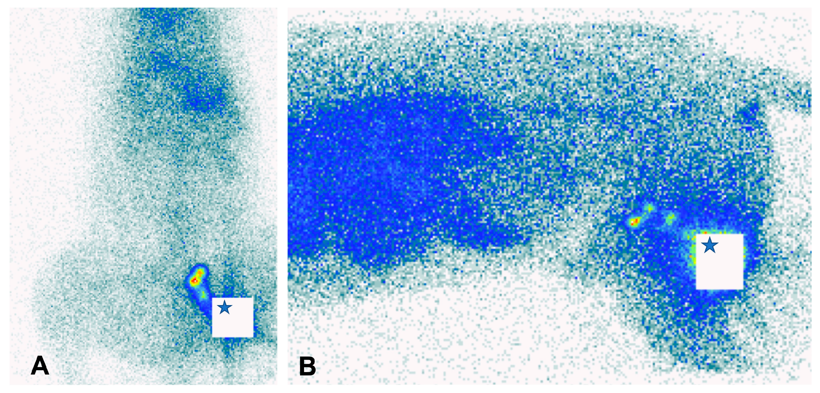
3.1.1. Lymphoscintigraphy

The use of radioactive tracers for SLN mapping is currently considered the gold standard [2,10,43,44,53,54].
Radio-guided SLN mapping includes preoperative planar imaging (Figure 1) and/or the intraoperative application of a hand-held gamma probe. The radiotracers typically used are technetium-99m (Tc-99m) labeled colloids [20,44,45,54,55]. These tracers are retained at the injection site for prolonged periods due to their particulate nature when administered peritumorally (intradermal, subcutaneous). Accumulating radioactivity is detected in SLNs during the acquisition of a preoperative scan [43,44,45,54,55]. A hand-held gamma probe guides the surgeon intraoperatively to the radiolabeled “hot” SLNs even if located in deep tissue layers [43]. Whether these “hot” SLNs are affected by metastasis, however, cannot be confirmed through lymphoscintigraphy [56].
In 2002, Balogh et al. investigated the feasibility of scintigraphy for SLN mapping in dogs with spontaneous tumors [43]. They were able to detect 89% of SLNs using preoperative scintigraphy and 97% with the combination of a gamma probe and blue dye in surgery, but using only the blue dye resulted in the identification of 77% of SLNs [43]. A combination of radioisotopes and methylene blue dye in dogs was reported in 2014 [20]. In 19 patients, 18 SLNs were detected using preoperative lymphoscintigraphy, 19 SLNs were found by the intraoperative use of a gamma probe, and 18 LNs were stained blue [2]. In healthy dogs, the feasibility of scintigraphy for SLN mapping has been confirmed by studies investigating the drainage of the prostate and mammary glands and pulmonary and thoracic tissue [44,45,55]. Manfredi et al. published a study evaluating lymphoscintigraphy in 51 dogs with 60 solid tumors and found that SLNs were detectable in 95% of the cases [54]. The same group also published a study evaluating lymphoscintigraphy in head and neck tumors, for which a lower detection rate of 83% was observed [13]. Finally, a recent study revealed the successful usage of lymphoscintigraphy for the detection of SLN in cats with various solid tumors [26].
Unfortunately, only a scarce number of veterinary clinics currently have access to radioactive tracers, and this, together with the associated expensive equipment, poses a major drawback. In addition, lymphoscintigraphy does not offer a special resolution, therefore making the identification of individual nodes and adjacent nodes in proximity (such as in the head and neck area) difficult. Future studies evaluating alternative methods for SLN mapping might result in the establishment of a new gold standard in the future.

3.1.2. Indirect Radiographic Lymphography

The peritumoral injection of a radio-opaque contrast agent has been utilized as one of the first techniques for the detection of SLNs in veterinary cancer patients [2,36,57]. After injection, the contrast is drained towards SLNs via the lymphatic system and can be visualized by taking serial radiographs [36,57]. The SLNs stay radio-opaque for several days to months, until the contrast is cleared from the lymphatic system [38,58]. Patsikas et al. and Collivignarelli showed that the lymphatic drainage pattern in canine mammary cancer differed from that in normal glands, as LNs not expected to be sentinel received lymph from the tumor [57,59]. Brissot and colleagues compared the corresponding detection rates between radiographically identified SLNs and those detected intraoperatively via blue dye mapping [36]. SLNs were successfully identified in 29 out of 30 tumors using the radiographic lymphography technique (96.6%), and an agreement was observed between blue-stained SLNs and those detected with radiographic lymphography in 84.6% (23/26) of the cases [36]. Insufficient staining visible on radiographs required a contrast medium to be reinjected in 4 out of 25 patients. Both techniques were reportedly safe, with a minor complication only noted in 1 out of 30 dogs [36]. In contrast, Mayer and others observed swelling and erythema of the injection site in 50% (10/20) and swelling of the LNs in 30% (6/20) of healthy dogs when injecting the same contrast agent subcutaneously or submucosally [38]. Severe complications, such as allergic reactions or pulmonary embolisms, are reported in humans but have not been observed in small animals to date.
The major advantages of radiographic lymphography are the general availability of its equipment, being a noninvasive procedure, and being a simple technique, which makes it feasible for clinical practice [2]. However, relatively low detection rates, compared with other SLN mapping techniques, have been reported, in addition to discrepancies in suspected SLNs. Hlusko et al. compared indirect lymphography to the current gold standard, lymphoscintigraphy, in eight healthy dogs, and found discrepancies in five of the eight dogs [37]. This presents a major limitation and should be considered before applying these techniques.

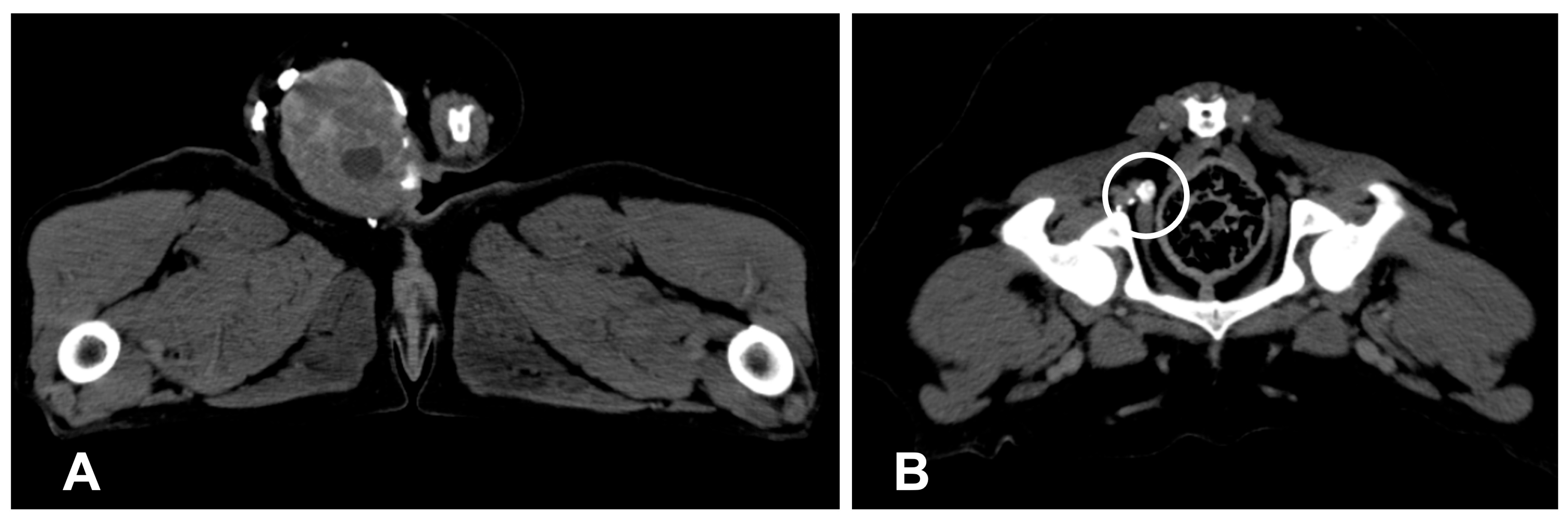
3.1.3. Computed Tomography Lymphography

The main rationale for CT lymphography was to develop a contrast-based technique that accurately displays the anatomical location of the SLNs and the detail of the surrounding tissue (Figure 2) [20,39,41,60]. The injection technique, the contrast agents, their lymphatic uptake and distribution, and potential complications are principally similar between CT and radiographic lymphography, with uptake in SLNs occurring minutes after injection [20,30,41,60].
Recently, four veterinary studies proved the successful implementation of CT lymphography in clinical canine patients with mammary tumors [42], anal gland sac cancer [40], tumors of the head [39], and tumors at various sites [41]. The contrast-filled afferent lymph vessels could be followed to one or more SLNs in several studies [39,40,41,42]. In a pilot study including 18 dogs with variable tumors on the head, indirect CT lymphography was successfully performed in identifying SLNs in 16 cases (89%) with 2 patients having more than 1 SLN (13%) [39]. Soultani et al. published a study on the evaluation of the value of contrast-enhanced CT imaging after IV contrast application and indirect SLN mapping to detect metastatic nodes [42]. While a homogenous contrast distribution was observed in the SLNs free from metastases, a heterogenous pattern was associated with metastases [42]. Likewise, a low Hounsfield unit (HU) in the center of SLNs was associated with high sensitivity and specificity for SLN metastases [42]. The authors concluded that CT lymphography may be a helpful tool to predict the presence of nodal metastasis and determined that a cutoff of 59.5 HU maximum density in the periphery of the LNs displays high sensitivity and specificity (87.5% and 89.3%, respectively) [42]. Brissot et al. also described CT lymphography to localize SLNs in cancer-bearing canine patients [36]. However, they did not specify how specific CT lymphography was compared with other techniques [36]. Wan et al. published a study comparing CT lymphography and intraoperative colorimetric mapping using ICG or methylene blue in patients with oral neoplasms [47]. While a combination of CT and colorimetric mapping resulted in a detection rate of 100%, CT alone only identified 42.1% of SLNs, followed by methylene blue (50.8%). The method that showed the best detection rate as a stand-alone technique was NIR imaging using ICG, with a detection rate of 91% [43]. In a similar study comparing CT lymphography, methylene blue, and lymphoscintigraphy, a comparably low success rate of 55% was described for CT lymphography [59]. Lastly, Rossi et al. published a large study on 45 dogs with several solid malignancies and were able to detect at least 1 SLN after a maximum of 3 min in 60% of dogs [37].
The great advantage of CT lymphography is its broad availability in veterinary centers. However, clinicians need to be aware that its success rate is considerably low, and the technique does not help with the intraoperative identification of the nodes in question.

3.1.4. Contrast-Enhanced Ultrasound (CEUS)

CEUS is a popular technique in human medicine and is becoming increasingly common in veterinary medicine. It is mainly used to examine the blood flow and tissue perfusion of abdominal organs such as the liver, the spleen, the pancreas, or the kidneys [60,61,62,63]. In order to use CEUS for the visualization of the lymphatics, small gas-filled microbubbles trapped in a lipid shell are injected into the peritumoral tissue [48,64,65]. The high affinity of the lipid contrast to the lymphatic system means it enters the lymphatics within seconds and drains to the SLNs within a few minutes, depending on the distance between the injection site and the SLNs [64,65]. The microbubbles are restricted to the first LN of the basin with minimal spill-over of contrast agents to the second-tier LNs [65,66]. After excitation via grey-scale or Doppler ultrasound, the microbubbles start oscillating, thereby reflecting a signal several times higher than that of body tissue [67].
Wang et al. described an SLN detection rate of 91.3%, compared with SLN mapping, using blue dye as a gold standard in healthy dogs [65]. However, given the relatively low sensitivity of blue dye compared with other techniques, this rate has to be considered with caution. Lurie et al. detected SLNs in dogs with spontaneous head and neck tumors in 8 out of 10 cases with lymphoscintigraphy as the reference method [48].
CEUS has similar disadvantages in the intraoperative localization of SLNs as CT lymphography. To address this shortcoming, Sever et al. developed a guidewire that can be inserted into the SLNs during the preoperative ultrasound procedure that enables the surgeon to identify the correct SLNs during resection [68].

3.2. Intraoperative SLN Mapping Techniques

The resection and identification of an individual LN can be challenging, especially in unenlarged LNs. All methods for presurgical mapping are of limited value in the identification of the node of interest during surgery. Two independent studies evaluating the success rate of unguided lymphadenectomy in dogs found a high failure rate of 28% [69] and 26% [14]. Therefore, presurgical mapping is mostly combined with some sort of intraoperative imaging to help find the SLN during resection. As an alternative, the LNs of interest can be directly marked using methylene blue injection into the LNs (this decreases failure to identify the LNs during resection to 13%) or a specially designed anchor (failure rate 6%) [69].

3.2.1. Colorimetric SLN Mapping Using Blue Dye

One of the first techniques utilized to allow intraoperative SLN mapping in dogs was methylene blue injection. This technique was also one of the first SLN techniques in humans, as described by Morton et al. in the 1960s [23]. Unlike other techniques, colorimetric SLN mapping is based on the direct visualization of the SLNs without the need for specialized detection equipment. The dyes mostly used are methylene blue dye, isosulfan blue, and patent blue dye. Within 5–10 min of peritumoral injection, the ascending lymph vessels and their corresponding SLNs are stained blue and can be visualized if exposed or directly beneath the skin [20,36,70]. However, to identify SLNs using blue dyes, the approach has to be more extensive to follow the lymphatics, and a metareview in human medicine found the lowest overall detection rate of SLNs using blue dyes alone was 84% [70]. Due to its poor sensitivity and specificity, this technique is not recommended as a stand-alone technique but is mostly used as an intraoperative add-on to CT lymphography and/or lymphoscintigraphy [70].
Similar conclusions were drawn for cancer-bearing dogs by Balogh and colleagues, who reported the lack of sensitivity for blue dye alone and recommended a dual application of blue dye plus TC-99m, highlighting the fact that only using the blue dye resulted in the least amount of SLNs identified [43].
Likewise, Worley et al. and Randall et al. provided evidence for a successful application of SLN mapping with blue dye and radioisotopes [20,61]. Randall et al. were able to detect SLNs using methylene blue in 17/18 dogs (94%) and reported that this technique was superior to CT lymphography (success rate 11/20 dogs, 55%) but still inferior to lymphoscintigraphy (success rate 100%, 20/20 dogs) [61]. Brissot and Edery evaluated blue dye and radiographic lymphography in cancer-bearing dogs. The latter observed an agreement in the rate of SLNs identified between the two techniques, both detecting 22/26 cases (84.6%) [36].

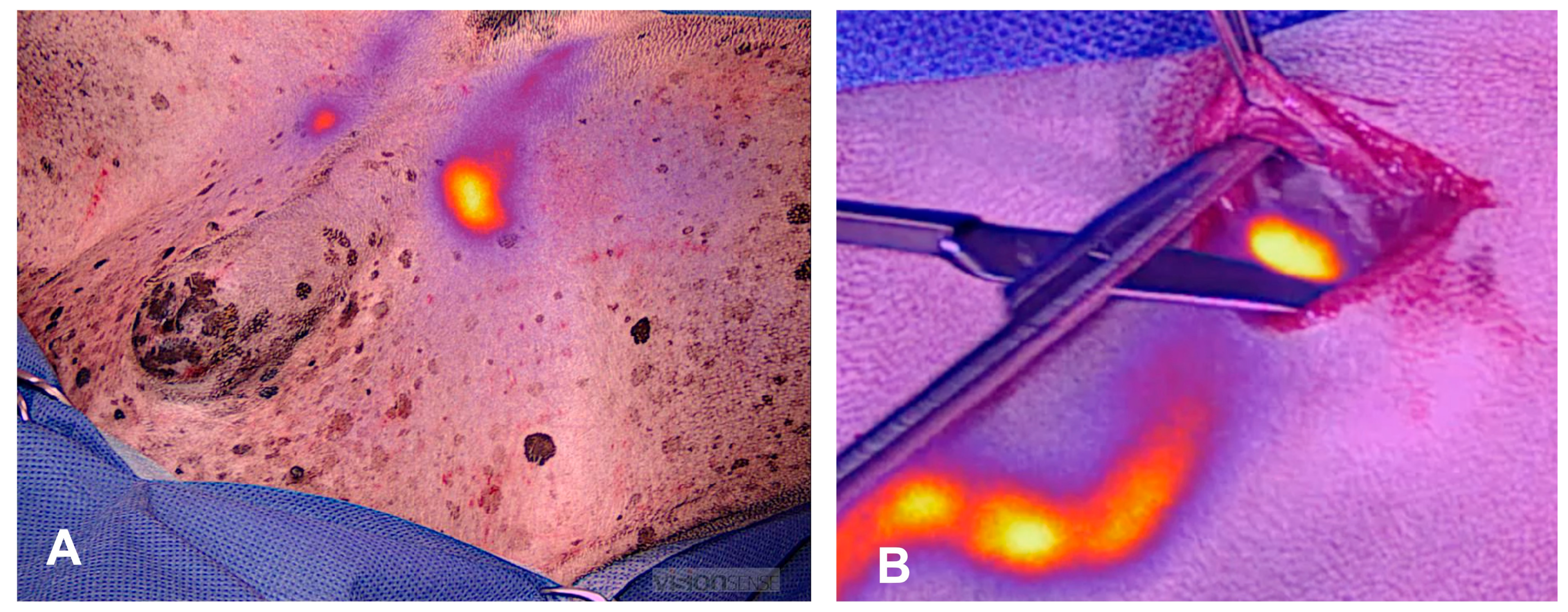
3.2.2. Near-Infrared Lymphography

NIR for SLN mapping is a rapidly developing field in human and veterinary surgical oncology. It offers the opportunity of optical image-guided surgery to provide real-time visualization of lymph vessels and SLNs [71]. The near-infrared light spectrum has a wavelength of 700–900 nm, which is invisible to the human eye [72]. NIR imaging systems emit excitation light in the low near-infrared spectrum that is absorbed by the fluorescent agent and shifted to a lower wavelength. The emitted light can be detected using a NIR camera device and translated to a real-time video displayed on a screen [72,73]. In contrast to visible light, the light of the near-infrared spectrum is hardly absorbed by the body tissue, which allows tissue penetration up to a few centimeters, making NIR suitable for the transcutaneous identification of SLNs in humans and animals (Figure 3) [71,72,73]. The most frequently used dye is ICG, which was approved for clinical use by the FDA in 1959 [72]. It was shown that the identification of SLNs via NIR is technically simpler, more reliable, and associated with increased SLN detection rates, compared with other techniques, which may be explained by the smaller particle size of fluorescent agents facilitating a faster spill into secondary lymph nodes [74,75,76]. Lately, several metareviews in human medicine have confirmed NIR imaging using ICG as a reliable mapping technique compared with the gold standard lymphoscintigraphy [35,77].
After NIR SLN mapping in dogs was initially described in experimental studies in dogs and pigs [50], it is now gaining increased interest in veterinary surgical oncology [14,49,52]. Beer et al. published a retrospective study on NIR lymphography in dogs with mast cell tumors. NIR lymphography significantly improved the success rate of lymphadenectomies and increased the detection rate of metastatic disease (68% of dogs were diagnosed with at least one metastatic node using NIR lymphography compared with 33% only when regional lymphadenectomies were performed) [14]. The improved detection rate of metastatic disease was achieved by a combination of improved resection success and the identification of the SLNs different from the expected regional nodes [14]. A first case report showing the feasibility of SLN mapping in a cat also bearing a mast cell tumor has just been published, followed by a pilot study in cats with different solid neoplasms [26,52]. Finally, Gariboldi et al. very recently published a study that proved that the SLN mapping of scars from previous tumor excision is also possible using lymphoscintigraphy or NIR lymphography [49].
Taken together, optical imaging with NIR offers several distinctive advantages. In cases of superficial tumors, NIR camera systems enable a transcutaneous visualization of fluorescent dyes, making a massive surgical dissection of the overlying tissue unnecessary and allowing distinction between the first- and second-tier nodes [14,71,78]. The improved visualization of small and nonpalpable nodes results in a significantly increased success rate of lymphadenectomies compared with unguided lymphadenectomies in dogs [14]. No mapping-related complications have been reported in dogs or cats [14,49,52,78,79]. The disadvantages of NIR include its low depth of penetration, limited to a few millimeters to centimeters, and that it does not allow the identification of sentinel nodes in body cavities without surgical access to allow visual inspection [72].
To summarize, NIR lymphography has been identified as one of the most sensitive and specific SLN mapping techniques, compared with the gold standard lymphoscintigraphy. As NIR lymphography does not require specific legal allowances and is possible with significantly less financial investment than lymphoscintigraphy, it represents a simple and cost-effective alternative for accurate mapping. As in human medicine, or perhaps more so, in veterinary medicine, the application of simple and inexpensive, but accurate and safe, techniques is of great importance, and an important requirement to enable the broad-based implementation of mapping in veterinary surgical oncology.
The results of the available studies for SLN mapping using the above-mentioned techniques in tumor-bearing dogs and cats are summarized in Table 1.

4. Role of Lymphadenectomies and SLN Mapping in Selected Cancer Types in Dogs and Cats

4.1. Mammary Carcinoma

The tumors of the mammary glands are the most common neoplastic condition in dogs, accounting for 25–50% of all diagnosed neoplasms in dogs [59,82]. Approximately 50% are malignant, and in 35–70% of these, the local recurrence of metastatic spread is observed, often leading to the death of the patient [59,82]. As with other neoplasms in dogs, there are conflicting results on the therapeutic effect of lymphadenectomy. However, most of these studies were conducted by removing the proposed RLN: This corresponds to the ipsilateral axillary lymph node for the first two mammary complexes, the ipsilateral axillar and inguinal node (+/− medial iliac) for tumors in the third complex, and the ipsilateral inguinal (+/− medial iliac) for the fourth complex [17,59]. Of these, the deep LNs (sternal, medial iliac) are not routinely removed.
The SLN mapping of the mammary glands in dogs and cats has been performed using CT lymphography, radiographic lymphography, and scintigraphy [42,44,57,58,59,60]. Pereira et al. (2008) used lymphoscintigraphy and identified a complex heterogeneous draining pattern with wide variations between patients, with a high number of cross-connections between the lymphatics [40]. The cranial two complexes drained to various centers including the axillary, sternal, and superficial cervical while the caudal three glands drained to the inguinal and/or medial iliac lymphocenters [44]. Similarly, unpredictive and very individual draining patterns often draining contralateral LNs have been described by other studies using SLN mapping [42,57,58,59].
Whether routine SLN mapping and removal of SLN impact therapeutic outcomes in affected dogs is yet to be proven.

4.2. Apocrine Gland Anal Sac Adenocarcinoma (AGASAC)

Another condition in which the resection of metastatic LNs has proven therapeutic benefits is AGASAC [1,83,84]. AGSAC accounts for 17% of perianal neoplasia, and 2% of skin tumors [8,17]. These tumors tend to metastasize to the regional draining lymph center in up to 96%, and nodal metastasis represents an important negative prognostic factor [11,12,21,83,84,85]. The removal of metastatic LNs is recommended, and several studies documented the improvement in clinical symptoms and survival times when the lymphadenectomy of metastatic LNs was performed in addition to the surgical removal of the primary tumor [12]. Despite the high incidence of nodal metastasis, the current treatment standard mainly involves the removal of grossly enlarged or structurally altered LN [1], rather than the routine resection of the entire lymphocenter [1,12]. Associated complications vary depending on the size of the nodes and range between 5% and 41%, mostly in the form of hemorrhage [12].
Given the high metastatic rate, the early detection of the affected unenlarged LNs might help control disease; however, currently, no information exists regarding the metastatic rate of unenlarged nonstructurally altered LNs. A recent study evaluating owner compliance after the diagnosis of AGASAC documented that anal sacculectomy was performed in 93% of cases; however, only 22% of dogs underwent iliosacral lymphadenectomy [86]. This seems to be a relatively low number in a condition with such a high metastatic rate, and it underlines a lack of routine strategy for lymph node management in this disease.
Routine SLN mapping could potentially lead to improved detection and management of early metastatic LNs; however, this must be validated in future studies. Thus far, SLN mapping has been reported in few studies. In 2018, Linden et al. compared two different injection techniques to map the SLN of the anal glands in healthy dogs and found that intramural injection was more reliable than perigland injection [21]. The medial iliac node was identified as sentinel in 62.5% of dogs, the lateral sacral node was sentinel in 37.5% of the cases, and the inguinal node was sentinel in 12.5% [21]. Majeski et al. published a study involving 13 dogs with AGASAC that were subjected to indirect CT lymphography [40]. They were able to detect at least one SLN in 92% of the cases, and 67% were ipsilateral, whereas 33% were contralateral. SLNs were medial iliac (42%), sacral (25%), or internal iliac (8%), and 25% of dogs had more than one tier-1 SLN (tier-2 SLNs were detected in 42% of the cases) [40]. Unfortunately, the authors failed to report the histologic status and size of the SLNs.

4.3. Mast Cell Tumor

Solid MCTs are among the most frequent skin tumors encountered in dogs [87] and cats [88,89]. During the past five years, our understanding of the importance of LN metastasis has greatly improved this condition. Studies have demonstrated a metastatic rate of 56% to 68% when SLN mapping was performed [14,20,25,80,90]. In 2014, Weishaar et al. developed a nodal staging system and were able to prove that nodal status is an important prognostic factor for survival [91]. Finally, the removal of nodal metastasis has proven relevant for therapeutic reasons. Patients whose lymph nodes are routinely removed showed longer survival and recurrence-free times than patients in whom lymphadenectomies were not performed during tumor resection [3,4,5,92,93].
LN status is also relevant to plan adjuvant chemotherapy, as it has been proven that patients with HN3 metastatic nodes benefit from chemotherapy irrespective of the primary grade of the tumor, whereas there is no benefit expected in patients with low-grade MCTs, i.e., grade-1 and -2 mast cell tumors with HN0-HN2 nodes [5].
Due to the obvious impact of lymphadenectomy in patients with mast cell tumors, and the high incidence of LN metastasis, this condition is one of the most frequently used cancer types to evaluate SLN mapping in dogs. In one of the earlier studies, Worley et al. performed lymphoscintigraphy combined with intrasurgical methylene blue injection. They were able to detect SLNs in 95% of the cases using preoperative lymphoscintigraphy and in 100% of the cases using an intraoperative gamma probe [20]. They also demonstrated that SLNs did not correspond to the expected regional lymph node in 42% of the cases using lymphoscintigraphy [20]. Beer et al. supported this finding using NIR lymphography and also documented that SLNs did not correspond to the regional node in 42% of dogs, and Ferrari et al. even documented a discrepancy of 63% [14,24]. Furthermore, we were able to demonstrate that NIR lymphography detected at least one SLN in 100% of cases and resulted in higher success rates for lymphadenectomies [14]. As a result, significantly more metastatic LNs were identified when NIR mapping was compared with unguided lymphadenectomy (68% metastatic rate versus 33%) [14].
Fournier et al. evaluated the performance of CEUS in the detection of SLNs in dogs with MCTs. They were able to detect SLNs in 95.2% of cases, with a discrepancy rate between SLNs and a regional node prediction of 35.6%, and an overall metastatic rate of 60% [25]. Notably, only 55% of SLNs were then evaluated using histopathology, but the authors did not indicate whether the reason for not submitting the remaining nodes was because of unsuccessful resection or other reasons [25]. Disturbingly, the authors also found metastatic disease in 50% of regional nodes, which were nonsentinel [25]. This raises the question of whether CEUS failed to detect these as sentinel, or if the concept of SLNs must be revised. Beer et al. were also able to identify metastatic disease in a subset of nonsentinel nodes, although their false negative rate was much lower (36% of non-SLN were metastatic), potentially due to the superior sensitivity of NIR lymphography [14]. These studies highlight the necessity to investigate whether SLN-based resections are suitable to fully address metastatic disease in the future, or if resections of the entire basin are still needed.
Most recently, De Bonis et al. demonstrated that indirect lymphography using lipidiol and serial radiographs resulted in the detection of at least one SLN in 90% of cases [81]. Their study also described a high discrepancy between RLNs and SLNs, with only 27% full agreement, and interestingly, they also described a considerably lower rate of 50% than other studies utilizing more efficient SLN methods [94]. This drawback has already been described by Hlusko et al., who demonstrated a significant discrepancy between indirect lymphography and lymphoscintigraphy [37]. Thus, although this technique is technically feasible, it is not recommended.

4.4. Cancers of the Head and Neck

In dogs with tumors of the head and neck area, metastases to the lymph nodes other than the mandibular are reported in nearly half of the cases, and contralateral dissemination occurs in up to 62% of animals with oral tumors [22,94]. While the early detection of cervical LN metastases is considered one of the mainstays for the correct management in humans [95], comparable evidence is still limited for dogs and cats, mainly due to inconsistent recommendations for the management of the cN0 neck in the veterinary practice [96].
Traditionally, elective neck dissection (END) is recommended for tumors with a risk of occult LN metastases >20% in humans [97], but high rates of functional morbidity have led to the investigation of SLN biopsy as a less invasive approach [98,99]. In veterinary medicine, END was first described in 1995 in six carcasses and three dogs and consisted of the ipsilateral or bilateral excision of the retropharyngeal, parotid, and mandibular nodes [100]. When this technique was reapplied a few years later in dogs and cats with spontaneous head and neck malignancies, the metastases not involving the mandibular LNs were detected in nearly half of the dogs [94]. More recently, Skinner et al. described 31 clinical cases, and LN metastases were detected in 11 retropharyngeal nodes without concurrent mandibular LN involvement, with 61% of metastasis occurring in the contralateral to the primary tumor [22]. Complications reported after the radical lymphadenectomy of the head and neck region are rare and self-limiting in dogs, mainly consisting of the transitory edema of the muzzle or lip [22,94,96,100]. However, unguided END frequently fails to remove the parotid nodes, which can harbor metastasis in 9–16% of dogs [13,94]. Only a few studies are available for SLN biopsy in dogs with malignancies of the head and neck, but the technique seems promising, with high detection rates and diagnostic accuracy [13,39,48,94]. In 2009, Luire et al. described, for the first time, SLN biopsy in dogs with spontaneous tumors of the head and neck and reported a detection rate of 100% with radiopharmaceutical and 80% with CEUS [48]. Indirect CT lymphography was also used to map the cN0 neck in cancer dogs, allowing for the detection of contralateral dissemination in 8% of animals in one study [39]. The technique was compared with lymphoscintigraphy in a more recent study, with detection rates of 55% and 100%, respectively [61]. The diagnostic accuracy of SLN biopsy using lymphoscintigraphy was confirmed in a recent study on 23 dogs with naturally occurring malignancies of the head and neck and the absence of clinically evident LN disease [13]. In that study, a detection rate of 83% was recorded, whereas sensitivity and specificity were 88.9% and 100%, respectively [13]. Lastly, Wan et al. (2021) described the combined use of indirect CT lymphography and vital dyes for SLN biopsy in dogs with oral tumors and reported a detection rate of 100% with the combined technique. However, when only one technique was assessed, the detection rate of NIR lymphography using ICG was much higher than CT lymphography [47].
SLN biopsy in head and neck cancer is still at its infancy in veterinary oncology, and although some studies have demonstrated the feasibility of different mapping techniques, the actual impact of a targeted nodal approach and its clinical impact compared with END need to be further explored in the future.

5. Future Perspectives

Future structured studies need to identify the therapeutic impact of extended versus SLN resections in dogs with different types of cancer. In addition, techniques that can not only detect the SLN but also reliably show metastatic nodes should be further investigated. In this context, the use of SPECT-CT, iron-oxide ultra-small particles, or targeted fluorophores might be of interest in the future.

6. Conclusions

LN staging and resection are gaining prominence in veterinary surgical oncology, but further studies are needed to evaluate the concrete therapeutic impact for most conditions. Among the several malignancies for which therapeutic lymphadenectomies are considered potentially beneficial, their concrete impact is best established for canine mast cell tumors. SLN mapping could help to develop standardized approaches for lymphadenectomy in various conditions, setting the stage for the subsequent evaluation of potential therapeutic benefits. While numerous techniques for SLN mapping have been described, NIR lymphography seems to have the highest SLN detection rate, compared to the gold standard lymphoscintigraphy, and thus presents a noteworthy technique for future implementation in veterinary medicine.

Author Contributions

Conceptualization, M.C.N.; writing—original draft preparation, P.B., L.E.C. and M.C.N.; writing—review and editing, M.C.N., P.B., L.E.C.; supervision, M.C.N.; All authors have read and agreed to the published version of the manuscript.

Funding

This research received no external funding.

Conflicts of Interest

The authors declare no conflict of interest.

List of Abbreviations

AGASACApocrine gland anal sac adenocarcinoma
CEUSContrast-enhanced ultrasound
CTComputed tomography
ENDElective neck dissection
ICGIndocyanine green
LNLymph node
MCT Mast cell tumor
NIR Near-infrared
NIR-LNear-infrared fluorescent lymphography
RLNRegional lymph node
SLNSentinel lymph node
Tc-99mTechnetium-99m

References

  1. Liptak, J.M.; Boston, S.E. Nonselective Lymph Node Dissection and Sentinel Lymph Node Mapping and Biopsy. Vet. Clin. N. Am. Small Anim. Pract. 2019, 49, 793–807. [Google Scholar] [CrossRef] [PubMed]
  2. Beer, P.; Pozzi, A.; Rohrer Bley, C.; Bacon, N.; Pfammatter, N.S.; Venzin, C. The role of sentinel lymph node mapping in small animal veterinary medicine: A comparison with current approaches in human medicine. Vet. Comp. Oncol. 2018, 16, 178–187. [Google Scholar] [CrossRef] [PubMed]
  3. Chalfon, C.; Sabattini, S.; Finotello, R.; Faroni, E.; Guerra, D.; Pisoni, L.; Ciammaichella, L.; Vasconi, M.E.; Annoni, M.; Marconato, L. Lymphadenectomy improves outcome in dogs with resected Kiupel high-grade cutaneous mast cell tumours and overtly metastatic regional lymph nodes. J. Small Anim. Pract. 2022, 63, 661–669. [Google Scholar] [CrossRef]
  4. Marconato, L.; Polton, G.; Stefanello, D.; Morello, E.; Ferrari, R.; Henriques, J.; Tortorella, G.; Benali, S.L.; Bergottini, R.; Vasconi, M.E.; et al. Therapeutic impact of regional lymphadenectomy in canine stage II cutaneous mast cell tumours. Vet. Comp. Oncol. 2018, 16, 580–589. [Google Scholar] [CrossRef] [PubMed]
  5. Marconato, L.; Stefanello, D.; Kiupel, M.; Finotello, R.; Polton, G.; Massari, F.; Ferrari, R.; Agnoli, C.; Capitani, O.; Giudice, C.; et al. Adjuvant medical therapy provides no therapeutic benefit in the treatment of dogs with low-grade mast cell tumours and early nodal metastasis undergoing surgery. Vet. Comp. Oncol. 2020, 18, 409–415. [Google Scholar] [CrossRef] [PubMed]
  6. Mendez, S.E.; Drobatz, K.J.; Duda, L.E.; White, P.; Kubicek, L.; Sorenmo, K.U. Treating the locoregional lymph nodes with radiation and/or surgery significantly improves outcome in dogs with high-grade mast cell tumours. Vet. Comp. Oncol. 2020, 18, 239–246. [Google Scholar] [CrossRef]
  7. Halsted, W.S.I. The Results of Operations for the Cure of Cancer of the Breast Performed at the Johns Hopkins Hospital from June 1889, to January 1894. Ann. Surg. 1894, 20, 497–555. [Google Scholar] [CrossRef]
  8. Loukas, M.; Bellary, S.S.; Kuklinski, M.; Ferrauiola, J.; Yadav, A.; Shoja, M.M.; Shaffer, K.; Tubbs, R.S. The lymphatic system: A historical perspective. Clin. Anat. 2011, 24, 807–816. [Google Scholar] [CrossRef]
  9. Dogan, N.U.; Dogan, S.; Favero, G.; Kohler, C.; Dursun, P. The Basics of Sentinel Lymph Node Biopsy: Anatomical and Pathophysiological Considerations and Clinical Aspects. J. Oncol. 2019, 2019, 3415630. [Google Scholar] [CrossRef] [Green Version]
  10. Liptak, J.M. Histologic margins and the residual tumour classification scheme: Is it time to use a validated scheme in human oncology to standardise margin assessment in veterinary oncology? Vet. Comp. Oncol. 2020, 18, 25–35. [Google Scholar] [CrossRef]
  11. Barnes, D.C.; Demetriou, J.L. Surgical management of primary, metastatic and recurrent anal sac adenocarcinoma in the dog: 52 cases. J. Small Anim. Pract. 2017, 58, 263–268. [Google Scholar] [CrossRef] [PubMed]
  12. Repasy, A.B.; Selmic, L.E.; Kisseberth, W.C. Canine Apocrine Gland Anal Sac Adenocarcinoma: A Review. Top. Companion Anim. Med. 2022, 50, 100682. [Google Scholar] [CrossRef] [PubMed]
  13. Chiti, L.E.; Stefanello, D.; Manfredi, M.; Zani, D.D.; De Zani, D.; Boracchi, P.; Giudice, C.; Grieco, V.; Di Giancamillo, M.; Ferrari, R. To map or not to map the cN0 neck: Impact of sentinel lymph node biopsy in canine head and neck tumours. Vet. Comp. Oncol. 2021, 19, 661–670. [Google Scholar] [CrossRef] [PubMed]
  14. Beer, P.; Rohrer Bley, C.; Nolff, M.C. Near-infrared fluorescent image-guided lymph node dissection compared with locoregional lymphadenectomies in dogs with mast cell tumours. JSAP 2022, 63, 670–678. [Google Scholar] [CrossRef] [PubMed]
  15. Ferrari, R.; Boracchi, P.; Chiti, L.E.; Manfredi, M.; Giudice, C.; De Zani, D.; Spediacci, C.; Recordati, C.; Grieco, V.; Gariboldi, E.M.; et al. Assessing the Risk of Nodal Metastases in Canine Integumentary Mast Cell Tumors: Is Sentinel Lymph Node Biopsy Always Necessary? Animals 2021, 11, 2373. [Google Scholar] [CrossRef]
  16. Haley, P.J. The lymphoid system: A review of species differences. J. Toxicol. Pathol. 2017, 30, 111–123. [Google Scholar] [CrossRef] [Green Version]
  17. Baum, H. Das Lymphgefäßsystem des Hundes; Hirschwald: Berlin, Germnay, 1918. [Google Scholar]
  18. Suami, H.; Yamashita, S.; Soto-Miranda, M.A.; Chang, D.W. Lymphatic territories (lymphosomes) in a canine: An animal model for investigation of postoperative lymphatic alterations. PLoS ONE 2013, 8, e69222. [Google Scholar] [CrossRef] [Green Version]
  19. Suami, H.; Shin, D.; Chang, D.W. Mapping of lymphosomes in the canine forelimb: Comparative anatomy between canines and humans. Plast. Reconstr. Surg. 2012, 129, 612–620. [Google Scholar] [CrossRef]
  20. Worley, D.R. Incorporation of sentinel lymph node mapping in dogs with mast cell tumours: 20 consecutive procedures. Vet. Comp. Oncol. 2014, 12, 215–226. [Google Scholar] [CrossRef]
  21. Linden, D.S.; Cole, R.; Tillson, D.M.; Boothe, H.W.; Matz, B.M. Sentinel lymph node mapping of the canine anal sac using lymphoscintigraphy: A pilot study. Vet. Radiol. Ultrasound Off. J. Am. Coll. Vet. Radiol. Int. Vet. Radiol. Assoc. 2019, 60, 346–350. [Google Scholar] [CrossRef]
  22. Skinner, O.T.; Boston, S.E.; Souza, C.H.M. Patterns of lymph node metastasis identified following bilateral mandibular and medial retropharyngeal lymphadenectomy in 31 dogs with malignancies of the head. Vet. Comp. Oncol. 2017, 15, 881–889. [Google Scholar] [CrossRef] [PubMed]
  23. Morton, D.L.; Wen, D.R.; Wong, J.H.; Economou, J.S.; Cagle, L.A.; Storm, F.K.; Foshag, L.J.; Cochran, A.J. Technical details of intraoperative lymphatic mapping for early-stage melanoma. Arch. Surg. 1992, 127, 392–399. [Google Scholar] [CrossRef] [PubMed]
  24. Ferrari, R.; Chiti, L.E.; Manfredi, M.; Ravasio, G.; De Zani, D.; Zani, D.D.; Giudice, C.; Gambini, M.; Stefanello, D. Biopsy of sentinel lymph nodes after injection of methylene blue and lymphoscintigraphic guidance in 30 dogs with mast cell tumors. Vet. Surg. 2020, 49, 1099–1108. [Google Scholar] [CrossRef]
  25. Fournier, Q.; Thierry, F.; Longo, M.; Malbon, A.; Cazzini, P.; Bisson, J.; Woods, S.; Liuti, T.; Bavcar, S. Contrast-enhanced ultrasound for sentinel lymph node mapping in the routine staging of canine mast cell tumours: A feasibility study. Vet. Comp. Oncol. 2021, 19, 451–462. [Google Scholar] [CrossRef] [PubMed]
  26. Chiti, L.E.; Gariboldi, E.M.; Stefanello, D.; De Zani, D.; Grieco, V.; Nolff, M.C. Sentinel Lymph Node Mapping and Biopsy in Cats with Solid Malignancies: An Explorative Study. Animals 2022, 12, 3116. [Google Scholar] [CrossRef] [PubMed]
  27. Ashikaga, T.; Krag, D.N.; Land, S.R.; Julian, T.B.; Anderson, S.J.; Brown, A.M.; Skelly, J.M.; Harlow, S.P.; Weaver, D.L.; Mamounas, E.P.; et al. Morbidity results from the NSABP B-32 trial comparing sentinel lymph node dissection versus axillary dissection. J. Surg. Oncol. 2010, 102, 111–118. [Google Scholar] [CrossRef] [Green Version]
  28. Schrenk, P.; Rieger, R.; Shamiyeh, A.; Wayand, W. Morbidity following sentinel lymph node biopsy versus axillary lymph node dissection for patients with breast carcinoma. Cancer 2000, 88, 608–614. [Google Scholar] [CrossRef]
  29. Cabanas, R.M. An approach for the treatment of penile carcinoma. Cancer 1977, 39, 456–466. [Google Scholar] [CrossRef]
  30. Hayashi, H.; Tangoku, A.; Suga, K.; Shimizu, K.; Ueda, K.; Yoshino, S.; Abe, T.; Sato, T.; Matsunaga, N.; Oka, M. CT lymphography-navigated sentinel lymph node biopsy in patients with superficial esophageal cancer. Surgery 2006, 139, 224–235. [Google Scholar] [CrossRef]
  31. Motomura, K.; Sumino, H.; Noguchi, A.; Horinouchi, T.; Nakanishi, K. Sentinel nodes identified by computed tomography-lymphography accurately stage the axilla in patients with breast cancer. BMC Med. Imaging 2013, 13, 42. [Google Scholar] [CrossRef]
  32. Sever, A.; Jones, S.; Cox, K.; Weeks, J.; Mills, P.; Jones, P. Preoperative localization of sentinel lymph nodes using intradermal microbubbles and contrast-enhanced ultrasonography in patients with breast cancer. Br. J. Surg. 2009, 96, 1295–1299. [Google Scholar] [CrossRef] [PubMed]
  33. Naaman, Y.; Pinkas, L.; Roitman, S.; Ikher, S.; Oustinov, N.; Vaisbuch, E.; Yachnin, A.; Ben-Arie, A. The Added Value of SPECT/CT in Sentinel Lymph Nodes Mapping for Endometrial Carcinoma. Ann. Surg. Oncol. 2016, 23, 450–455. [Google Scholar] [CrossRef] [PubMed]
  34. Heusner, T.A.; Kuemmel, S.; Hahn, S.; Koeninger, A.; Otterbach, F.; Hamami, M.E.; Kimmig, K.R.; Forsting, M.; Bockisch, A.; Antoch, G.; et al. Diagnostic value of full-dose FDG PET/CT for axillary lymph node staging in breast cancer patients. Eur. J. Nucl. Med. Mol. Imaging 2009, 36, 1543–1550. [Google Scholar] [CrossRef] [PubMed]
  35. Jeremiasse, B.; van den Bosch, C.H.; Wijnen, M.; Terwisscha van Scheltinga, C.E.J.; Fiocco, M.F.; van der Steeg, A.F.W. Systematic review and meta-analysis concerning near-infrared imaging with fluorescent agents to identify the sentinel lymph node in oncology patients. Eur. J. Surg. Oncol. 2020, 46, 2011–2022. [Google Scholar] [CrossRef]
  36. Brissot, H.N.; Edery, E.G. Use of indirect lymphography to identify sentinel lymph node in dogs: A pilot study in 30 tumours. Vet. Comp. Oncol. 2016, 15, 740–753. [Google Scholar] [CrossRef]
  37. Hlusko, K.C.; Cole, R.; Tillson, D.M.; Boothe, H.W.; Almond, G.; Coggeshall, W.S.; Matz, B.M. Sentinel lymph node detection differs when comparing lymphoscintigraphy to lymphography using water soluble iodinated contrast medium and digital radiography in dogs. Vet. Radiol. Ultrasound 2020, 61, 659–666. [Google Scholar] [CrossRef]
  38. Mayer, M.N.; Silver, T.I.; Lowe, C.K.; Anthony, J.M. Radiographic lymphangiography in the dog using iodized oil. Vet. Comp. Oncol. 2013, 11, 151–161. [Google Scholar] [CrossRef]
  39. Grimes, J.A.; Secrest, S.A.; Northrup, N.C.; Saba, C.F.; Schmiedt, C.W. Indirect computed tomography lymphangiography with aqueous contrast for evaluation of sentinel lymph nodes in dogs with tumors of the head. Vet. Radiol. Ultrasound Off. J. Am. Coll. Vet. Radiol. Int. Vet. Radiol. Assoc. 2017, 58, 559–564. [Google Scholar] [CrossRef]
  40. Majeski, S.A.; Steffey, M.A.; Fuller, M.; Hunt, G.B.; Mayhew, P.D.; Pollard, R.E. Indirect Computed Tomographic Lymphography for Iliosacral Lymphatic Mapping in a Cohort of Dogs with Anal Sac Gland Adenocarcinoma: Technique Description. Vet. Radiol. Ultrasound Off. J. Am. Coll. Vet. Radiol. Int. Vet. Radiol. Assoc. 2017, 58, 295–303. [Google Scholar] [CrossRef]
  41. Rossi, F.; Korner, M.; Suarez, J.; Carozzi, G.; Meier, V.S.; Roos, M.; Rohrer Bley, C. Computed tomographic-lymphography as a complementary technique for lymph node staging in dogs with malignant tumors of various sites. Vet. Radiol. Ultrasound Off. J. Am. Coll. Vet. Radiol. Int. Vet. Radiol. Assoc. 2018, 59, 155–162. [Google Scholar] [CrossRef]
  42. Soultani, C.; Patsikas, M.N.; Karayannopoulou, M.; Jakovljevic, S.; Chryssogonidis, I.; Papazoglou, L.; Papaioannou, N.; Papadopoulou, P.; Pavlidou, K.; Ilia, G.M.; et al. Assessment of Sentinel Lymph Node Metastasis in Canine Mammary Gland Tumors Using Computed Tomographic Indirect Lymphography. Vet. Radiol. Ultrasound Off. J. Am. Coll. Vet. Radiol. Int. Vet. Radiol. Assoc. 2017, 58, 186–196. [Google Scholar] [CrossRef] [PubMed]
  43. Balogh, L.; Thuroczy, J.; Andocs, G.; Mathe, D.; Chaudhari, P.; Perge, E.; Biksi, I.; Polyak, A.; Kiraly, R.; Janoki, G.A. Sentinel lymph node detection in canine oncological patients. Nucl. Med. Rev. Cent. East. Eur. 2002, 5, 139–144. [Google Scholar] [PubMed]
  44. Pereira, C.T.; Luiz Navarro Marques, F.; Williams, J.; Wlademir De Martin, B.; Primo Bombonato, P. 99mTc-labeled dextran for mammary lymphoscintigraphy in dogs. Vet. Radiol. Ultrasound Off. J. Am. Coll. Vet. Radiol. Int. Vet. Radiol. Assoc. 2008, 49, 487–491. [Google Scholar] [CrossRef]
  45. Tuohy, J.L.; Worley, D.R. Pulmonary lymph node charting in normal dogs with blue dye and scintigraphic lymphatic mapping. Res. Vet. Sci. 2014, 97, 148–155. [Google Scholar] [CrossRef]
  46. Hlusko, K.C.; Cole, R.; Tillson, D.M.; Boothe, H.W.; Almond, G.T.; Coggeshall, W.S.; Matz, B.M. The effect of surgery on lymphoscintigraphy drainage patterns from the canine brachium in a simulated tumor model. Vet. Surg. 2020, 49, 1118–1124. [Google Scholar] [CrossRef]
  47. Wan, J.; Oblak, M.L.; Ram, A.; Singh, A.; Nykamp, S. Determining agreement between preoperative computed tomography lymphography and indocyanine green near infrared fluorescence intraoperative imaging for sentinel lymph node mapping in dogs with oral tumours. Vet. Comp. Oncol. 2021, 19, 295–303. [Google Scholar] [CrossRef]
  48. Lurie, D.M.; Seguin, B.; Schneider, P.D.; Verstraete, F.J.; Wisner, E.R. Contrast-assisted ultrasound for sentinel lymph node detection in spontaneously arising canine head and neck tumors. Investig. Radiol. 2006, 41, 415–421. [Google Scholar] [CrossRef] [PubMed]
  49. Gariboldi, E.M.; Stefanello, D.; Nolff, M.C.; De Zani, D.; Zani, D.; Grieco, V.; Giudice, C.; Recordati, C.; Ferrari, F.; Ferrari, R.; et al. Sentinel Lymph Node Biopsy Is Feasible in Dogs with Scars from Prior Local Excision of Solid Malignancies. Animals 2022, 12, 2195. [Google Scholar] [CrossRef] [PubMed]
  50. Knapp, D.W.; Adams, L.G.; Degrand, A.M.; Niles, J.D.; Ramos-Vara, J.A.; Weil, A.B.; O’Donnell, M.A.; Lucroy, M.D.; Frangioni, J.V. Sentinel lymph node mapping of invasive urinary bladder cancer in animal models using invisible light. Eur. Urol. 2007, 52, 1700–1708. [Google Scholar] [CrossRef] [Green Version]
  51. Townsend, K.L.; Milovancev, M.; Bracha, S. Feasibility of near-infrared fluorescence imaging for sentinel lymph node evaluation of the oral cavity in healthy dogs. Am. J. Vet. Res. 2018, 79, 995–1000. [Google Scholar] [CrossRef]
  52. Arz, R.; Seehusen, F.; Meier, V.S.; Nolff, M.C. Indocyanine-based near-infrared lymphography for real-time detection of lymphatics in a cat with multiple mast cell tumours. JFMS Open Rep. 2022, 8, 20551169221074961. [Google Scholar] [CrossRef]
  53. Niebling, M.G.; Pleijhuis, R.G.; Bastiaannet, E.; Brouwers, A.H.; van Dam, G.M.; Hoekstra, H.J. A systematic review and meta-analyses of sentinel lymph node identification in breast cancer and melanoma, a plea for tracer mapping. Eur. J. Surg. Oncol. 2016, 42, 466–473. [Google Scholar] [CrossRef] [PubMed]
  54. Manfredi, M.; De Zani, D.; Chiti, L.E.; Ferrari, R.; Stefanello, D.; Giudice, C.; Pettinato, V.; Longo, M.; Di Giancamillo, M.; Zani, D.D. Preoperative planar lymphoscintigraphy allows for sentinel lymph node detection in 51 dogs improving staging accuracy: Feasibility and pitfalls. Vet. Radiol. Ultrasound Off. J. Am. Coll. Vet. Radiol. Int. Vet. Radiol. Assoc. 2021, 62, 602–609. [Google Scholar] [CrossRef] [PubMed]
  55. Wawroschek, F.; Wengenmair, H.; Senekowitsch-Schmidtke, R.; Hamm, M.; Henke, J.; Schonberger, T.; Hauser, A.; Erhardt, W.; Harzmann, R. Prostate lymphoscintigraphy for sentinel lymph node identification in canines: Reproducibility, uptake, and biokinetics depending on different injection strategies. Urol. Res. 2003, 31, 152–158. [Google Scholar] [CrossRef] [PubMed]
  56. Alex, J.C.; Krag, D.N. Gamma-probe guided localization of lymph nodes. Surg. Oncol. 1993, 2, 137–143. [Google Scholar] [CrossRef]
  57. Patsikas, M.N.; Karayannopoulou, M.; Kaldrymidoy, E.; Papazoglou, L.G.; Papadopoulou, P.L.; Tzegas, S.I.; Tziris, N.E.; Kaitzis, D.G.; Dimitriadis, A.S.; Dessiris, A.K. The lymph drainage of the neoplastic mammary glands in the bitch: A lymphographic study. Anat. Histol. Embryol. 2006, 35, 228–234. [Google Scholar] [CrossRef]
  58. Papadopoulou, P.L.; Patsikas, M.N.; Charitanti, A.; Kazakos, G.M.; Papazoglou, L.G.; Karayannopoulou, M.; Chrisogonidis, I.; Tziris, N.; Dimitriadis, A. The lymph drainage pattern of the mammary glands in the cat: A lymphographic and computerized tomography lymphographic study. Anat. Histol. Embryol. 2009, 38, 292–299. [Google Scholar] [CrossRef] [PubMed]
  59. Collivignarelli, F.; Tamburro, R.; Aste, G.; Falerno, I.; Del Signore, F.; Simeoni, F.; Patsikas, M.; Gianfelici, J.; Terragni, R.; Attorri, V.; et al. Lymphatic Drainage Mapping with Indirect Lymphography for Canine Mammary Tumors. Animals 2021, 11, 115. [Google Scholar] [CrossRef] [PubMed]
  60. Patsikas, M.N.; Papadopoulou, P.L.; Charitanti, A.; Kazakos, G.M.; Soultani, C.B.; Tziris, N.E.; Tzegas, S.I.; Jakovljevic, S.; Savas, I.; Stamoulas, K.G. Computed tomography and radiographic indirect lymphography for visualization of mammary lymphatic vessels and the sentinel lymph node in normal cats. Vet. Radiol. Ultrasound Off. J. Am. Coll. Vet. Radiol. Int. Vet. Radiol. Assoc. 2010, 51, 299–304. [Google Scholar] [CrossRef]
  61. Randall, E.K.; Jones, M.D.; Kraft, S.L.; Worley, D.R. The development of an indirect computed tomography lymphography protocol for sentinel lymph node detection in head and neck cancer and comparison to other sentinel lymph node mapping techniques. Vet. Comp. Oncol. 2020, 18, 634–644. [Google Scholar] [CrossRef]
  62. Haers, H.; Saunders, J.H. Review of clinical characteristics and applications of contrast-enhanced ultrasonography in dogs. J. Am. Vet. Med. Assoc. 2009, 234, 460–470. [Google Scholar] [CrossRef] [PubMed]
  63. Seiler, G.S.; Brown, J.C.; Reetz, J.A.; Taeymans, O.; Bucknoff, M.; Rossi, F.; Ohlerth, S.; Alder, D.; Rademacher, N.; Drost, W.T.; et al. Safety of contrast-enhanced ultrasonography in dogs and cats: 488 cases (2002–2011). J. Am. Vet. Med. Assoc. 2013, 242, 1255–1259. [Google Scholar] [CrossRef] [PubMed]
  64. Goldberg, B.B.; Merton, D.A.; Liu, J.B.; Murphy, G.; Forsberg, F. Contrast-enhanced sonographic imaging of lymphatic channels and sentinel lymph nodes. J. Ultrasound Med. 2005, 24, 953–965. [Google Scholar] [CrossRef] [PubMed]
  65. Wang, Y.; Cheng, Z.; Li, J.; Tang, J. Gray-scale contrast-enhanced ultrasonography in detecting sentinel lymph nodes: An animal study. Eur. J. Radiol. 2010, 74, e55–e59. [Google Scholar] [CrossRef]
  66. Wisner, E.R.; Ferrara, K.W.; Short, R.E.; Ottoboni, T.B.; Gabe, J.D.; Patel, D. Sentinel node detection using contrast-enhanced power Doppler ultrasound lymphography. Investig. Radiol. 2003, 38, 358–365. [Google Scholar] [CrossRef]
  67. Blomley, M.J.; Cooke, J.C.; Unger, E.C.; Monaghan, M.J.; Cosgrove, D.O. Microbubble contrast agents: A new era in ultrasound. BMJ 2001, 322, 1222–1225. [Google Scholar] [CrossRef] [PubMed]
  68. Sever, A.R.; Mills, P.; Jones, S.E.; Cox, K.; Weeks, J.; Fish, D.; Jones, P.A. Preoperative sentinel node identification with ultrasound using microbubbles in patients with breast cancer. AJR Am. J. Roentgenol. 2011, 196, 251–256. [Google Scholar] [CrossRef]
  69. Rossanese, M.; Pierini, A.; Pisani, G.; Freeman, A.; Burrow, R.; Booth, M.; Marchetti, V.; Finotello, R. Ultrasound-guided placement of an anchor wire or injection of methylene blue to aid in the intraoperative localization and excision of peripheral lymph nodes in dogs and cats. J. Am. Vet. Med. Assoc. 2021, 260, S75–S82. [Google Scholar] [CrossRef]
  70. Qiu, S.Q.; Zhang, G.J.; Jansen, L.; de Vries, J.; Schroder, C.P.; de Vries, E.G.E.; van Dam, G.M. Evolution in sentinel lymph node biopsy in breast cancer. Crit. Rev. Oncol. Hematol. 2018, 123, 83–94. [Google Scholar] [CrossRef]
  71. Troyan, S.L.; Kianzad, V.; Gibbs-Strauss, S.L.; Gioux, S.; Matsui, A.; Oketokoun, R.; Ngo, L.; Khamene, A.; Azar, F.; Frangioni, J.V. The FLARE intraoperative near-infrared fluorescence imaging system: A first-in-human clinical trial in breast cancer sentinel lymph node mapping. Ann. Surg. Oncol. 2009, 16, 2943–2952. [Google Scholar] [CrossRef]
  72. Schaafsma, B.E.; Mieog, J.S.; Hutteman, M.; van der Vorst, J.R.; Kuppen, P.J.; Lowik, C.W.; Frangioni, J.V.; van de Velde, C.J.; Vahrmeijer, A.L. The clinical use of indocyanine green as a near-infrared fluorescent contrast agent for image-guided oncologic surgery. J. Surg. Oncol. 2011, 104, 323–332. [Google Scholar] [CrossRef] [PubMed] [Green Version]
  73. Sevick-Muraca, E.M. Translation of near-infrared fluorescence imaging technologies: Emerging clinical applications. Annu. Rev. Med. 2012, 63, 217–231. [Google Scholar] [CrossRef] [PubMed]
  74. Fujisawa, Y.; Nakamura, Y.; Kawachi, Y.; Otsuka, F. Indocyanine green fluorescence-navigated sentinel node biopsy showed higher sensitivity than the radioisotope or blue dye method, which may help to reduce false-negative cases in skin cancer. J. Surg. Oncol. 2012, 106, 41–45. [Google Scholar] [CrossRef] [PubMed]
  75. Sugie, T.; Kinoshita, T.; Masuda, N.; Sawada, T.; Yamauchi, A.; Kuroi, K.; Taguchi, T.; Bando, H.; Yamashiro, H.; Lee, T.; et al. Evaluation of the Clinical Utility of the ICG Fluorescence Method Compared with the Radioisotope Method for Sentinel Lymph Node Biopsy in Breast Cancer. Ann. Surg. Oncol. 2016, 23, 44–50. [Google Scholar] [CrossRef] [PubMed]
  76. Jewell, E.L.; Huang, J.J.; Abu-Rustum, N.R.; Gardner, G.J.; Brown, C.L.; Sonoda, Y.; Barakat, R.R.; Levine, D.A.; Leitao, M.M., Jr. Detection of sentinel lymph nodes in minimally invasive surgery using indocyanine green and near-infrared fluorescence imaging for uterine and cervical malignancies. Gynecol. Oncol. 2014, 133, 274–277. [Google Scholar] [CrossRef] [Green Version]
  77. Van Heumen, S.; Riksen, J.J.M.; Bramer, W.M.; van Soest, G.; Vasilic, D. Imaging of the Lymphatic Vessels for Surgical Planning: A Systematic Review. Ann. Surg. Oncol. 2022, 2022, 1–18. [Google Scholar] [CrossRef]
  78. Hirche, C.; Murawa, D.; Mohr, Z.; Kneif, S.; Hunerbein, M. ICG fluorescence-guided sentinel node biopsy for axillary nodal staging in breast cancer. Breast Cancer Res. Treat. 2010, 121, 373–378. [Google Scholar] [CrossRef] [Green Version]
  79. Kim, H.; Lee, S.K.; Kim, Y.M.; Lee, E.H.; Lim, S.J.; Kim, S.H.; Yang, J.; Lim, J.S.; Hyung, W.J. Fluorescent iodized emulsion for pre- and intraoperative sentinel lymph node imaging: Validation in a preclinical model. Radiology 2015, 275, 196–204. [Google Scholar] [CrossRef]
  80. Lapsley, J.; Hayes, G.M.; Janvier, V.; Newman, A.W.; Peters-Kennedy, J.; Balkman, C.; Sumner, J.P.; Johnson, P. Influence of locoregional lymph node aspiration cytology vs sentinel lymph node mapping and biopsy on disease stage assignment in dogs with integumentary mast cell tumors. Vet. Surg. 2021, 50, 133–141. [Google Scholar] [CrossRef]
  81. De Bonis, A.; Collivignarelli, F.; Paolini, A.; Falerno, I.; Rinaldi, V.; Tamburro, R.; Bianchi, A.; Terragni, R.; Gianfelici, J.; Frescura, P.; et al. Sentinel Lymph Node Mapping with Indirect Lymphangiography for Canine Mast Cell Tumour. Vet. Sci. 2022, 9, 484. [Google Scholar] [CrossRef]
  82. Stratmann, N.; Failing, K.; Richter, A.; Wehrend, A. Mammary tumor recurrence in bitches after regional mastectomy. Vet. Surg. 2008, 37, 82–86. [Google Scholar] [CrossRef]
  83. Polton, G.A.; Brearley, M.J. Clinical stage, therapy, and prognosis in canine anal sac gland carcinoma. J. Vet. Intern. Med. 2007, 21, 274–280. [Google Scholar] [CrossRef]
  84. Williams, L.E.; Gliatto, J.M.; Dodge, R.K.; Johnson, J.L.; Gamblin, R.M.; Thamm, D.H.; Lana, S.E.; Szymkowski, M.; Moore, A.S.; Veterinary Cooperative Oncology, G. Carcinoma of the apocrine glands of the anal sac in dogs: 113 cases (1985–1995). J. Am. Vet. Med. Assoc. 2003, 223, 825–831. [Google Scholar] [CrossRef] [PubMed]
  85. Wong, H.; Byrne, S.; Rasotto, R.; Drees, R.; Taylor, A.; Priestnall, S.L.; Leo, C. A Retrospective Study of Clinical and Histopathological Features of 81 Cases of Canine Apocrine Gland Adenocarcinoma of the Anal Sac: Independent Clinical and Histopathological Risk Factors Associated with Outcome. Animals 2021, 11, 3327. [Google Scholar] [CrossRef] [PubMed]
  86. Chambers, A.R.; Skinner, O.T.; Mickelson, M.A.; Schlag, A.N.; Butler, J.R.; Wallace, M.L.; Moyer, A.L.; Vinayak, A.; Samuel, N.; Kennedy, K.C.; et al. Adherence to follow-up recommendations for dogs with apocrine gland anal sac adenocarcinoma: A multicentre retrospective study. Vet. Comp. Oncol. 2020, 18, 683–688. [Google Scholar] [CrossRef] [PubMed]
  87. Graf, R.; Pospischil, A.; Guscetti, F.; Meier, D.; Welle, M.; Dettwiler, M. Cutaneous Tumors in Swiss Dogs: Retrospective Data from the Swiss Canine Cancer Registry, 2008–2013. Vet. Pathol. 2018, 55, 809–820. [Google Scholar] [CrossRef]
  88. Miller, M.A.; Nelson, S.L.; Turk, J.R.; Pace, L.W.; Brown, T.P.; Shaw, D.P.; Fischer, J.R.; Gosser, H.S. Cutaneous neoplasia in 340 cats. Vet. Pathol. 1991, 28, 389–395. [Google Scholar] [CrossRef] [Green Version]
  89. Sabattini, S.; Bettini, G. Grading Cutaneous Mast Cell Tumors in Cats. Vet. Pathol. 2019, 56, 43–49. [Google Scholar] [CrossRef]
  90. Fournier, Q.; Cazzini, P.; Bavcar, S.; Pecceu, E.; Ballber, C.; Elders, R. Investigation of the utility of lymph node fine-needle aspiration cytology for the staging of malignant solid tumors in dogs. Vet. Clin. Pathol. 2018, 47, 489–500. [Google Scholar] [CrossRef]
  91. Weishaar, K.M.; Thamm, D.H.; Worley, D.R.; Kamstock, D.A. Correlation of nodal mast cells with clinical outcome in dogs with mast cell tumour and a proposed classification system for the evaluation of node metastasis. J. Comp. Pathol. 2014, 151, 329–338. [Google Scholar] [CrossRef]
  92. Hume, C.T.; Kiupel, M.; Rigatti, L.; Shofer, F.S.; Skorupski, K.A.; Sorenmo, K.U. Outcomes of dogs with grade 3 mast cell tumors: 43 cases (1997–2007). J. Am. Anim. Hosp. Assoc. 2011, 47, 37–44. [Google Scholar] [CrossRef] [PubMed] [Green Version]
  93. Sabattini, S.; Kiupel, M.; Finotello, R.; Stefanello, D.; Faroni, E.; Bertazzolo, W.; Bonfanti, U.; Rigillo, A.; Del Magno, S.; Foglia, A.; et al. A retrospective study on prophylactic regional lymphadenectomy versus nodal observation only in the management of dogs with stage I, completely resected, low-grade cutaneous mast cell tumors. BMC Vet. Res. 2021, 17, 331. [Google Scholar] [CrossRef] [PubMed]
  94. Herring, E.S.; Smith, M.M.; Robertson, J.L. Lymph node staging of oral and maxillofacial neoplasms in 31 dogs and cats. J. Vet. Dent. 2002, 19, 122–126. [Google Scholar] [CrossRef] [PubMed]
  95. Rodrigo, J.P.; Grilli, G.; Shah, J.P.; Medina, J.E.; Robbins, K.T.; Takes, R.P.; Hamoir, M.; Kowalski, L.P.; Suarez, C.; Lopez, F.; et al. Selective neck dissection in surgically treated head and neck squamous cell carcinoma patients with a clinically positive neck: Systematic review. Eur. J. Surg. Oncol. 2018, 44, 395–403. [Google Scholar] [CrossRef]
  96. Congiusta, M.; Lawrence, J.; Rendahl, A.; Goldschmidt, S. Variability in Recommendations for Cervical Lymph Node Pathology for Staging of Canine Oral Neoplasia: A Survey Study. Front. Vet. Sci. 2020, 7, 506. [Google Scholar] [CrossRef]
  97. Ren, Z.H.; Xu, J.L.; Li, B.; Fan, T.F.; Ji, T.; Zhang, C.P. Elective versus therapeutic neck dissection in node-negative oral cancer: Evidence from five randomized controlled trials. Oral Oncol. 2015, 51, 976–981. [Google Scholar] [CrossRef]
  98. Costantino, A.; Canali, L.; Festa, B.M.; Spriano, G.; Mercante, G.; De Virgilio, A. Sentinel lymph node biopsy in high-risk cutaneous squamous cell carcinoma of the head and neck: Systematic review and meta-analysis. Head Neck 2022, 44, 2288–2300. [Google Scholar] [CrossRef]
  99. Den Toom, I.J.; Boeve, K.; Lobeek, D.; Bloemena, E.; Donswijk, M.L.; de Keizer, B.; Klop, W.M.C.; Leemans, C.R.; Willems, S.M.; Takes, R.P.; et al. Elective Neck Dissection or Sentinel Lymph Node Biopsy in Early Stage Oral Cavity Cancer Patients: The Dutch Experience. Cancers 2020, 12, 1783. [Google Scholar] [CrossRef]
  100. Smith, M.M. Surgical approach for lymph node staging of oral and maxillofacial neoplasms in dogs. J. Vet. Dent. 2002, 19, 170–174. [Google Scholar] [CrossRef]
Figure 1. Lymphoscintigraphy in a dog with a cutaneous mast cell tumor located at the left upper thigh of the hindlimb: (A) dorsoventral and (B) lateral view. The injection site is covered by a lead sheet, to avoid interference with radioactive signals (star).
Lymphatics 01 00002 g001
Figure 2. A CT lymphangiography in a dog with AGASAC: (A) the peritumoral injection pattern; (B) the SLN is identified as the sacral node on the ipsilateral site (encircled).
Lymphatics 01 00002 g002
Figure 3. NIR lymphography in two dogs with cutaneous mast cell tumors: (A) two tier-1 SLN (bilateral superficial inguinal nodes) that become visible through the skin after peritumoral injection of the primary tumor located on the right perineum. Two separate lymphatic ducts can be visualized; (B) due to the transcutaneous visible signal, subsequent precise lymphadenectomy is possible under visual guidance. The nodes were unenlarged (<1 cm) and nonpalpable in both patients.
Lymphatics 01 00002 g003
Table 1. Veterinary studies that reported SLN mapping and detection rates in tumor-bearing animals.
StudyTechniqueDetection RateTumor TypeSpecies
Balogh et al. 2002 [43] *Lymphoscintigraphy89%variousdog
Methylene Blue 77%
Gamma probe and Methylene Blue97%
Brissot et al. 2016 [36] *Radiographic lymphography96%variousdog
Blue dye86%
Correspondence rate between techniques84%
Rossi et al. 2018 [41]CT Lymphography60%variousdog
Manfredi et al. 2021 [54]Lymphoscintigraphy95%variousdog
Chiti et al. 2022 [26]Lymphoscintigraphy + gamma probe + Methylene Blue100%variouscat
NIR Lymphography100%
Gariboldi et al. 2022 [49]Lymphoscintigraphy and Methylene Blue87%various
scar revisions
dog
NIR Lymphography100%
Lurie et al. 2006 [48]CEUS80%head and neckdog
Grimes et al. 2017 [39]CT lymphography89%head and neckdog
Randall et al. 2020 [61] *Lymphoscintigraphy100%head and neckdog
Methylene Blue94%
CT Lymphography55%
Wan et al. [47]CT Lymphography42%Head and neck (oral)dog
Methylene Blue50%
NIR Lymphography91%
CT and Methylene Blue100%
Chiti et al. 2021 [13]Lymphoscintigraphy83%various
head and neck
dog
Lurie et al. 2006 [48]CEUS80%head and neckdog
Collivignarelli et al. 2021 [59]Radiographic lymphography100%mammadog
Majeski et al. 2017 [40]CT lymphography92%AGASACdog
Worley et al. 2014 [20] *Lymphoscintigraphy and Methylene Blue95%MCTdog
Gamma probe100%
Lapsley et al. 2020 [80]CT Lymphography 90%MCT dog
Ferrari et al. 2020 [24]Lymphoscintigraphy and Methylene Blue91%MCTdog
Fournier et al. 2021 [25]CEUS95%MCTdog
De Bonis et al. 2022 [81]Radiographic lymphography90%MCTdog
Beer et al. 2022 [14]NIR Lymphography100%MCTdog
* Study directly compared different techniques.
Disclaimer/Publisher’s Note: The statements, opinions and data contained in all publications are solely those of the individual author(s) and contributor(s) and not of MDPI and/or the editor(s). MDPI and/or the editor(s) disclaim responsibility for any injury to people or property resulting from any ideas, methods, instructions or products referred to in the content.

