Article

Isolation and Characterization of Prophylactic Antimalarial Agents from Ochna kibbiensis Leaves

by
Amina J. Yusuf
1,*,
Musa I. Abdullahi
1,
Ibrahim Nasir
1,
Abdulmajeed Yunusa
2,
Celestina O. Alebiosu
1 and
Abubakar A. Muhammad
1
1
Department of Pharmaceutical and Medicinal Chemistry, Faculty of Pharmaceutical Sciences, Usmanu Danfodiyo University, Sokoto P.M.B. 2346, Nigeria
2
Department of Pharmacology and Therapeutics, College of Health Sciences, Usmanu Danfodiyo University, Sokoto P.M.B. 2346, Nigeria
*
Author to whom correspondence should be addressed.
Drugs Drug Candidates 2023, 2(1), 37-51; https://doi.org/10.3390/ddc2010003
Submission received: 20 October 2022 / Revised: 16 December 2022 / Accepted: 25 December 2022 / Published: 24 January 2023
(This article belongs to the Section Drug Candidates from Natural Sources)

Abstract

:
Ochna kibbiensis (Family: Ochnaceae) has been employed in ethnomedicine for the treatment of malaria and inflammation, among others. The aim of this study was to isolate and characterize prophylactic antimalarial agents from the leaves of O. kibbiensis against Plasmodium berghei, in vivo and in silico. The median lethal dose (LD50) of the methanol extract and its fractions (hexane, dichloromethane, ethylacetate and butanol) was determined according to Lorke’s method while the antimalarial effect of the extract and its fractions was investigated according to the method described by Peters prophylactic test using Chloroquine-sensitive Plasmodium berghei (NK65). All the extract/fractions exhibited LD50 values ≥ 5000 mg/kg with the exception of the n-butanol fraction (1702.94 mg/kg), which indicate that the plant is non-toxic. Dichloromethane fraction exhibited a significant (p < 0.05) and dose-dependent prophylactic effect with 47.62, 85.12, and 100.0% prophylaxis (at 500, 250, and 125 mg/kg), while the least effect was observed by the butanol fraction with a percentage prophylaxis of 64.29 and 76.19, respectively; the standard drug, pyrimethamine, had 95.24% prophylaxis. Based on the results obtained, dichloromethane fraction of O. kibbiensis was subjected to chromatographic purification, which led to the isolation of a mixture of two compounds identified as stigmasterol and β-sitosterol by analysis of the NMR spectral data and comparison with existing literature; the compounds exhibited good binding affinities (−5.129 and −4.889 kcal/mol) against pfLDH and a favorable ADMET profile. In conclusion, the leaves of O. kibbiensis have demonstrated a significant prophylactic antimalarial activity and the two known steroids (stigmasterol and β-sitosterol) were isolated from the dichloromethane fraction for the first time.

Graphical Abstract

1. Introduction

Malaria is a life-threatening disease caused by parasites that are transmitted to people through the bites of infected female Anopheles mosquitoes [1]. According to the World Malaria Report, there were 241 million cases of malaria in 2020 with an estimated mortality of 627,000 compared to 227 million cases in 2019 leading to an increase of over 69,000 deaths which were due to disruptions during the COVID-19 pandemic and a recent change in WHO’s methodology for calculating malaria mortality [1,2]. Four African countries accounted for just over half of all malaria deaths worldwide: Nigeria (31.9%), the Democratic Republic of the Congo (13.2%), the United Republic of Tanzania (4.1%) and Mozambique (3.8%) [1]. Children under 5 years of age accounted for about 80% of all malaria deaths in the African regions.
An estimated 50 million travelers visit malaria endemic areas annually and about 30,000 malaria cases in non-endemic industrialized countries are reported yearly. Imported malaria remains a public health problem associated with high fatality rates in European countries, with the United Kingdom, France, Italy and Germany accounting for about 70% [3,4,5]. Malaria can be prevented via vaccine, personal protection and chemoprophylaxis. However, the malaria vaccine is not on the near horizon, despite the reports of new data [6,7]. Personal protection, albeit an important tool, is often not sufficient, thus chemoprophylaxis remains the principal means to prevent malaria [8]. Prevention of malaria in travelers to endemic areas has been confined and is fully dependent on chemoprophylaxis [5]. Although malaria chemoprophylaxis refers to all malaria species, it is important to note that there is a distinction between falciparum malaria prophylaxis and the prophylaxis of the relapsing malaria species (vivax and ovale); thus, falciparum prophylaxis use has been complicated due to emergence of drug resistant strains, and virtually high costs and adverse reactions to medications, and there are virtually no drugs available for vivax prophylaxis, except primaquine [5].
Traditionally, antimalarial drugs have been developed as agents for dual indications (treatment and prophylaxis). There are at least three prophylaxis strategies of administration that have been utilized; the most common strategy is the administration of casual or suppressive drugs at efficacious prophylaxis doses throughout the period of exposure to malaria which must be continuous [9], a post-exposure PART regime which is required to prevent subsequent relapse of P. vivax [10] and an alternative approach called ‘fire and forget’ prophylaxis, or ‘pre-exposure prophylaxis’, in which travelers are given a single dose or short course regime of a long half-life drug at a treatment dose that will protect them throughout the duration of exposure [11]. However, this approach is currently unproven in clinical practice and no drug for malaria prevention is adequate and effective in all respects [12]. Thus, there is a need to search and develop new prophylactic antimalarial drugs.
Ochna kibbiensis (OK) belonging to the Ochnaceae family is a shrub or small tree found in tropical Africa from Guinea to southern and northern Nigeria with brilliant red calyx in fruit; the plant has been used in ethnomedicine to treat and/or manage wound infections, pain, inflammation and malaria [3]; it is also used as a laxative, antiseptic, stimulant and febrifuge, among others [13]. We have reported the antimicrobial [14] and anti-proliferative [15] activities of the plant. Previous phytochemical investigations on the leaf of the plant revealed the isolation of ochnaflavone as one of the major bioactive constituents of the ethyl-acetate fraction [
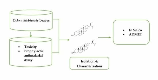
15]. In this paper, we report the prophylactic antimalarial properties of Ochna kibbiensis leaves, isolation and characterization of two known steroids, stigmasterol and β-sitosterol and their effect against Plasmodium falciparum Lactate Dehydrogenase (pfLDH) in silico.

2. Results

2.1. Acute Toxicity Studies

The median lethal dose of the methanol leaf extract of OK and its fractions were found to be safe based on the estimated LD50 value of ≥5000 mg/kg, i.p. with the exception of BFL which had 1702.94 mg/kg (Table 1).

2.2. Antiplasmodial Activity of O. kibbiensis—Prophylactic Test

The crude extract and fractions of OK exerted a significant (p < 0.05) prophylactic effect against P. berghei (Table 2). The highest % prophylaxis (97.62%) was recorded by MLE at the highest dose (500 mg/kg), which subsequently reduced to 61.31% at the medium dose (250 mg/kg), and there was a slight increase (73.81%) at the lowest dose (125 mg/kg). A decrease in the level of parasitaemia was observed with the standard drug, pyrimethamine (25 mg/kg) at 95.24% prophylaxis.
Of all the fractions tested, DFL was the most active while BFL was the least active. DFL exhibited a significant (p < 0.05) and dose-dependent effect with 47.62, 85.12 and 100% prophylaxis at 500, 250 and 125 mg/kg, respectively, while BFL recorded its highest % prophylaxis (76.19%) at the lowest (125 mg/kg) and medium (250 mg/kg) doses and reduction of the prophylactic effect (64.24%) at the highest dose (500 mg/kg). EFL exerted a similar effect to that of BFL; however, the highest (500 mg/kg) and the medium (250 mg/kg) doses of the fraction had % prophylaxis of 79.17% while the lowest dose (125 mg/kg) exhibited a higher prophylactic effect (90.48%) compared to the standard drug, pyrimethamine (95.24%). HFL had 85.71, 35.71 and 73.81% prophylaxis at the graded doses (500, 250 and 125 mg/kg) employed in this study (Table 2).

2.3. Phytochemical Screening of DFL of OK

Preliminary phytochemical screening of DFL revealed the presence of flavonoids, steroids and triterpenes (Table 3).

2.4. Characterization of K4

Compound K4 (28.4 mg) was isolated as a white crystalline substance with an uncorrected melting point range between 136–137 °C, and it tested positive for Liebermann–Burchard’s reagents; comparison of the NMR data of the compound with literature is summarized in Table 4 and Table 5, respectively.

2.5. In Silico Studies of Compound Stigmasterol and β-Sitosterol

2.5.1. Molecular Docking Analysis

Stigmasterol and β-sitosterol from O. kibbiensis were screened against pfLDH, and the docking scores are indicated in Table 6. The binding energies for the best poses of the compounds range from −5.129 to −4.889 kcal/mol, while the co-crystalline ligand NADH had the highest binding affinity (−10.106 kcal/mol).

2.5.2. Biological Interactions

The 2D- and 3D-view of the molecular interactions of the clinically important amino acid residues of pfLDH, with stigmasterol and β-sitosterol, and NADH, are shown in Table S1, Supplementary Materials, Figure 1 and Figure 2, respectively. The co-crystallized ligand, NADH, formed a conventional hydrogen bond with Gly29, Met30, Ile31, Asp53, Asn140, Phe100 and His195 in addition to the other hydrophobic interactions with Gly27, Ser28, Gly32, Ile54, Val55, Met58, Thr97, Ala98, Gly99, Thr101, Ile123, Ile119, Val138, Thr139, Leu163, Leu167, Arg171, Pro246, Tyr247 and Pro250, while the compounds from OK (stigmasterol and β-sitosterol) interacted with Asp53 via hydrogen bonds in addition to other major hydrophobic interactions with Gly29, Met30, Ile31, Asp53, Asn140, Phe100 and His195, among others.

2.5.3. ADMET Analysis

Drug-Likeness, Oral Bioavailability, and Pharmacokinetic Properties

The results of the in silico ADMET screening and drug-likeness properties of stigmasterol and β-sitosterol from O. kibbiensis are indicated in Table 7; the molecular weight of the compounds range from 412.69 to 414.71 g/mol. Both compounds indicated one hydrogen bond acceptor and donor with molar refractivity of 132.75 and 133.23, respectively, and TPSA value of 20.23. The water solubility values of the compounds expressed in terms of LogSw were between −5.47 and −6.19, and the compounds were predicted to be poorly soluble. Consensus log p-values representing lipophilicity were between 6.97 and 7.19. Both compounds had one Lipinski and Egan violations, two Muegge and three Ghose violations, zero Veber’s violations and a bioavailability score of 0.55. The compounds had low GI absorption, and none of them are BBB permeant or Pgp substrates; none of the compounds are potential inhibitors of cytochrome P450 enzymes, though stigmasterol is a potential inhibitor of CYP2C9.

Toxicity Profile

The predicted LD50 of Stigmasterol and β-sitosterol is 890 mg/kg, and they fall under the oral toxicity class 4; none of the compounds showed any indication of hepatotoxicity, carcinogenicity, mutagenicity, or cytotoxicity, but both compounds have a tendency of being immunotoxic (Table 8).

3. Discussion

Considering the widespread usage of medicinal plants as remedies by a large proportion (>80%) of the world population as complementary and alternative medicines with little idea about their toxicity [18], it is very vital to conduct toxicity tests in order to evaluate the possible harmful side effects of these products. The increased use of these natural medicines is attributed to the fact that they are considered safe with little or no side effects compared to the orthodox medicines [19]. Thus, OK was subjected to an acute toxicity test to evaluate its median lethal dose (LD50), and the findings demonstrated that the plant is relatively safe and acceptable. These values recorded for the methanol extract and its fractions agreed with the values reported for a related Ochna species (O. schweinfurthiana) at 5000 mg/kg orally [20].
The methanol leaf extract of OK and its fractions exhibited significant (p < 0.05) activity, which was comparable to pyrimethamine in the prophylaxis assay. DFL exhibited a very strong prophylactic antimalarial effect against P. berghei. In a study by Mil-Homens et al. [21], dichloromethane fraction was shown to exhibit the highest antimalarial activity compared to the other extracts and fractions studied. A decline in the prophylactic activity of MLE and HFL of OK at the medium dose (250 mg/kg) might be related to saturation at the active sites or the action of other endogenous substances in the body or activity on other body organs of the experimental animals [20]. Even more so, increasing the dose of BFL will lead to a decrease in prophylactic effect as clearly indicated in this study.
The extracts of OK may act by inhibiting the multiplication of the parasites as well as direct cytotoxic effect on the parasites [22] or by modulating the membrane properties of the erythrocytes there by preventing parasite invasion [23]. Pyrimethamine acts by inhibiting the dihydrofolate reductase of plasmodia, thereby blocking the biosynthesis of purines and pyrimidines, which are essential for DNA synthesis and cell multiplication, which leads to a failure of nuclear division at the time of schizont formation in erythrocytes and liver [24,25,26,27]. Generally, prophylactic antimalarials work by either disrupting the initial development of malaria parasites in the liver (casual activity), suppressing the emergent asexual blood stages of parasite (suppressive activity) or preventing the relapses induced by the latent liver forms (hypnozoites) of relapsing P. vivax and P. ovale malaria (presumptive antirelapse therapy) [10,27,28]. Thus, O. kibbiensis might act via the same mechanism. In addition, a higher prophylactic effect exhibited by DFL of OK might also be linked to the presence of secondary metabolites such as steroids, triterpenes and flavonoids that were present in the fraction [13].
Following the promising effects obtained in the prophylactic antimalarial effect using the repository test, the DFL of OK being the most active fraction was subjected to chromatographic studies which resulted in the isolation of a mixture of known steroids (K4). The isolated compound(s) appeared as a white crystalline substance with an uncorrected melting point ranging from 136–137 °C and gave a positive reaction when tested with Liebermann–Burchard’s reagent, an indication of the presence of a steroid [16,29]. The 1H-NMR spectrum of K4 (600 Hz, in CDCl3) revealed the presence of three olefinic resonances at δH 5.28, 5.10 and 5.05, an oxy-methine proton at δH 3.45 and six methyl signals at δH 0.74, 1.00, 0.94, 0.85, 0.79 and 0.80, characteristic of a steroidal nucleus [16]. The 13C-NMR and APT experiments of K4 showed 29 carbon signals constituting four olefinic carbons at δC 140.76, 121.72, 138.31 and 129.28 corresponding to C-5, C-6, C-22 and C-23, respectively [30] and two angular methyl groups at δC 12.05 and 21.20 corresponding to C-18 and C-19, respectively [16,31]. The signal at δC 71.83 clearly indicated the presence of a β-hydroxyl group at C-3 typical of stigmasterol [16,30]. Additional peaks observed at δC 33.95 and 26.08 which were assigned to C-22 and C-23 indicated that 
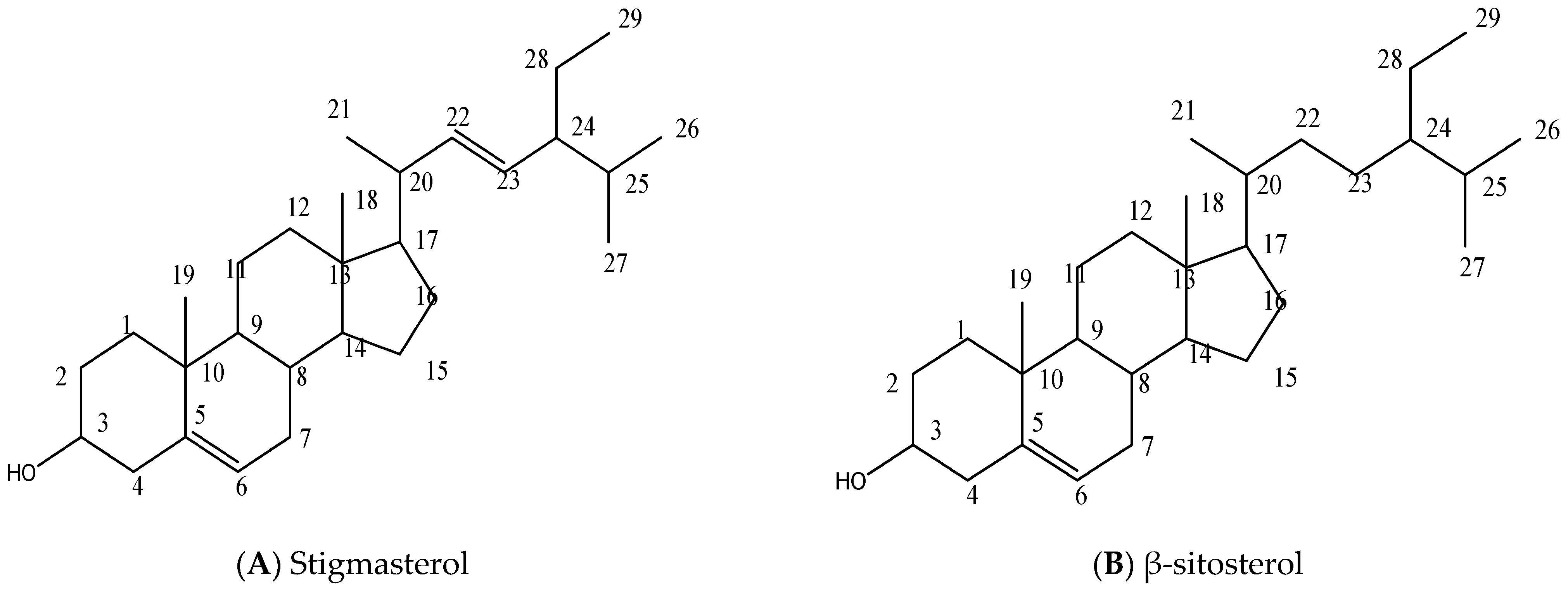
K4 is a mixture of stigmasterol and β-sitosterol. Based on the spectral data of compound K4, physicochemical tests and a direct comparison with existing data in the literature (Table 3 and Table 4) [16,30,31,32], compound K4 was elucidated and confirmed as a mixture of two compounds stigmasterol and β-sitosterol (Figure 3). Although the isolated compound(s) could not be tested for prophylactic antimalarial activity due to insufficient quantity, the compounds have been reported to possess antimalarial activity [33,34,35]. Further bio-assay-guided isolation of the bioactive constituents responsible for the observed effect is presently ongoing in our lab.
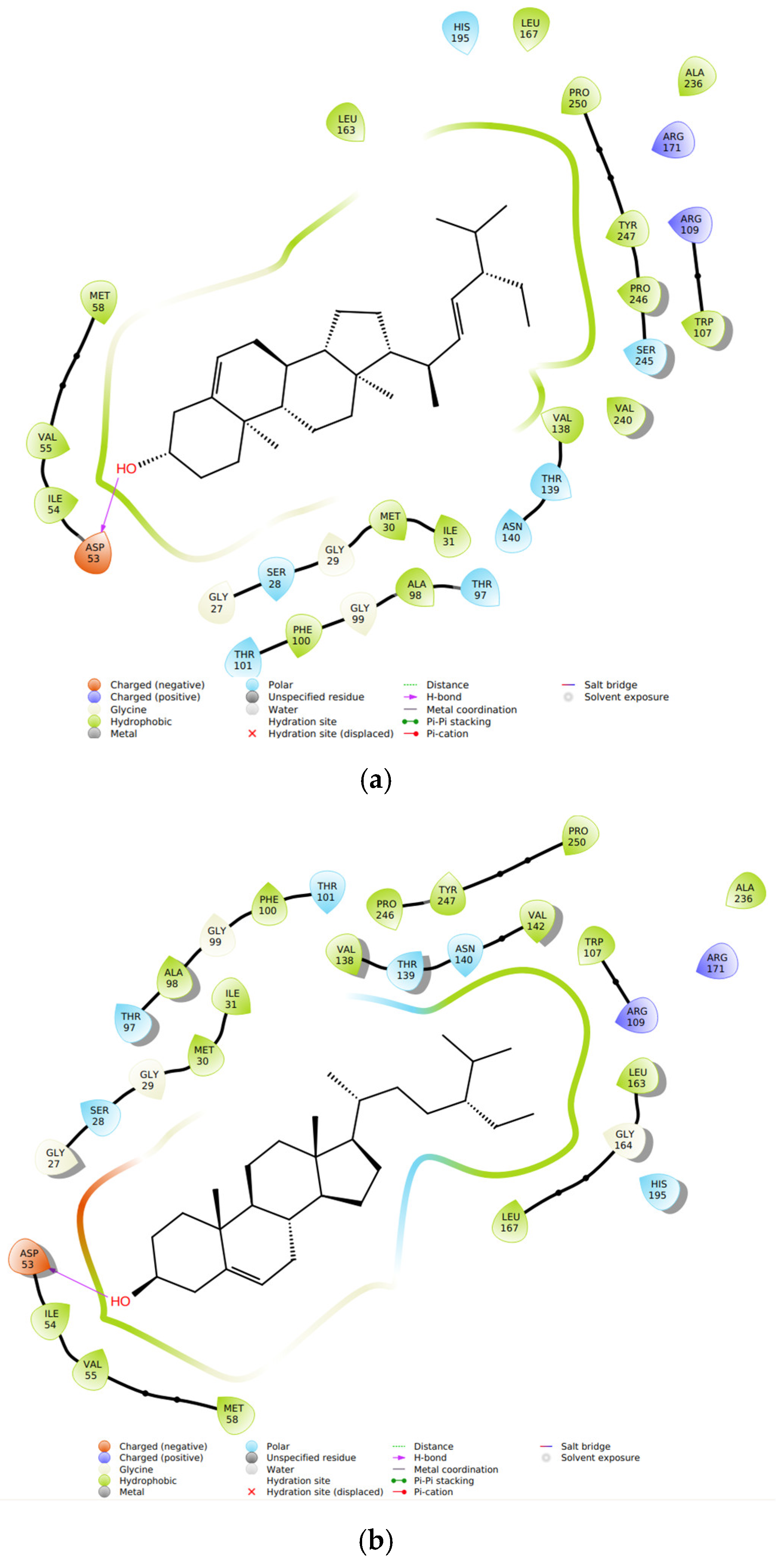
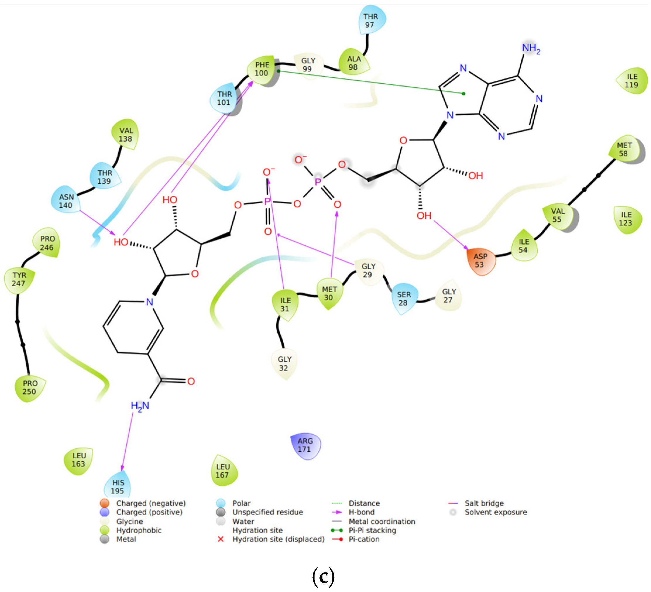
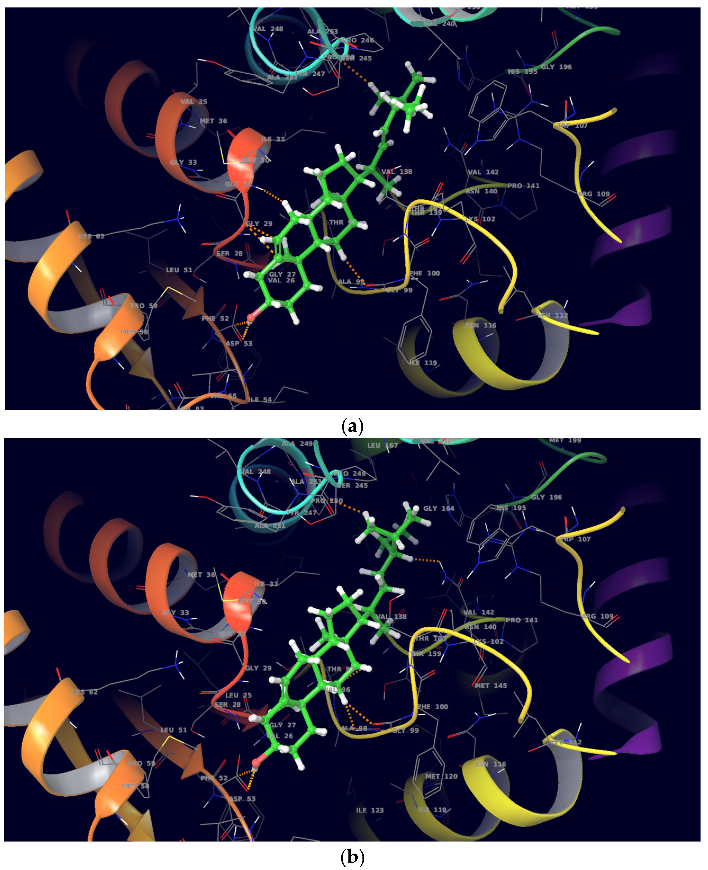
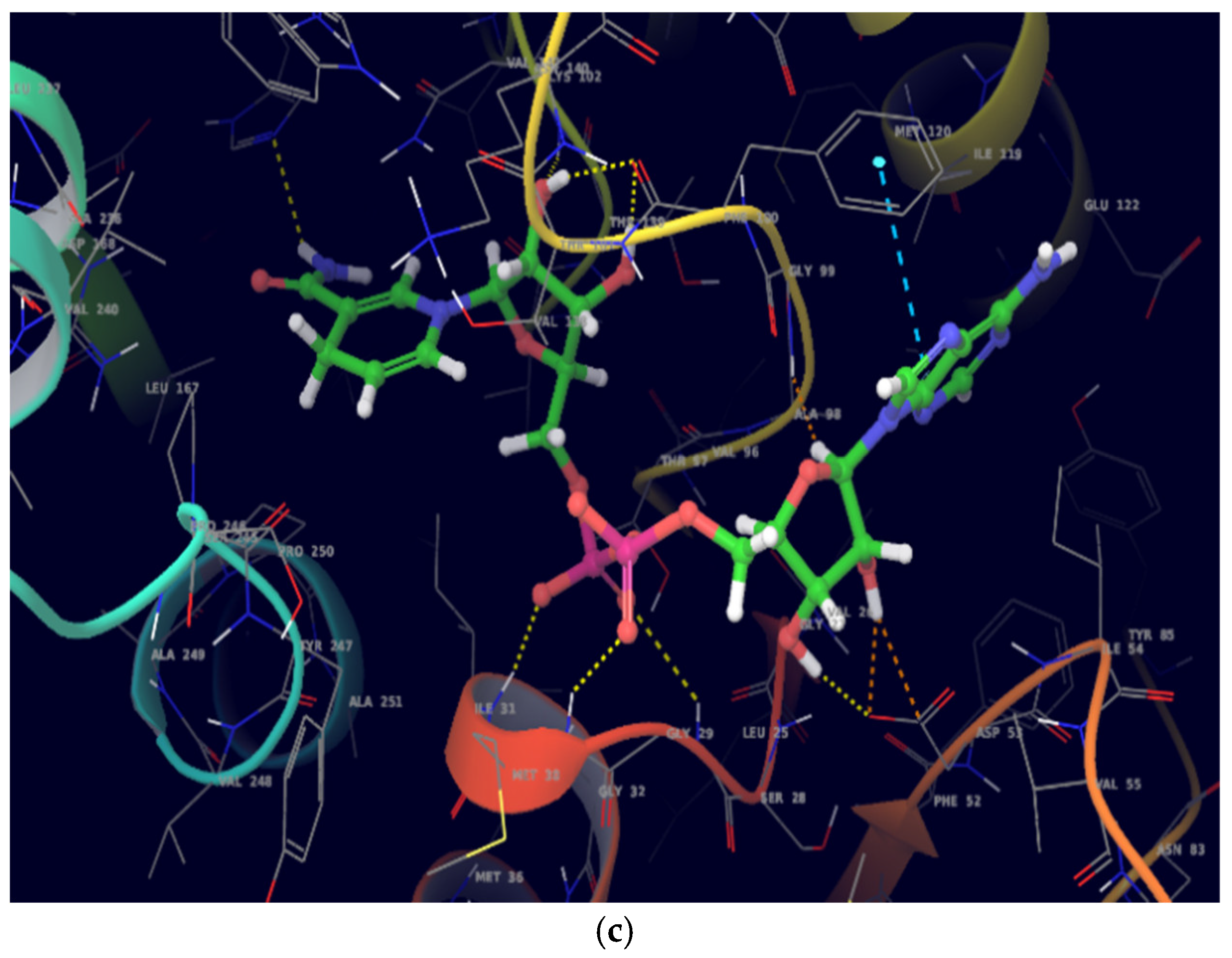
On molecular docking analysis, stigmasterol and β-sitosterol showed lower docking scores against pfLDH compared to the co-crystallized ligand, NADH. Studies have shown that pfLDH is composed of NADH-binding sites with amino acid residues such as Gly-27, Ser-28, Gly-29, Phe-52, Asp-53, Ile-54, Thr-97, Ala-98, Gly-99, Phe-100, Thr-139 and Asn-140, and the substrate-binding site constituting Lys-198, Met-199, Val-200, Leu-201, Glu-226, Phe-229, Asp-230, Val-233, Lys-314 and Glu-317 [36]. In this study, stigmasterol and β-sitosterol interacted with clinically important amino acid residues similar to that of NADH; a similar study was reported by Read et al. [37].
ADMET analysis indicated stigmasterol and β-sitosterol to be poorly soluble with low GI absorption and none of the compounds are either BBB permeant or Pgp substrate. Both compounds had one Lipinski violation for drug-likeness. None of the compounds violate the general rule of five or Lipinski’s rule for drug-likeness and both compounds are potential inhibitors of cytochrome P450 enzymes (CYP1A2, CYP2C19, CYP2D6 and CYP3A4); however, stigmasterol is a potential inhibitor of CYP2C9 and thus could lead to alteration of metabolism, which may lead to drug–drug interaction [38]. Moderate toxicity was recorded for both compounds based on the predicted LD50 value 890 mg/kg and toxicity class of 4. However, both compounds have a tendency to be immunotoxic with a percentage probability of 99%.

4. Materials and Methods

4.1. Collection and Identification Plant Materials

The whole plant of OK was collected from Samaru Zaria, Northern Nigeria in the month of December, 2020. It was identified and authenticated by Mal. Namadi Sanusi at the Herbarium unit Department of Botany, Ahmadu Bello University Zaria, Nigeria by comparing with a voucher specimen number (573).

4.2. Extraction and Partitioning

The leaves of the plant were air dried, pulverized and preserved according to the methods described in the African Pharmacopoeia [39]. The pulverized plant material (3.73 kg) was extracted with 90% methanol (25 L) using a maceration method for 8 days, and the solvent was evaporated in vacuo using a rotary evaporator at 40 °C to yield a residue (825.51 g) referred to as the methanol leaf extract (MLE). The extract (770 g) obtained was suspended in distilled water to obtain water soluble and water insoluble portions; the water-soluble portion was then partitioned using solvents of increasing polarity to obtain dichloromethane (DFL, 2.16 g), ethyl acetate (EFL, 34.69 g), butanol (BFL, 129.28 g) and residual aqueous fractions. The water insoluble portion (370.99 g) was washed with n-hexane to obtain a hexane (HFL, 5.59 g) fraction.

4.3. Antimalarial Studies

4.3.1. Experimental Animals

Locally bred adult Swiss albino mice of either sex (19–30 g body weight) were acquired from the Animal House Facility of the Department of Pharmacology and Therapeutics, Ahmadu Bello University Zaria, Nigeria. The animals were fed with laboratory diet and water ad libitum and maintained under standard conditions in clean cages under normal 12 h light, 12 h dark cycles. All experimental procedures followed the ethical guidelines for the care and use of laboratory animals as provided by the Usmanu Danfodiyo University Research Policy and accepted internationally (UDUS/UREC/2020/001).

4.3.2. Malaria Parasite

A mouse-infective chloroquine-sensitive strain of Plasmodium berghei NK-65 was obtained from the National Institute of Medical Research, Lagos. The parasites were kept alive by continuous intra-peritoneal passage in mice.

4.3.3. Acute Toxicity Studies

The median lethal dose (LD50) of MLE of OK, its n-hexane, dichloromethane, ethylacetate, and n-butanol fractions was determined using Lorke’s method [40]. The study was divided into two phases. In phase one, nine (9) mice were divided into three groups containing three mice each. Groups 1, 2 and 3 received 10, 100 and 1000 mg/kg of each of the extract and fraction separately. Based on the results of the first phase, each treatment group received the following doses; MLE, EFL, DFL and HFL = 1600, 2900 and 5000 mg/kg; BFL = 600, 1000, 1600 and 2900 mg/kg. Animals in all groups were observed for any sign and symptoms of toxicity and mortality for 24 h, and the route of administration was intraperitoneal.

4.3.4. Antiplasmodial Screening

Parasite Induction

A standard inoculum of 1 × 10⁷ of parasitized erythrocytes from a donor mouse in volume of 0.2 mL was used to infect the experimental animals intra-peritoneally.

Prophylactic Test—Residual Infection

The prophylactic activity of MLE of OK was tested according to the method described by Ryley and Peters [41]. Twenty-five (25) mice were randomly divided into 5 groups of 5 mice each. Groups 1, 2 and 3 were treated orally with 500, 250 and 125 mg/kg of MLE respectively, daily. Group 4 received 25 mg/kg of Pyrimethamine (positive control), while group 5 received 10 mL/kg of distilled water and served as the negative control. Treatments continued daily for four (4) consecutive days, and all the mice were infected with the parasite on the 5th day. Blood smears were then made from each mouse 72 h after inoculation/infection. Increase or decrease in parasitaemia was then determined microscopically. The average percentage prophylaxis was calculated as 100 × [(P-Q)]/P, where P is the average parasitaemia in the negative control group, and Q is the average parasitaemia in the test group [42]. The above procedure was repeated for the hexane, dichloromethane, ethylacetate and butanol fractions of O. kibbiensis. DFL was the most active fraction, and thus it was subjected to further fractionation using chromatographic techniques.

4.4. Chromatographic Fractionation of DFL

4.4.1. General Experimental Procedures

Thin-layer chromatography (TLC) was carried out using silica gel 60 GF254 precoated aluminum sheets (Sigma Aldrich, Germany). Column chromatography was conducted using LOBA Cheme silica gel (60–200) mesh. Spots on TLC plates were visualized by spraying with 10% H2SO4 followed by heating at 105 °C for 10 min. The melting point was determined on an Electro thermal melting point apparatus. NMR data were recorded on a Bruker AVANCE (III) spectrometer (600 MHz) with a residual solvent as the internal standard.

4.4.2. Phytochemical Screening of DFL

Fraction DFL was subjected to phytochemical tests to determine the presence of different secondary metabolites such as flavonoids, alkaloids, steroids and triterpenes, among others [13].

4.4.3. Isolation Procedures

DFL of the water-soluble portion of the MLE was loaded over a well packed silica gel column and eluted gradiently with the mixtures of n-hexane and ethyl acetate (9:1, 7:1, 5:1, 3:1 and 1:1) as mobile phase. Eluents were collected in 30 mL and were monitored using TLC. Nine major fractions (A to I) were obtained from the pooling of 120 collections made. Fractions C and D were subjected to purification using a silica gel column repeatedly, which afforded a white crystalline substance (14 mg) coded K4. Compound K4 was subjected to TLC analysis using hexane: ethylacetate (6:1 and 5:1) as solvent systems, which revealed a single homogenous pink spot after spraying with 10% sulphuric acid and heated in an oven at 105 °C; the compound was also subjected to a Liebermann–Burchard’s test, melting point determination and NMR analysis to elucidate its chemical structure.

4.5. Molecular Docking Studies

4.5.1. Protein Preparation

The Protein, Plasmodium falciparum lactate dehydrogenase (pfLDH), was prepared as previously described by Iwaloye et al. [43] and Johnson et al. [36]. The crystal structure of the protein obtained from the Protein Data Bank (PDB: 1T2C) repository was prepared in Glide (Schrödinger Suite 2021-2) using the protein preparation wizard panel. During the process, hydrogen was added, bond orders were assigned, disulfide bonds were created, and the missing side chains and loops were replaced with prime. Water molecules outside 3.0 Å of the heteroatoms were eliminated, and the protein structure was minimized and optimized using OPLS4 and PROPKA, respectively.

4.5.2. Generation of Receptor Grid

The receptor grid was developed to define the location and size of the active site of the protein for ligand docking. This was accomplished with Schrödinger Maestro 12.8’s receptor grid generation tool. The active site of the protein’s co-crystalized ligand (1,4-dihydronicotinamide adenine dinucleotide) was employed as the scoring grid [43,44].

4.5.3. Ligand Preparation

The crystal structures of stigmasterol and and β-sitosterol isolated from the leaves of O. kibbiensis were retrieved from Dr. Duke’s Phytochemical and Ethnobotanical Databases, and the standard ligands (NADH) were prepared using the Ligprep panel of Maestro 12.8, Schrödinger Suite 2021-2, as previously reported [36,45]. Low-energy 3D structures with acceptable chiralities were generated. Each ligand’s ionization state was formed at a physiological pH of 7.2 ± 0.2. Stereoisomers of each ligand were computed by keeping certain chiralities constant while varying others.

4.5.4. Protein–Ligand Docking

The molecular docking analysis was performed on Schrödinger Suite 2021-2 utilizing the Glide–Ligand Docking panel of Maestro 12.8. The prepared ligands and the receptor grid file were loaded into Maestro’s work space, and the ligands were docked into the protein’s binding site. The vdW radius scaling factor was set to 0.80 for ligand atoms, with a partial charge cut-off of 0.15, and the flexible ligand sampling option was employed [45].

4.5.5. ADMET Profiling

In silico predictive models were used to determine the ADMET properties of the test compounds i.e., stigmasterol and and β-sitosterol. The SwissADME server was used to assess the ADME properties of the compounds [46,47] while the ProTox-II online server was used to predict the toxicity profile of the compounds [48].

4.6. Statistical Analysis

Results were expressed as mean ± standard error of mean (SEM). Statistical analysis of control and test data was based on simple one-way ANOVA, and Dunnett’s post hoc test was used for different doses within a group. A Bonferroni post hoc test was used to compare results between the means at the significance level considered at p < 0.05.

5. Conclusions

The leaves of O. kibbiensis were found to be relatively safe based on the LD50 values obtained, and the plant has demonstrated significant antimalarial activity with prophylactic effects at the graded doses employed in this study. In addition, a mixture of Stigmasterol and β-Sitosterol were isolated from the leaf of O. kibbiensis, and the compounds have shown good binding affinities against Plasmodium falciparum lactate dehydrogenase and a favorable pharmacokinetic profile in silico. Thus, the observed effect could be partly attributed to the presence of these compounds.

Supplementary Materials

The following supporting information can be downloaded at: https://www.mdpi.com/article/10.3390/ddc2010003/s1, Table S1: Molecular interactions of Stigmasterol, β-sitosterol, Chloroquine, Pyrimethamine and NADH with pfLDH.

Author Contributions

Conceptualization, A.J.Y.; methodology, A.J.Y., M.I.A., I.N. and A.Y.; formal analysis, A.Y.; investigation, A.J.Y., A.A.M. and C.O.A.; data curation, A.J.Y.; writing—original draft preparation, A.J.Y.; writing—review and editing, all authors; supervision, M.I.A. All authors have read and agreed to the published version of the manuscript.

Funding

This research work was funded by the Nigerian Tertiary Education Trust Fund (TETFund) Research Projects (RP) Intervention via the Institutional Based Research (IBR) Grant with Grant reference No. TETFUND/DR&D/CE/UNIV/SOKOTO/RP/VOL.1, Batch A, Serial no. 46.

Institutional Review Board Statement

The animal study protocol was approved by the Usmanu Danfodiyo University Research and Ethics Committee (UDUS/UREC/2020/002, 8 January 2022).

Informed Consent Statement

Not applicable.

Data Availability Statement

Not applicable.

Acknowledgments

We acknowledged the efforts of Yusuf Sani from the Department of Pharmacology and Therapeutics, College of Health Sciences, UDU Sokoto for his diligent support in conducting the antimalarial assay, Mal. Hamza Muhammad from the Department of Pharmaceutical and Medicinal Chemistry, UDU Sokoto for participating in the extraction protocols, and Vuyisa Mzozoyana, School of Chemistry and Physics, University of Kwa-Zulu Natal-Durban, South Africa, for running the NMR spectroscopy.

Conflicts of Interest

The authors declare no conflict of interest.

References

  1. World Health Organization. Malaria. 2022. Available online: https://www.who.int/news-room/fact-sheets/detail/malaria (accessed on 1 July 2022).
  2. Centres for Disease Control and Prevention, Malaria. Available online: https://www.cdc.gov/parasites/malaria/index.html (accessed on 8 June 2022).
  3. Muentener, P.; Schlagenhauf, P.; Steffen, R. Imported malaria (1985–95): Trends and perspectives. Bull. World Health Organ. 1999, 77, 560–566. [Google Scholar] [PubMed]
  4. Robert, S. Malaria Prophylaxis: Setting the Scene. J. Travel. Med. 2003, 10, s3–s7. [Google Scholar] [CrossRef]
  5. Schwartz, E. Prophylaxis of malaria. Mediterr. J. Hematol. Infect. Dis. 2012, 4, e2012045. [Google Scholar] [CrossRef] [PubMed]
  6. Schwartz, E.; Parise, M.; Kozarsky, P.; Cetron, M. Delayed onset of malaria—Implications for chemoprophylaxis in travelers. N. Engl. J. Med. 2003, 349, 1510–1516. [Google Scholar] [CrossRef] [PubMed]
  7. Agnandji, S.T.; Lell, B.; Soulanoudjingar, S.S.; Fernandes, J.F.; Abossolo, B.P.; Conzelmann, C.; Methogo, B.G.; Doucka, Y.; Flamen, A.; Mordmüller, B.; et al. First results of phase 3 trial of RTS, S/AS01 malaria vaccine in African children. N. Engl. J. Med. 2011, 365, 1863–1875. [Google Scholar] [CrossRef] [Green Version]
  8. Soto, J.; Medina, F.; Dember, N.; Berman, J. Efficacy of permethrin-impregnated uniforms in the prevention of malaria and leishmaniasis in Colombian soldiers. Clin. Infect. Dis. 1995, 21, 599–602. [Google Scholar] [CrossRef]
  9. Baird, J.; Schwartz, E.; Hoffman, S. Prevention and treatment of vivax malaria. Curr. Infect. Dis. Rep. 2007, 9, 39–46. [Google Scholar] [CrossRef]
  10. Hill, D.R.; Baird, J.K.; Parise, M.E.; Lewis, L.S.; Ryan, E.T.; Magill, A.J. Primaquine: Report from CDC expert meeting on malaria chemoprophylaxis I. Am. J. Trop. Med. Hyg. 2006, 75, 402–415. [Google Scholar] [CrossRef] [Green Version]
  11. Shanks, G.D.; Magill, A.J.; Freedman, D.O. Drug free holidays: Pre-travel versus during travel malaria prophylaxis. Am. J. Trop. Med. Hyg. 2007, 77, 1–2. [Google Scholar] [CrossRef]
  12. Dow, G.S.; Magill, A.J.; Ohrt, C. Clinical development of new prophylactic antimalarial drugs after the 5th Amendment to the Declaration of Helsinki. Ther. Clin. Risk Manag. 2008, 4, 803–819. [Google Scholar] [CrossRef]
  13. Abdullahi, M.I.; Haruna, A.K.; Musa, M.A.; Alhassan, M.A.; Umar, A.; Yusuf, A.J.; Uba, A.; Ibrahim, Z.Y.Y. Preliminary phytochemical and antimicrobial evaluation of the methanolic leaf extract of Ochna kibbiensis Hutz and Dalz (Ochnaceae). J. Chem. Pharm. 2015, 7, 626–631. [Google Scholar]
  14. Burkill, H.M. The Useful Plants of West Tropical Africa; Royal Botanic Gardens, Kew: Richmond, UK, 1985; Volume 4, p. 445. Available online: https://plants.jstor.org/stable/10.5555/al.ap.flora.fwta1194 (accessed on 20 October 2022).
  15. Abdullahi, M.I.; Musa, A.M.; Nasir, T.; Magaji, M.G.; Yusuf, A.J.; Iliya, I. Anti-proliferative study and isolation of ochnaflavone from the ethyl acetate-soluble fraction of Ochna kibbiensis Hutch and Dalziel. Nat. Prod. Res. 2017, 31, 2149–2152. [Google Scholar] [CrossRef]
  16. Yusuf, A.J.; Abdullahi, M.I.; Haruna, A.K.; Idris, A.Y.; Musa, A.M. Isolation and Characterization of Stigmasterol and Bis-(5,7-diacetyl-catechin-4’-α-rhamnopyranoside) from the stem bark of Neocarya macrophylla (Sabine) Prance (Chrysobalanaceae). Nig. J. Basic Appl. Sci. 2015, 23, 15–22. [Google Scholar] [CrossRef] [Green Version]
  17. Abdulmalik, I.A.; Sule, I.M.; Musa, A.M.; Yaro, A.H.; Abdullahi, M.I.; Abdulkadir, M.F.; Yusuf, H. Isolation of Steroids from Acetone Extract of Ficus iteophylla. Br. J. Pharmacol. 2011, 2, 270–272. Available online: https://maxwellsci.com/print/bjpt/v2-270-272.pdf (accessed on 5 September 2022).
  18. Tiza, N.; Jeremy, A.K.; Burtram, C.F. Acute toxicity studies of the South African medicinal plant Galenia africana. Toxicol. Rep. 2018, 5, 813–818. [Google Scholar] [CrossRef]
  19. Park, H.; Hwang, Y.H.; Yang, H.J.; Kim, H.K.; Song, K.S.; Ma, J.Y. Acute toxicity and genotoxicity study of fermented traditional herb formula Guibi-tang. J. Ethnopharmacol. 2014, 156, 182–189. [Google Scholar] [CrossRef]
  20. Ibrahim, Z.Y.Y.; Abdullahi, M.I.; Musa, A.M.; Maje, I.M.; Yusuf, A.J.; Aliyu, I.M. Acute Toxicity Profile and Plasmodium berghei Inhibitory Activity of Ochna schweinfurthiana (F. Hoffm) Ochnaceae Leaf Extract in Laboratory Animals. Int. J. Pharm. Sci. Res. 2015, 6, 1302–1306. Available online: http://www.ijpsr.info/ (accessed on 21 October 2022).
  21. Abrantes, M.; Mil-Homens, T.; Duarte, N.; Lopes, D.; Cravo, P.; Madureira, M.d.; Ferreira, M.J. Antiplasmodial activity of lignans and extracts from Pycnanthus angolensis. Planta Med. 2008, 74, 1408–1412. [Google Scholar] [CrossRef]
  22. Golenser, J.; Waknine, J.H.; Krugliak, M.; Hunt, N.H.; Grau, G.E. Current perspectives on the mechanism of action of artemisinins. Int. J. Parasitol. 2006, 36, 1427–1441. [Google Scholar] [CrossRef]
  23. Hansen, D.S. Inflammatory Responses Associated with the Induction of Cerebral Malaria: Lessons from Experimental Murine Models. PLoS Pathog. 2012, 8, e1003045. [Google Scholar] [CrossRef] [Green Version]
  24. Fohl, L.M.; Roos, D.S. Fitness effects of DHFR-TS mutations associated with pyrimethamine resistance in apicomplexan parasites. Mol. Microbiol. 2003, 50, 1319–1327. [Google Scholar] [CrossRef] [Green Version]
  25. Gatton, M.L.; Martin, L.B. Cheng Q: Evolution of resistance to sulfadoxine-pyrimethamine in Plasmodium falciparum. Antimicrob. Agents Chemother. 2004, 48, 2116–2123. [Google Scholar] [CrossRef] [Green Version]
  26. Imming, P.; Sinning, C.; Meyer, A. Drugs, their targets and the nature and number of drug targets. Nat. Rev. Drug Discov. 2006, 5, 821–834, Erratum in Nat. Rev. Drug Discov. 2007, 6, 126. [Google Scholar] [CrossRef] [PubMed]
  27. Overington, J.P.; Al-Lazikani, B.; Hopkins, A.L. How many drug targets are there? Nat. Rev. Drug Discov. 2006, 5, 993–996. [Google Scholar] [CrossRef] [PubMed]
  28. United States Center for Diseases Control and Prevention [USCDC]. CDC Health Information for International Travel 2008. 2008. Available online: https://wwwnc.cdc.gov/travel/yellowbook/2020/introduction/introduction-to-travel-health-and-the-cdc-yellow-book (accessed on 11 July 2007).
  29. Silva, J.L.; Chai, H.; Farnworth, N.I.; Gypta, M.P. Natural Product Isolation; Carnell, R., Ed.; Humara Press Publication: Totowa, NJ, USA, 1998; pp. 349–359. [Google Scholar]
  30. Pateh, U.U.; Haruna, A.K.; Garba, M.; Iliya, I.; Sule, I.M.; Abubakar, M.S.; Ambi, A.A. Isolation of Stigmasterol, Beta-Sitosterol and 2-hydroxyhexadecamic acid methyl ester from the Rhizomes of Stylochiton lancifolius Pyer and Kotchy (Araceae). Niger. J. Pharm. Sci. 2009, 8, 19–25. [Google Scholar]
  31. Habib, M.R.; Nikkon, F.; Rahman, M.; Haque, M.E.; Karim, M.R. Isolation of Stigmasterol and Beta-sitosterol from methanol extract of root bark of Calotropis gigantean (Linn.). Pak. J. Biol. Sci. 2007, 10, 4174–4176. [Google Scholar] [CrossRef] [Green Version]
  32. Khanam, S.; Sultana, R. Isolation of β-Sitosterol and Stigmasterol as active immunomodulatory constituents from fruits of Solanium xanthocarpum (Solanaceae). Int. J. Pharm. Sci. Res. 2012, 3, 1057–1060. [Google Scholar] [CrossRef]
  33. Lagnika, L.; Attioua, B.; Weniger, B.; Kaiser, M.; Sanni, A.; Vonthron-Senecheau, C. Phytochemical Study and Antiprotozoal Activity of Compounds Isolated from Thalia geniculate. Pharm. Biol. 2008, 46, 162–165. [Google Scholar] [CrossRef]
  34. Ntie-Kang, F.; Onguéné, P.A.; Lifongo, L.L.; Ndom, J.C.; Sippl, W.; Mbaze, L.M. The potential of anti-malarial compounds derived from African medicinal plants, part II: A pharmacological evaluation of non-alkaloids and non-terpenoids. Malar. J. 2014, 13, 81. [Google Scholar] [CrossRef] [Green Version]
  35. Philip, K.; Cheplogoi, P.K.; Elizabeth, M.; Hoseah, M.; Langat, M.K. Assessment of Antiplasmodial Activity and Toxicity of Crude Extracts and Isolated Compounds from Oncoba spinosa (Flacourtiaceae). J. Adv. Med. Pharm. Sci. 2019, 21, 1–14. [Google Scholar] [CrossRef] [Green Version]
  36. Johnson, T.O.; Adegboyega, A.E.; Ojo, O.A.; Yusuf, A.J.; Iwaloye, O.; Ugwah-Oguejiofor, C.J.; Asomadu, R.O.; Chukwuma, I.F.; Ejembi, S.A.; Ugwuja, E.I.; et al. A Computational Approach to Elucidate the Interactions of Chemicals from Artemisia annua Targeted Toward SARS-CoV-2 Main Protease Inhibition for COVID-19 Treatment. Front. Med. 2022, 15, 907583. [Google Scholar] [CrossRef]
  37. Read, J.A.; Wilkinson, K.W.; Tranter, R.; Sessions, R.B.; Brady, R.L. Chloroquine binds in the cofactor binding site of Plasmodium falciparum lactate dehydrogenase. J. Biol. Chem. 1999, 274, 10213–10218. [Google Scholar] [CrossRef] [Green Version]
  38. Johnson, T.O.; Adegboyega, A.E.; Iwaloye, O.; Eseola, O.A.; Plass, W.; Afolabi, B.; Rotimi, D.; Ahmed, E.I.; Albrakati, A.; Batiha, G.E.; et al. Computational study of the therapeutic potentials of a new series of imidazole derivatives against SARS-CoV-2. J. Pharmacol. Sci. 2021, 147, 62–71. [Google Scholar] [CrossRef]
  39. African Pharmacopoeia; Organization of African Unity, Scientific, Technical & Research Commission: Lagos, Nigeria, 1985; Volume 1.
  40. Lorke, D. A new approach to practical acute toxicity testing. Arch. Toxicol. 1983, 54, 275–287. [Google Scholar] [CrossRef]
  41. Ryley, J.F.; Peters, W. The antimalarial activity of some quinolone esters. Ann. Trop. Med. Parasitol. 1970, 64, 209–222. [Google Scholar] [CrossRef] [PubMed]
  42. Dacie, J.V.; Lewis, S.M. Practical Haematology, 9th ed.; Churchill Livingstone: London, UK, 2000; p. 663. [Google Scholar]
  43. Iwaloye, O.; Elekofehinti, O.O.; Momoh, A.I.; Babatomiwa, K.; Ariyo, E.O. In silico molecular studies of natural compounds as possible anti-Alzheimer’s agents: Ligand-based design. Netw. Model. Anal. Health Inform. Bioinform. 2020, 9, 54. [Google Scholar] [CrossRef]
  44. Olsson, M.H.M.; SØndergaard, C.R.; Rostkowski, M.; Jensen, J.H. PROPKA3: Consistent treatment of internal and surface residues in empirical pKa predictions. J. Chem. Theory Comput. 2011, 7, 525–537. [Google Scholar] [CrossRef]
  45. Ojo, O.A.; Adegboyega, A.E.; Johnson, G.I.; Umedum, N.L.; Onuh, K.; Adeduro, M.N.; Nwobodo, V.O.; Elekan, A.O.; Alemika, T.E.; Johnson, T.O. Deciphering the interactions of compounds from Allium sativum targeted towards identification of novel PTP 1B inhibitors in diabetes treatment: A computational approach. Inform. Med. Unlocked 2021, 26, 100719. [Google Scholar] [CrossRef]
  46. Daina, A.; Michielin, O.; Zoete, V. SwissADME: A free web tool to evaluate pharmacokinetics, drug-likeness and medicinal chemistry friendliness of small molecules. Sci. Rep. 2017, 7, 42717. [Google Scholar] [CrossRef] [Green Version]
  47. Yusuf, A.J.; Abdullahi, M.I.; Musa, A.M.; Haruna, A.K.; Vuyisa, M. Biochemical evaluation and molecular docking assessment of Glucosamines from Neocarya macrophylla fruits against Naja nigricollis venom. Carbohydr. Res. 2021, 509, 108436. [Google Scholar] [CrossRef]
  48. Banerjee, P.; Eckert, A.O.; Schrey, A.K.; Preissner, R. ProTox-II: A webserver for the prediction of toxicity of chemicals. Nucleic Acids Res. 2018, 46, W257–W263. [Google Scholar] [CrossRef] [PubMed]
Figure 1. 2D view of the molecular interactions of (a) Stigmasterol; (b) β-sitosterol; (c) NADH with pfLDH.
Ddc 02 00003 g001aDdc 02 00003 g001b
Figure 2. Three-dimensional view of the molecular interactions of (a) Stigmasterol; (b) β-sitosterol; (c) NADH with pfLDH.
Ddc 02 00003 g002aDdc 02 00003 g002b
Figure 3. Structures of Stigmasterol and β-Sitosterol.
Ddc 02 00003 g003
Table 1. Median lethal dose (LD50) of extract/fractions of O. kibbiensis.
Extract/FractionLD50 Value (mg/kg)
Methanol≥5000.00
Hexane≥5000.00
Dichloromethane≥5000.00
Ethylacetate≥5000.00
Butanol1702.94
Table 2. Prophylactic effect of OK against P. berghei berghei infection in mice.
Treatment (mg/kg/day)Average Parasitaemia ± SEM% Prophylaxis
1NS 0.2 mL8.40 ± 0.93-
2MLE   5000.20± 0.20 *97.62
MLE   2503.25 ± 0.48 *61.31
MLE   1252.20 ± 0.80 *73.81
3HFL   5002.20 ± 1.02 *73.81
HFL   2505.40 ± 1.57 **35.71
HFL   1251.20 ± 0.58 *85.71
4DFL   5004.40 ± 1.63 ***47.62
DFL   2501.25 ± 0.95 *85.12
DFL   1250.00 ± 0.00 *100
5EFL   5001.75 ± 0.63 *79.17
EFL   2501.75 ± 1.03 *79.17
EFL   1250.80 ± 0.58 *90.48
6BFL   5003.00 ± 1.73 *64.29
BFL   2502.00 ± 0.91 *76.19
BFL   1252.00 ± 0.77 *76.19
7PM   250.40 ± 0.2495.24
Key: NS = Normal saline; MLE = Methanol leaf extract of OK; HFL = Hexane fraction of OK; DFL = Dichloromethane fraction of OK; EFL= Ethylacetate fraction of OK; BFL = Butanol fraction of OK; PM = Pyrimethamine; OK = Ochna kibbiensis. Values were expressed as Mean ± SEM (n = 5). Values of the group with * are statistically significantly (p < 0.05) different from NS treated. Values of the group with superscript ** are statistically significantly different from Pyrimethamine treated groups. Values of the group with superscript *** are statistically significantly different from NS and Pyrimethamine treated groups.
Table 3. Phytochemical constituents of DFL of OK.
ConstituentsTestObservationInference
AlkaloidsMayer’sWhite-yellowish ppt-
Dragendorff’sOrange-brownish ppt
CarbohydratesMolisch’sReddish colored ring-
Fehling’sRed precipitate
FlavonoidsFerric chlorideGreen precipitate+
NaOHYellow colour
SaponinsFrothingFroth persists for 15 mins-
TanninsLead Sub-acetateCream ppt-
Steroids/terpenesLieberman-Buchard’sBrown ring at interface+
Salkowski’sReddish color
AnthraquinonesBontrager’sPinkish color-
Key: + = present; - = absent.
Table 4. Comparison of spectral data of Stigmasterol with literature.
Position13C-NMR K413C-NMR1H-NMR K41H-NMRDEPT K4
Yusuf et al. [16]Yusuf et al. [16]
137.2737.26-1.85CH2
231.6731.671.441.46CH2
371.8371.813.453.52CH
442.3142.312.212.27CH2
5140.76140.76--C
6121.72121.715.285.35CH
731.9331.901.921.96CH2
831.8831.90-1.48CH
950.1650.160.940.93CH
1036.5136.51--C
1121.0921.21-1.49CH2
1239.739.68-1.16CH2
1342.2342.22--C
1456.8856.871.091.05CH
1524.3724.361.561.56CH2
1628.9128.921.771.7CH2
1755.9855.96-1.13CH
1812.0512.050.740.69CH3
1921.221.0811.03CH3
2040.4840.49-2.02CH
2123.0623.070.941.02CH3
22138.31138.315.105.10CH
23129.3129.285.055.03CH
2451.2551.24-1.53CH
2529.229.15-1.65CH
2618.9818.980.850.82CH3
2719.3919.400.790.78CH3
2825.425.40-1.15CH2
2912.2412.250.800.80CH3
Table 5. Comparison of spectral data of β-Sitosterol with literature.
Position13C-NMR K413C-NMR1H-NMR K41H-NMRDEPT K4
Abdulmalik et al. [17]Abdulmalik et al. [17]
137.1137.261.091.08CH2
231.6831.66-1.51CH2
371.8371.823.453.52CH
442.3242.32.212.28CH2
5140.76140.76--C
6121.72121.725.285.35CH
731.9231.91-1.99CH2
823.0923.07-1.25CH
924.3124.3-1.18CH
1036.51---C
1121.0921.081.441.46CH2
1239.839.78-1.16CH2
1342.32---C
1456.7956.77-0.98CH
1529.229.161.561.58CH2
1628.2428.241.771.85CH2
1750.1650.14-1.09CH
1811.9811.980.740.69CH3
1911.8611.8611.03CH3
2037.2737.25-1.35CH
2119.0519.030.940.92CH3
2233.9833.95-1.32CH2
2326.1426.08-1.17CH2
2445.8645.841.921.91CH
2529.729.69-1.65CH
2618.7818.780.790.82CH3
2718.7818.780.790.82CH3
2823.0923.07-1.22CH2
2919.8119.810.850.85CH3
Table 6. Docking scores of Stigmasterol, β-sitosterol and some standard ligands against pfLDH.
CompoundPubChem IDDocking Score (in kcal/mol)
Stigmasterol5280794−5.129
β-sitosterol222284−4.889
NADH (Co-crystalline ligand)439153−10.106
Table 7. The drug-likeness, oral bioavailability, and pharmacokinetic properties of Stigmasterol and β-sitosterol from O. kibbiensis.
ParameterStigmasterolβ-Sitosterol
Molecular weight (g/mol)412.69414.71
H-bond acceptors11
H-bond donors11
Molecular refractivity132.75133.23
TPSA (Å2)20.2320.23
Silicos-IT LogSw−5.47−6.19
Silicos-IT classPoorly solublePoorly soluble
Consensus Log P6.977.19
Lipinski violations11
Ghose violations33
Veber’s violations00
Egan violations11
Muegge violations22
Bioavailability Score0.550.55
log Kp (skin permeation) (cm/s)−2.74−2.20
GI absorptionLowLow
BBB permeantNoNo
Pgp substrateNoNo
CYP1A2 inhibitorNoNo
CYP2C19 inhibitorNoNo
CYP2C9 inhibitorYesNo
CYP2D6 inhibitorNoNo
CYP3A4 inhibitorNoNo
Table 8. Toxicity profile of Stigmasterol, β-sitosterol from O. kibbiensis.
Stigmasterolβ-Sitosterol
Predicted LD50 (mg/kg)890890
Acute Oral Toxicity (c)44
HepatotoxicityInactiveInactive
CarcinogenicityInactiveInactive
ImmunotoxicityActive (* 99)Active (* 99)
MutagenicityInactiveInactive
CytotoxicityInactiveInactive
* Percentage probability.
Disclaimer/Publisher’s Note: The statements, opinions and data contained in all publications are solely those of the individual author(s) and contributor(s) and not of MDPI and/or the editor(s). MDPI and/or the editor(s) disclaim responsibility for any injury to people or property resulting from any ideas, methods, instructions or products referred to in the content.

