

Indonesian Vegetables: Searching for Antioxidant and Antidiabetic Therapeutic Agents
 , , , , , and
Abstract
:1. Introduction
2. Results
3. Discussion
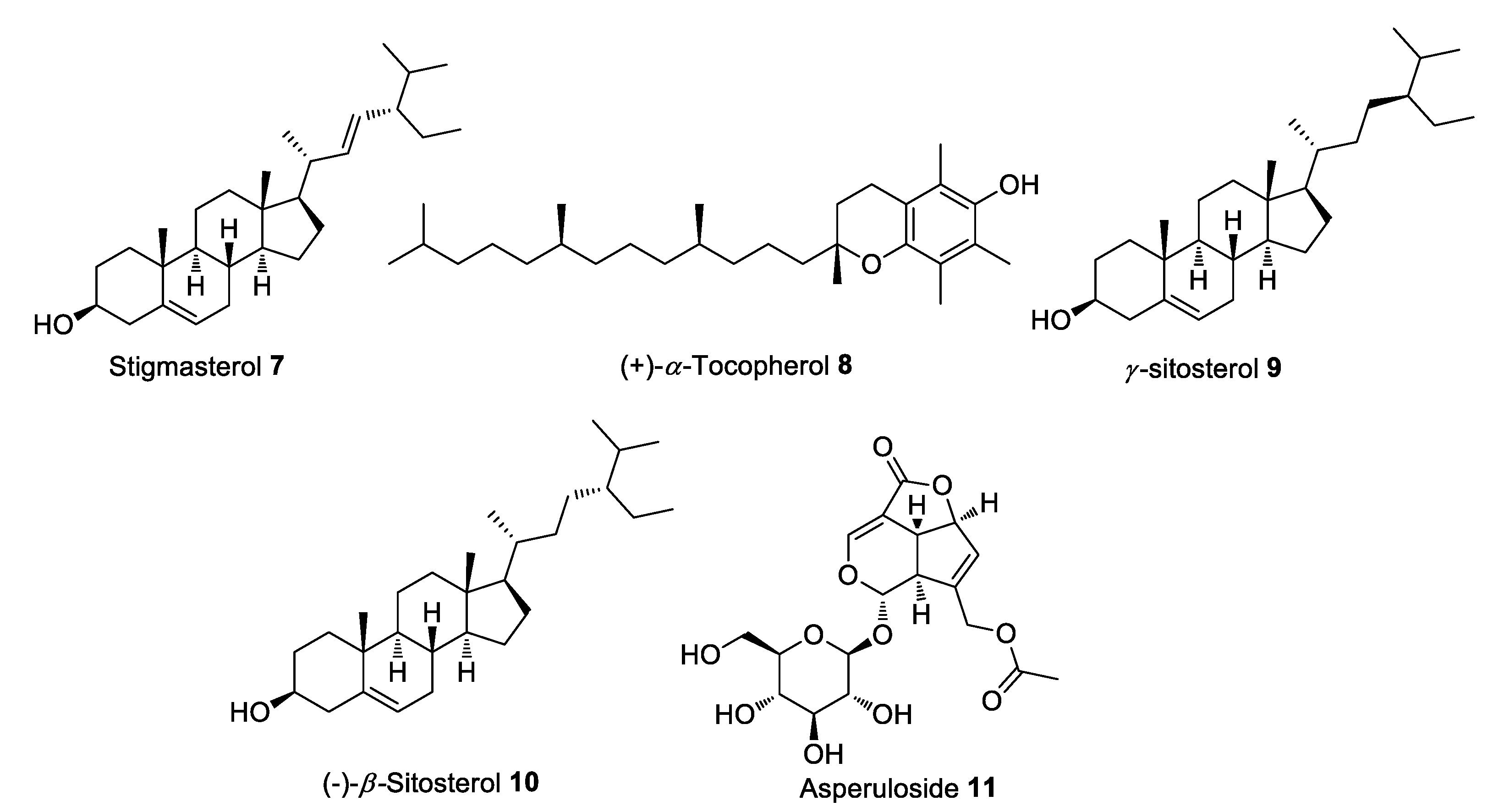
3.1. Ipomoea aquatica
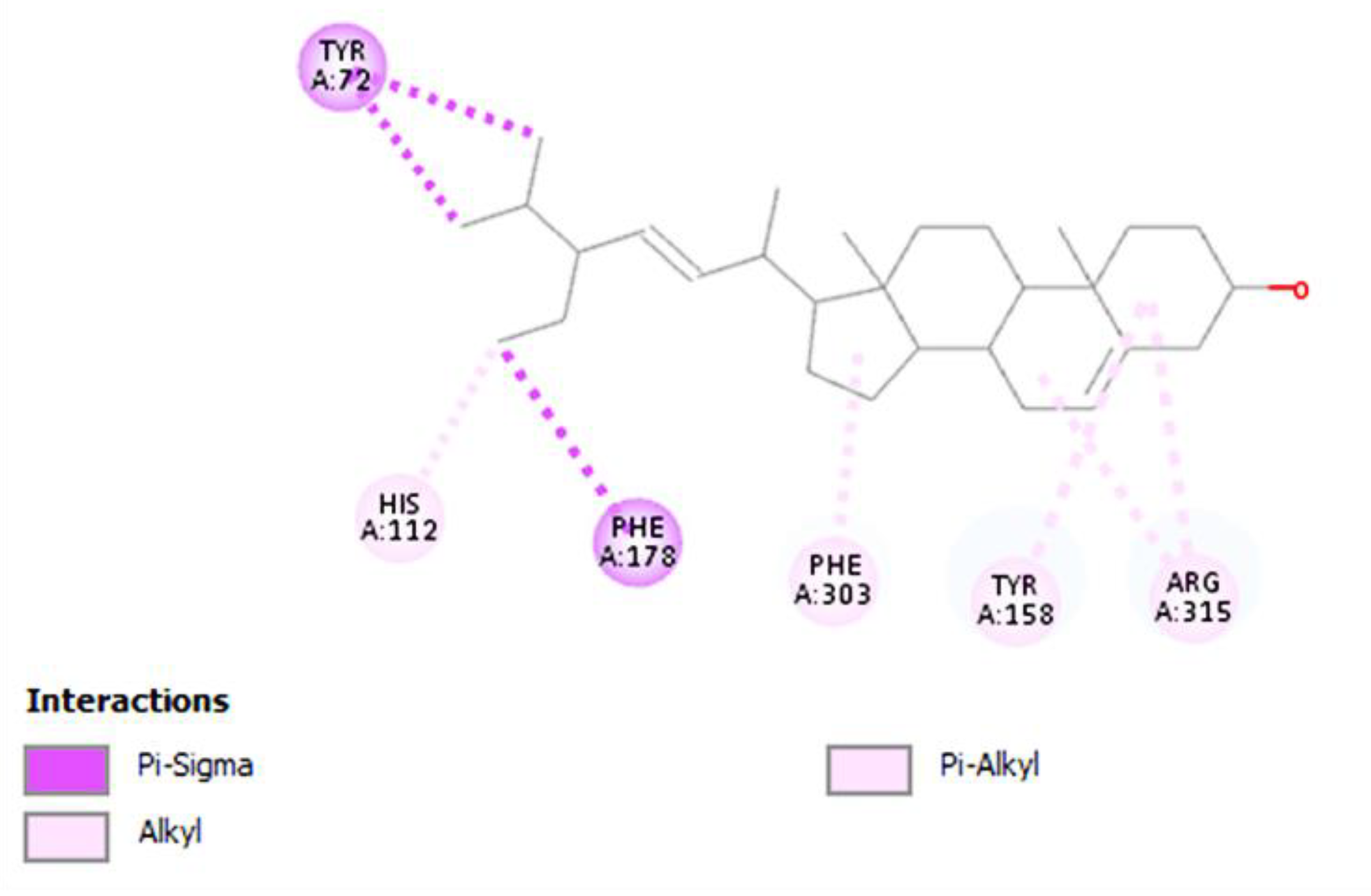
3.2. Paederia foetida
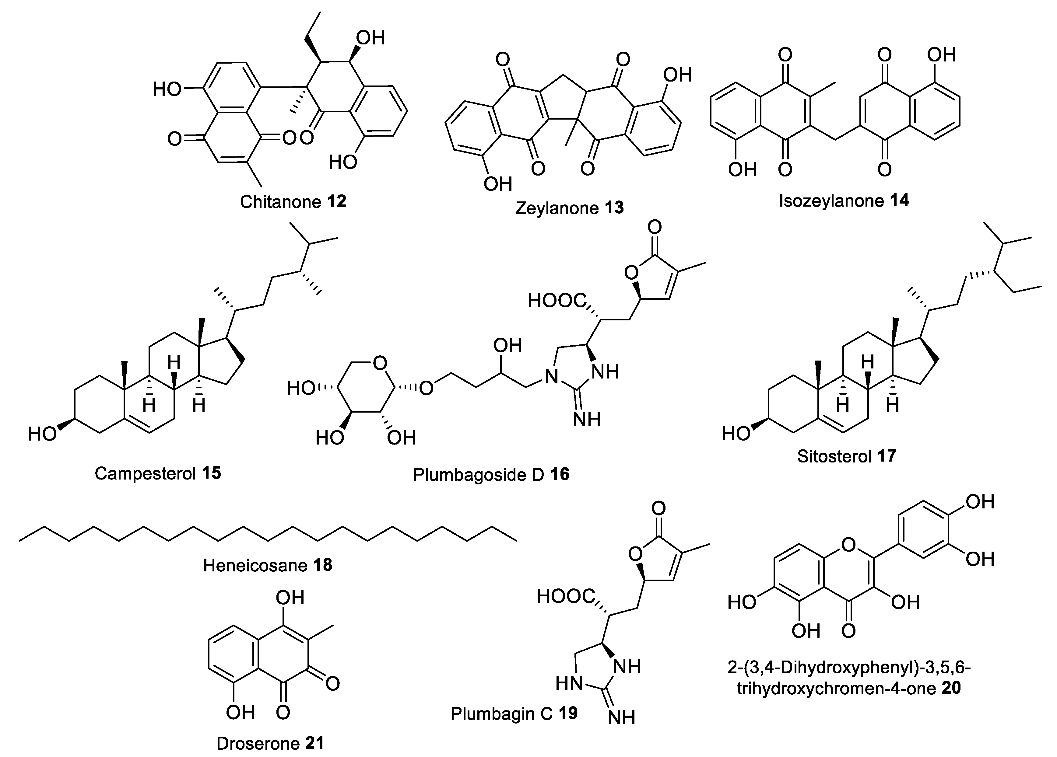
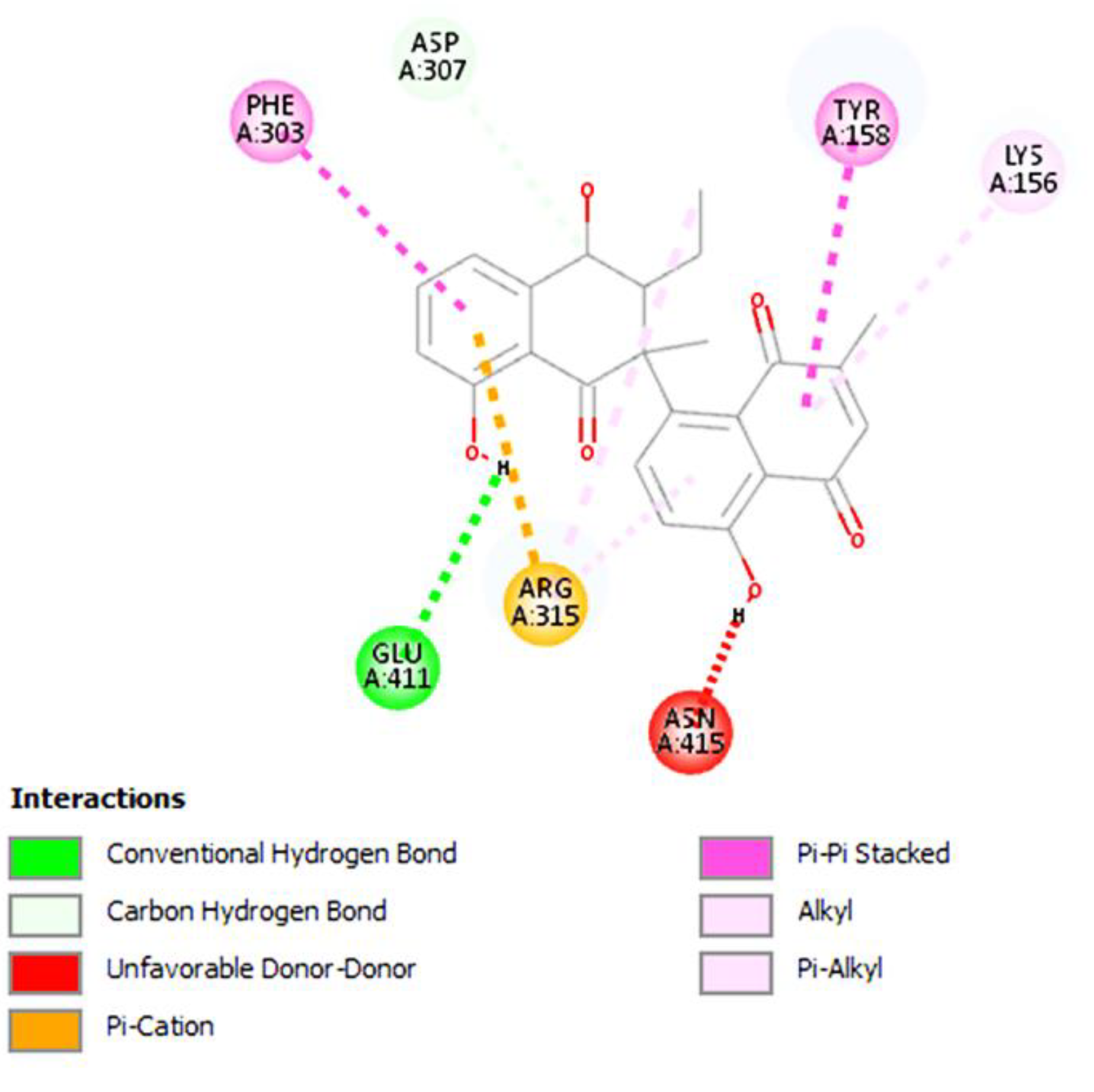
3.3. Plumbago zeylanica
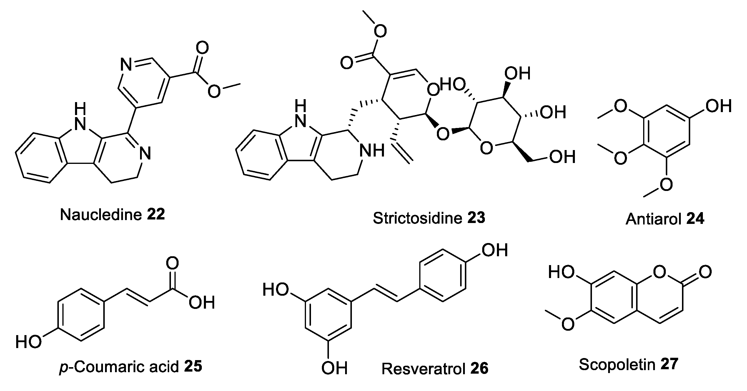
3.4. Nauclea pallida Reinw
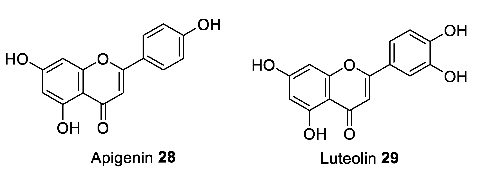
3.5. Sauropus androgynus
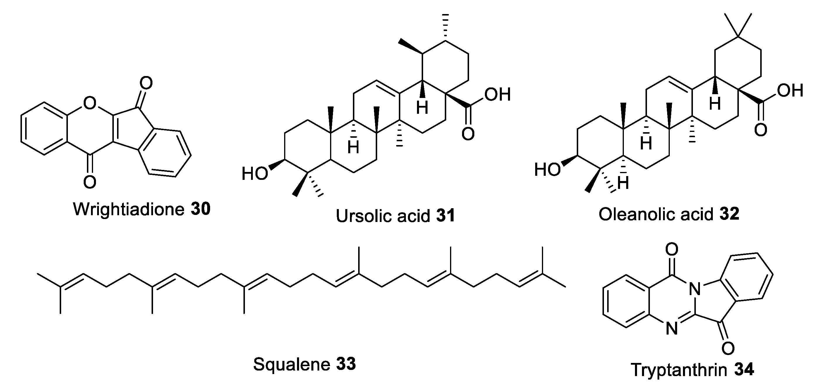
3.6. Wrightia pubescens
3.7. Psophocarpus tetragonolobus
4. Materials and Methods
4.1. Plant Materials
4.2. Extract Preparation
4.3. Antioxidant Determination
4.3.1. 2-2-Diphenyl-1-picrylhydrazyl (DPPH) Assay
4.3.2. Superoxide Anion Radical Scavenging Assay
4.3.3. Hydroxyl Radical Scavenging Assay
4.4. Molecular Docking
5. Conclusions
Author Contributions
Funding
Institutional Review Board Statement
Informed Consent Statement
Data Availability Statement
Acknowledgments
Conflicts of Interest
References
- Schleicher, E.; Gerdes, C.; Petersmann, A.; Müller-Wieland, D.; Müller, U.A.; Freckmann, G.; Heinemann, L.; Nauck, M.; Landgraf, R. Definition, Classification and Diagnosis of Diabetes Mellitus. Exp. Clin. Endocrinol. Diabetes 2022, 130, S1–S8. [Google Scholar] [CrossRef] [PubMed]
- Sun, H.; Saeedi, P.; Karuranga, S.; Pinkepank, M.; Ogurtsova, K.; Duncan, B.B.; Stein, C.; Basit, A.; Chan, J.C.; Mbanya, J.C.; et al. IDF Diabetes Atlas: Global, regional and country-level diabetes prevalence estimates for 2021 and projections for 2045. Diabetes Res. Clin. Pract. 2022, 183, 109119. [Google Scholar] [CrossRef] [PubMed]
- Kementerian Kesehatan Republik Indonesia. Hasil Utama Riset Kesehatan Dasar; Departemen Kesehatan RI: Jakarta, Indonesia, 2018. [Google Scholar]
- Asmat, U.; Abad, K.; Ismail, K. Diabetes mellitus and oxidative stress—A concise review. Saudi Pharm. J. 2016, 24, 547–553. [Google Scholar] [CrossRef] [PubMed] [Green Version]
- Li, Y.; Wang, D.D.; Ley, S.H.; Vasanti, M.; Howard, A.G.; He, Y.; Hu, F.B. Time trends of dietary and lifestyle factors and their potential impact on diabetes burden in China. Diabetes Care 2017, 40, 1685–1694. [Google Scholar] [CrossRef] [Green Version]
- Papatheodorou, K.; Banach, M.; Bekiari, E.; Rizzo, M.; Edmonds, M. Complications of diabetes 2017. J. Diabetes Res. 2018, 2018, 3086167. [Google Scholar] [CrossRef]
- Zheng, Y.; Ley, S.H.; Hu, F.B. Global aetiology and epidemiology of type 2 diabetes mellitus and its complications. Nat. Rev. Endocrinol. 2018, 14, 88–98. [Google Scholar] [CrossRef]
- Ćorković, I.; Gašo-Sokač, D.; Pichler, A.; Šimunović, J.; Kopjar, M. Dietary Polyphenols as Natural Inhibitors of α-Amylase and α-Glucosidase. Life 2022, 12, 1692. [Google Scholar] [CrossRef]
- Meneses, M.J.; Silva, B.M.; Sousa, M.; Sa, R.; Oliveira, P.F.; Alves, M.G. Antidiabetic drugs: Mechanisms of action and potential outcomes on cellular metabolism. Curr. Pharm. Des. 2015, 21, 3606–3620. [Google Scholar] [CrossRef]
- Chaudhury, A.; Duvoor, C.; Reddy Dendi, V.S.; Kraleti, S.; Chada, A.; Ravilla, R.; Marco, A.; Shekhawat, N.S.; Montales, M.T.; Kuriakose, K.; et al. Clinical review of antidiabetic drugs: Implications for type 2 diabetes mellitus management. Front. Endocrinol. 2017, 8, 6. [Google Scholar] [CrossRef] [Green Version]
- Saeed, F.; Sultan, M.T.; Riaz, A.; Ahmed, S.; Bigiu, N.; Amarowicz, R.; Manea, R. Bitter melon (Momordica charantia L.) fruit bioactives charantin and vicine potential for diabetes prophylaxis and treatment. Plants 2021, 10, 730. [Google Scholar] [CrossRef]
- Martín, M.Á.; Ramos, S. Dietary flavonoids and insulin signaling in diabetes and obesity. Cells 2021, 10, 1474. [Google Scholar] [CrossRef] [PubMed]
- Rahimi-Madiseh, M.; Malekpour-Tehrani, A.; Bahmani, M.; Rafieian-Kopaei, M. The research and development on the antioxidants in prevention of diabetic complications. Asian Pac. J. Trop. Med. 2016, 9, 825–831. [Google Scholar] [CrossRef] [PubMed] [Green Version]
- Preshaw, P.M.; Alba, A.L.; Herrera, D.; Jepsen, S.; Konstantinidis, A.; Makrilakis, K.; Taylor, R. Periodontitis and diabetes: A two-way relationship. Diabetologia 2012, 55, 21–31. [Google Scholar] [CrossRef] [PubMed] [Green Version]
- Chee, B.; Park, B.; Bartold, P.M. Periodontitis and type II diabetes: A two-way relationship. Int. J. Evid. Based Healthc. 2013, 11, 317–329. [Google Scholar] [CrossRef]
- Wu, Y.; Zhang, D.; Jiang, X.; Jiang, W. Fruit and vegetable consumption and risk of type 2 diabetes mellitus: A dose-response meta-analysis of prospective cohort studies. Nutr. Metab. Cardiovasc. Dis. 2015, 25, 140–147. [Google Scholar] [CrossRef]
- Wallace, T.C.; Bailey, R.L.; Blumberg, J.B.; Burton-Freeman, B.; Chen, C.O.; Crowe-White, K.M.; Drewnowski, A.; Hooshmand, S.; Johnson, E.; Lewis, R.; et al. Fruits, vegetables, and health: A comprehensive narrative, umbrella review of the science and recommendations for enhanced public policy to improve intake. Crit. Rev. Food Sci. Nutr. 2020, 60, 2174–2211. [Google Scholar] [CrossRef] [Green Version]
- Hartanti, D.; Budipramana, K. Traditional antidiabetic plants from Indonesia. Ethnobot. Res. Appl. 2020, 19, 1–24. [Google Scholar] [CrossRef]
- Ri, K.K. Fitofarmaka Berpotensi Jadi Produk Farmasi Utama Dalam Negeri. Available online: https://sehatnegeriku.kemkes.go.id/baca/umum/20211108/5038813/fitofarmaka-berpotensi-jadi-produk-farmasi-utama-dalam-negeri/ (accessed on 24 January 2022).
- Hutapea, J.R.; Djumidi, S. Inventaris Tanaman Obat Indonesia I Jilid 2; Departemen Kesehatan dan Kesejahteraan Sosial RI: Jakarta, Indonesia, 2001; p. 355. [Google Scholar]
- Djumidi, S. Inventaris Tanaman Obat Indonesia IV; Departemen Kesehatan RI: Jakarta, Indonesia, 1997. [Google Scholar]
- Mustofa, F.I.; Rahmawati, N. Studi etnofarmakologi tumbuhan obat yang digunakan oleh penyehat tradisional untuk mengatasi diare di Sulawesi Selatan. J. Tumbuh. Obat Indones. 2018, 11, 17–32. [Google Scholar] [CrossRef]
- Permatasari, D.; Diniatik, D.; Hartanti, D. Studi Etnofarmakologi Obat Tradisional Sebagai Anti Diare Di Kecamatan Baturaden Kabupaten Banyumas. Pharm. J. Farm. Indones. 2011, 8, 44–64. [Google Scholar] [CrossRef]
- Hutapea, J.R. Inventaris Tanaman Obat Indonesia I Jilid I; Departemen Kesehatan dan Kesejahteraan Sosial RI: Jakarta, Indonesia, 2000. [Google Scholar]
- Hutapea, J.R. Inventaris Tanaman Obat Indonesia II; Departemen Kesehatan RI: Jakarta, Indonesia, 1993. [Google Scholar]
- Rizki, R.; Chairunnisak, C.; Alfina, R.; Darlis, O.; Rasdanelwati, R. Potential of Medicinal Plants in the Asteraceae Family Found in Harau Lima Puluh Kota Regency. Sainstek J. Sains Teknol. 2021, 13, 73–83. [Google Scholar] [CrossRef]
- Ito, M.F. Kajian Etnofarmakologi Penggunaan Tanaman Obat Oleh Masyarakat Di Kecamatan Soa Kabupaten Ngada; Poltekkes Kemenkes: Kupang, Indonesia, 2019. [Google Scholar]
- Hutapea, J.R. Inventaris Tanaman obat Indonesia III; Departemen Kesehatan RI: Jakarta, Indonesia, 1994. [Google Scholar]
- Kodir, R.A.; Moektiwardoyo, M.; Iskandar, Y. Etnofarmasi dan ulasan bioprospektif tumbuhan obat liar dalam pengobatan tradisional kampung adat cikondang, kecamatan pangalengan, kabupaten bandung, jawa barat. Farmaka 2017, 15, 26–44. [Google Scholar]
- Eisai Indonesia. Herb Index Indonesia; Eisai Indonesia: Jakarta, Indonesia, 1986. [Google Scholar]
- Qasrin, U.; Setiawan, A.; Yulianty, Y.; Bintoro, A. Studi Etnobotani Tumbuhan Berkhasiat Obat yang Dimanfaatkan Masyarakat Suku Melayu Kabupaten Lingga Provinsi Kepulauan Riau. J. Belantara 2020, 3, 139–152. [Google Scholar] [CrossRef]
- Gunarti, N.S. Studi Etnobotani Tumbuhan Obat di Desa Kutalanggeng dan Kutamaneuh Kecamatan Tegalwaru Kabupaten Karawang Jawa Barat. Maj. Farmasetika 2021, 6, 14–23. [Google Scholar] [CrossRef]
- Hefny Gad, M.; Tuenter, E.; El-Sawi, N.; Younes, S.; El-Ghadban, E.M.; Demeyer, K.; Pieters, L.; Vander Heyden, Y.; Mangelings, D. Identification of some bioactive metabolites in a fractionated methanol extract from Ipomoea aquatica (aerial parts) through TLC, HPLC, UPLC-ESI-QTOF-MS and LC-SPE-NMR fingerprints analyses. Phytochem. Anal. 2018, 29, 5–15. [Google Scholar] [CrossRef]
- Lawal, U.; Leong, S.W.; Shaari, K.; Ismail, I.S.; Khatib, A.; Abas, F. α-glucosidase inhibitory and antioxidant activities of Different Ipomoea aquatica cultivars and LC–MS/MS profiling of the active cultivar. J. Food Biochem. 2017, 41, e12303. [Google Scholar] [CrossRef]
- Lawal, U.; Mediani, A.; Maulidiani, H.; Shaari, K.; Ismail, I.S.; Khatib, A.; Abas, F. Metabolite profiling of Ipomoea aquatica at different growth stages in correlation to the antioxidant and α-glucosidase inhibitory activities elucidated by 1H NMR-based metabolomics. Sci. Hortic. 2015, 192, 400–408. [Google Scholar] [CrossRef]
- Prasad, K.N.; Divakar, S.; Shivamurthy, G.R.; Aradhya, S.M. Isolation of a free radical-scavenging antioxidant from water spinach (Ipomoea aquatica Forsk). J. Sci. Food Agric. 2005, 85, 1461–1468. [Google Scholar] [CrossRef]
- Xiao, M.; Ying, L.; Li, S.; Fu, X.; Du, G. Progress on research and development of Paederia scandens as a natural medicine. Int. J. Clin. Exp. Med. 2019, 12, 158–167. [Google Scholar]
- Nile, S.H.; Park, S.W. Biologically active compounds from Plumbago zeylanica. Chem. Nat. Compd. 2014, 50, 905–907. [Google Scholar] [CrossRef]
- Cong, H.J.; Zhang, S.W.; Shen, Y.; Zheng, Y.; Huang, Y.J.; Wang, W.Q.; Leng, Y.; Xuan, L.-J. Guanidine alkaloids from Plumbago zeylanica. J. Nat. Prod. 2013, 76, 1351–1357. [Google Scholar] [CrossRef]
- Vanitha, V.; Vijayakumar, S.; Nilavukkarasi, M.; Punitha, V.; Vidhya, E.; Praseetha, P. Heneicosane—A novel microbicidal bioactive alkane identified from Plumbago zeylanica L. Ind. Crops Prod. 2020, 154, 112748. [Google Scholar] [CrossRef]
- Shukla, B.; Saxena, S.; Usmani, S.; Kushwaha, P. Phytochemistry and pharmacological studies of Plumbago zeylanica L.: A medicinal plant review. Clin. Phytoscience 2021, 7, 1–11. [Google Scholar] [CrossRef]
- Andarwulan, N.; Batari, R.; Sandrasari, D.A.; Bolling, B.; Wijaya, H. Flavonoid content and antioxidant activity of vegetables from Indonesia. Food Chem. 2010, 121, 1231–1235. [Google Scholar] [CrossRef] [PubMed] [Green Version]
- Andarwulan, N.; Kurniasih, D.; Apriady, R.A.; Rahmat, H.; Roto, A.V.; Bolling, B.W. Polyphenols, carotenoids, and ascorbic acid in underutilized medicinal vegetables. J. Funct. Foods 2012, 4, 339–347. [Google Scholar] [CrossRef]
- Traxler, F.; Iamprasertkun, N.; Tschigg, A.M.; Vajrodaya, S.; Valant-Vetschera, K.; Brecker, L.; Schinnerl, J. Specialized plant metabolites from indolic and polyphenolic biosynthetic pathways in Wrightia religiosa (Teijsm. & Binn.) Benth. and Wrightia pubescens R. Br. (Apocynaceae). S. Afr. J. Bot. 2021, 137, 242–248. [Google Scholar]
- Ragasa, C.Y.; Ng, V.; de Los Reyes, M.M.; Mandia, E.H.; Shen, C.C. An isoflavone from Wrightia pubescens. Int. J. Pharmacogn. Phytochem. Res. 2015, 7, 353–355. [Google Scholar]
- Ragasa, C.Y.; Ng, V.A.S.; Mariquit, M.; Mandia, E.H.; Shen, C.-C. Chemical constituents of Wrightia pubescens (R. Br.). Delta 2014, 18, 14–19. [Google Scholar]
- Yaribeygi, H.; Sathyapalan, T.; Atkin, S.L.; Sahebkar, A. Molecular mechanisms linking oxidative stress and diabetes mellitus. Oxidative Med. Cell. Longev. 2020, 2020, 8609213. [Google Scholar] [CrossRef] [Green Version]
- Syabana, M.A.; Yuliana, N.D.; Batubara, I.; Fardiaz, D. Antidiabetic activity screening and nmr profile of vegetable and spices commonly consumed in Indonesia. Food Sci. Technol. 2020, 41, 254–264. [Google Scholar] [CrossRef]
- Kashihara, N.; Haruna, V.K.; Kondeti, Y.S.; Kanwar, Y. Oxidative stress in diabetic nephropathy. Curr. Med. Chem. 2010, 17, 4256–4269. [Google Scholar] [CrossRef] [Green Version]
- Kowluru, R.A.; Chan, P.S. Oxidative stress and diabetic retinopathy. Exp. Diabetes Res. 2007, 2007, 043603. [Google Scholar] [CrossRef] [PubMed] [Green Version]
- Pang, L.; Lian, X.; Liu, H.; Zhang, Y.; Li, Q.; Cai, Y.; Ma, H.; Yu, X. Understanding diabetic neuropathy: Focus on oxidative stress. Oxidative Med. Cell. Longev. 2020, 2020, 9524635. [Google Scholar] [CrossRef] [PubMed]
- Srivastava, G.P.; Yadav, N.; Yadav, B.N.; Yadav, R.K.; Yadav, D.K. Pan-interactomics and its applications. In Pan-Genomics: Applications, Challenges, and Future Prospects; Elsevier: Amsterdam, The Netherlands, 2020; pp. 397–435. [Google Scholar]
- Elfrida, E.; Tarigan, N.S.; Suwardi, A.B. Ethnobotanical study of medicinal plants used by community in Jambur Labu Village, East Aceh, Indonesia. Biodiversitas J. Biol. Divers. 2021, 22, 2893–2900. [Google Scholar] [CrossRef]
- Hefny Gad, M.; Demeyer, K.; Vander Heyden, Y.; Mangelings, D. Cytotoxic, Antioxidant, and Antidiabetic Activities versus UPLC-ESI-QTOF-MS Chemical-Profile Analysis of Ipomoea aquatica Fractions. Planta Med. 2021, 87, 1089–1100. [Google Scholar] [CrossRef]
- Vuolo, M.M.; Lima, V.S.; Junior, M.R.M. Phenolic compounds: Structure, classification, and antioxidant power. In Bioactive Compounds; Elsevier: Amsterdam, The Netherlands, 2019; pp. 33–50. [Google Scholar]
- Choi, J.; Kang, H.J.; Kim, S.Z.; Kwon, T.O.; Jeong, S.I.; Jang, S.I. Antioxidant effect of astragalin isolated from the leaves of Morus alba L. against free radical-induced oxidative hemolysis of human red blood cells. Arch. Pharmacal Res. 2013, 36, 912–917. [Google Scholar] [CrossRef]
- Li, P.; Yin, Y.; Pan, G.; Zhao, F.; Wang, Q. Effect of astragalin on paraoxon-induced vascular endothelium dysfunction. Plant Dis. Pests 2011, 2, 73–76. [Google Scholar]
- Hung, T.M.; Na, M.; Thuong, P.T.; Su, N.D.; Sok, D.; Song, K.S.; Seong, Y.H.; Bae, K. Antioxidant activity of caffeoyl quinic acid derivatives from the roots of Dipsacus asper Wall. J. Ethnopharmacol. 2006, 108, 188–192. [Google Scholar] [CrossRef]
- Ding, S.; Jiang, H.; Fang, J. Regulation of Immune Function by Polyphenols. J. Immunol. Res. 2018, 2018, 1264074. [Google Scholar] [CrossRef] [Green Version]
- Wang, L.; Jiang, Y.; Han, T.; Zheng, C.; Qin, L. A phytochemical, pharmacological and clinical profile of Paederia foetida and P. scandens. Nat. Prod. Commun. 2014, 9. [Google Scholar] [CrossRef] [Green Version]
- Osuntokun, O.T.; Oluduro, A.; Idowu, T.; Omotuyi, A. Assessment of nephrotoxicity, anti-inflammatory and antioxidant properties of epigallocatechin, epicatechin and stigmasterol phytosterol (synergy) derived from ethyl acetate stem bark extract of Spondias mombin on Wistar rats using molecular method of analysis. J. Mol. Microbiol. 2017, 1, 103. [Google Scholar]
- Pattarachotanant, N.; Prasansuklab, A.; Tencomnao, T. Momordica charantia L. Extract Protects Hippocampal Neuronal Cells against PAHs-Induced Neurotoxicity: Possible Active Constituents Include Stigmasterol and Vitamin, E. Nutrients 2021, 13, 2368. [Google Scholar] [CrossRef] [PubMed]
- Takayama, F.; Fujihara, Y. How does Eucommia leaf extract prevent smooth muscle cell proliferation induced by high-fat diets at the aortic tunica media? Hypertens. Res. 2017, 40, 541–543. [Google Scholar] [CrossRef] [PubMed]
- El-Najjar, N.; Gali-Muhtasib, H.; Ketola, R.A.; Vuorela, P.; Urtti, A.; Vuorela, H. The chemical and biological activities of quinones: Overview and implications in analytical detection. Phytochem. Rev. 2011, 10, 353–370. [Google Scholar] [CrossRef]
- Shao, Y.; Dang, M.; Lin, Y.; Xue, F. Evaluation of wound healing activity of plumbagin in diabetic rats. Life Sci. 2019, 231, 116422. [Google Scholar] [CrossRef] [PubMed]
- Yoshida, Y.; Niki, E. Antioxidant effects of phytosterol and its components. J. Nutr. Sci. Vitaminol. 2003, 49, 277–280. [Google Scholar] [CrossRef]
- Haudecoeur, R.; Peuchmaur, M.; Pérès, B.; Rome, M.; Taïwe, G.S.; Boumendjel, A.; Boucherle, B. Traditional uses, phytochemistry and pharmacological properties of African Nauclea species: A review. J. Ethnopharmacol. 2018, 212, 106–136. [Google Scholar] [CrossRef]
- King, F.; Jurd, L. 239. The Chemistry of extractives from hardwoods. Part XII. The cyclitols and steroids of opepe (Sarcocephalus diderrichii). J. Chem. Soc. 1953, 1192–1195. [Google Scholar] [CrossRef]
- Abreu, P.; Pereira, A. New indole alkaloids from Sarcocephalus latifolius. Nat. Prod. Lett. 2001, 15, 43–48. [Google Scholar] [CrossRef]
- Kuete, V.; Sandjo, L.P.; Mbaveng, A.T.; Seukep, J.A.; Ngadjui, B.T.; Efferth, T. Cytotoxicity of selected Cameroonian medicinal plants and Nauclea pobeguinii towards multi-factorial drug-resistant cancer cells. BMC Complement. Altern. Med. 2015, 15, 1–9. [Google Scholar] [CrossRef] [Green Version]
- McLean, S.; Murray, D. The constituents of Nauclea diderrichii. Part IV. Miscellaneous substances; biogenetic considerations. Can. J. Chem. 1972, 50, 1496–1501. [Google Scholar] [CrossRef] [Green Version]
- Awah, F.M.; Uzoegwu, P.N.; Ifeonu, P.; Oyugi, J.O.; Rutherford, J.; Yao, X.; Fehrmann, F.; Fowke, K.R.; Eze, M.O. Free radical scavenging activity, phenolic contents and cytotoxicity of selected Nigerian medicinal plants. Food Chem. 2012, 131, 1279–1286. [Google Scholar] [CrossRef]
- Iwueke, A.; Nwodo, O.; Ojiako, O.; Okwu, G.; Nwogu, L.; Igwe, C. Modification of lipid peroxidation and oxidative stress in hepatocytes of diabetic rats treated with root extract of Sarcocephalus latifolius and Daniella oliveri. Aust. J. Basic Appl. Sci. 2010, 4, 3578–3584. [Google Scholar]
- Gidado, A.; Ameh, D.; Atawodi, S. Effect of Nauclea latifolia leaves aqueous extracts on blood glucose levels of normal and alloxan-induced diabetic rats. Afr. J. Biotechnol. 2005, 4, 91–93. [Google Scholar]
- Zhang, B.D.; Cheng, J.X.; Zhang, C.F.; Bai, Y.D.; Liu, W.Y.; Li, W.; Koike, K.; Akihisa, T.; Feng, F.; Zhang, J.; et al. Sauropus androgynus L. Merr.-A phytochemical, pharmacological and toxicological review. J. Ethnopharmacol. 2020, 257, 112778. [Google Scholar] [CrossRef]
- Zhao, L.; Wang, J.L.; Liu, R.; Li, X.X.; Li, J.F.; Zhang, L. Neuroprotective, anti-amyloidogenic and neurotrophic effects of apigenin in an Alzheimer’s disease mouse model. Molecules 2013, 18, 9949–9965. [Google Scholar] [CrossRef] [PubMed] [Green Version]
- Kang, J.; Li, Z.; Wu, T.; Jensen, G.S.; Schauss, A.G.; Wu, X. Anti-oxidant capacities of flavonoid compounds isolated from acai pulp (Euterpe oleracea Mart.). Food Chem. 2010, 122, 610–617. [Google Scholar] [CrossRef]
- Taek, M.M.; Ew, B.P.; Agil, M. Ethnomedicinal Plants Used for the Treatment of Malaria in Malaka, West Timor. J. Young Pharm. 2018, 10, 187–192. [Google Scholar] [CrossRef] [Green Version]
- Moon, S.Y.; Lee, J.H.; Choi, H.Y.; Cho, I.J.; Kim, S.C.; Kim, Y.W. Tryptanthrin protects hepatocytes against oxidative stress via activation of the extracellular signal-regulated kinase/NF-E2-related factor 2 pathway. Biol. Pharm. Bull. 2014, 37, 1633–1640. [Google Scholar] [CrossRef] [Green Version]
- Fatihah, H.N.; Maxted, N.; Arce, L.R. Cladistic analysis of Psophocarpus Neck. ex DC.(Leguminosae, Papilionoideae) based on morphological characters. S. Afr. J. Bot. 2012, 83, 78–88. [Google Scholar] [CrossRef]
- Bassal, H.; Merah, O.; Ali, A.M.; Hijazi, A.; El Omar, F. Psophocarpus tetragonolobus: An Underused Species with Multiple Potential Uses. Plants 2020, 9, 1730. [Google Scholar] [CrossRef]
- Tang, X.; He, Z.; Dai, Y.; Xiong, Y.L.; Xie, M.; Chen, J. Peptide fractionation and free radical scavenging activity of zein hydrolysate. J. Agric. Food Chem. 2010, 58, 587–593. [Google Scholar] [CrossRef] [PubMed]
- Halliwell, B.; Gutteridge, J.M.; Aruoma, O.I. The deoxyribose method: A simple “test-tube” assay for determination of rate constants for reactions of hydroxyl radicals. Anal. Biochem. 1987, 165, 215–219. [Google Scholar] [CrossRef] [PubMed]


| No | Local Name | Part Used | Species Name | Family | Distribution in Indonesia | Ethnopharmacology Indication | Ref. |
|---|---|---|---|---|---|---|---|
| 1 | Bayam duri | Whole plant (young) | Amaranthus spinosus L. | Amaranthaceae | Sumatra, Jawa, Madura, Bali, Sulawesi, Maluku, Halmahera, Tidore | Blister, asthma, fever | [20] |
| 2 | Kluwih | Young fruit | Artocarpus Altilis (Park.) FSB. | Moraceae | Sumatera, Jawa, Madura, Bali, Nusa Tenggara, Timor, Sulawesi, Selayar, Maluku, Seram, Halmahera | Toothache, skin infection | [21] |
| 3 | Nangka | Young fruit | Artocarpus heterophyllus Lam. | Moraceae | Sulawesi, Jawa | Diarrhea | [22,23] |
| 4 | Gude | Seeds | Cajanus cajan Millspaugh | Leguminosae | Sumatera, Jawa, Madura, Bali, Nusa Tenggara, Timor, Sulawesi, Maluku, Halmahera, Ternate, Tidore | Scabies, cough, blood cleansing | [20] |
| 5 | Pepaya | Flower | Carica papaya L. | caricaceae | Sumatera, Kalimantan, Nusa Tenggara, Jawa, Sulawesi, maluku, Papua | Malaria, anthelmintic, stomachache, | [24] |
| 6 | Telang | Flower | Clitoria ternatea L. | Papilonaceae | Sumatra, Jawa, Sulawesi, Maluku, Halmahera, Ternate | Blister, eye irritation | [25] |
| 7 | Sintrong | Whole plant (young) | Crassocephalum crepidioides (Benth.) S.Moore | Asteraceae | Sumatera, Jawa | Stomachache, headache, blister | [26,27] |
| 8 | Pakis | Whole plant (young) | Diplazium esculentum Swartz. | Polypodiaceae | Sumatra, Jawa, Bali, Sulawesi, Maluku, Ambon | Rub | [28] |
| 9 | Kecombrang | Flower | Etlingera elatior (Jack) R.M.Sm. | Zingiberaceae | Jawa | Vitality | [29] |
| 10 | Kangkung | Whole plant (young) | Ipomoea aquatica Forssk. | Convolvulaceae | Sumatera, Jawa, Nusa Tenggara, Sulawesi, Gorontalo, Maluku, Halmahera, Tidore, Buru | Sedative agent | [20] |
| 11 | Genjer | Whole plant (young) | Limnocharis flava (L.) Buch | Butomaceae | Sumatera, Jawa, Madura | Food | [30] |
| 12 | Gambas | Young Fruit | Luffa acutangular (L.) Roxb. | Cucurbitaceae | Sumatera, Jawa, Madura, Maluku, Ternate | Fever, nausea, stomachache, toothache | [29] |
| 13 | Semanggi | Whole plant (young) | Marsilea minuta L. | Marsilaceae | Jawa | diuretic agents | [28] |
| 14 | Kelor | Young Leaves Fruit, seeds | Moringa oleifera L. | Moringaceae | Sumatera, Jawa, Madura, Bali, Nusa Tenggara, Maluku, Buru, Ternate, Tidore | Gum bleeding, period, headache, asthma, rheumatism, anti-emetic, spasm | [20] |
| 15 | Pisang | Stem | Musa paradisiaca L. | Musaceae | Sumatera, Jawa, Ambon | Tonsil and gastro inflammation, anemia, trachoma inflammation, diarrhea, fever, food poisoning | [30] |
| 16 | Selada Air | Whole plant (young) | Nasturtium officinale W.T. Aiton | Sumatera | Excema | [31] | |
| 17 | Kolpo | Young Leaves | Nauclea pallida Reinw | Rubiaceae | Jawa | Diuretic | [30] |
| 18 | Mangkokan | Young Leaves | Nothopanax scutellarium Merr. | Araliaceae | Jawa, Sulawesi, Mena, Maluku, Ambon, Roti, Halmahera, Ternate | Hari tonic, anti-inflammation (edema), diuretic agents | [20] |
| 19 | Sembukan | Leaves | Paederia foetida L. | Rubiaceae | Sumatera, Jawa, Madura, Maluku, Ternate | Sore | [32] |
| 20 | Beluntas | Young Leaves | Pluchea indica L. | Asteraceae | Sumatera, Jawa, Madura, Sulawesi, Nusa, Tenggara Timor | Fever, cough, body odor | [20] |
| 21 | Kareka | Young Leaves | Plumbago zeylanica L. | Plumbaginaceae | Sumatera, Jawa, Bali, Timor | Rheumatism, headache | [24] |
| 22 | Krokot | Whole plant (young) | Portulaca oleracea L. | Portulaceae | Sumatera, Jawa, Madura, Maluku, Ternate | Diarrhea, fever, stomachache | [20] |
| 23 | Kecipir | Fruit and seeds | Psophocarpus tetragonolobus (L.) D.C. | Fabaceae | Sumatera, Jawa, Bali, Maluku, Ternate | Ear inflammation | [20] |
| 24 | Katuk | Young Leaves | Sauropus Androgynus (L.) Merr. | Euphorbiaceae | Sumatera, Jawa, Madura | Breas milk stimulant, acne, fever, blister, eczema | [20] |
| 25 | Labu Siam | Fruit (young) | Sechium Edule (Jacq.) Sw. | Cucurbitaceae | Sumatera, Jawa, Madura | Food | |
| 26 | Turi | Flower | Sesbania grandiflora Pers. | Leguminosae | Sumatera, Jawa, Madura, Bali, Sulawesi, Nusa tenggara, Timor, Ternate, Tidore | Gastro-inflammation, mouth blister, scabies | [20] |
| 27 | Kenikir | Young leaves | Tagetes erecta L. | Compositae | Jawa | Insect repellant | [20] |
| 28 | Kacang panjang | Young Fruit | Vigna cylindrica (L.) Walp. | Fabaceae | Jawa | Diuretic | [28] |
| 29 | Bintaos | Young Leaves | Wrightia pubescens R.Br. | Apocynaceae | Sumatera, Jawa, Madura, Nusa Tenggara, Bali, Timor | Eye irritation | [30] |
| No | Sample | Plant Parts | Inhibition Percentage (%) | ||
|---|---|---|---|---|---|
| DPPH | SO | Hydroxyl | |||
| 1 | Carica papaya | Flos | 47.2 ± 0.2 | 60.3 ± 1.0 | 59.3 ± 0.1 |
| 2 | Clitoria ternatea | Flos | 87.5 ± 0.2 | 53.7 ± 1.8 | 73.2 ± 0.1 |
| 3 | Etlingera elatior | Flos | 66.2 ± 0.2 | 93.6 ± 1.2 | 80.4 ± 0.1 |
| 4 | Ipomoea aquatica | All parts | 92.6 ± 0.2 | 97.6 ± 1.0 | 80.2 ± 0.1 |
| 5 | Limnocharis Flava | All parts | 27.7 ± 0.3 | 83.4 ± 1.0 | 79.2 ± 0.1 |
| 6 | Luffa acutangular | Fructus | 12.6 ± 0.2 | 72.7 ± 1.0 | 80.2 ± 0.1 |
| 7 | Marsilea minuta | All parts | 72.2 ± 0.1 | 74.5 ± 2.7 | 86.0 ± 0.1 |
| 8 | Moringa Oleifera | All parts | 5.3 ± 0.4 | 66.8 ± 2.1 | 78.6 ± 0.2 |
| 9 | Moringa Oleifera | Flos | 24.4 ± 0.2 | 95.1 ± 0.8 | 73.9 ± 0.1 |
| 10 | Nasturtium officinale | All parts | 36.3 ± 0.2 | 88.1 ± 0.8 | 86.3 ± 0.1 |
| 11 | Nauclea pallida Reinw. | All parts | 89.1 ± 0.1 | 72.3 ± 2.2 | 86.1 ± 0.1 |
| 12 | Nothopanax scutellarium | All parts | 1.1 ± 0.2 | 79.7 ± 0.6 | 84.2 ± 0.1 |
| 13 | Paederia foetida | All parts | 90.3 ± 0.2 | 93.5 ± 2.1 | 85.7 ± 0.1 |
| 14 | Pluchea indica | All parts | 86.4 ± 0.2 | 95.3 ± 2.1 | 57.9 ± 0.1 |
| 15 | Plumbago zeylanica | All parts | 90.0 ± 0.6 | 84.6 ± 2.1 | 89.2 ± 0.1 |
| 16 | Psophocarpus tetragonolobus | Fructus | 83.8 ± 0.2 | 87.5 ± 1.8 | 90.0 ± 0.1 |
| 17 | Sauropus Androgynus | All parts | 20.3 ± 0.2 | 34.5 ± 1.9 | 91.1 ± 0.1 |
| 18 | Vigna cylindrica | All parts | 22.0 ± 0.2 | 82.2 ± 1.8 | 82.9 ± 0.1 |
| 19 | Wrightia pubescens | All parts | 72.8 ± 0.1 | 96.5 ± 0.5 | 90.0 ± 0.1 |
| 20 | Diplazium esculentum | All parts | 25.0 ± 0.2 | 77.5 ± 2.1 | 90.2 ± 0.1 |
| 21 | Vitamin C (standard) | - | 88.8 ± 0.1 | 79.9 ± 2.6 | 38.1 ± 0.5 |
| Name of Structure | Affinity (kcal/mol) |
|---|---|
| Ipomoea aquatica | |
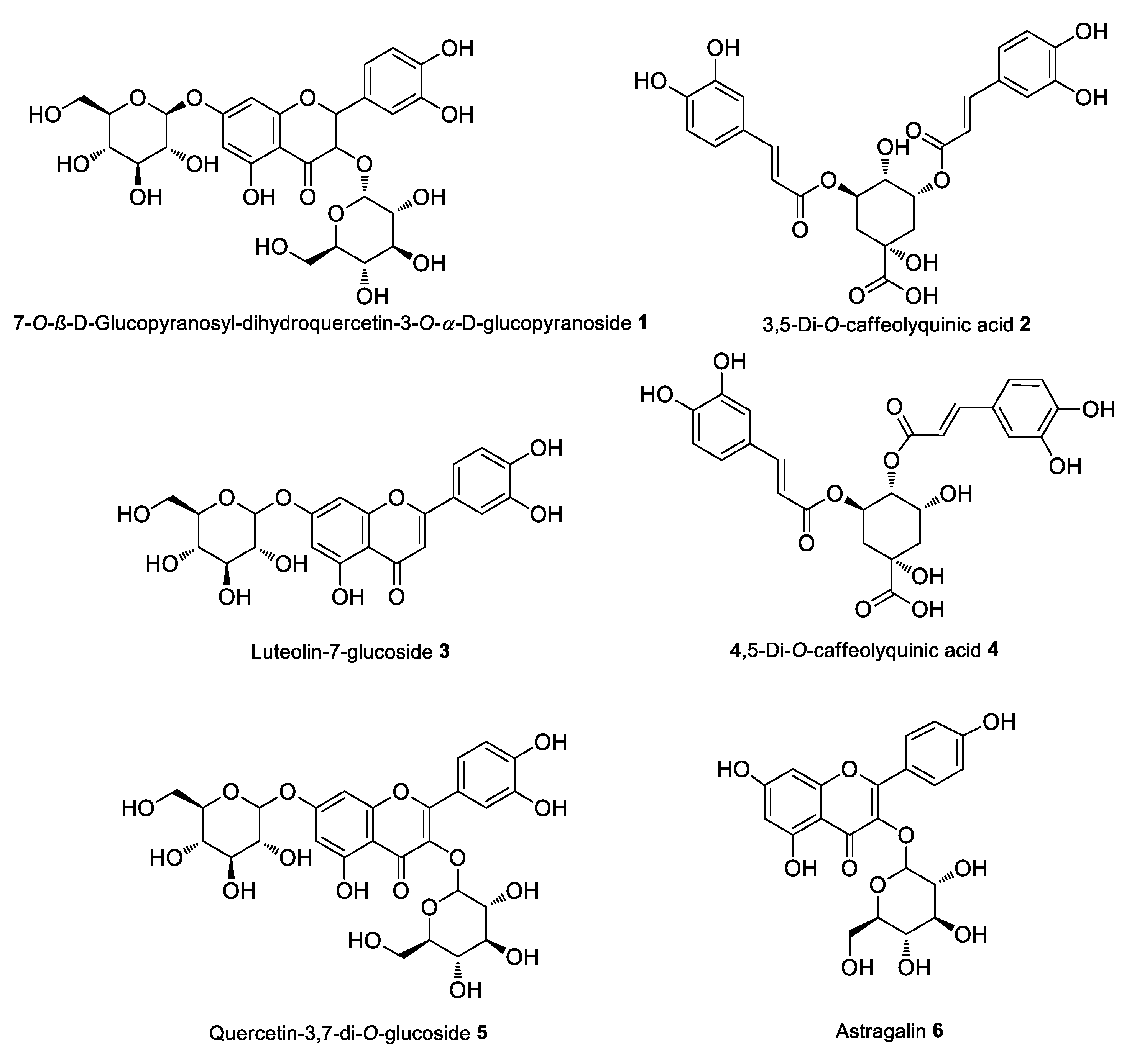
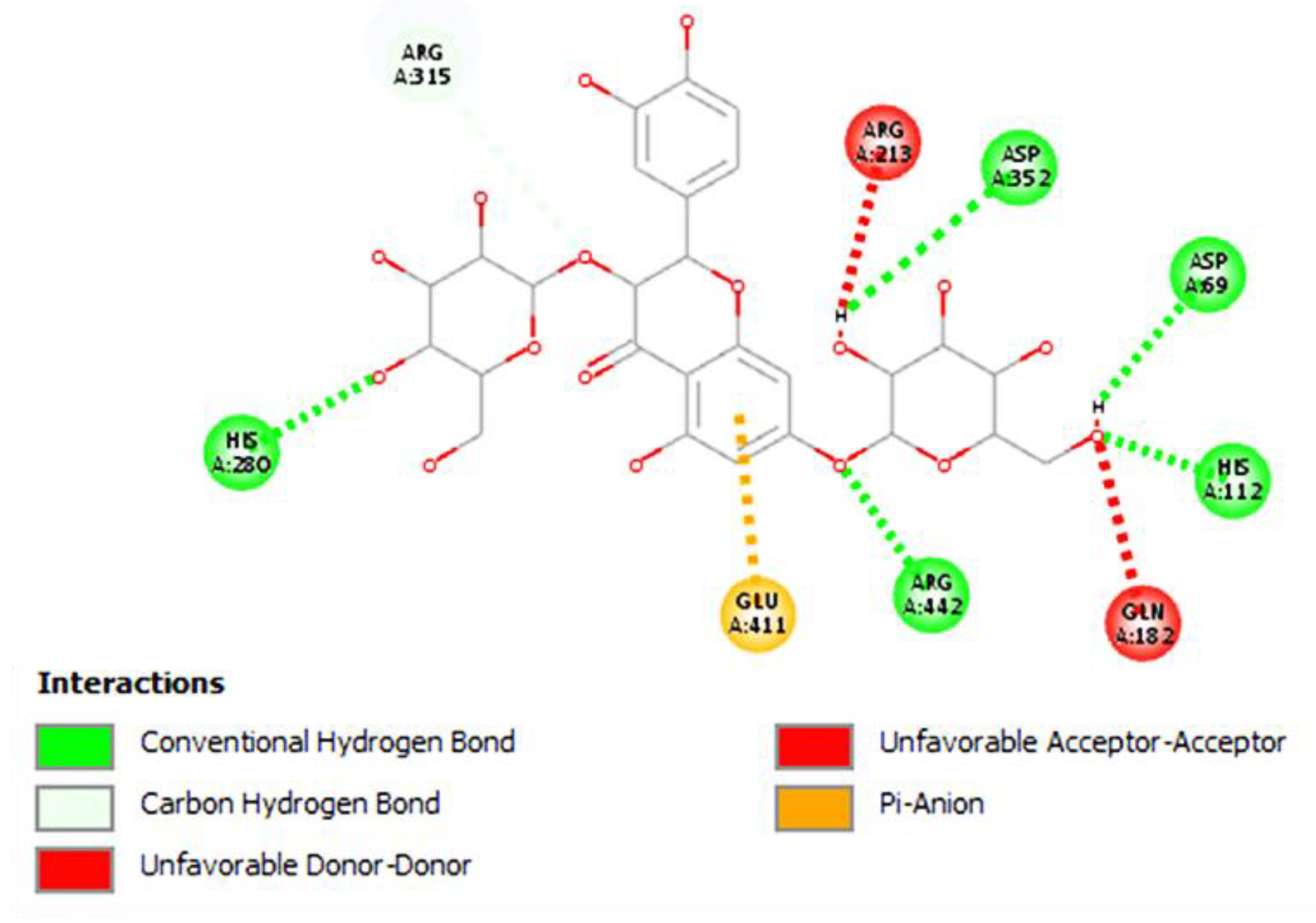
| 7-O-β-D-Glucopyranosyl-dihydroquercetin-3-O-α-D-glucopyranoside | −11.98 |
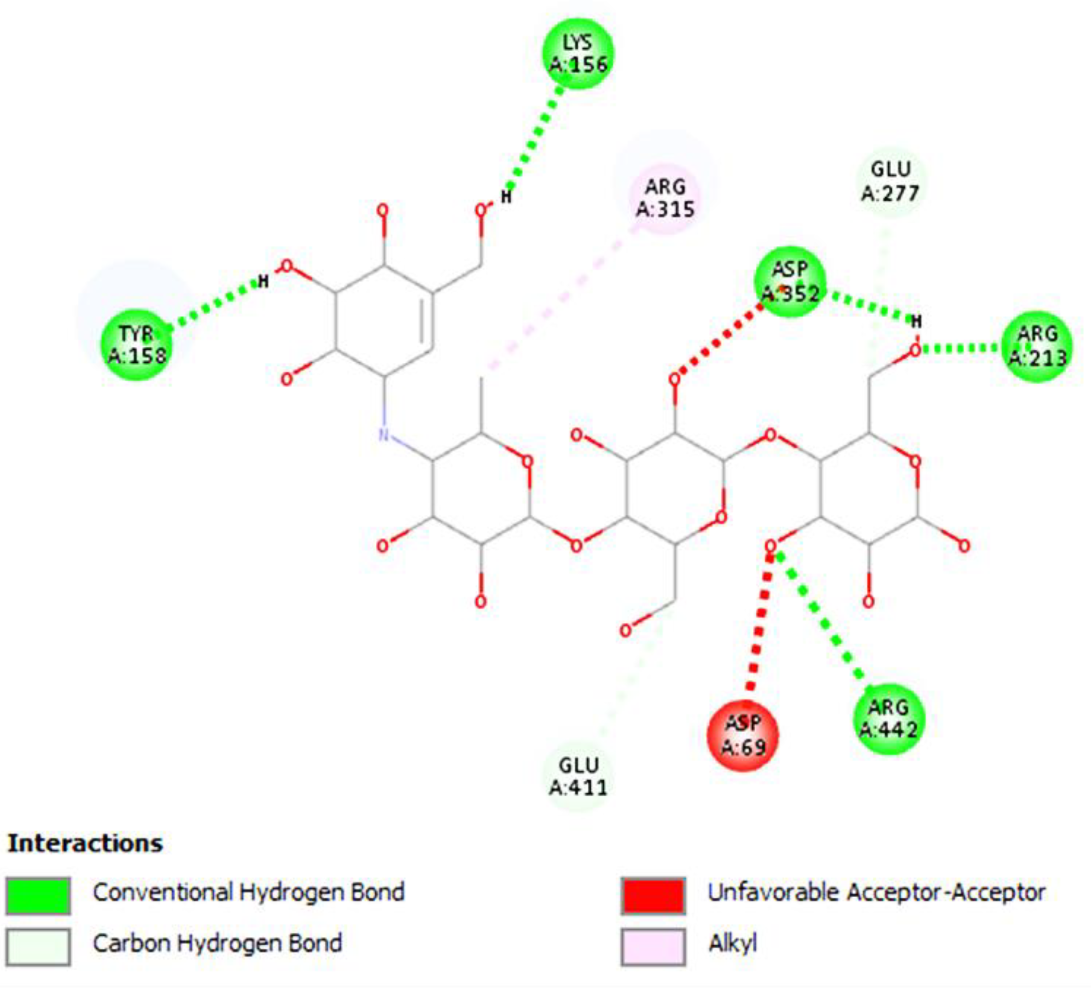
| 3,5-di-O-Caffeolyquinic acid | −10.75 |
| Luteolin-7-glucoside | −10.31 |
| 4,5-Di-O-caffeolyquinic acid | −9.67 |
| Quercetin-3,7-di-O-glucoside | −9.45 |
| Astragalin | −9.06 |
| Quercetin-3-O-β-D-glucoside | −8.71 |
| Rutin | −8.34 |
| Isoquercetin | −8.24 |
| Dihydroxybenzoic acid di-pentoside | −8.23 |
| Nicotiflorin | −7.52 |
| Isorhamnetin-3-O-rutinoside | −7.40 |
| Nomilinic acid glucoside | −6.97 |
| Quercetin-3-O-sophoroside | −6.93 |
| Dihydroxybenzoic acid pentoside | −6.65 |
| Paederia foetida | |
| Stigmasterol | −10.18 |
| (+)-α-Tocopherol | −9.94 |
| γ-Sitosterol | −9.81 |
| (−)-β-Sitosterol | −9.53 |
| Asperuloside | −9.20 |
| Benzbromarone | −8.88 |
| Kaempferol 3-O -rutinoside | −8.68 |
| Quercetin 3-glucoside | −8.63 |
| Caffeic acid 4-O-β-D-glucopyranoside | −8.50 |
| Deacetylasperuloside | −8.48 |
| Scandoside | −8.44 |
| 2-Anthraquinonecarboxylic acid | −8.39 |
| Rutin | −8.35 |
| 2-Hydroxy-3-methylanthraquinone | −8.12 |
| Anthra [1,2-d]-1,3-dioxole-6,11-dione | −8.10 |
| Rubiadin | −8.09 |
| 2-(Hydroxymethyl)anthraquinone | −8.00 |
| Geniposide | −7.99 |
| 3-Hydroxy-2-(hydroxymethyl)anthraquinone | −7.98 |
| 2-Hydroxy-1,3,4-trimethoxy-9,10-anthracenedione | −7.96 |
| 1,4-Dihydroxy-2-(hydroxymethyl)-9,10-anthracenedione | −7.88 |
| N-(4-Methylphenyl) benzenepropanamide | −7.86 |
| 6α-Hydroxygeniposide | −7.80 |
| 1-Hydroxy-2-(hydroxymethyl)-9,10-anthracenedione | −7.79 |
| 2-(Ethoxymethyl)-3-hydroxy-1-methoxy-9,10-anthracenedione | −7.71 |
| 1,3-Dihydroxy-2-methoxyanthraquinone | −7.69 |
| Alizarin | −7.69 |
| Capsaicin | −7.38 |
| Friedelin | −7.01 |
| Glutathione | −6.82 |
| β-Carotene | −6.64 |
| 2,6-Di-tert-butyl-4-methylphenol | −6.58 |
| Fraxidin | −6.48 |
| Ethyl (E)-4-methoxycinnamate | −6.45 |
| (±)-Pentobarbital | −6.39 |
| Plumbago zeylanica L. | |
| Chitanone | −12.19 |
| Zeylanone | −10.38 |
| Isozeylanone | −10.38 |
| Campesterol | −9.70 |
| Plumbagoside D | −9.70 |
| Stigmasterol | −9.50 |
| Sitosterol | −9.42 |
| Heneicosane | −9.41 |
| Plumbagin C | −9.39 |
| 2-(3,4-Dihydroxyphenyl)-3,5,6-trihydroxychromen-4-one | −9.11 |
| Droserone | −9.05 |
| 2-(2,4-Dihydroxy-phenyl)-3,6,8-trihydroxy-chromen-4-one | −8.54 |
| Plumbagoside C | −8.13 |
| Plumbagoside B | −7.95 |
| 3-(2,5-Dimethylphenyl)-1-(2-hydroxyphenyl)-propenone | −7.75 |
| Plumbagin F | −7.68 |
| Plumbagoside A | −7.51 |
| Plumbagin D | −7.26 |
| Plumbagin B | −7.16 |
| Plumbagin G | −7.16 |
| Plumbagin E | −7.11 |
| 2,5-Dimethyl-7-hydroxychromone | −6.98 |
| Plumbagic acid | −6.84 |
| Isoshinanolone | −6.75 |
| Indole-3-carboxaldehyde | −5.83 |
| Plumbagin A | −5.78 |
| Vanilic acid | −5.78 |
| trans-cinnamic acid | −5.76 |
| 4-Hydroxybenzaldehyde | −5.03 |
| Acarbose (standard drug) | −7.80 |
| No | Species | Local Name | Voucher Code |
|---|---|---|---|
| 1 | Carica papaya | Bunga papaya | CP |
| 2 | Clitoria ternatea L. | Bunga telang | CT |
| 3 | Etlingera elatior | Bunga kecombrang | EE |
| 4 | Ipomoea aquatica | Kangkung | IA |
| 5 | Limnocharis Flava | Genjer | LF |
| 6 | Luffa acutangular | Buah gambas | LA |
| 7 | Marsilea minuta L. | Semanggi | MM |
| 8 | Moringa Oleifera | Daun kelor | MO |
| 9 | Nasturtium officinale | Selada air | NO |
| 10 | Nauclea pallida Reinw. | Daun kolpo | NP |
| 11 | Nothopanax scutellarium Merr. | Daun mangkokan | NS |
| 12 | Paederia foetida Linn | Daun sembukan | PF |
| 13 | Pluchea indica | Daun beluntas | PI |
| 14 | Plumbago zeylanica L. | Daun encok | PZ |
| 15 | Psophocarpus tetragonolobus | Kecipir | PT |
| 16 | Sauropus androgynus | Daun katuk | SA |
| 17 | Vigna cylindrica (L.) Skeels | Kacang panjang | VC |
| 18 | Wrightia pubescens R.Br | Daun dan tangkai Bintaos | WP |
| 19 | Diplazium esculentum | Pakis | DE |
Disclaimer/Publisher’s Note: The statements, opinions and data contained in all publications are solely those of the individual author(s) and contributor(s) and not of MDPI and/or the editor(s). MDPI and/or the editor(s) disclaim responsibility for any injury to people or property resulting from any ideas, methods, instructions or products referred to in the content. |
© 2023 by the authors. Licensee MDPI, Basel, Switzerland. This article is an open access article distributed under the terms and conditions of the Creative Commons Attribution (CC BY) license (https://creativecommons.org/licenses/by/4.0/).
Share and Cite
Rani, D.M.; Hanafi, N.; Sudarko; Rachmawati, D.; Siswoyo, T.A.; Christianty, F.M.; Dewi, I.P.; Nugraha, A.S. Indonesian Vegetables: Searching for Antioxidant and Antidiabetic Therapeutic Agents. Drugs Drug Candidates 2023, 2, 14-36. https://doi.org/10.3390/ddc2010002
Rani DM, Hanafi N, Sudarko, Rachmawati D, Siswoyo TA, Christianty FM, Dewi IP, Nugraha AS. Indonesian Vegetables: Searching for Antioxidant and Antidiabetic Therapeutic Agents. Drugs and Drug Candidates. 2023; 2(1):14-36. https://doi.org/10.3390/ddc2010002
Chicago/Turabian StyleRani, Dinar Mutia, Nur Hanafi, Sudarko, Dessy Rachmawati, Tri Agus Siswoyo, Fransiska Maria Christianty, Ika Puspita Dewi, and Ari Satia Nugraha. 2023. "Indonesian Vegetables: Searching for Antioxidant and Antidiabetic Therapeutic Agents" Drugs and Drug Candidates 2, no. 1: 14-36. https://doi.org/10.3390/ddc2010002