Review

Angiogenesis under Opioids Preconditioning in Renal Ischemia Reperfusion

by
Maritza G. Verdugo-Molinares
1,2,
Adriana Franco-Acevedo
3,
Cesar I. Ortiz
4,
José L. Cerino-Recinos
2,
Bibiana Moreno-Carranza
5 and
Zesergio Melo
6,*
1
Unidad de Biotecnología Médica y Farmacéutica, Centro de Investigación y Asistencia en Tecnología y Diseño del Estado de Jalisco, Guadalajara 44270, Mexico
2
Centro de Investigación Biomédica de Occidente, Instituto Mexicano del Seguro Social, Guadalajara 44340, Mexico
3
Department of Pathology and Laboratory Medicine, University of California, Los Angeles, CA 90095, USA
4
Anesthesiology Department, Hospital Country 2000, Guadalajara 44620, Mexico
5
División de Ciencias de la Salud, Universidad Anáhuac Querétaro, Querétaro 76246, Mexico
6
CONACYT-Centro de Investigación Biomédica de Occidente, Instituto Mexicano del Seguro Social, Guadalajara 44340, Mexico
*
Author to whom correspondence should be addressed.
Drugs Drug Candidates 2023, 2(1), 1-13; https://doi.org/10.3390/ddc2010001
Submission received: 18 November 2022 / Revised: 26 December 2022 / Accepted: 28 December 2022 / Published: 7 January 2023
(This article belongs to the Section Clinical Research)

Abstract

:
Renal ischemia reperfusion (IR) presents a common challenge for organ graft and function after transplantation. In the kidney, although there are several mechanisms involved in the IR injury, some studies have pointed to angiogenesis as an important process in the pathophysiology of IR and, therefore, as a possible target mechanism to reduce IR damage. Angiogenesis can be modulated by different molecules and recent evidence has shown that opioids are among these molecules. Angiogenesis preconditioning with opioids is a useful and non-invasive strategy to increase the transplant success rate. Although some results have suggested an interaction between the opioid system and VEGF-mediated angiogenesis, more studies are required to fully elucidate the specific mechanisms involved in these actions. The present review summarizes the recent findings on kidney IR-related mechanisms, with as special emphasis on vascular changes. Finally, the evidence about the modulation of angiogenesis by opioids in a preconditioning scheme will be addressed.

1. Introduction

An intricate network of blood vessels cooperates within the nephron to maintain kidney function. The structure of the renal vascular system is complex, but perfectly distributed throughout the organ. This network is composed of vessels that go through the glomerulus. The well-established vasa recta and a set of peritubular vessels maintain the oxygen, ions, and nutrients supply all over the nephron. Kidneys are extremely sensitive to changes in oxygen availability, which predisposes them to severe organ damage in the case of hypoxia [1].
Oxygenated tissues, such as the kidney, liver, and heart, among others, are susceptible to hypoxia and ischemia-reperfusion [2]. Ischemia-reperfusion (IR) consists of a restriction of the blood flow to the organ followed by the restoration of vascularization and oxygenation [3]. It is known that both episodes have adverse consequences for the cells and, in fact, IR is one of the major causes of delayed function recovery immediately after kidney transplantation, and is associated with acute rejection and chronic graft disfunction in the long term [4]. Early studies demonstrated that brief periods of ischemia before a long ischemic insult reduced the infarct size, introducing the term ischemic preconditioning (IPC) [5]. Since this discovery, the benefits of IPC have been shown in several tissues such as the kidney, brain, retina, intestine, liver, spinal cord, and skeletal muscles [6].
Research on the mechanisms of IPC revealed that the protective effect is mediated by receptors, and it then became important to identify the triggers, mediators, and end effectors involved [7]. With the discoveries that followed, it became apparent that this protection could also be activated by different stimuli, for example, pharmacological agents [8]. After preconditioning, the cell is able to resist the damage caused by IR, including the oxidative stress produced by this phenomenon. The production of new vessels, known as angiogenesis, is a natural response to oxidative stress, and the availability of angiogenic factors affects the tolerance to this kind of cellular stress [9,10].
Opioids are extremely useful and effective drugs for the treatment of pain. They are universally used in patients with severe acute pain and chronic pain. Their mechanism of action is triggered through a specific interaction with their cellular receptors, through which they conduct pain modulation. However, there are other functions of opioids that depend on other interactions [11].
Angiogenesis is a naturally occurring process that involves the formation and growth of new blood vessels. It is essential for embryonic and tissue development, and organ repair and regeneration after damage. In the kidney, angiogenesis is known to sustain organ function. A hypoxic stimulus promotes detrimental vessel formation and organ damage. Additionally, the occurrence of blood vessel rarefaction in the kidney due to hypoxia is still under investigation.
In this review, we summarize the relevant evidence on the role of angiogenesis in the kidney and the possible benefits of pharmacological preconditioning with opioids as a potential angiogenic therapy to protect the organ from hypoxia-induced damage.

2. Kidney Physiology

The most essential functions of the kidneys are to filter the plasma, to remove unwanted substances from the filtrate by excreting them into urine, and to return the necessary substances back into the blood. These functions maintain the stable internal environment necessary for cells to perform their various activities. The kidneys support an adequate bodily blood pressure by regulating the sodium and water balance, controlling the activation of the renin–angiotensin–aldosterone system, and releasing endothelin and prostaglandins, mechanisms that in turn help to maintain the intricate network of kidney blood vessels [12]. By performing these functions, the kidneys establish a remarkably close physiological relationship with the endocrine, circulatory, and respiratory systems. In addition to these excretory functions, kidneys produce and secrete important regulatory molecules such as erythropoietin, renin, the most active metabolic form of vitamin D, and certain prostaglandins, among others [13,14].
The kidneys perform ultrafiltration of plasma and, through processes of reabsorption and secretion, selectively excrete or conserve water and solutes. The kidneys perform these functions thanks to their special macroscopic, histological, cytological, and chemical architecture. The first step in the formation of urine is the filtration process. The basic functional and morphological unit of the kidney is the nephron. It is composed of a system of tubules that interact closely with blood vessels to ensure the proper fulfillment of the functions of the organ [15]. The filtering membrane present in the glomeruli retains the large proteins in the blood. It is a passive process based on the molecular size and electric charge of ions and molecules [12]. As it passes through the tubule, the filtrate is modified through the secretion of additional substances that the tubule lining cells carry from the circulating renal interstitium into the filtrate within the tubular lumen, and the rest of the filtered fluid is reabsorbed through the tubular epithelium and re-enters the blood vascular system [15].
A kidney can filter the blood plasma up to 60 times a day, a physiological process that can be measured. In this regard, the concept of the glomerular filtration rate (GFR) refers to the amount of glomerular filtration that is formed in all renal corpuscles of both kidneys per minute and represents a transcendent index of global renal function. The GFR is considered one of the most important parameters of the physiology of this organ. Physiologically, the GFR ranges from 120 to 125 mL/min/m2, and during the day only 0.75–1% of the GFR will be excreted in the form of urine, while the rest will be reabsorbed [16]. The GFR value is obtained using different mechanisms including both myogenic and glomerular tubule feedback. Those mechanisms depend directly on cellular intrinsic factors of the filtration membrane as well as on the permeability, changes in blood hydrostatic pressure, hydrostatic pressure of the glomerular capsule, and colloidal osmotic pressure of the glomerular capillaries [17].

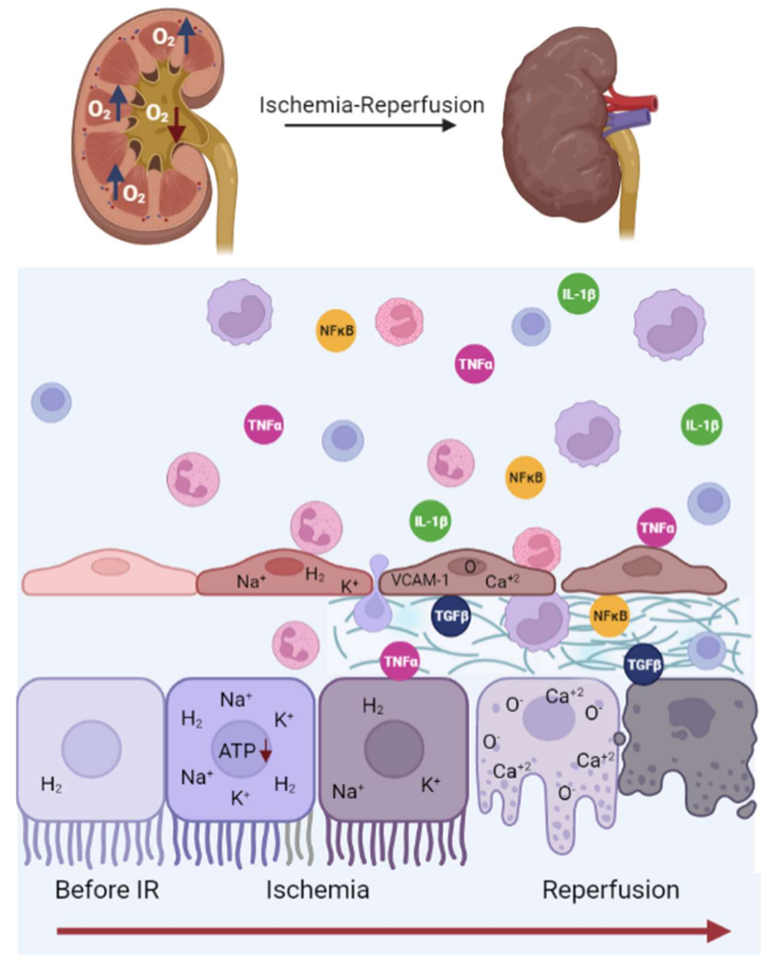
3. Renal Ischemia-Reperfusion Injury

The kidney is susceptible to oxygen variations. With a body mass of only 1% of the total body weight, the kidney is perfused by approximately 25% of the cardiac output [18]. More than half of the renal oxygen consumption is required to manage the active sodium transport, one of the kidneys main activities [18].
The kidney’s structure comprises two zones, a medullary and a cortical one. Cortical cells are highly oxygenated compared to medullary cells, which operate almost in anaerobic states [19]. Medullar cells are highly sensitive to pathological oxygen supply reductions [20].
An IR episode is an oxygen depletion event with the subsequent restoration of oxygen levels [20]. IR is a deleterious incident for the cells. In the kidney, IR is associated with acute kidney injury (AKI), chronic kidney disease (CKD), delayed graft function, and graft loss [21]. The IR episode duration is directly related to the organ damage degree [22]. In a porcine model, kidney function and creatinine levels decreased with the largest IR episodes [23]. In a murine model, Na+–K+–ATPase was released from the cytoskeleton complex after 15 min of IR injury [24]. Thirty minutes after IR, tight junctions were lost in nearly 60% of the cells, leading to a polarity disruption [25]. After more than 30 min, renal tissue showed nuclear extrusion and microfilament disruption associated with actin breakdown [26].
IR mechanisms are defined by a shift from an aerobic to an anaerobic environment resulting in several metabolic changes [22]. Anaerobic metabolism creates an acidosis state through the accumulation of lactic acid [27]. As a consequence of the acidosis state, the Na+/H+ exchanger pump (NHE) activates to modify the sodium and hydrogen ion influx in an effort to restore the intracellular pH and control the ion disturbance [28]. Moreover, anaerobic metabolism leads to a fall in ATP production, affecting the Na+–K+–ATPase and Ca+2–ATPase pumps, and the distribution of sodium, hydrogen, and calcium [27]. These ions accumulate in the cytosol, increasing osmolarity and cell swelling (see Figure 1) [29,30].
In the reperfusion period, ion homeostasis and pH are restored but the organ damage expands [3,27]. The reperfusion mechanism causes enhanced activity of the NHE pump, leading to a sodium and calcium intracellular overload [30]. The increased calcium concentration and exacerbated reactive oxygen species (ROS) formation affects mitochondrial function [3,31]. The mitochondrial transition pore is opened and cell death programs are activated [32]. Mitochondrial disruption occurs in the first minutes of reperfusion and continues for a prolonged period, leading to endothelial damage and inflammatory responses [29].
Afterwards, numerous factors influence the response to damage caused by IR, such as genetic factors, pathological history, and sex, among others [33].

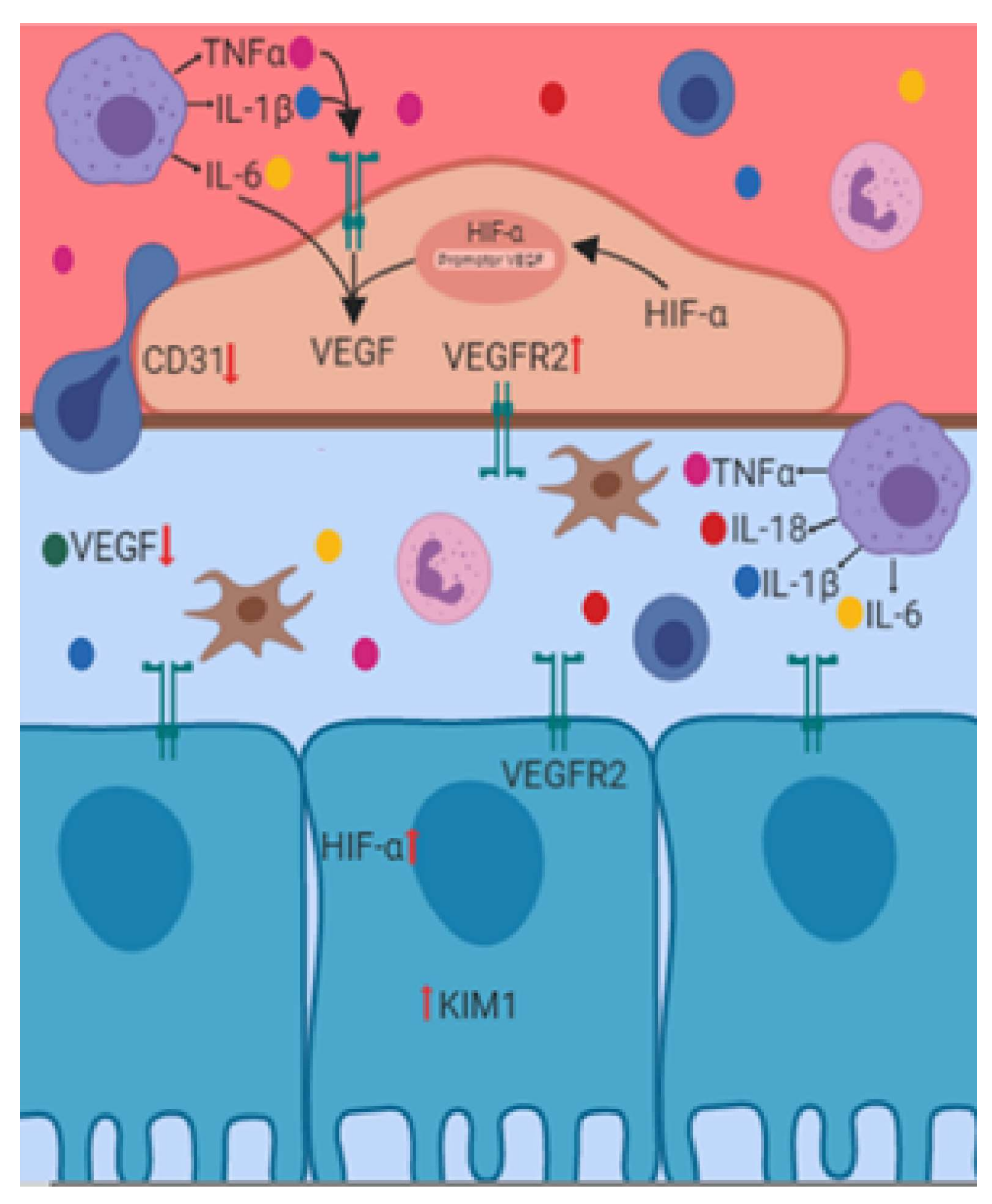
4. Renal Endothelial Damage Associated with IR

The endothelium is very susceptible to an IR event. Vascular damage is evidenced by the alteration of the capillary barrier function, elevation of vascular resistance, and activation of inflammatory pathways [34,35]. Diverse research has documented these alterations in vitro and in vivo. With the use of intravital microscopy, an IR event has been shown to cause a capillary blood flow decrease and a shift of blood flow direction. Yamamoto et al. described ischemia as an effector of vasomotor nephropathy [36]. In vitro, the population of endothelial cells decreased by half after 24 h of oxygen deprivation, and in vivo, IR damage increased kidney vascular permeability, particularly in the cortex and outer medulla [36]. Interestingly, secondary to reperfusion, the renal blood flow decreased, leading to vasoconstriction and endothelial dysfunction two days after the IR event [37]. Moreover, the kidney endothelium showed an enhanced expression of vascular cell adhesion molecule 1 (VCAM-1), indicating an endothelial cell detachment and interaction with inflammatory cells [38]. IR exacerbates endothelium inflammation pathways, increasing the expression of adhesion molecules, cytokines, and polymorphonuclear cell infiltration [39].
The results from Basile et al. revealed that most kidney impairments associated with an IR event returned to normal approximately e4 weeks after the injury [40]. However, in contrast, vascularity showed a reduction of fifty percent. In this study, rarefaction events were maintained up to 40 weeks postinjury [40]. These results agree with an IR-induced AKI model that reported a 45% capillary density loss and the activation of proapoptotic proteases [41]. The pathological features of AKI contribute to develop CKD [15]. CKD progression was related to peritubular capillary (PTC) retraction [42]. Menshikh et al., discussed how PTC, more than fibrosis, could be associated with CKD progression and considered in long-term kidney damage recovery [42]. On the other hand, fibrosis exacerbated kidney injuries, which was stimulated by the retraction of microvessels [43,44]. Therefore, angiogenesis is an alternative to counteract endothelial damage and improve kidney function [45].

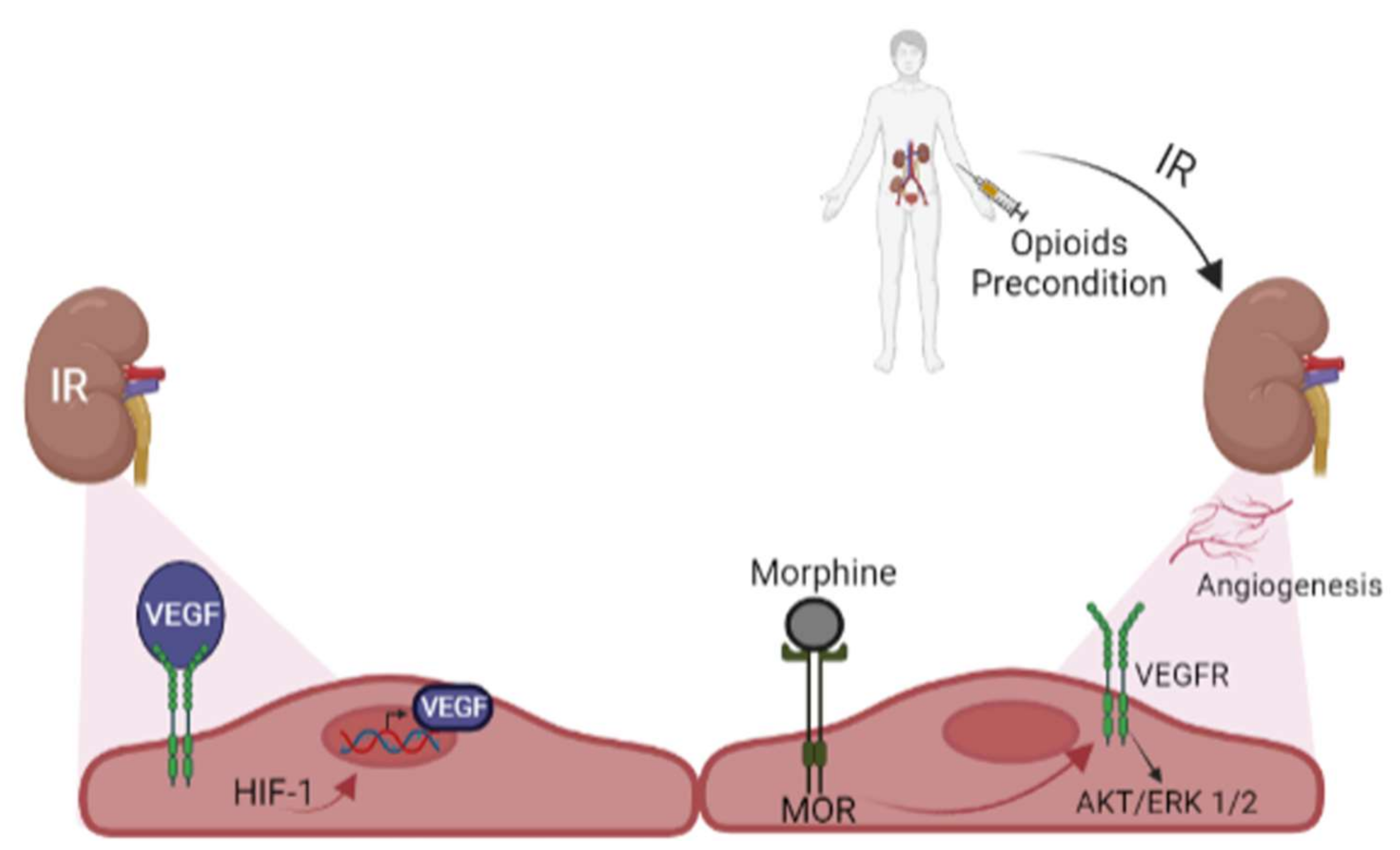
5. Angiogenesis and Renal IR

Angiogenesis is the sprouting of new capillaries from existing vessels that contribute to the expansion of the vascular tree [46]. Angiogenesis begins with vessel wall and extracellular matrix disruption [47]. Endothelial cells change into tip and stalk cells and migrate and elongate to form new capillary networks which later mature and stabilize [48]. The angiogenic process is regulated by various cytokines and growth factors [49]. Vascular endothelial growth factor (VEGF) is the main cytokine in the angiogenic process; however, fibroblast growth factor (FGF), transforming growth factor-beta (TGF-β), and angiopoietins collaborate actively in it [49].
The formation of new capillaries is a compensatory mechanism during IR events to decrease hypoxia damage [50]. The lower oxygen levels enhance the activity and expression of hypoxia inducible factor 1 (HIF-1) in an oxygen-concentration- and time-dependent manner [51]. In vitro studies demonstrated that HIF-1 is the first molecule to be activated, reaching its maximum level two to six hours after the hypoxia event. Although HIF-1 has been associated with a protector role in kidney injury [52], it is essential in blood vessel formation and promotes angiogenesis via the upregulation of VEGF expression [53]. HIF-1 null embryonic stem cells had low capacity to express VEGF (see Figure 2) [54].
VEGF promotes endothelial cell survival, increases permeability, and induces vascular relaxation [55,56]. A VEGF single allele loss in mouse embryonic cells caused death and abnormal blood vessels formation [57,58]. In a remnant kidney (RK) model, the administration of VEGF prevented renal function deterioration, increased endothelial cell proliferation, preserved glomerular capillary endothelium, and decreased blood vessel rarefaction [59]. However, interestingly, several studies [60,61,62,63] support the assumption that after an IR event there is a decrease in VEGF mRNA levels and VEGF receptor (VEGFR) protein expression, suggesting the absence of the VEGF system during the early phase of the IR event. On the other hand, Kanellis et al. described that after IR injury there is no change in VEGF mRNA and protein levels, but there is a VEGF protein relocation to the basolateral proportion of the tubular epithelial cell [61]. The authors discussed this redistribution as a cell protection mechanism to counteract IR damage [61]. Additionally, postischemic kidney rats demonstrated an elevation in VEGF protein 24 h after reperfusion. This result suggests a VEGF post-transcriptional regulation after an IR insult [62].
Receptors VEGFR-1 and VEGFR-2 mediate the angiogenic VEGF actions [64]. VEGFR-1 is a positive regulator of angiogenesis; however, its soluble form (sFlt-1) acts as a VEGF inhibitor. In a study that followed a cohort of 136 patients with a recent kidney transplant, sFlt-1 expression was associated with delayed graft function and vascular rarefaction [65]. These results agreed with Wewers et al., who reported that a higher concentration of sFlt-1 is associated with an independent risk factor for graft rejection and peritubular capillary rarefaction [66]. Furthermore, in an IR murine model, the treatment with recombinant sFlt-1 for 7 days led to peritubular capillary loss, contrary to VEGF-121 administration [66] On the other hand, it is known that VEGFR-2 is expressed in vascular endothelial cells, stimulates angiogenesis [67], and is upregulated after IR in the glomerulus and peritubular capillary endothelium [68].
A renal IR event is a determinant factor to developing kidney damage [69], and a disbalance between angiogenic and antiangiogenic factors may collaborate in the progression of nephropathies due to the IR [34]. Therefore, the endothelium is a key player to developing pharmacological strategies to decrease the vascular damage associated with IR.

6. Opioids

Opioids are analgesic drugs routinely used for the treatment of chronic and perioperative pain [70]. Codeine, morphine, and thebaine are naturally occurring opioids derived from opium. Over the years, their structures have been modified to reduce their side effects [71]. Buprenorphine and oxycodone are examples of semisynthetic opioids derived from thebaine. However, there are also synthetic opioids, such as methadone and fentanyl, whose structure are not similar to any natural opioids, but still have analgesic effects [72]. Figure 3 shows the structure of common opioids, where structural similarities and differences can be noticed.
There also exist more than 20 endogenous opioid peptides derived from proopiomelanocortin, proenkephalin, and prodynorphin. All these peptides are generated after post-translational modifications performed by multiple enzymes [73,74]. Each of the different peptides has a specific expression pattern, resulting in the localization of endogenous opioids in both the nervous system and peripheral organs [62].
All opioids exert their biological actions via G-protein coupled receptors (GPCRs). Opioid receptors are classified based on their ligand and action into µ-opioid receptors (MORs), к-opioid receptors (KORs), and δ-opioid receptor (DORs) [75], which are found in the nervous system and peripheral tissues [12]. Thus, endothelial and other cell types express the µ-opioid receptor (MOR) [76]. In the kidney, expression of the MOR, K-opioid receptor (KOR), and δ-opioid receptor (DOR) was found along the nephron [77].

7. Opioids and Kidney Preconditioning

Even with the advances in the field of transplant therapy to prevent acute graft rejection and promote the prolonged acceptance of transplanted organs, it is challenging to discover new therapies to prevent rejection with as few side effects as possible and reduce or avoid the use of lifelong immunosuppressive drugs [78]. One of the causes that contributes to organ rejection is IR; however, it is inevitable in the transplant process [79].
A therapeutic strategy to decrease renal damage associated with IR is to prepare the organ for the damage. This process is called preconditioning and is defined as the induction of mechanisms to improve organ adaptation to adverse events [12]. In this regard, ischemic preconditioning uses a short hypoxic episode before organ transplant to reduce IR-related damage. For extensive information on this topic, please refer to the review by Palomino et al. [12].
The cardioprotective effects of opioid receptor stimulation have been studied for a few years, and it has been demonstrated that endogenous opioids are produced in response to ischemia and there is a diminution of the protective effect with the administration of opioid antagonists [80,81,82].
The mechanism by which opioids exert a function on the kidney is still poorly understood; however, a study by our group suggested that anesthetic preconditioning with morphine and fentanyl reduces kidney injury markers, such as creatinine and KIM-1, after IR and modulates sirtuin 2 gene expression. In an IR animal model, opioid administration before ischemia increased the sirtuin 2 expression and was correlated with improved renal function [83].
In renal transplantation, the prognosis of the graft depends on various factors. Some of these factors are associated with characteristics of the recipient and others are related to the surgical process itself. The use of trans-surgical anesthetic drugs is one of the factors that is least considered when monitoring the patient. The use of opioids during surgery is controversial for some anesthesiologists and surgeons. The chronic use of this group of drugs is associated with a poor prognosis for the graft and readmission of the patient after transplant surgery. However, fentanyl and its analogs are mainly used for analgesia in renal transplantation. They are also used for postsurgical pain, albeit with some caution as a risk of addictive behavior has been found in opioid painkiller users compared to non-users.
In the following table, we recapitulate some studies where opioids have been used in renal transplantation surgery and their main effects (see Table 1).

8. Opioids Preconditioning and Angiogenesis in Kidney

Opioids are not only involved in pain modulation: they also participate in physiological and pathophysiological activities such as regulation of ionic membrane homeostasis, cell proliferation, emotional response, immune function, cardiovascular control, and neurodegenerative diseases [89].
In vitro evidence has shown that morphine at low concentrations stimulates endothelial cell proliferation [90], activates endothelial cell migration, and promotes VEGFR-2 mediated signaling [91]. These effects are mediated by the activation of the mitogen-activated protein kinases (MAPK) cascade (see Figure 4) [92]. However, at high concentrations, the effect is the opposite, stimulating apoptotic signals and decreasing cell viability [93]. The effects of opioids on angiogenesis have also been extensively studied in models of cancer and tumor growth, and have been reviewed in the literature [94,95].
In the kidney, evidence suggests that opioids increase the urinary flow rate and influence sodium excretion [96,97,98]. However, the actions of opioid receptors on renal angiogenesis remain poorly understood. Increasing evidence shows that opioids impact tumor growth and wound healing [90,99]. The effect on angiogenesis of MOR agonists, such as morphine, fentanyl, oxycodone, and codeine, has been extensively studied, but the findings are contradictory [91]. The role of opioid receptors in angiogenesis has been studied in endothelial cells [91,92]. VEGF increased MOR expression and the ability of morphine to activate VEGF receptors and protein kinase B (AKT) and ERK ½-related (extracellular-signal-regulated kinase) downstream pathways. This result suggested an interaction between the opioid system and VEGF-mediated angiogenesis [90].
Previous results from our group demonstrated that opioid preconditioning modulates the expression of HIF-1, VEGF, VEGF receptor 2 (VEGF-R2), Cathepsin D, CD31, and IL-6 in the kidney. These molecules are considered important effectors of angiogenesis and tissue repair mechanisms that directly influence the development of new blood vessels [100]. This study also corroborated that opioid preconditioning offers protection to the kidney against IR injury, reducing the expression of markers of renal functions. Our study encouraged the use of opioids in interventions that present a period of ischemia for transplantation or other surgical procedures. Thus far, the consequences of IR on endothelial cells, such as microvascular rarefaction, have been poorly studied. Nevertheless, this study demonstrated the stimulation of proangiogenic molecules to avoid renal injury after IR [100].

9. Conclusions

In this review, we analyzed the relevant literature on IR, angiogenesis, and the influence of opioids. Preconditioning with opioids is a proven strategy to improve renal function after an IR injury. Tissue preparation through pharmacological preconditioning with opioids may be essential to extend the graft life in kidney transplant recipients and to prevent cell loss. Although little is known about the mechanism of opioids in the kidney, recent research evidence suggests that, after preconditioning with opioids, new blood vessels can be generated, and this would help neutralize damage after a hypoxic stimulus.

Author Contributions

Conceptualization, C.I.O. and Z.M.; literature review, M.G.V.-M., J.L.C.-R. and Z.M.; writing—original draft preparation, A.F.-A., M.G.V.-M., B.M.-C. and Z.M.; writing—review and editing, A.F.-A., M.G.V.-M., B.M.-C. and Z.M. All authors have read and agreed to the published version of the manuscript.

Funding

This review was funded by the Consejo Nacional de Ciencia y Tecnología (CONACyT) México Grant “Ciencia de Frontera” 345366 FORDECYT-PRONACES awarded to Z.M. M.V.M. is an MSc student awarded with a fellowship from CONACYT.

Conflicts of Interest

The authors declare no conflict of interest.

References

  1. Evans, R.G.; Smith, D.W.; Lee, C.J.; Ngo, J.P.; Gardiner, B.S. What Makes the Kidney Susceptible to Hypoxia? Anat. Rec. 2020, 303, 2544–2552. [Google Scholar] [CrossRef]
  2. Soares, R.O.S.; Losada, D.M.; Jordani, M.C.; Evora, P.; Castro, E.S.O. Ischemia/Reperfusion Injury Revisited: An Overview of the Latest Pharmacological Strategies. Int. J. Mol. Sci. 2019, 20, 5034. [Google Scholar] [CrossRef] [PubMed] [Green Version]
  3. Malek, M.; Nematbakhsh, M. Renal ischemia/reperfusion injury; from pathophysiology to treatment. J. Ren. Inj. Prev. 2015, 4, 20–27. [Google Scholar] [CrossRef] [PubMed]
  4. Nieuwenhuijs-Moeke, G.J.; Pischke, S.E.; Berger, S.P.; Sanders, J.S.F.; Pol, R.A.; Struys, M.; Ploeg, R.J.; Leuvenink, H.G.D. Ischemia and Reperfusion Injury in Kidney Transplantation: Relevant Mechanisms in Injury and Repair. J. Clin. Med. 2020, 9, 253. [Google Scholar] [CrossRef] [Green Version]
  5. Nadarajah, L.; Yaqoob, M.M.; McCafferty, K. Ischemic conditioning in solid organ transplantation: Is it worth giving your right arm for? Curr. Opin. Nephrol. Hypertens 2017, 26, 467–476. [Google Scholar] [CrossRef] [PubMed]
  6. Theodoraki, K.; Tympa, A.; Karmaniolou, I.; Tsaroucha, A.; Arkadopoulos, N.; Smyrniotis, V. Ischemia/reperfusion injury in liver resection: A review of precondittioning methods. Surg. Today 2011, 41, 620–629. [Google Scholar] [CrossRef]
  7. Iliodromitis, E.K.; Lazou, A.; Kremastinos, D.T. Ischemic preconditioning: Protection against myocardial necrosis and apoptosis. Vasc. Health Risk Manag. 2007, 3, 629–637. [Google Scholar]
  8. Das, M.; Das, D.K. Molecular mechanism of preconditioning. IUBMB Life 2008, 60, 199–203. [Google Scholar] [CrossRef] [PubMed]
  9. Maulik, N. Ischemic preconditioning mediated angiogenic response in the heart. Antioxid. Redox Signal. 2004, 6, 413–421. [Google Scholar] [CrossRef] [PubMed]
  10. Ranjbar, K. Improved Cardiac Function Following Ischemia Reperfusion Injury Using Exercise Preconditioning and L-Arginine Supplementation via Oxidative Stress Mitigation and Angiogenesis Amelioration. Cardiovasc. Toxicol. 2022, 22, 736–745. [Google Scholar] [CrossRef] [PubMed]
  11. Rosenblum, A.; Marsch, L.A.; Joseph, H.; Portenoy, R.K. Opioids and the treatment of chronic pain: Controversies, current status, and future directions. Exp. Clin. Psychopharmacol. 2008, 16, 405–416. [Google Scholar] [CrossRef] [PubMed]
  12. Palomino, J.; Echavarria, R.; Franco-Acevedo, A.; Moreno-Carranza, B.; Melo, Z. Opioids Preconditioning Upon Renal Function and Ischemia-Reperfusion Injury: A Narrative Review. Medicina 2019, 55, 522. [Google Scholar] [CrossRef] [PubMed] [Green Version]
  13. Shih, H.M.; Wu, C.J.; Lin, S.L. Physiology and pathophysiology of renal erythropoietin-producing cells. J. Formos. Med. Assoc. 2018, 117, 955–963. [Google Scholar] [CrossRef] [PubMed]
  14. Radi, Z.A. Kidney Pathophysiology, Toxicology, and Drug-Induced Injury in Drug Development. Int. J. Toxicol. 2019, 38, 215–227. [Google Scholar] [CrossRef] [PubMed]
  15. Scholz, H.; Boivin, F.J.; Schmidt-Ott, K.M.; Bachmann, S.; Eckardt, K.U.; Scholl, U.I.; Persson, P.B. Kidney physiology and susceptibility to acute kidney injury: Implications for renoprotection. Nat. Rev. Nephrol. 2021, 17, 335–349. [Google Scholar] [CrossRef]
  16. Mian, A.N.; Schwartz, G.J. Measurement and Estimation of Glomerular Filtration Rate in Children. Adv. Chronic. Kidney Dis. 2017, 24, 348–356. [Google Scholar] [CrossRef]
  17. Brenner, B.M.; Chertow, G.M. Congenital oligonephropathy and the etiology of adult hypertension and progressive renal injury. Am. J. Kidney Dis. 1994, 23, 171–175. [Google Scholar] [CrossRef]
  18. Nourbakhsh, N.; Singh, P. Role of renal oxygenation and mitochondrial function in the pathophysiology of acute kidney injury. Nephron Clin. Pract. 2014, 127, 149–152. [Google Scholar] [CrossRef] [Green Version]
  19. Evans, R.G.; Gardiner, B.S.; Smith, D.W.; O’Connor, P.M. Intrarenal oxygenation: Unique challenges and the biophysical basis of homeostasis. Am. J. Physiol. Renal. Physiol. 2008, 295, F1259–F1270. [Google Scholar] [CrossRef] [Green Version]
  20. Ray, S.C.; Mason, J.; O’Connor, P.M. Ischemic Renal Injury: Can Renal Anatomy and Associated Vascular Congestion Explain Why the Medulla and Not the Cortex Is Where the Trouble Starts? Semin. Nephrol. 2019, 39, 520–529. [Google Scholar] [CrossRef]
  21. Pefanis, A.; Ierino, F.L.; Murphy, J.M.; Cowan, P.J. Regulated necrosis in kidney ischemia-reperfusion injury. Kidney Int. 2019, 96, 291–301. [Google Scholar] [CrossRef]
  22. Chatauret, N.; Badet, L.; Barrou, B.; Hauet, T. Ischemia-reperfusion: From cell biology to acute kidney injury. Progrès Urol. 2014, 24, S4–S12. [Google Scholar] [CrossRef] [PubMed]
  23. Humphreys, M.R.; Castle, E.P.; Lohse, C.M.; Sebo, T.J.; Leslie, K.O.; Andrews, P.E. Renal ischemia time in laparoscopic surgery: An experimental study in a porcine model. Int. J. Urol. 2009, 16, 105–109. [Google Scholar] [CrossRef]
  24. Molitoris, B.A.; Dahl, R.; Geerdes, A. Cytoskeleton disruption and apical redistribution of proximal tubule Na+-K+-ATPase during ischemia. Am. J. Physiol. 1992, 263, F488–F495. [Google Scholar] [CrossRef] [PubMed]
  25. Molitoris, B.A.; Falk, S.A.; Dahl, R.H. Ischemia-induced loss of epithelial polarity. Role of the tight junction. J. Clin. Investig. 1989, 84, 1334–1339. [Google Scholar] [CrossRef] [Green Version]
  26. Kellerman, P.S.; Clark, R.A.; Hoilien, C.A.; Linas, S.L.; Molitoris, B.A. Role of microfilaments in maintenance of proximal tubule structural and functional integrity. Am. J. Physiol. 1990, 259, F279–F285. [Google Scholar] [CrossRef] [PubMed]
  27. Kalogeris, T.; Baines, C.P.; Krenz, M.; Korthuis, R.J. Cell biology of ischemia/reperfusion injury. Int. Rev. Cell Mol. Biol. 2012, 298, 229–317. [Google Scholar] [CrossRef] [Green Version]
  28. Piper, H.M.; Balser, C.; Ladilov, Y.V.; Schafer, M.; Siegmund, B.; Ruiz-Meana, M.; Garcia-Dorado, D. The role of Na+/H+ exchange in ischemia-reperfusion. Basic Res. Cardiol. 1996, 91, 191–202. [Google Scholar] [CrossRef]
  29. Wu, M.Y.; Yiang, G.T.; Liao, W.T.; Tsai, A.P.; Cheng, Y.L.; Cheng, P.W.; Li, C.Y.; Li, C.J. Current Mechanistic Concepts in Ischemia and Reperfusion Injury. Cell Physiol Biochem 2018, 46, 1650–1667. [Google Scholar] [CrossRef]
  30. Roberts, B.N.; Christini, D.J. NHE inhibition does not improve Na+ or Ca2+ overload during reperfusion: Using modeling to illuminate the mechanisms underlying a therapeutic failure. PLoS Comput. Biol. 2011, 7, e1002241. [Google Scholar] [CrossRef] [Green Version]
  31. Malis, C.D.; Bonventre, J.V. Mechanism of calcium potentiation of oxygen free radical injury to renal mitochondria. A model for post-ischemic and toxic mitochondrial damage. J. Biol. Chem. 1986, 261, 14201–14208. [Google Scholar] [CrossRef] [PubMed]
  32. Salvadori, M.; Rosso, G.; Bertoni, E. Update on ischemia-reperfusion injury in kidney transplantation: Pathogenesis and treatment. World J. Transplant. 2015, 5, 52–67. [Google Scholar] [CrossRef] [PubMed]
  33. Melo, Z.; Gutierrez-Mercado, Y.K.; Garcia-Martinez, D.; Portilla-de-Buen, E.; Canales-Aguirre, A.A.; Gonzalez-Gonzalez, R.; Franco-Acevedo, A.; Palomino, J.; Echavarria, R. Sex-dependent mechanisms involved in renal tolerance to ischemia-reperfusion: Role of inflammation and histone H3 citrullination. Transpl. Immunol. 2020, 63, 101331. [Google Scholar] [CrossRef] [PubMed]
  34. Basile, D.P. The endothelial cell in ischemic acute kidney injury: Implications for acute and chronic function. Kidney Int. 2007, 72, 151–156. [Google Scholar] [CrossRef] [Green Version]
  35. Sutton, T.A.; Fisher, C.J.; Molitoris, B.A. Microvascular endothelial injury and dysfunction during ischemic acute renal failure. Kidney Int. 2002, 62, 1539–1549. [Google Scholar] [CrossRef] [Green Version]
  36. Yamamoto, T.; Tada, T.; Brodsky, S.V.; Tanaka, H.; Noiri, E.; Kajiya, F.; Goligorsky, M.S. Intravital videomicroscopy of peritubular capillaries in renal ischemia. Am. J. Physiol. Renal. Physiol. 2002, 282, F1150–F1155. [Google Scholar] [CrossRef] [Green Version]
  37. Jankauskas, S.S.; Andrianova, N.V.; Alieva, I.B.; Prusov, A.N.; Matsievsky, D.D.; Zorova, L.D.; Pevzner, I.B.; Savchenko, E.S.; Pirogov, Y.A.; Silachev, D.N.; et al. Dysfunction of Kidney Endothelium after Ischemia/Reperfusion and Its Prevention by Mitochondria-Targeted Antioxidant. Biochemistry 2016, 81, 1538–1548. [Google Scholar] [CrossRef]
  38. Seron, D.; Cameron, J.S.; Haskard, D.O. Expression of VCAM-1 in the normal and diseased kidney. Nephrol. Dial. Transplant. 1991, 6, 917–922. [Google Scholar] [CrossRef]
  39. Takada, M.; Nadeau, K.C.; Shaw, G.D.; Marquette, K.A.; Tilney, N.L. The cytokine-adhesion molecule cascade in ischemia/reperfusion injury of the rat kidney. Inhibition by a soluble P-selectin ligand. J. Clin. Investig. 1997, 99, 2682–2690. [Google Scholar] [CrossRef] [Green Version]
  40. Basile, D.P.; Donohoe, D.; Roethe, K.; Osborn, J.L. Renal ischemic injury results in permanent damage to peritubular capillaries and influences long-term function. Am. J. Physiol. Renal. Physiol. 2001, 281, F887–F899. [Google Scholar] [CrossRef]
  41. Horbelt, M.; Lee, S.Y.; Mang, H.E.; Knipe, N.L.; Sado, Y.; Kribben, A.; Sutton, T.A. Acute and chronic microvascular alterations in a mouse model of ischemic acute kidney injury. Am. J. Physiol. Renal. Physiol. 2007, 293, F688–F695. [Google Scholar] [CrossRef]
  42. Menshikh, A.; Scarfe, L.; Delgado, R.; Finney, C.; Zhu, Y.; Yang, H.; de Caestecker, M.P. Capillary rarefaction is more closely associated with CKD progression after cisplatin, rhabdomyolysis, and ischemia-reperfusion-induced AKI than renal fibrosis. Am. J. Physiol. Renal. Physiol. 2019, 317, F1383–F1397. [Google Scholar] [CrossRef] [PubMed]
  43. Kaissling, B.; Lehir, M.; Kriz, W. Renal epithelial injury and fibrosis. Biochim. Biophys. Acta 2013, 1832, 931–939. [Google Scholar] [CrossRef] [PubMed] [Green Version]
  44. Ballermann, B.J.; Obeidat, M. Tipping the balance from angiogenesis to fibrosis in CKD. Kidney Int. Suppl. 2014, 4, 45–52. [Google Scholar] [CrossRef] [PubMed] [Green Version]
  45. Chade, A.R. Renovascular disease, microcirculation, and the progression of renal injury: Role of angiogenesis. Am. J. Physiol. Regul. Integr. Comp. Physiol. 2011, 300, R783–R790. [Google Scholar] [CrossRef] [PubMed] [Green Version]
  46. Kolte, D.; McClung, J.A.; Aronow, W.S. Chapter 6—Vasculogenesis and Angiogenesis. In Translational Research in Coronary Artery Disease; Aronow, W.S., McClung, J.A., Eds.; Academic Press: Boston, MA, USA, 2016; pp. 49–65. [Google Scholar] [CrossRef]
  47. Senger, D.R.; Davis, G.E. Angiogenesis. Cold Spring Harb. Perspect. Biol. 2011, 3, a005090. [Google Scholar] [CrossRef] [PubMed] [Green Version]
  48. Blanco, R.; Gerhardt, H. VEGF and Notch in tip and stalk cell selection. Cold Spring Harb. Perspect. Med. 2013, 3, a006569. [Google Scholar] [CrossRef] [Green Version]
  49. Ucuzian, A.A.; Gassman, A.A.; East, A.T.; Greisler, H.P. Molecular mediators of angiogenesis. J. Burn Care Res. 2010, 31, 158–175. [Google Scholar] [CrossRef]
  50. Lerman, L.O.; Chade, A.R. Angiogenesis in the kidney: A new therapeutic target? Curr. Opin. Nephrol. Hypertens. 2009, 18, 160–165. [Google Scholar] [CrossRef] [Green Version]
  51. Freeburg, P.B.; Abrahamson, D.R. Hypoxia-inducible factors and kidney vascular development. J. Am. Soc. Nephrol. 2003, 14, 2723–2730. [Google Scholar] [CrossRef]
  52. Nordquist, L.; Friederich-Persson, M.; Fasching, A.; Liss, P.; Shoji, K.; Nangaku, M.; Hansell, P.; Palm, F. Activation of hypoxia-inducible factors prevents diabetic nephropathy. J. Am. Soc. Nephrol. 2015, 26, 328–338. [Google Scholar] [CrossRef] [PubMed] [Green Version]
  53. Pallet, N.; Thervet, E.; Timsit, M.O. Angiogenic response following renal ischemia reperfusion injury: New players. Prog. Urol. 2014, 24 (Suppl. S1), S20–S25. [Google Scholar] [CrossRef] [PubMed]
  54. Ryan, H.E.; Lo, J.; Johnson, R.S. HIF-1 alpha is required for solid tumor formation and embryonic vascularization. EMBO J. 1998, 17, 3005–3015. [Google Scholar] [CrossRef] [PubMed] [Green Version]
  55. Ku, D.D.; Zaleski, J.K.; Liu, S.; Brock, T.A. Vascular endothelial growth factor induces EDRF-dependent relaxation in coronary arteries. Am. J. Physiol. 1993, 265, H586–H592. [Google Scholar] [CrossRef] [PubMed]
  56. Rattner, A.; Williams, J.; Nathans, J. Roles of HIFs and VEGF in angiogenesis in the retina and brain. J. Clin. Investig. 2019, 129, 3807–3820. [Google Scholar] [CrossRef] [Green Version]
  57. Ferrara, N.; Carver-Moore, K.; Chen, H.; Dowd, M.; Lu, L.; O’Shea, K.S.; Powell-Braxton, L.; Hillan, K.J.; Moore, M.W. Heterozygous embryonic lethality induced by targeted inactivation of the VEGF gene. Nature 1996, 380, 439–442. [Google Scholar] [CrossRef] [Green Version]
  58. Carmeliet, P.; Ferreira, V.; Breier, G.; Pollefeyt, S.; Kieckens, L.; Gertsenstein, M.; Fahrig, M.; Vandenhoeck, A.; Harpal, K.; Eberhardt, C.; et al. Abnormal blood vessel development and lethality in embryos lacking a single VEGF allele. Nature 1996, 380, 435–439. [Google Scholar] [CrossRef]
  59. Kang, D.H.; Hughes, J.; Mazzali, M.; Schreiner, G.F.; Johnson, R.J. Impaired angiogenesis in the remnant kidney model: II. Vascular endothelial growth factor administration reduces renal fibrosis and stabilizes renal function. J. Am. Soc. Nephrol. 2001, 12, 1448–1457. [Google Scholar] [CrossRef]
  60. Basile, D.P.; Fredrich, K.; Chelladurai, B.; Leonard, E.C.; Parrish, A.R. Renal ischemia reperfusion inhibits VEGF expression and induces ADAMTS-1, a novel VEGF inhibitor. Am. J. Physiol. Renal. Physiol. 2008, 294, F928–F936. [Google Scholar] [CrossRef]
  61. Kanellis, J.; Mudge, S.J.; Fraser, S.; Katerelos, M.; Power, D.A. Redistribution of cytoplasmic VEGF to the basolateral aspect of renal tubular cells in ischemia-reperfusion injury. Kidney Int. 2000, 57, 2445–2456. [Google Scholar] [CrossRef]
  62. Vannay, A.; Fekete, A.; Adori, C.; Toth, T.; Losonczy, G.; Laszlo, L.; Vasarhelyi, B.; Tulassay, T.; Szabo, A. Divergence of renal vascular endothelial growth factor mRNA expression and protein level in post-ischaemic rat kidneys. Exp. Physiol. 2004, 89, 435–444. [Google Scholar] [CrossRef]
  63. Kramer, B.K.; Bucher, M.; Sandner, P.; Ittner, K.P.; Riegger, G.A.; Ritthaler, T.; Kurtz, A. Effects of hypoxia on growth factor expression in the rat kidney in vivo. Kidney Int. 1997, 51, 444–447. [Google Scholar] [CrossRef] [PubMed]
  64. Ferrara, N.; Gerber, H.P.; LeCouter, J. The biology of VEGF and its receptors. Nat. Med. 2003, 9, 669–676. [Google Scholar] [CrossRef] [PubMed]
  65. Chapal, M.; Neel, M.; Le Borgne, F.; Meffray, E.; Carceles, O.; Hourmant, M.; Giral, M.; Foucher, Y.; Moreau, A.; Fakhouri, F. Increased soluble Flt-1 correlates with delayed graft function and early loss of peritubular capillaries in the kidney graft. Transplantation 2013, 96, 739–744. [Google Scholar] [CrossRef] [PubMed]
  66. Wewers, T.M.; Mayer, A.B.; Pfleiderer, A.; Beul, K.; Schmidt, R.; Heitplatz, B.; Van Marck, V.; Nolte, I.; Pavenstadt, H.; Reuter, S.; et al. Increased soluble fms-like tyrosine kinase 1 after ischemia reperfusion contributes to adverse clinical outcomes following kidney transplantation. Kidney Int. 2019, 95, 1091–1102. [Google Scholar] [CrossRef] [PubMed]
  67. Shibuya, M. Differential roles of vascular endothelial growth factor receptor-1 and receptor-2 in angiogenesis. J. Biochem. Mol. Biol. 2006, 39, 469–478. [Google Scholar] [CrossRef] [PubMed]
  68. Kanellis, J.; Paizis, K.; Cox, A.J.; Stacker, S.A.; Gilbert, R.E.; Cooper, M.E.; Power, D.A. Renal ischemia-reperfusion increases endothelial VEGFR-2 without increasing VEGF or VEGFR-1 expression. Kidney Int. 2002, 61, 1696–1706. [Google Scholar] [CrossRef] [PubMed] [Green Version]
  69. Tanaka, S.; Tanaka, T.; Nangaku, M. Hypoxia and Dysregulated Angiogenesis in Kidney Disease. Kidney Dis. 2015, 1, 80–89. [Google Scholar] [CrossRef] [PubMed]
  70. Mahbuba, W.; Lambert, D.G. Opioids and neovascularization; pro or anti? Br. J. Anaesth. 2015, 115, 821–824. [Google Scholar] [CrossRef] [Green Version]
  71. Davis, M.P. Opioids in Cancer Pain, 2nd ed.; Oxford University Press: New York, NY, USA, 2009; 487p. [Google Scholar]
  72. Cruz, S.L. (Ed.) Opioids: Pharmacology, Abuse, and Addiction; Springer: Cham, Switzerland, 2022. [Google Scholar] [CrossRef]
  73. Abrimian, A.; Kraft, T.; Pan, Y.X. Endogenous Opioid Peptides and Alternatively Spliced Mu Opioid Receptor Seven Transmembrane Carboxyl-Terminal Variants. Int. J. Mol. Sci. 2021, 22, 3779. [Google Scholar] [CrossRef]
  74. Gomes, I.; Sierra, S.; Lueptow, L.; Gupta, A.; Gouty, S.; Margolis, E.B.; Cox, B.M.; Devi, L.A. Biased signaling by endogenous opioid peptides. Proc. Natl. Acad. Sci. USA 2020, 117, 11820–11828. [Google Scholar] [CrossRef] [PubMed]
  75. Zhang, L.; Zhang, J.T.; Hang, L.; Liu, T. Mu Opioid Receptor Heterodimers Emerge as Novel Therapeutic Targets: Recent Progress and Future Perspective. Front. Pharmacol. 2020, 11, 1078. [Google Scholar] [CrossRef] [PubMed]
  76. Stefano, G.B.; Hartman, A.; Bilfinger, T.V.; Magazine, H.I.; Liu, Y.; Casares, F.; Goligorsky, M.S. Presence of the μ3 opiate receptor in endothelial cells. Coupling to nitric oxide production and vasodilation. J. Biol. Chem. 1995, 270, 30290–30293. [Google Scholar] [CrossRef] [PubMed] [Green Version]
  77. Peng, J.; Sarkar, S.; Chang, S.L. Opioid receptor expression in human brain and peripheral tissues using absolute quantitative real-time RT-PCR. Drug Alcohol Depend. 2012, 124, 223–228. [Google Scholar] [CrossRef] [PubMed] [Green Version]
  78. Zhao, L.; Hu, C.; Han, F.; Cai, F.; Wang, J.; Chen, J. Preconditioning is an effective strategy for improving the efficiency of mesenchymal stem cells in kidney transplantation. Stem Cell Res. Ther. 2020, 11, 197. [Google Scholar] [CrossRef]
  79. Ha, H.; Park, J.; Kim, Y.S.; Endou, H. Oxidative stress and chronic allograft nephropathy. Yonsei Med. J. 2004, 45, 1049–1052. [Google Scholar] [CrossRef]
  80. Schultz, J.E.; Gross, G.J. Opioids and cardioprotection. Pharmacol. Ther. 2001, 89, 123–137. [Google Scholar] [CrossRef]
  81. Murphy, G.S.; Szokol, J.W.; Marymont, J.H.; Avram, M.J.; Vender, J.S. Opioids and cardioprotection: The impact of morphine and fentanyl on recovery of ventricular function after cardiopulmonary bypass. J. Cardiothorac. Vasc. Anesth. 2006, 20, 493–502. [Google Scholar] [CrossRef]
  82. Randhawa, P.K.; Jaggi, A.S. Opioids in Remote Ischemic Preconditioning-Induced Cardioprotection. J. Cardiovasc. Pharmacol. Ther. 2017, 22, 112–121. [Google Scholar] [CrossRef]
  83. Echavarria, R.; Garcia, D.; Figueroa, F.; Franco-Acevedo, A.; Palomino, J.; Portilla-Debuen, E.; Goldaraz-Monraz, M.P.; Moreno-Carranza, B.; Melo, Z. Anesthetic preconditioning increases sirtuin 2 gene expression in a renal ischemia reperfusion injury model. Minerva Urol. Nefrol. 2020, 72, 243–249. [Google Scholar] [CrossRef]
  84. Coupe, N.; O’Brien, M.; Gibson, P.; de Lima, J. Anesthesia for pediatric renal transplantation with and without epidural analgesia—A review of 7 years experience. Paediatr. Anaesth. 2005, 15, 220–228. [Google Scholar] [CrossRef] [PubMed]
  85. Kirvela, M.; Lindgren, L.; Seppala, T.; Olkkola, K.T. The pharmacokinetics of oxycodone in uremic patients undergoing renal transplantation. J. Clin. Anesth. 1996, 8, 13–18. [Google Scholar] [CrossRef] [PubMed]
  86. Freir, N.M.; Murphy, C.; Mugawar, M.; Linnane, A.; Cunningham, A.J. Transversus abdominis plane block for analgesia in renal transplantation: A randomized controlled trial. Anesth. Analg. 2012, 115, 953–957. [Google Scholar] [CrossRef] [PubMed]
  87. Farag, E.; Guirguis, M.N.; Helou, M.; Dalton, J.E.; Ngo, F.; Ghobrial, M.; O’Hara, J.; Seif, J.; Krishnamurthi, V.; Goldfarb, D. Continuous transversus abdominis plane block catheter analgesia for postoperative pain control in renal transplant. J. Anesth. 2015, 29, 4–8. [Google Scholar] [CrossRef] [Green Version]
  88. Soltani Mohammadi, S.; Dabir, A.; Shoeibi, G. Efficacy of transversus abdominis plane block for acute postoperative pain relief in kidney recipients: A double-blinded clinical trial. Pain Med. 2014, 15, 460–464. [Google Scholar] [CrossRef]
  89. Feng, Y.; He, X.; Yang, Y.; Chao, D.; Lazarus, L.H.; Xia, Y. Current research on opioid receptor function. Curr. Drug Targets 2012, 13, 230–246. [Google Scholar] [CrossRef] [Green Version]
  90. Ondrovics, M.; Hoelbl-Kovacic, A.; Fux, D.A. Opioids: Modulators of angiogenesis in wound healing and cancer. Oncotarget 2017, 8, 25783–25796. [Google Scholar] [CrossRef] [Green Version]
  91. Feng, T.; Zeng, S.; Ding, J.; Chen, G.; Wang, B.; Wang, D.; Li, X.; Wang, K. Comparative analysis of the effects of opioids in angiogenesis. BMC Anesthesiol. 2021, 21, 257. [Google Scholar] [CrossRef]
  92. Leo, S.; Nuydens, R.; Meert, T.F. Opioid-induced proliferation of vascular endothelial cells. J. Pain Res. 2009, 2, 59–66. [Google Scholar] [CrossRef]
  93. Tuerxun, H.; Cui, J. The dual effect of morphine on tumor development. Clin. Transl. Oncol. 2019, 21, 695–701. [Google Scholar] [CrossRef] [Green Version]
  94. Novy, D.M.; Nelson, D.V.; Koyyalagunta, D.; Cata, J.P.; Gupta, P.; Gupta, K. Pain, opioid therapy, and survival: A needed discussion. Pain 2020, 161, 496–501. [Google Scholar] [CrossRef] [PubMed]
  95. Grandhi, R.K.; Lee, S.; Abd-Elsayed, A. Does Opioid Use Cause Angiogenesis and Metastasis? Pain Med. 2017, 18, 140–151. [Google Scholar] [CrossRef] [PubMed] [Green Version]
  96. Kapusta, D.R.; Kenigs, V.A. Cardiovascular and renal responses produced by central orphanin FQ/nociceptin occur independent of renal nerves. Am. J. Physiol. 1999, 277, R987–R995. [Google Scholar] [CrossRef] [PubMed]
  97. Kapusta, D.R.; Dzialowski, E.M. Central mu opioids mediate differential control of urine flow rate and urinary sodium excretion in conscious rats. Life Sci. 1995, 56, PL243-8. [Google Scholar] [CrossRef] [PubMed]
  98. Shweta, A.; Malpas, S.C.; Anderson, W.P.; Evans, R.G. Effects of naloxone on the haemodynamic and renal functional responses to plasma volume expansion in conscious rabbits. Pflügers Arch. 1999, 439, 150–157. [Google Scholar] [CrossRef] [PubMed]
  99. Zhou, Q.; Zhang, Z.; Long, S.; Li, W.; Wang, B.; Liang, N. Opioids in cancer: The kappaopioid receptor (Review). Mol. Med. Rep. 2022, 25, 44. [Google Scholar] [CrossRef] [PubMed]
  100. Franco-Acevedo, A.; Echavarria, R.; Moreno-Carranza, B.; Ortiz, C.I.; Garcia, D.; Gonzalez-Gonzalez, R.; Bitzer-Quintero, O.K.; Portilla-De Buen, E.; Melo, Z. Opioid Preconditioning Modulates Repair Responses to Prevent Renal Ischemia-Reperfusion Injury. Pharmaceuticals 2020, 13, 387. [Google Scholar] [CrossRef] [PubMed]
Figure 1. Kidney damage is associated with IR. Detrimental responses take place in the nephron endothelial and tubular zones with the presence of acidosis, polarity disruption, cell swelling, oxidative stress, increased vascular permeability, endothelial dysfunction, cell detachment, inflammation, and apoptosis. VCAM-1, vascular cell adhesion molecule 1; NFκB, nuclear factor κB; TNF-α, tumor necrosis factor alpha; TGFβ, transforming growth factor-beta; IL-18, interleukin 18; TNFα, tumor necrosis factor alpha.
Ddc 02 00001 g001
Figure 2. Proangiogenic molecules in ischemia-reperfusion. Renal expression of molecules involved in tissue repair and angiogenesis and its impact on damage reduction. TNF-α, tumor necrosis factor alpha; IL-1β, interleukin 1 beta; IL-6, interleukin 6; IL-18, interleukin 18; VEGF, vascular endothelial growth factor, VEGF-R2, vascular endothelial growth factor receptor 2; HIF-α, hypoxia inducible factor alpha; KIM1, kidney injury molecule 1.
Ddc 02 00001 g002
Figure 3. Structure of common opioids.
Ddc 02 00001 g003
Figure 4. Opioids exert angiogenic actions on endothelial cells. Influence of opioid preconditioning on renal angiogenesis. After acute ischemia, HIF-1 translocates to the nucleus and induces VEGF transcription. Morphine and other opioids are able to induce similar signals to promote angiogenesis. IR, ischemia-reperfusion; VEGF, vascular endothelial growth factor; VEGFR, vascular endothelial growth factor receptor; HIF-1, hypoxia inducible factor 1; MOR, µ opioid receptor; AKT, protein kinase B; ERK ½, extracellular-signal-regulated kinase ½.
Ddc 02 00001 g004
Table 1. Opioids in kidney transplantation and their effects.
ReferenceOpioidEffectRecipients
Coupe et al. [84]Morphine, bupivacaine,
and fentanyl
Improve intraoperative hemodynamic stabilityFifty-three pediatrics
Kirvela et al. [85]OxycodoneOpioid metabolites accumulationTen uremic transplant recipients
Freir et al. [86]Morphine and LevobupivacaineRisk of nauseaSixty-five adults
Farag et al. [87]Morphine and RopivacaineNausea, hospital staySixty-three adults
Mohammadi et al. [88]Fentanyl and bupivacaine Postrenal transplantation pain and amount of opioids consumptionSixty-seven adults
Disclaimer/Publisher’s Note: The statements, opinions and data contained in all publications are solely those of the individual author(s) and contributor(s) and not of MDPI and/or the editor(s). MDPI and/or the editor(s) disclaim responsibility for any injury to people or property resulting from any ideas, methods, instructions or products referred to in the content.

