Case Report

Alternative Approaches to Osteoarthritis-Related Knee Pain: Transvenous Arteriovenous Malformation Embolization

1
Department of Medicine, University of Miami/Jackson Health System, Miami, FL 33136, USA
2
Department of Research, NYC Surgical Associates, New York City, NY 10022, USA
3
Department of Surgery, NYC Surgical Associates, New York City, NY 10022, USA
4
Kidney Transplant Program, Tampa General Hospital, Tampa, FL 33606, USA
5
Department of Surgery, University of South Florida Morsani College of Medicine, Tampa, FL 33602, USA
*
Authors to whom correspondence should be addressed.
These authors contributed equally to this paper.
J. Vasc. Dis. 2023, 2(2), 212-221; https://doi.org/10.3390/jvd2020015
Submission received: 30 December 2022 / Revised: 26 January 2023 / Accepted: 8 February 2023 / Published: 5 April 2023
(This article belongs to the Section Peripheral Vascular Diseases)

Abstract

:
Background: Osteoarthritis (OA) of the knee is an inflammatory joint disorder of the cartilage, joint capsule, synovium, and surrounding bone. Intraarticular inflammation induces angiogenesis leading to pain and inflammation. Research suggests a relationship between patients with osteoarthritis of the knee and venous insufficiency or contributing arteriovenous malformations (AVMs). Similar to genicular artery embolization (GAE), transvenous or transmalformation cannulation and embolization offers a minimally invasive treatment modality for patients with mild to moderate osteoarthritis. Case presentations: Here, we present the successful treatment of OA of the knee using direct puncture transmalformation cannulation and embolization in five patients. Direct puncture, a technique used to embolize peripheral AVMs, is a safe, less invasive method for the treatment of osteoarthritis with associated AVMs. Conclusions: For patients with OA-related knee pain and associated AVMs, the advancement of endovascular techniques offers alternative approaches to the treatment of mild to moderate OA. Direct puncture and transmalformation cannulation and embolization reduce the risk of intra- and post-operative complications, improve recovery time, and minimize operating time and operating costs. In review of the literature, this is one of the first published reports describing the use of direct puncture transvenous or transmalformation cannulation and embolization to treat OA-related pain.

1. Introduction

Osteoarthritis (OA) is the most common inflammatory joint disorder in the developing world with a lifetime risk of about 45% [1]. It is reported that more than 50% of those diagnosed are younger than 65 [1,2,3]. Risk factors include age, female sex, obesity, sports participation, knee injury, genetic predisposition, repetitive squatting, standing and kneeling, muscle weakness, and joint laxity [4,5,6].
Osteoarthritis is a multifactorial disease caused by trauma, inflammation, local metabolic changes, biochemical changes, and mechanical forces [7,8]. New research suggests pain and inflammation are localized to the joint capsule, synovium, surrounding bone, muscles, and ligaments [7,9]. As the disease progresses, neovascularization exacerbates the pathogenesis of OA, creating a state of chronic inflammation and pain [10,11]. This state causes cartilage breakdown and synovial inflammation as macrophages stimulate proinflammatory cytokines [12,13]. This leads to abnormal growth of sensory nerves and blood vessels, termed angiogenesis, a hallmark of OA [14,15].
One, often overlooked, aspect of osteoarthritis of the knee is research linking OA with venous insufficiency and arteriovenous malformations (AVM) [16,17,18]. Although it is unknown if OA of the knee precedes or develops because of venous insufficiency or AVM, researchers now know there is a relationship. This relationship between increased venous pressure and abnormal arterial and venous tributaries adds to the growing repository that treatment methods targeting neovascularization with embolization are worthwhile [19,20,21].
Geniculate artery embolization (GAE) is one minimally invasive treatment choice traditionally used for refractory knee hemarthrosis following knee arthroplasty [21,22,23,24]. Okuno et al. were the first to report on the clinically successful treatment of mild to moderate knee OA by way of transcatheter arterial embolization, thus reducing blood flow to the synovium [25]. Since then, GAE has demonstrated promising results [19,20]. Traditional methods involve endovascular entry through the radial artery or contralateral femoral artery maneuvered under fluoroscopy. Although effective, this procedure comes with inherent risks, including non-target embolization, arterial damage, contrast-induced nephropathy, and puncture site bleeding. An alternative approach for patients with OA and connecting AVMs is transvenous cannulation and embolization, similar to how brain, intrarenal, and some internal and peripheral AVMs are treated [26,27,28,29,30].
Here, we report five successfully treated cases of OA knee-related pain utilizing direct puncture embolization of contributing AVM. Transvenous and/or transmalformation cannulation and embolization offers a safe, less invasive method for treatment [31,32]. This technique reduces the risk for patients, improves recovery time, and minimizes costs. To the best of our knowledge, this is the first report that describes the use of transvenous or transmalformation cannulation and embolization to treat OA-related knee pain.

2. Materials and Methods

We performed a retrospective review of five cases of OA-related knee pain identified from April 2021 to December 2021 at our outpatient surgical institutions (four females and one male; age range, 36–69). Each patient reported mild to moderate OA of the knee, with symptoms including pain, joint stiffness, and pain after walking, running, and/or climbing flights of stairs affecting the patient’s quality of life. Each patient had failed conservative therapies (including weight loss, exercise, physical therapy, and intra-articular injections). Informed consent was performed including risks, possible complications, benefits, and alternatives, and written consent was obtained for inclusion in this case report. Clinical success was defined by the resolution of symptoms post-procedure as well as on follow-up. Institutional review board approval was obtained for the conduct of this research.
As an image-guided procedure, GAE traditionally involves angiography of the genicular arteries by way of radial artery or femoral artery access [25]. Once the specific artery that supplies blood to the synovium is identified, arterial infarction occurs by way of embolization from the same site. In this report, patients underwent direct cannulation of malformations associated with surrounding vasculature of the symptomatic knee, rather than gaining access by way of the femoral or radial arteries. We herein describe our experience with direct transvenous or transmalformation cannulation to treat OA knee pain.
After informed consent was obtained, each patient elected to either undergo general anesthesia or local anesthesia and was then brought to the operating room and placed in the supine position on the operating room table. They were then prepped and draped using conventional sterile technique. On-table ultrasound mapping was used to view the arteries and delineate each patient’s anatomy. The diameters and blood flow velocity of the geniculate arteries were measured. In conjunction with ultrasound guidance, a 4-French micro-puncture, Cobra C2 catheter, and hydrophilic J curve guidewire were utilized to gain arterial entry. Gelfoam was employed for embolization including SURGIFOAM absorbable Gelatin Powder by Ethicon (J&J), Microsphere Hydropearls by Terumo, or EmboGold or Embosphere by Merit; sizes and quantity varied amongst each patient. Real-time ultrasound mapping was used to document the complete cessation of blood flow. No post-procedure side effects consistent with non-target embolization occurred in any procedure. After the withdrawal of sheaths, repeat duplex ultrasonography was performed of the lesion, which showed no evidence of major deep vein thrombosis (DVT), appropriate embolic agent in the lesion, no embolic material escape, and sufficient cessation of flow. Compression dressings were applied in the post-anesthesia care unit (PACU) following another duplex ultrasound.

3. Case Presentations
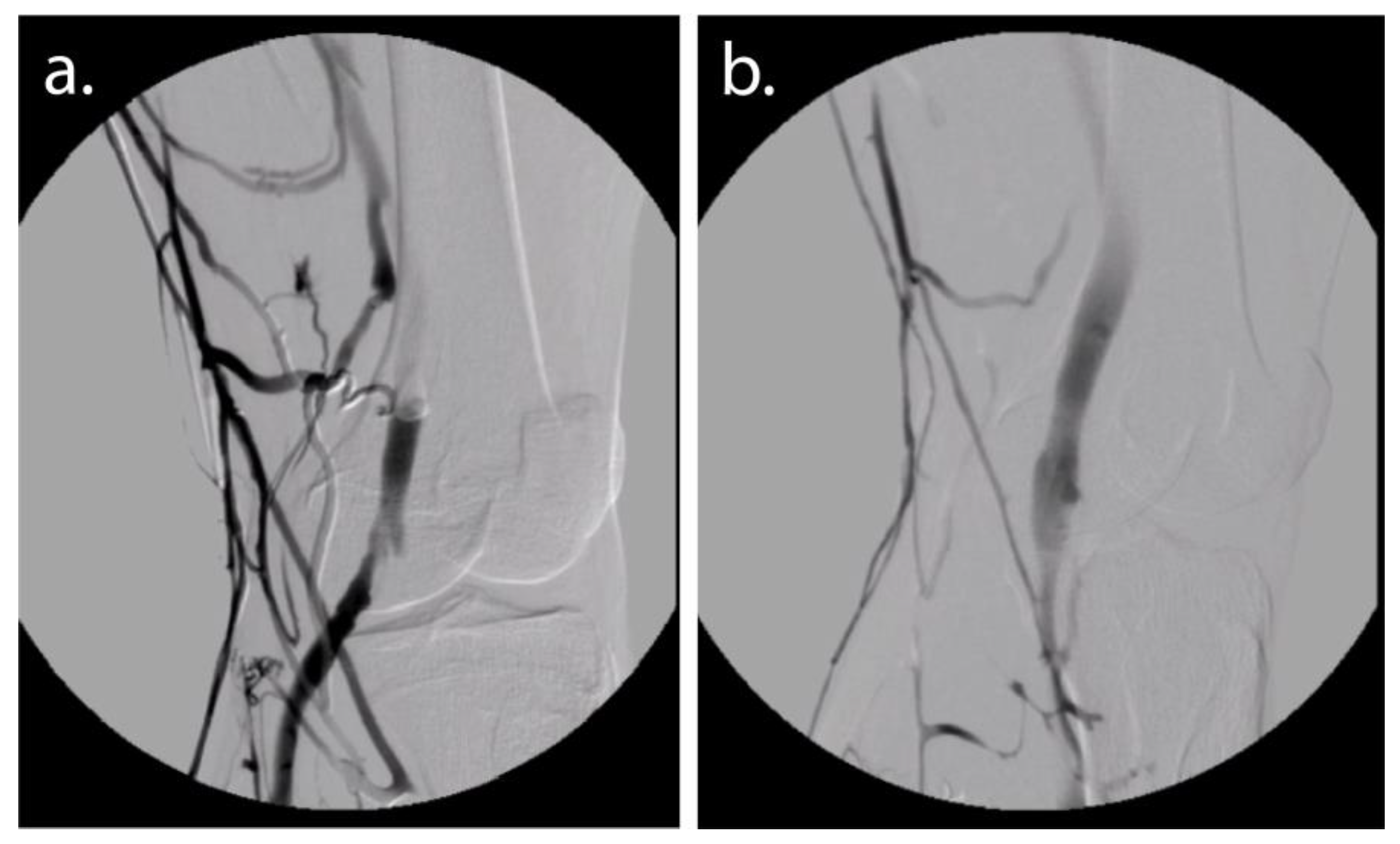
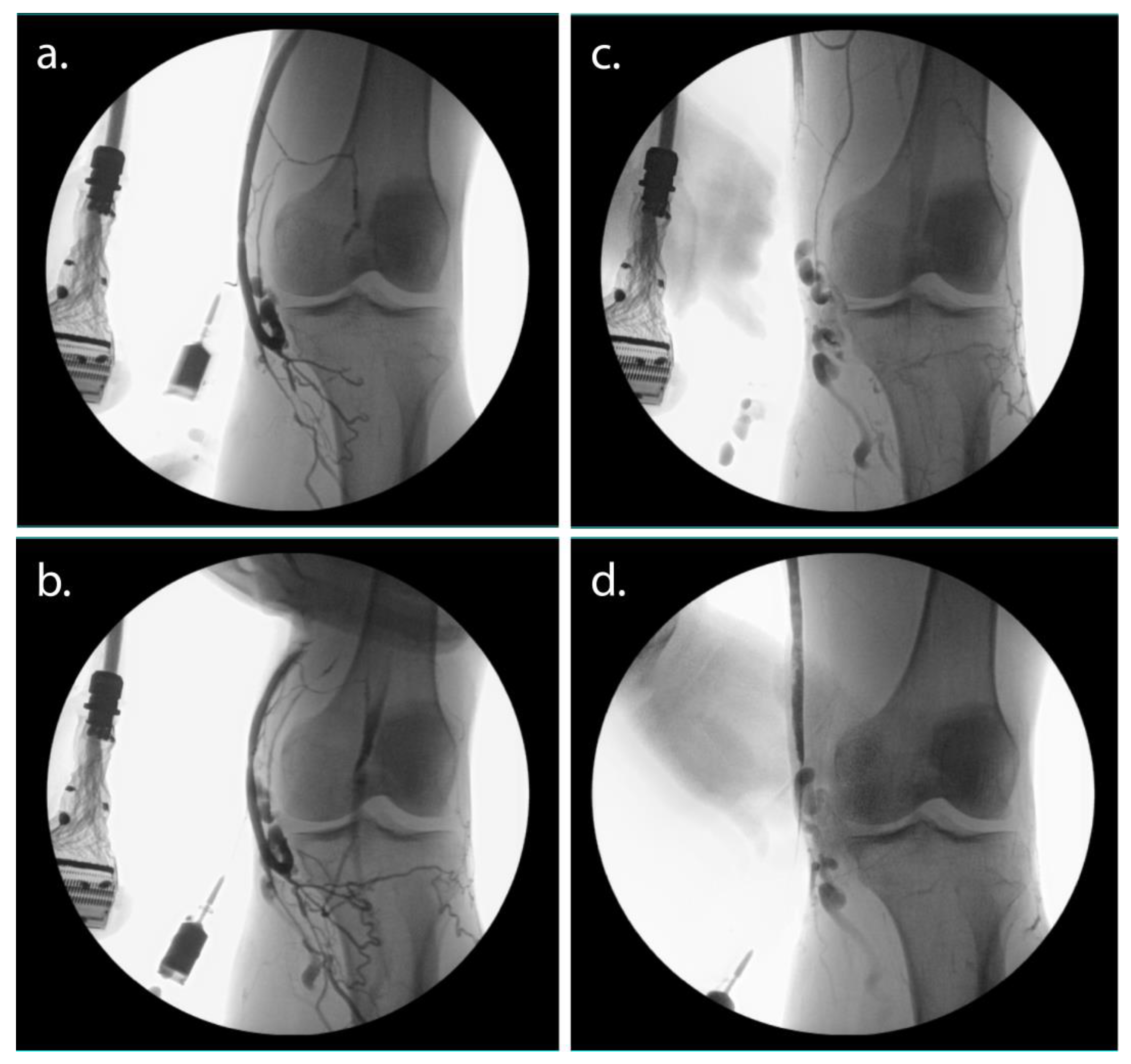
3.1. Case 1

A 57-year-old female with a past medical history of osteoarthritis, back pain, asthma, pedal edema, sciatica, restless leg syndrome, and stress incontinence presented with worsening right knee pain exacerbated by walking upstairs and worse late in the day. The patient has been following conservative management protocols for over 10 years, including non-steroidal anti-inflammatory medications (NSAIDs), attempted weight loss, leg elevation, and lifestyle interventions. The patient reports having to stop a task or activity at least 5 times a day due to experiencing pain rated as 9 out of 10 in severity. X-ray imaging of bilateral knees revealed mild joint space narrowing. Doppler ultrasonography showed low-flow vasculature and venous malformation in the vastus lateralis and tensor fasciae latae on the right lower extremity with perforators to the deep venous system.
Intraoperative ultrasound mapping identified two separate AVM niduses above and below the right knee in conjunction with small caliber geniculate arteries with significant neovascularization consistent with OA (Figure 1). After adequate IV sedation, the AVMs were cannulated directly and embolized with gelfoam. The patient noted significant improvement in pain immediately following the procedure. One week later, the patient elected to treat her less symptomatic left knee. Intraoperative ultrasound mapping showed AVMs from the inferior geniculate artery on the lateral side of the left knee. Under general anesthesia, direct cannulation of both the superior and inferolateral genicular arteries and the AVM was performed followed by embolization with gelfoam. Postembolization ultrasound mapping of the dorsalis pedis and posterior tibial vessels was performed to ensure patent flow.
The patient was seen for a follow-up two weeks after her second procedure. According to her arterial post-intervention patient survey, the patient reported an extreme alleviation of symptoms, such that she noted a 1 out of 10 in pain severity in both knees. The patient was once again seen for a two-month follow-up, where she reported a 5 and 6 out of 10 in knee pain improvement. Overall, the patient noted an improvement in quality of life.

3.2. Case 2

A 36-year-old male with a past medical history of peripheral edema, chronic low back pain, status post stent placement for severe iliofemoral stenosis in the bilateral lower extremities, and recurrent chronic venous insufficiency presented with bilateral knee osteoarthritis. The diagnosis was made by an outside provider. Pain severity was noted to be 6 and 8 out of 10 of his left and right knees respectively. He was noted to have symptomatic AVMs of the left and right lower extremities worsened with exercise and increased circulatory demand. Doppler ultrasonography revealed several non-dominant vessels with moderate flow, likely the cause of a significant portion of his pain.
This case was performed under local anesthesia. First, under direct ultrasound guidance and fluoroscopy, the right geniculate artery was embolized with gelfoam using direct cannulation. The additional symptomatic AVM was subsequently cannulated directly and embolized (Figure 2). The complete cessation of blood flow was confirmed by intraoperative angiography. The patient reported a significant improvement in symptoms perioperatively. At the 2-week follow-up, the patient reported pain severity of 2 out of 10 of the right knee. The patient elected to undergo a similar embolization procedure of the AVMs for OA of the less symptomatic left knee and associated genicular vasculature at a later date.

3.3. Case 3

A 54-year-old female presented with bilateral leg pain, heaviness and swelling of the lower extremities, and restless leg syndrome. Over the past 15 years, the patient has been subject to aggressive, prolonged non-operative management for her discomfort, including NSAIDs, weight loss, and leg elevation. Her symptoms have worsened over the past four years, such that five to ten times per day she notes an 8 out of 10 pain score. Her symptoms are worse after a prolonged period of sitting and standing. The clinical diagnosis of osteoarthritis was made given her activity-related joint pain and morning stiffness that lasted no longer than 30 min. Doppler ultrasonography revealed bilateral greater saphenous vein insufficiency as well as multiple areas of anomalous vascularity in the bilateral lower extremities.
A bubble study was obtained preoperatively. The patient underwent sedation, and the two left genicular arteries were targeted laterally and medially by way of the left greater saphenous vein and direct puncture of the corresponding AVM (Figure 3). Under real-time ultrasound guidance, embolization was subsequently performed using gelfoam.
Complete cessation of flow at the symptomatic AVM was noted with no evidence of postoperative non-target embolization and confirmed with an on-table duplex. Postoperatively, the pedal vessels were found to be significantly higher flowing. Immediately following the procedure, the patient reported a significant improvement in symptoms and described her symptoms as less than 4 out of 10 on the pain scale.

3.4. Case 4

A 69-year-old female with a past medical history of high blood pressure, pelvic congestion syndrome, cholelithiasis, and OA presented with back pain and bilateral knee pain. Outside imaging showed Grade 3 osteoarthritis. Pain severity was noted to be between 7 and 8 out of 10 in the knee, depending on the day. After outpatient evaluation and conservative management failure, lower extremity duplex ultrasonography revealed vascular malformations associated with her native genicular artery and venous vasculature. Duplex ultrasonography identified the lesions of interest and after sedation was achieved, the patient underwent embolization of the superior and inferior genicular arteries, with gelfoam using lateral direct cannulation. A moderately high-flow AVM located distally on the lateral lower leg was also embolized as it was a tributary to the inferior genicular system (Figure 4). Postembolization ultrasound mapping of the dorsalis pedis and posterior tibial vessels was performed to ensure patent flow.
The patient opted for a postoperative telephone interview one day after the procedure. She reported major pain relief and no discomfort, so much so that the day after surgery she did not need to take Tylenol/Motrin. Two months after her procedure she reported continued relief, and pain had improved to a 2 out of 10 in pain severity.

3.5. Case 5

A 56-year-old female presented with bilateral calf cramping and swelling, restless leg syndrome, and leg heaviness over the past 14 months. She also experiences back pain, hip pain, pelvic pain, leg pain, leg numbness, foot/toe numbness, urinary symptoms, and urinary frequency or the feeling of incomplete emptying. Conservative management protocols failed to cease recurrent bilateral leg symptoms, including leg elevation, weight loss, physical therapy, NSAIDs, and lifestyle modifications. Her symptoms have been resistant to vein stripping, hormone therapy, and iliac stent placement. She reports episodic 9 out of 10 knee pain, with episodes occurring five to ten times a day. X-ray imaging confirmed Grade 3 osteoarthritis. A duplex ultrasound ruled out DVT in the lower extremities but noted two venovenous malformations in the bilateral posterior calves.
After additional workup and evaluation, the patient underwent embolization under local anesthesia. Angiography and duplex ultrasonography were used to obtain direct intraarterial access to the right medial and lateral geniculate arteries. Venous tributaries of the inferior lateral genicular vessels were embolized using gelfoam (Figure 5). Angiography as well as on-table duplex ultrasound confirmed the immediate cessation of flow. Following the procedure, the patient reported complete alleviation of knee-related symptoms.

4. Discussion

Direct cannulation and embolization is a safe and effective treatment modality for mild to moderate OA-related knee pain after initial conservative therapies fail. Arteriovenous embolization offers an additional treatment option for the alleviation of pain. Endovascular techniques allow high-risk patients to opt for less-invasive procedures when conservative treatment fails. Direct puncture of an AVM nidus or transvenous cannulation with embolization for OA-related knee pain holds the potential to achieve better results with less risk to the patient [29,32]. For patients with AVM-associated OA, AVM cannulation offers direct treatment of the dominant venous outflow without the risk of non-target embolization into normal arterial circulation [29,30,32]. Direct puncture cannulation via transvenous and transmalformation limits the risk of the traditional arterial endovascular approach and is more reliable and reproducible than transarterial direct cannulation, limited by the tortuosity and small caliber of most arteries of the knee [33].
This case report offers a safe, minimally invasive treatment option for OA-related knee pain. Traditionally, treatment options for OA vary depending on the severity and history [34]. Initial conservative managements include weight loss, exercise, physiotherapy, systemic analgesic treatment, and NSAID treatment. Secondary treatment methods include intra-articular steroids, platelet-rich plasma injections, and hyaluronic injections [6,35]. For recurrent mild or moderate osteoarthritis, arthroscopic debridement has historically been the next best option for patients. Surgical reconstruction or partial or total knee replacement is reserved for severe OA and treatment-resistant osteoarthritis. These newer, conservative, less invasive treatment options, such as endovascular genicular artery embolization and percutaneous embolization, expand the treatment alternatives for patients [19,36].
In this present study, no minor or major complications occurred. Direct puncture transmalformation cannulation and embolization inherently runs the risk of infection and puncture site bleeding. This technique reduces the risk of traditional GAE, including arterial dissection, hemarthrosis, transient cutaneous color changes, tissue ischemia, and non-target embolization [20]. Given the limited data in this case report, however, we are unable to conclude if there was a reduced risk profile compared to traditional geniculate artery embolization and if venous embolization offers the same long-term outcomes as traditional treatment approaches. Limitations of this case report also include limited post-surgical follow-up. Although a novel treatment that tackles the same pathology as traditional geniculate artery embolization, future research will have to ensure that the long-term results mirror those of GAE. Future research will focus on prospective studies utilizing these techniques with at least two years of follow-up. As patients with AVMs and OA often have additional malformations, subsequent procedures may be needed to ensure full resolution and improvement of OA-related symptoms. We hope future studies include blinded treatment cohorts with an appropriate control group.
Although follow-up magnetic resonance imaging was not obtained, we anticipate similar findings to that of traditional GAE. Okuno et al. found that after two years, there was an improvement in the synovitis score, a predictor of the structural progression of OA of the knee [19,20].
In the present study, each patient reported a high patient satisfaction score, including immediate knee pain relief following the procedure and satisfaction with the procedural experience. These treated patients self-reported an improvement in pre-interventional symptoms following their procedures. Future research should focus on using validated improvement scores of OA, such as the Western Ontario MacMaster Questionnaire for patients with OA, rheumatoid arthritis, and fibromyalgia [37].

5. Conclusions

Here, we report five consecutive cases of transmalformation cannulation and embolization for the mitigation of OA-related knee pain using an alternative approach to GAE or direct geniculate artery cannulation and embolization. Direct puncture transvenous and transmalformation cannulation and embolization may offer a safe, less invasive method for the treatment of osteoarthritis of the knee [31]. In our review of the literature, this is the first case report that describes the use of direct genicular artery cannulation and direct AVM cannulation with embolization to treat OA-related knee pain for patients with associated AVM.

Author Contributions

Conceptualization, F.A.C. and D.A.G.; methodology, F.A.C., M.C., A.V., C.M. and C.Z.; validation, N.C., A.V., C.M., C.Z. and G.H.; formal analysis, F.A.C., M.C. and D.A.G.; resources, N.G., N.C. and A.T.; data curation, F.A.C., M.C., N.C. and N.G.; writing—original draft preparation, F.A.C. and M.C.; writing—review and editing, G.H., A.W., A.T. and D.A.G.; visualization, F.A.C. and D.A.G.; supervision, A.W., A.T. and D.A.G.; project administration, N.C., N.G. and A.T. All authors have read and agreed to the published version of the manuscript.

Funding

This research received no external funding.

Institutional Review Board Statement

The ethics committee of NYC Surgical Associates gave ethical approval for this work. The study was conducted in accordance with the Declaration of Helsinki and deemed exempt for full review by the Institutional Review Board of NYC Surgical Associates as it was deemed to be an unsystematic clinical observation for educational purposes. All authors use of protected health information complied with HIPAA and patient identifiers were not collected.

Informed Consent Statement

Consent was obtained from all five patients for treatment and publication of this report including the use of images.

Data Availability Statement

All data generated or analyzed during this study are included in this article. Further inquiries can be directed to the corresponding author.

Conflicts of Interest

F.A.C., M.C., N.C., A.V., C.M., C.Z., G.H., N.G., and A.W. declare no conflicts of interest or competing financial interests. A.T. and D.A.G. own and manage NYC Surgical Associates.

References

  1. Wallace, I.J.; Worthington, S.; Felson, D.T.; Jurmain, R.D.; Wren, K.T.; Maijanen, H.; Woods, R.J.; Lieberman, D.E. Knee osteoarthritis has doubled in prevalence since the mid-20th century. Proc. Natl. Acad. Sci. USA 2017, 114, 9332–9336. [Google Scholar] [CrossRef] [PubMed] [Green Version]
  2. Felson, D.T.; Lawrence, R.C.; Dieppe, P.A.; Hirsch, R.; Helmick, C.G.; Jordan, J.M.; Kington, R.S.; Lane, N.E.; Nevitt, M.C.; Zhang, Y.; et al. Osteoarthritis: New insights. Part 1: The disease and its risk factors. Ann. Intern. Med. 2000, 133, 635–646. [Google Scholar] [CrossRef]
  3. Driban, J.B.; Harkey, M.S.; Liu, S.-H.; Salzler, M.; McAlindon, T.E. Osteoarthritis and Aging: Young Adults with Osteoarthritis. Curr. Epidemiol. Rep. 2020, 7, 9–15. [Google Scholar] [CrossRef]
  4. Plotnikoff, R.; Karunamuni, N.; Lytvyak, E.; Penfold, C.; Schopflocher, D.; Imayama, I.; Johnson, S.T.; Raine, K. Osteoarthritis prevalence and modifiable factors: A population study. BMC Public Health 2015, 15, 1195. [Google Scholar] [CrossRef] [PubMed] [Green Version]
  5. Heidari, B. Knee osteoarthritis prevalence, risk factors, pathogenesis and features: Part I. Caspian J. Intern. Med. 2011, 2, 205–212. [Google Scholar]
  6. Wehling, P.; Evans, C.; Wehling, J.; Maixner, W. Effectiveness of intra-articular therapies in osteoarthritis: A literature review. Ther. Adv. Musculoskelet. Dis. 2017, 9, 183–196. [Google Scholar] [CrossRef]
  7. Ayhan, E.; Kesmezacar, H.; Akgun, I. Intraarticular injections (corticosteroid, hyaluronic acid, platelet rich plasma) for the knee osteoarthritis. World J. Orthop. 2014, 5, 351–361. [Google Scholar] [CrossRef]
  8. Mora, J.C.; Przkora, R.; Cruz-Almeida, Y. Knee osteoarthritis: Pathophysiology and current treatment modalities. J. Pain Res. 2018, 11, 2189–2196. [Google Scholar] [CrossRef] [Green Version]
  9. Sharma, V.; Anuvat, K.; John, L.; Davis, M. Scientific American Pain Management—Arthritis of the Knee; Decker: Toronto, ON, Canada, 2017. [Google Scholar]
  10. Robinson, W.H.; Lepus, C.M.; Wang, Q.; Raghu, H.; Mao, R.; Lindstrom, T.M.; Sokolove, J. Low-grade inflammation as a key mediator of the pathogenesis of osteoarthritis. Nat. Rev. Rheumatol. 2016, 12, 580–592. [Google Scholar] [CrossRef]
  11. Sellam, J.; Berenbaum, F. The role of synovitis in pathophysiology and clinical symptoms of osteoarthritis. Nat. Rev. Rheumatol. 2010, 6, 625–635. [Google Scholar] [CrossRef]
  12. Little, M.W.; Gibson, M.; Briggs, J.; Speirs, A.; Yoong, P.; Ariyanayagam, T.; Davies, N.; Tayton, E.; Tavares, S.; MacGill, S.; et al. Genicular artEry embolizatioN in patiEnts with oSteoarthrItiS of the Knee (GENESIS) Using Permanent Microspheres: Interim Analysis. Cardiovasc. Intervent. Radiol. 2021, 44, 931–940. [Google Scholar] [CrossRef] [PubMed]
  13. Casadaban, L.C.; Mandell, J.C.; Epelboym, Y. Genicular Artery Embolization for Osteoarthritis Related Knee Pain: A Systematic Review and Qualitative Analysis of Clinical Outcomes. Cardiovasc. Intervent. Radiol. 2021, 44, 1–9. [Google Scholar] [CrossRef] [PubMed]
  14. Mapp, P.I.; Walsh, D.A. Mechanisms and targets of angiogenesis and nerve growth in osteoarthritis. Nat. Rev. Rheumatol. 2012, 8, 390–398. [Google Scholar] [CrossRef] [PubMed]
  15. Bijlsma, J.W.J.; Berenbaum, F.; Lafeber, F.P.J.G. Osteoarthritis: An update with relevance for clinical practice. Lancet 2011, 377, 2115–2126. [Google Scholar] [CrossRef] [PubMed]
  16. Güneş, S.; Şehim, K.; Cüneyt, K.; Gökmen, D.; Küçükdeveci, A.A. Is there a relationship between venous insufficiency and knee osteoarthritis? Turk. J. Phys. Med. Rehabil. 2020, 66, 40–46. [Google Scholar] [CrossRef] [PubMed]
  17. Labott, J.R.; Wyles, C.C.; Houdek, M.T.; Tollefson, M.M.; Driscoll, D.J.; Shaughnessy, W.J.; Sierra, R.J. Total Knee Arthroplasty Is Safe and Successful in Patients with Klippel-Trénaunay Syndrome. J. Arthroplast. 2019, 34, 682–685. [Google Scholar] [CrossRef]
  18. Hauert, J.; Loose, D.A.; Westphal, F.M. Joint Involvement in Patients with Vascular Malformations. Destructive Angiodysplastic Arthritis. In Hemangiomas and Vascular Malformations: An Atlas of Diagnosis and Treatment; Mattassi, R., Loose, D.A., Vaghi, M., Eds.; Springer: Milan, Italy, 2009; pp. 299–303. ISBN 978-88-470-0569-3. [Google Scholar]
  19. Okuno, Y.; Korchi, A.M.; Shinjo, T.; Kato, S. Transcatheter arterial embolization as a treatment for medial knee pain in patients with mild to moderate osteoarthritis. Cardiovasc. Intervent. Radiol. 2015, 38, 336–343. [Google Scholar] [CrossRef] [Green Version]
  20. Okuno, Y.; Korchi, A.M.; Shinjo, T.; Kato, S.; Kaneko, T. Midterm Clinical Outcomes and MR Imaging Changes after Transcatheter Arterial Embolization as a Treatment for Mild to Moderate Radiographic Knee Osteoarthritis Resistant to Conservative Treatment. J. Vasc. Interv. Radiol. 2017, 28, 995–1002. [Google Scholar] [CrossRef]
  21. Bagla, S.; Piechowiak, R.; Hartman, T.; Orlando, J.; Del Gaizo, D.; Isaacson, A. Genicular Artery Embolization for the Treatment of Knee Pain Secondary to Osteoarthritis. J. Vasc. Interv. Radiol. 2020, 31, 1096–1102. [Google Scholar] [CrossRef]
  22. Guevara, C.J.; Lee, K.A.; Barrack, R.; Darcy, M.D. Technically Successful Geniculate Artery Embolization Does Not Equate Clinical Success for Treatment of Recurrent Knee Hemarthrosis after Knee Surgery. J. Vasc. Interv. Radiol. 2016, 27, 383–387. [Google Scholar] [CrossRef]
  23. Yoo, J.-H.; Oh, H.-C.; Park, S.-H.; Lee, S.; Lee, Y.; Kim, S.-H. Treatment of Recurrent Hemarthrosis after Total Knee Arthroplasty. Knee Surg. Relat. Res. 2018, 30, 147–152. [Google Scholar] [CrossRef] [Green Version]
  24. Given, M.F.; Smith, P.; Lyon, S.M.; Robertson, D.; Thomson, K.R. Embolization of spontaneous hemarthrosis post total knee replacement. Cardiovasc. Intervent. Radiol. 2008, 31, 986–988. [Google Scholar] [CrossRef]
  25. Okuno, Y.; Oguro, S.; Iwamoto, W.; Miyamoto, T.; Ikegami, H.; Matsumura, N. Short-term results of transcatheter arterial embolization for abnormal neovessels in patients with adhesive capsulitis: A pilot study. J. Shoulder Elbow Surg. 2014, 23, e199–e206. [Google Scholar] [CrossRef] [PubMed]
  26. Chen, C.-J.; Norat, P.; Ding, D.; Mendes, G.A.C.; Tvrdik, P.; Park, M.S.; Kalani, M.Y. Transvenous embolization of brain arteriovenous malformations: A review of techniques, indications, and outcomes. Neurosurg. Focus 2018, 45, E13. [Google Scholar] [CrossRef] [PubMed] [Green Version]
  27. Lee, S.Y.; Do, Y.S.; Kim, C.W.; Park, K.B.; Kim, Y.H.; Cho, Y.J. Efficacy and Safety of Transvenous Embolization of Type II Renal Arteriovenous Malformations with Coils. J. Vasc. Interv. Radiol. 2019, 30, 807–812. [Google Scholar] [CrossRef]
  28. Nassiri, N.; Thomas, J.; Rahimi, S. Fibrodysplastic implications for transvenous embolization of a high-flow pelvic arteriovenous malformation in Osler-Weber-Rendu syndrome. J. Vasc. Surg. Cases 2015, 1, 16–19. [Google Scholar] [CrossRef] [Green Version]
  29. Pimpalwar, S. Vascular malformations: Approach by an interventional radiologist. Semin. Plast. Surg. 2014, 28, 91–103. [Google Scholar] [CrossRef] [PubMed] [Green Version]
  30. Müller-Wille, R.; Wildgruber, M.; Sadick, M.; Wohlgemuth, W.A. Vascular Anomalies (Part II): Interventional Therapy of Peripheral Vascular Malformations. ROFO Fortschr. Geb. Rontgenstr. Nuklearmed. 2018, 190, 927–937. [Google Scholar] [CrossRef] [Green Version]
  31. Kim, R.; Do, Y.S.; Park, K.B. How to Treat Peripheral Arteriovenous Malformations. Korean J. Radiol. 2021, 22, 568–576. [Google Scholar] [CrossRef]
  32. Khurana, A.; Hangge, P.T.; Albadawi, H.; Knuttinen, M.-G.; Alzubaidi, S.J.; Naidu, S.G.; Kriegshauser, J.S.; Oklu, R.; Chong, B.W. The Use of Transarterial Approaches in Peripheral Arteriovenous Malformations (AVMs). J. Clin. Med. 2018, 7, 109. [Google Scholar] [CrossRef] [Green Version]
  33. Hyun, D.; Do, Y.S.; Park, K.B.; Kim, D.-I.; Kim, Y.W.; Park, H.S.; Shin, S.W.; Song, Y.G. Ethanol embolotherapy of foot arteriovenous malformations. J. Vasc. Surg. 2013, 58, 1619–1626. [Google Scholar] [CrossRef] [Green Version]
  34. Goldman, D.T.; Piechowiak, R.; Nissman, D.; Bagla, S.; Isaacson, A. Current Concepts and Future Directions of Minimally Invasive Treatment for Knee Pain. Curr. Rheumatol. Rep. 2018, 20, 54. [Google Scholar] [CrossRef] [PubMed]
  35. Torkian, P.; Golzarian, J.; Chalian, M.; Clayton, A.; Rahimi-Dehgolan, S.; Tabibian, E.; Talaie, R. Osteoarthritis-Related Knee Pain Treated with Genicular Artery Embolization: A Systematic Review and Meta-analysis. Orthop. J. Sport. Med. 2021, 9, 23259671211021356. [Google Scholar] [CrossRef] [PubMed]
  36. Moseley, J.B.; O’Malley, K.; Petersen, N.J.; Menke, T.J.; Brody, B.A.; Kuykendall, D.H.; Hollingsworth, J.C.; Ashton, C.M.; Wray, N.P. A controlled trial of arthroscopic surgery for osteoarthritis of the knee. N. Engl. J. Med. 2002, 347, 81–88. [Google Scholar] [CrossRef] [PubMed]
  37. Wolfe, F.; Kong, S.X. Rasch analysis of the Western Ontario MacMaster questionnaire (WOMAC) in 2205 patients with osteoarthritis, rheumatoid arthritis, and fibromyalgia. Ann. Rheum. Dis. 1999, 58, 563–568. [Google Scholar] [CrossRef]
Figure 1. Case 1. (a) Direct cannulation and fluoroscopic imaging of the superficial and deep venous system with associated malformations with the superior lateral genicular vein and inferior lateral genicular vein before embolization. (b) Fluoroscopy after embolization with improved flow through the deep venous system and cessation of flow via the genicular venous tributaries.
Jvd 02 00015 g001
Figure 2. Case 2. (a) Direct fluoroscopic imaging of the symptomatic arteriovenous malformation of the lower leg just distal to the inferior genicular veins cannulated proximally. (b) Fluoroscopic imaging after embolization with cessation of flow through the proximal malformation and communicating inferior genicular tributaries.
Jvd 02 00015 g002
Figure 3. Case 3. (a) Direct cannulation of the great saphenous vein just proximal to the venous malformation along the medial knee with visualization of the inferior medial genicular vein and associated malformations and the superior medial genicular vein. (b) Fluoroscopic imaging of the venous system with tourniquet pressure applied to the proximal great saphenous venous for improved imaging through the venous system and backflow into the arterial system. (c) Fluoroscopic imaging during embolization, during the procedure, the lateral genicular system can be visualized. (d) Fluoroscopic imaging after embolization.
Jvd 02 00015 g003
Figure 4. Case 4. (a) Direct cannulation and fluoroscopic imaging of the veins of the lower leg with tributaries to the inferior medial genicular vein and associated venous malformations. (b) Fluoroscopic imaging after embolization.
Jvd 02 00015 g004
Figure 5. Case 5. (a) Fluoroscopic imaging of the venous system with cannulation through the great saphenous vein. (b) Post-embolization with cessation of flow to the inferior lateral genicular vein and improved flow on the native medial and deep venous system.
Jvd 02 00015 g005
Disclaimer/Publisher’s Note: The statements, opinions and data contained in all publications are solely those of the individual author(s) and contributor(s) and not of MDPI and/or the editor(s). MDPI and/or the editor(s) disclaim responsibility for any injury to people or property resulting from any ideas, methods, instructions or products referred to in the content.

