Systematic Review

Neural Network Modulation of Ayahuasca: A Systematic Review of Human Studies

by
Guilherme Henrique de Morais Santos
1,
Lucas Silva Rodrigues
1,
Juliana Mendes Rocha
1,
Giordano Novak Rossi
1,
Genís Ona
2,3,
José Carlos Bouso
1,2,3,
Jaime Eduardo Cecilio Hallak
1,2,4 and
Rafael Guimarães dos Santos
1,2,4,*
1
Department of Neurosciences and Behavior, Ribeirão Preto Medical School, University of São Paulo, Ribeirão Preto 14040-900, Brazil
2
ICEERS—International Center for Ethnobotanical Education, Research, and Service, 08015 Barcelona, Spain
3
Medical Anthropology Research Center (MARC), Universitat Rovira i Virgili, 43005 Tarragona, Spain
4
National Institute for Translational Medicine (INCT-TM), CNPq, Ribeirão Preto, Brazil
*
Author to whom correspondence should be addressed.
Psychoactives 2023, 2(1), 76-91; https://doi.org/10.3390/psychoactives2010006
Submission received: 6 February 2023 / Revised: 14 March 2023 / Accepted: 17 March 2023 / Published: 20 March 2023
(This article belongs to the Special Issue Natural Hallucinogens in Mental Health)

Abstract

:
Background: Ayahuasca is a serotoninergic hallucinogen that plays a central role in the Amazonian traditional medicine. Its psychoactive effects are associated with the presence of N,N-dimethyltryptamine (DMT), and monoamine oxidase inhibitors (MAO-A). Advances in neuroimaging investigations have provided insight into ayahuasca’s neurobiological mechanisms of action. Methods: Selecting only studies with neuroimaging results related to human ayahuasca consumption, we included six articles from a previous systematic review of serotoninergic hallucinogen neuroimaging studies up to 2016. Furthermore, we updated the data with a new systematic search from 2016 to 2022. We searched the PubMed, SciELO, and LILACS databases using the search terms “(ayahuasca OR DMT) AND (MRI OR fMRI OR PET OR SPECT OR imaging OR neuroimaging)”. Results: Our updated search provided five new articles for a total of 11 included in this review. The results on the Default Mode Network (DMN) are evident and may indicate a path to short term neuromodulation. Acutely, local neural networks appeared to become expanded, while overall brain connectivity declined. On chronic consumers, anatomical changes were reported, most notably related to cingulate cortex. Conclusion: Ayahuasca seems to change acute brain connectivity similarly to other psychedelics. The results are preliminary and further studies are warranted.

1. Introduction

Ayahuasca is a decoction traditionally used by Indigenous people from the Amazonian rainforest and in syncretic religious rituals in South America and around the world [1]. In Brazil, since the regulation of its use for religious and scientific purposes in 2010, it is officially prepared by the decoction of two plants, Psychotria viridis and Banisteriopsis caapi. Pharmacologically, ayahuasca is classified as a serotoninergic hallucinogen due to the presence of N,N-dimethyltryptamine (DMT, contained in P. viridis). It also contains monoamine oxidase type A inhibitors (MAO-A, contained in B. caapi) in its composition, namely harmine, tetrahydroharmine, and harmline [2]. Other substances considered serotonergic hallucinogens (or psychedelics) are lysergic acid diethylamide (LSD), mescaline, and psilocybin since they all share the agonism for serotonin 5-HT2A receptor as their main mechanism of action. On one hand, serotonergic hallucinogens have a long tradition of use in their original cultures and in the recreational context for their mind-altering effects. On the other, preliminary scientific evidence suggests that they possess anxiolytic, antidepressant, and anti-addictive properties, which has generated a growing interest regarding their possible therapeutic potential for treating mental disorders [1,3].
Concerning the modulation of neural networks, ayahuasca’s (and other similar drugs) mechanisms of action on affecting mood and self-perception remain to be fully elucidated. However, the literature points to a possible relationship between these effects with modulation of the Default Mode Network (DMN) [4], although it is still unknown how central the DMN is for the mechanisms of action of hallucinogens [5]. The DMN is a network composed of the medial prefrontal cortex, cingulate/precuneus cortex, and angular gyrus. It is involved with self-perception and self-awareness and is active when a person is not focused on the outside world, such as during daydreams, remembering the past, self-judgment, and divagation [6]. Its hyperactivation is associated with various psychiatric disorders, such as depression, anxiety, post-traumatic stress disorder, and attention deficit/hyperactivity disorder [5,6]. Furthermore, frontocortical activation of glutamate receptors, a downstream effect secondary to 5-HT2A agonism, seems to be a common pathway through which serotonergic and other hallucinogens act [7,8], but more investigations are necessary.
In one of our previous articles [1], we conducted a systematic review of human trials applying neuroimaging techniques to analyze the effects of serotonergic hallucinogens. In the present study, we focused on neural network modulation by ayahuasca exclusively. Thus, this systematic review aimed to assess changes in brain anatomy and neural networks activation with acute, subacute, or chronic use of ayahuasca through neuroimaging techniques.

2. Materials and Methods

The data of the present systematic review was collected according to the Systematic Reviews and Meta-Analyses guidelines (PRISMA) [9]. This study was not registered in PROSPERO. After extracting from our previous review all citations that reported results evaluating the effects of ayahuasca with any neuroimaging technique [1], we conducted a new search in the PubMed, SciElO, and LILACS databases from 2016 (the last year included in our previous review) to 1 December 2022, using the search terms “(ayahuasca OR DMT) AND (MRI OR fMRI OR PET OR SPECT OR imaging OR neuroimaging)”. Inclusion criteria consisted of any observational, case-series, and clinical trial studies with humans that analyzed acute, subacute, or prolonged effects of ayahuasca using any type of neuroimaging technique. Only studies written in English, Spanish, or Portuguese were included. Single case reports, letters to the editor, reviews, and pre-clinical studies were excluded.

2.1. Data Extraction

Two independent reviewers screened and selected studies for inclusion, with discrepancies resolved by a third reviewer. From the articles included, we recorded the names of authors, year of publication, study location (city and country), study design (open-label and controlled trials, observational studies, case-series), sample characteristics (size, age, and gender), drug used, dosage, neuroimaging techniques, and main outcomes.

Quality Evaluation of Selected Studies

To evaluate the quality of the studies selected from our search in a standardized manner, we have utilized the National Heart, Lung, and Blood Institute (NIH) checklists [10]. Per each study design, we used three checklists: “Quality assessment for observational cohort”, “Quality assessment of controlled interventions studies”, and “Quality Assessment Tool for Before-After (Pre-Post) Studies With No Control Group”. All articles were analyzed independently by 2 reviewers to verify whether they contained or not the items presented in the respective checklist. Items from the checklists that were present within each article provided one positive point for the respective article. The overall grade of each article was calculated by dividing the positive points by the difference between the total number of points less the not applicable points. Grades go from 0 to 1, with 0 being the worst and 1 being the best. In the case of the “Quality assessment for observational cohort”, there were a total of 14 points applicable; in the “Quality Assessment Tool for Before-After (Pre-Post) Studies With No Control Group”, there were a total of 12 points; and in the “Quality assessment of controlled interventions studies”, 14 points. In cases of disagreement on scoring, the reviewers discussed their reasons for giving positive, negative, or not applicable points, and if a consensus was not reached, a third author/reviewer was consulted.

3. Results

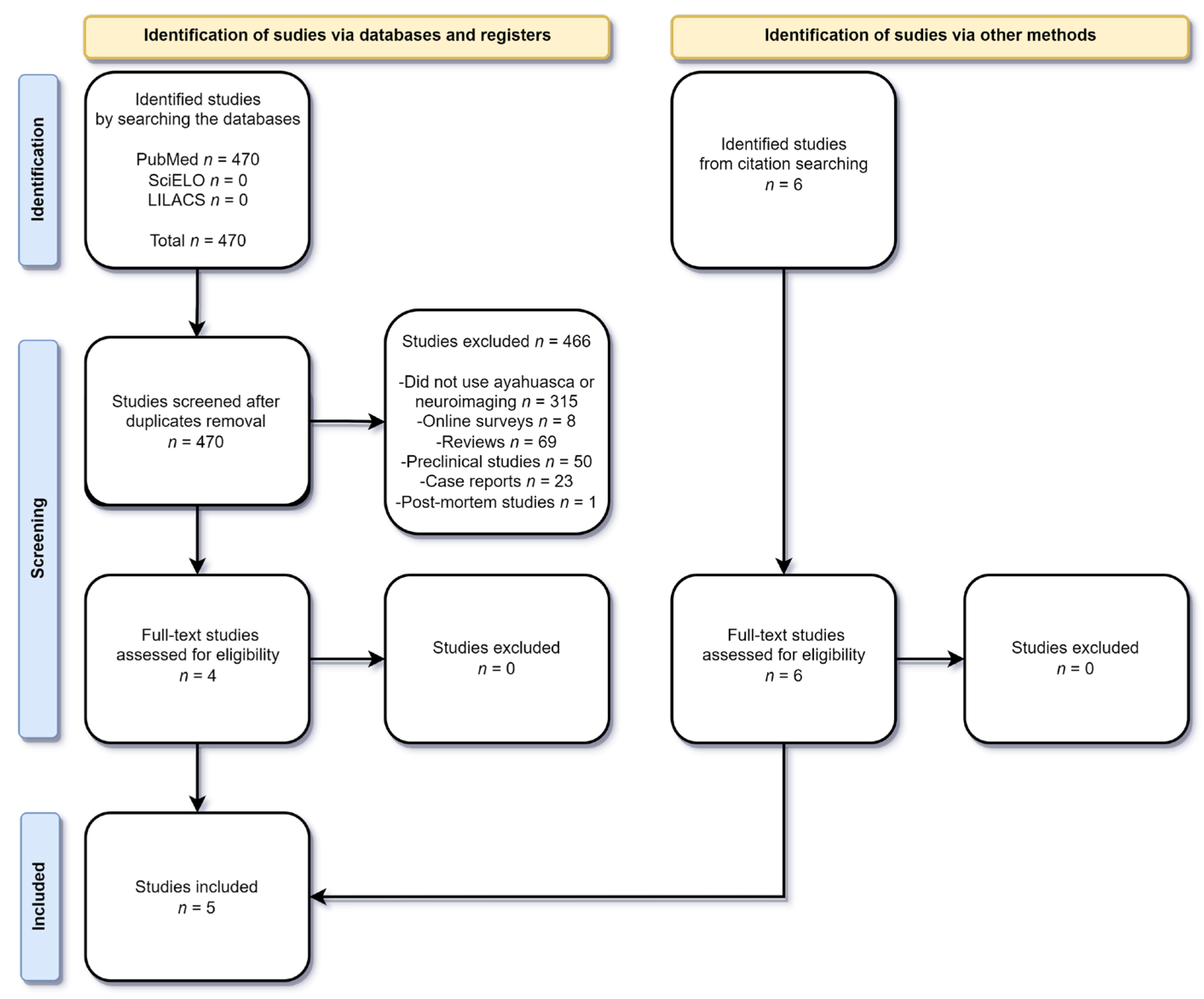
3.1. Article Screening and Inclusion

From our previous systematic review [1], six articles met our selection criteria [11,12,13,14,15,16]. We further searched the databases to include studies that were published after our previous review, resulting in 470 articles, of which none were duplicates. From these, 466 were excluded according to our inclusion and exclusion criteria. Thus, four articles were selected for full reading, and all of them were included in this review. Moreover, during the citation search one more article was also screened and selected. Therefore, a total of five new articles that were not present in our last review were included. Of these included papers, one used magnetic resonance spectroscopy (MRS) [17], three used functional magnetic resonance imaging (fMRI) [18,19,20], and one used magnetic resonance imaging (MRI) [21]. A flow diagram illustrating the different phases of the systematic search update from 2016 to 2022 is presented in Figure 1. The Cohen’s kappa coefficient for selecting the studies was 0.95.

3.2. Results from Selected Studies

In this review, we worked with 11 studies that investigated the effects of ayahuasca combined with different neuroimaging techniques. Results were divided into neuroimaging techniques (SPECT, MRS, MRI, and fMRI). In 10 articles, the volunteers were healthy, with previous experience with ayahuasca [11,12,13,14,15,17,18,19,21] or without previous experience [20]. One article [16] evaluated the effects of ayahuasca in patients with major depressive disorder and did not specify previous use by the volunteers. Acute effects were investigated in 8 studies [11,12,13,15,16,17,18,19], subacute effects in 1 study [20], and long-term effects in 2 studies [14,21]. The results found are consistent in demonstrating that there were brain anatomical and functional changes caused by ayahuasca in acute or chronic consumption. Regarding clinical trials, four different articles used the same sample of volunteers, differing in the types of data analyzed [13,15,18,19]. Concerning observational studies, two articles used the same sample of volunteers [14,21]. We will discuss the results in detail in the following paragraphs. A summary figure with the main results discussed is presented in Figure 2.

3.2.1. Single Photon Emission Computed Tomography (SPECT) Acute Effects (Molecular Imaging)

Riba et al., 2006 [11] performed a double-blind, randomized clinical study involving 15 healthy male volunteers with previous experience with psychedelics. They administered a single oral, encapsulated, and lyophilized dose, equivalent to 1.0 mg DMT/kg of ayahuasca or placebo. Acute effects were measured between 100 and 110 min after administration, using SPECT. Significant activations of the frontal and paralimbic regions of the brain were observed (p < 0.002). Increased blood perfusion was reported bilaterally in the anterior insula, with high intensity in the right hemisphere, and in the anterior/frontomedial cingulate cortex of the same hemisphere. Activity increases were noted in the left amygdala and the parahippocampal gyrus, a structure also involved in emotional arousal. Thus, the results suggest that ayahuasca interacts with important neural systems in interoception and emotional processing related to the DMN.
Sanches et al., 2016 [16] performed an open-label study to evaluate the possible acute, subacute, and prolonged antidepressant effects of a single dose of ayahuasca (120 to 200 mL, mean concentrations of 0.8 mg/mL DMT and 0.21 mg/mL harmine). The sample consisted of 17 volunteers diagnosed with major depressive disorder. To assess symptoms of depression, the researchers used the Hamilton Depression Rating Scale (HAMS-D) and the Montgomery–Åsberg Depression Rating Scale (MADRS). The Clinician-Administered Dissociative States Scale (CADSS) was used to assess dissociative symptoms, the Young Mania Rating Scale (YMRS) for mania symptoms, and the Brief Psychiatric Rating Scale (BPRS) for general psychiatric symptoms. The SPECT technique was used to assess brain network blood perfusion changes. Significant decreases were observed during the acute effects of ayahuasca (80 to 180 min) on the HAM-D and MADRS scales compared to baseline (p < 0.01). Furthermore, significant prolonged reduction effects (21 days after the experimental session) were observed on both scales (p < 0.001). Regarding the BPRS scale, the researchers observed significant acute and prolonged changes in the anxious-depression (p < 0.01), thinking disorder, and withdrawal-retardation (p < 0.05) subscales. Finally, the study authors noted that there was an increase in acute effects on the CADSS scale (p < 0.01) [16]. In regard to acute SPECT results (assessed eight hours after ayahuasca administration), an increase in blood perfusion of the subungual areas, nucleus accumbens, and insula was noted (p < 0.01).

3.2.2. Magnetic Resonance Spectroscopy (MRS) and Functional Magnetic Resonance Imaging (fMRI) Subacute Effects

In Sampedro et al.’s 2017 [17] study, the sample consisted of 16 healthy volunteers, including 10 men and six women, with previous experience using ayahuasca. Through a single average dose of 148 mL ± 29 mL of ayahuasca (0.3 mg/mL DMT, 0.86 mg/mL harmine, 0.17 mg/mL of tetrahydroharmine, and 0.04 mg/mL of harmaline), the neuroimaging data were evaluated through MRS in two sessions (24 h before and 24 h after the intervention). Mindfulness scores were assessed before and after tea drinking using Spanish versions of three instruments: The Five Facet Mindfulness Questionnaire (FFMQ), the Experiences Questionnaire (EQ), and the short version of the Self Compassion Questionnaire (SC). The MINDSENS index was also used to assess the extent to which ayahuasca-induced changes were comparable to those induced by meditation practice (this index is calculated with items from the FFMQ and EQ questionnaires). Using two different MRS techniques, neuro-metabolic (p ≤ 0.075) and functional (p < 0.05) changes were evidenced hours after the acute effects of ayahuasca had disappeared [17]. The results indicated the involvement of glutamatergic neurotransmitters in the participants’ psychedelic effects. They also suggest neuro-metabolic changes in the posterior cingulate cortex (PCC), a key region within the DMN, and an increase in connectivity between the anterior cingulate cortex (ACC) and the medial temporal lobe. Results show that ayahuasca (and probably other psychedelics) induce neural changes beyond the acute period, suggesting a biological basis related to therapeutic effects that occur after ayahuasca’s psychoactive effects have ceased.

3.2.3. Structural Magnetic Resonance Imaging (MRI) Long-Term Effects

Bouso et al., 2015 [14], performed an observational study with 22 regular users of ayahuasca from the Santo Daime religion compared to a control group of 22 matched participants and assessed the cortical thickness of brain regions using MRI. Psychometric scales were used to measure personality traits (Temperamental and Character Inventory-Revised/TCI-R) and psychopathological aspects (Symptom Check-List-90-Revised/SCL-90-R). Neuropsychological aspects were assessed using two computer tests: a two-back test to assess working memory and the Wisconsin’s Card-sorting Test (WCST) to assess executive function. Furthermore, a switching task was performed to evaluate the set change. Ayahuasca users showed significant differences in cortical thickness (p < 0.002) in the midline structures of the brain. Thinning was reported in the ayahuasca group in the middle frontal, inferior frontal, superior frontal, and superior occipital gyri, the precuneus, and the PCC. Thickening was found in the precentral gyrus and in the ACC. Cortical thickness values in the PCC were inversely correlated with the intensity and duration of previous ayahuasca use and with scores on self-transcendence, an important personality trait for measuring religiosity, transpersonal feelings, and spirituality. Ayahuasca users scored below the control group on the harm prevention characteristic (p = 0.044). This effect is driven by the score also below the “anticipatory concern” subscale (p = 0.005). Scores on other subscales were not significantly different. Moreover, regarding harm prevention, ayahuasca users scored significantly higher on self-transcendence (p < 0.001). Three other subscales demonstrated the significance of this result in relation to the control group: self-forgetfulness (p < 0.001), transpersonal identification (p < 0.001), and spiritual acceptance (p < 0.001). The data suggest that regular use of psychedelic drugs is correlated with structural changes in the brain in areas related to attentional processes, self-referential thinking, and internal mentalization.
Simonsson et al., 2022 [21], using the same sample as Bouso et al., 2015 [14], sought to investigate links between ayahuasca use and callosal structure. They compared the thickness of the corpus callosum using structural imaging data from 22 ayahuasca users and 22 matched controls. In addition, they investigated point-wise correlations between callosal thickness and the number of past ayahuasca sessions. In the isthmus, the mean corpus callosum was thicker in the ayahuasca group (5.47 ± 0.71 mm) compared to the control group (4.78 ± 1.01 mm, p = 0.006, d = 0.85). The ayahuasca group reported 123 past ayahuasca sessions on average (range 30–352), and it was observed a significant positive correlation between callosal thickness and the number of sessions within the rostral body (r = 0.45, p = 0.026), classified by the authors as a medium effect size. However, the statistical significance of these findings disappeared after multiple comparisons correction.

3.2.4. Functional Magnetic Resonance Imaging (fMRI) Acute Effects

The sample from Almeida Prado et al., 2009 [12], was composed of 10 healthy volunteers (five men and five women). All were ayahuasca users from the União do Vegetal (UDV) church. They received 150 mL of ayahuasca with a concentration of 0.65 mg/kg of DMT. The fMRI technique was used during a task in which the volunteers were asked to mentally generate words that started with the letters F, A, and S, with one minute for each word, with the objective of evaluating the effects of ayahuasca during the execution of the task.
They observed significant acute effects, increasing the scores of the BPRS (from 0.5 (SD: 1.01) at baseline to 9.44 (SD: 5.74) at the second scan time, when ayahuascas’ effects were more intense (F: 15.48, df: 2.00, p < 0.001)), YMRS (from 0.22 (SD: 0.66) at baseline to 6.88 (SD: 2.14) at second scan (F: 23.84, df: 2.00, p < 0.001)), and CADSS (from 0.90 (SD: 1.10) at baseline to 9.70 (SD: 6.18)) at the peak of effect. Moreover, independent of the language task, the researchers also noted acute activation in the bilateral anterior and posterior brain regions of the cingulate, superior, medial, and medial frontal gyrus, medial and superior temporal gyrus, and the precuneus. On the other hand, when applying verbal fluency tasks, reductions in the activation of the Broca area, frontal lobe, occipital temporal, temporal, cingulate areas, and some limbic structures were observed. A possible interaction of acute effects of ayahuasca with the language fluency task was evidenced in the temporal gyrus and cingulate activated bilaterally and in the right medial frontal lobe, left inferior, and left cingulate lobe activated unilaterally.
Three years later, de Araujo et al., 2012 [13], conducted an open-label study to investigate the neural bases involved in image processing during the effects of ayahuasca using the fMRI technique. The sample consisted of 10 healthy volunteers from the Santo Daime church (mean age 29 years), with at least 5 years of regular use of ayahuasca (twice a month), and compared them with a control group of 26 people. Each participant received 120 to 200 mL (2.2 mL/kg) of ayahuasca containing 0.8 mg/mL of DMT and 0.21 mg/mL of harmine. Each subject performed two fMRI sessions, one at baseline and one during the peak of the ayahuasca effect. Moreover, the BPRS and YMRS were used. The imaging task was divided into three blocks, with subjects scanned before and after ayahuasca intake. In the first block, volunteers passively saw images of people, animals, or trees. In the second, subjects were asked to close their eyes and mentally generate the same image seen in the first block. Finally, subjects were presented with a scrambled version of the first block. The last block was used as a baseline for the analysis. Significant effects were observed from 40 to 80 min after ayahuasca administration on the BPRS and YMRS (p = 0.036 and p = 0.036). An increase in blood-oxygen-level-dependent (BOLD) signal was reported in the bilateral precuneus, cuneus, and lingual, fusiform, middle occipital, parahippocampal, posterior cingulate, superior temporal, superior and middle frontal, and inferior frontal gyri (comprising Brodmann areas 7, 8, 9, 10, 17, 18, 19, 22, 23, 29, 30, 31, 37, 42, and 47) when comparing before and after ayahuasca intake. Most notably, when analyzing average time courses of BOLD responses data during block 2 (mental image formation) after ayahuasca intake, authors reported the same pattern of visual brain areas activation as in block 1 (seeing the image with eyes open) before and after ayahuasca intake, indicating that subjects were experiencing vivid mentally visualized pictures as if they were seeing the images with their eyes open. This effect was most evident in Brodmann areas 7, 10, 17, 18, 19, 30, and 37, related amongst other phenomena with the manifestation of hallucinations, REM sleep, episodes of memory retrieval, and contextual associations.
Palhano-Fontes et al., 2015 [15], carried out a study with the objective of verifying the relationship between the use of ayahuasca and the notion of altered state of consciousness, modulation of activity, and DMN connectivity to define the regions of interest (ROI) using fMRI. The sample was the same as from the previous article [13]. The ayahuasca dose used was 2.2 mL/kg, with concentrations of 0.8 mg/mL for DMT and 0.21 mg/mL for harmine. The DMN activation signal changed significantly (p < 0.01) when compared with rest and activation periods. There was a significant reduction in brain activity (p < 0.001) after ayahuasca administration in the following areas: ACC, PCC, medial prefrontal cortex, precuneus, and inferior parietal lobe. There was also a considerable decrease in PCC and precuneus functional connectivity in study subjects after ayahuasca ingestion (p < 0.001).
Using the same sample of the previous articles [13,15], Viol et al., 2017 [18], studied the increase in brain entropy associated with whole-brain functional connectivity under the influence of a single dose of ayahuasca (2.2 mL/kg containing 0.8 mg/mL of DMT and 0.21 mg/mL of harmine). They analyzed the functional connectivity of fMRI in relation to the increase in Shannon entropy. Data analysis consisted of two steps: first, fMRI data was used to generate complex networks representing the connectivity patterns of functional areas of the brain. In the second stage, the data created in the first stage was used as input and the characteristics of neural networks were calculated as output, using techniques from the theory of complex networks. The main result found was an increase in Shannon Entropy of the degree of distribution of the functional networks after ayahuasca ingestion. It was reported increases in the geodesic distance during the effects of ayahuasca, i.e., the neural networks qualitatively became locally “wider” while the brain’s global integration became less prominent.
In a later study with the same volunteers, Viol et al., 2019 [19], sought to analyze how nodes locally contribute to the global connectivity of the neural network, aiming to elucidate the individual role of these nodes. Each node interacts with its neighboring nodes and, indirectly, with the neighbors of neighbors, forming a large interaction radius. In this work, they sought to quantify the diversity of rays—as a “network of influences”—in each node exerted by all other nodes in the network. For this, they also used the concept of Shannon’s Entropy in addition to geodesic entropy (a statistical quantity that measures, in the frame of reference of a given node, the level of restrictions on the influences of neighboring nodes). In this study, it was found that ayahuasca ingestion tends to lead to higher geodesic entropy compared to before ingestion. Here, the geodesic entropy becomes less constrained after substance use, making the network wider. It was concluded that the ingestion of ayahuasca, compared to the ordinary state, led to greater diversity within the network of brain nodes.
Pasquini et al., 2020 [20], performed a randomized, placebo-controlled study with 50 healthy volunteers to assess the effects of a single dose of ayahuasca (1 mL/kg, 0.01 mg/mL DMT, 1.86 mg/mL harmine, 0.24 mg/mL of 0.03 mg/mL harmaline, and 1.20 mg/mL of tetrahydroharmine), or a dose of 1 mL/kg of placebo to map the saliency areas of the DMN, visual areas, and sensorimotor connections, and analyze possible subacute changes in the connectivity of these areas one day after administration of ayahuasca (or placebo). They used a task-free fMRI technique. To assess the acute psychedelic effect, the Hallucinogen Rating Scale (HRS) was used. There was a significant increase in the HRS scale between groups (p < 0.009). Functional connectivity increased in the area of interest located in the ACC and superior frontal gyrus (p < 0.05), with a tendency to the left hemisphere. On the other hand, there was a decrease in DMN’s activation in the ayahuasca group, predominantly affecting the PCC (p < 0.05). This result may reflect the change in the volition of the ayahuasca user, related to the subject’s ability to voluntarily interact with his ‘self’ during the psychedelic experience. The functional intra-network and inter-network connectivity of the primary sensory network (visual and sensorimotor) did not differ significantly between groups, suggesting some level of specificity of the subacute functional effect of ayahuasca on salience and on the DMN.
The summarized results for the articles included in this review are found below in Table 1.

3.2.5. Quality Assessment of Selected Studies

The selected studies obtained different scores. Regarding the studies obtained from our previous systematic review, the following scores were obtained: Riba et al., 2006 [11]—0.71; Almeida Prado et al., 2009 [12]—0.71; de Araujo et al., 2012 [13]—0.75; Bouso et al., 2015 [14]—0.41; Palhano-Fontes et al., 2015 [15]—0.41 and Sanches et al., 2016 [16]—0.75. Concerning articles from the updated systematic search present only in this review, the following scores were obtained: Sampedro et al., 2017 [17]—0.58; Viol et al., 2017 [18] and Viol et al., 2019 [19]—0.5; Pasquini et al., 2020 [20]—0.57; and Simonsson et al., 2022 [21]—0.66.
The articles selected for the present systematic review partially fulfilled the criteria of the quality analysis instruments used, except for the randomized placebo-controlled study conducted [20], which followed the CONSORT guidelines for randomized and controlled trials. Regarding open-label studies, the most common source of bias observed was the small sample sizes analyzed. Concerning clinical trials, the most common biases observed were the absence of details regarding randomization and study blinding. Moreover, since many studies utilized the same sample for different analyses, the resulting number of subjects is still limited when taking into consideration the number of variables analyzed. Finally, considering the report from Simonsson et al., 2022 [21], previous ayahuasca use is likely to cause changes in brain structures, and as most of the review articles included individuals with a history of previous ayahuasca use, this may also influence results.

4. Discussion

In our previous study involving neuroimaging techniques after the use of ayahuasca [1], results indicated excitatory effects in the frontolateral/frontomedial cortex, medial temporal lobe, and amygdala, affecting self-awareness, cognitive functioning, memory, and emotion processing. In the present review, we have mostly corroborated these findings and also the findings of a recent meta-analysis [22] by reporting excitatory effects (ACC, precuneus medial, superior temporal and frontomedial gyrus [10,11]) and increased connectivity (medial temporal lobe, ACC, PCC, superior frontal gyrus [16,19]) in similar regions, and decreased activation of the DMN [19]. On an anatomical perspective, Bouso et al., 2015 [13] reported thickening of the ACC, which may be a consequence of its frequent activation on chronic ayahuasca users.
The findings of Sanches et al., 2016 [16], and Sampedro et al., 2017 [17], corroborate an increased coupling between cortical networks. The agonist effect of hallucinogens on deep-layer pyramidal neurons rich in 5-HT2A receptors seems to be the main mechanism of action of these compounds, producing altered synchronization of cortical activity, “disintegration” of network connectivity, increased excitability of multimodal association hubs, and altered information flow, generating an “expanded awareness”, “ego-dissolution”, and “unconstrained cognition”. These effects are also reported with other psychedelics [23,24]. In this review, Viol et al., 2017 [18], and Viol et al., 2019 [19], provided further insight into this topic concerning ayahuasca. In the prior, the local neural network became enlarged while the global one became less connected. In the latest, ayahuasca caused higher geodesic entropy compared to the ordinary state. Geodesic entropy becomes less constrained after ayahuasca use, making the network wider, which leads to greater diversity within the network of brain nodes. These findings would corroborate the feelings such as “expanded awareness” provided using ayahuasca. In fact, increased brain entropy has been previously related with psilocybin administration and its effects [25].
The increased thickness in the corpus callosum reported by Simonsson et al., 2022 [21], in ayahuasca users compared to controls points to long-term anatomical changes in brain connectivity that could be a result of repeated exposure to acute changes reported by the previously mentioned authors. A thicker corpus callosum could imply a higher number of axons, thicker axons, and greater myelination, which could translate into greater anatomical interhemispheric connectivity. Given these findings, it is possible that ayahuasca could have a modulating impact and a positive effect on symptoms linked to impaired motor function, such as in neurodegenerative disorders. Ideas with this regard have recently been proposed [26,27]. However, these results are preliminary evidence, and a causality of the reported effects cannot be ascertained given the study design and the statistical analyses not surviving multiple comparisons correction.
Concerning the acute effects, Riba et al., 2006 [10], found increased blood perfusion in brain areas related to interoception (anterior insula) and emotional processing (ACC/frontomedial right hemisphere). These areas are also modulated by other psychedelics [22] and are also involved in the effects of traditional antidepressants. Importantly, these same areas were also activated by ayahuasca in depressed patients [15]. Moreover, increased blood perfusion was also observed in the subungual and nucleus accumbens areas, key areas for antidepressant action. From a cognitive point of view, De Araujo et al., 2012 [12], highlighted the potentiation of regions involved in memory and contextual associations, besides the occipital, temporal, and frontal areas. Taken together, these areas could be related to a reinterpretation of self and, thus, of the disorder, by allowing patients to access memories and thought patters related to their depressive state in a new manner. These effects could allow changes in behavior as well.
Finally, Palhano-Fontes et al., 2015 [15], reported similar acute and subacute effects through decreased activity in DMN regions, which modulate effects such as increased introspection, self-perception, and mind wandering. The researchers stipulated two hypotheses for the decrease in brain connectivity after the use of tea, not being mutually exclusive. The first hypothesis is related to the regions of the DMN that have activity reduced during the execution of a cognitive task. Thus, it would be possible to decrease the activity of DMN after ingesting ayahuasca due to mental effort and concentration during the experience. The second hypothesis is the consistent evidence that DMN activity is also reduced through meditative states, defined as a practice of rest in which the individual seeks to be aware of the thoughts that come to his mind. In fact, psychedelics and meditation share many similar physiological features. For example, they both increase introspection, self-awareness, and affect mind wandering. Moreover, two regions involved in language processing had an increase in signal, the left middle frontal gyrus and the left middle temporal gyrus. These regions are also associated with the verbal fluency test, which can be considered a confounding factor. Castelhano et al., 2021 [22], mention changes in PCC and precuneus (key regions of the DMN) with the use of other hallucinogenic substances such as psilocybin and LSD.
Working with the same sample, but with different data, Viol et al., 2017 [18], showed that the neural network became locally wider after the use of ayahuasca, while globally, the brain neural network became less connected. In a later study, Viol et al., 2019 [19], sought to work with the increase in geodesic entropy compared to the ordinary state of the individual. It was evidenced that the brain nodes became less restricted, showing, again, the acute effects of ayahuasca on the brain. A possible interpretation of these findings is that the increase in local connectivity and the decrease in global integration reflect the variation in the modular structure of the network. With the study of Shannon Entropy, innovative discoveries were made possible by using the study of the alteration of brain function at local and global levels, the analysis of the brain functional characteristics in altered states of consciousness, and the possibility of the method being applied immediately to study the variety of other phenomena, such as the effects of various medications for mental health disorders. The researchers point out that the finding may be related to the “mental expansion” event traditionally mentioned by ayahuasca users. In conclusion, they state that the results are striking when verifying the increase in entropy in brain functions after the use of ayahuasca. The increased brain entropy hypothesis may explain the increased flexibility, ease of accessing suppressed memories, and increased creativity, traits traditionally linked to tea use.
Regarding the medium and long-term effects among ayahuasca users, changes in the DMN regions, especially in the PCC, were observed to be associated with glutamatergic and neurometabolic changes by Sampedro et al., 2017 [17]. The results support consistent neural changes, especially in relation to memory and emotion. Regarding emotional processing data, these suggest significant changes in brain areas such as the insula and amygdala [22]. The amygdala is known to play an important role in symptoms such as fear and anxiety [28]. The results of Sampedro et al., 2017 [17], corroborate studies performed with psilocybin [22].
The changes visualized through neuroimaging may reflect on psychiatric scales. Almeida Prado et al., 2009 [12], observed significant results on the following scales: BPRS, CADSS, and YMRS, suggesting that drug-induced emotional and behavioral changes may be associated with improvements in symptoms of anxiety, depression, mania, and a change in dissociative pattern. Sanches et al., 2016 [16], reported significant changes in mean scores of the HAM-D, MADRS, and the depression-anxiety subscale of the BPRS during acute and prolonged effects that ranged from D1 to D21. In the first two scales, we went from a moderate level of depression to a mild level of depression. In addition, the authors also noted a significant change in fatigued affect and emotional withdrawal present on the Thinking Disorder and Withdrawal-Retardation subscale (p < 0.05). The researchers also observed an increase in blood perfusion in the subgenual, nucleus accumbens, and insula areas observed after ayahuasca administration. Studies associate hypoactivation of these areas with the emergence of depressive symptoms, while antidepressant effect is associated with increased activity of the mentioned areas. [28,29,30,31].
Bouso et al., 2015 [14], observed long-term behavioral and consistent changes in scales of spirituality, transpersonal identification, spiritual acceptance, and self-transcendence in chronic ayahuasca users compared to a control group. Pasquini et al., 2020 [20], also reported effects related to the potentiation of feelings related to spirituality in individuals after the use of ayahuasca, showing significant changes in the HRS scale, in addition to the effects seen in neuroimaging, such as a decrease in areas in the DMN, mainly affecting the PCC.
Clinical research involving the administration of ayahuasca, and other psychedelic compounds (such as LSD and psilocybin) has accumulated evidence that they possibly have beneficial effects for the treatment of psychopathologies [32,33]. Currently, preliminary studies suggest that the possible therapeutic effects of these substances are more evidence for the treatment of depression and anxiety [34,35,36,37,38,39] and substance dependence [40,41,42,43]. The results of these studies have been promising, with overall acceptable and expected adverse events, but they are still preliminary [44,45]. This is encouraging, especially when considering the lack of response to available treatments in a substantial percentage of patients. The most noticeable current limitation is still the lack of large-sample, placebo-controlled clinical studies to confirm these preliminary findings and further investigate less prevalent side effects and possible neural mechanisms. Nevertheless, the use of psilocybin for the treatment of major depression and treatment-resistant major depression was designated a breakthrough therapy by the United States Food and Drug Administration in 2019, demonstrating the scientific community’s recognition of the possible usefulness of this class of substances in the treatment of mental disorders [33]. Parallel to therapeutic effects assessments, mechanistic studies, such as neuroimaging investigations, will further help not only to unravel possible beneficial uses of psychedelics but also increase our understanding of their workings and that of the human brain. With the present data regarding ayahuasca, it seems that these drugs produce their therapeutic effects by modulating several networks involved in emotional processing, face recognition and empathy, introspection and self-awareness, and other higher-order cognitive processes. Several of these processes are modulated by limbic, para-limbic, and frontal areas of the brain, overlapping with the DMN.
There are some limitations regarding this review. Firstly, the average of included articles is of modest quality (0.59 overall), which is a potential source of bias. Secondly, the overall number of subjects is small, especially considering that some articles used the same sample for different analyses. Moreover, there is also the lack of other databases, such as Embase and Web of Science, within the search for studies, which may have resulted in missing other articles that could have been included [46].

5. Conclusions and Future Directions

In general, the results presented were supported by different neuroimaging tools, allowing the verification of the brain regions affected during acute or prolonged use of ayahuasca, identifying a correlation between tea use with different neuropsychiatric factors [12,14,16,20]. In addition to imaging studies, psychometric scales used to assess short and long-term effects have also shown promising results. Reviewed studies have linked reductions in DMN activation to acute, subacute, and prolonged antidepressant effects [14,15,20]. Investigations with psychedelics show preliminary but promising results, as they may become a possible pharmacological therapy for the treatment of a series of psychiatric disorders such as post-traumatic stress, resistant depression, substance use disorders, obsessive-compulsive disorder, and anxiety [22,47].
On the other hand, there are important biases to be highlighted in current research, such as the absence of a placebo in many trials, small samples, lack of transparency in sample selection description, and size calculation for adequate statistical power [48]. Therefore, it is recommended to carry out further studies with greater methodological rigor, use of inactive or active placebos, and larger samples to replicate and confirm the results described both in healthy volunteers and clinical samples.

Author Contributions

Original draft preparation and systematic search: G.H.d.M.S. and L.S.R. Review and editing: J.M.R., G.N.R., G.O. and J.C.B. Supervision: J.E.C.H. and R.G.d.S. All authors have read and agreed to the published version of the manuscript.

Funding

J.M.R. and L.S.R. received funding from Coordenação de Aperfeiçoamento de Pessoal de Nível Superior (CAPES, Brazil). G.N.R. received funding from Fundação de Amparo à Pesquisa do Estado de São Paulo (FAPESP). J.E.C.H. is recipient of CNPq 1A productivity fellowship. R.G.d.S. is Fellow of the Programa Nacional de Pós-Doutorado, Brazil (PNPD/CAPES).

Institutional Review Board Statement

Not applicable.

Informed Consent Statement

Not applicable.

Data Availability Statement

No new data were created or analyzed in this study. Data sharing is not applicable to this article.

Conflicts of Interest

The authors declare no conflict of interest.

References

  1. Dos Santos, R.G.; Balthazar, F.M.; Bouso, J.C.; Hallak, J.E. The current state of research on ayahuasca: A systematic review of human studies assessing psychiatric symptoms, neuropsychological functioning, and neuroimaging. J. Psychopharmacol. 2016, 30, 1230–1247. [Google Scholar] [CrossRef]
  2. McKenna, D.J.; Towers, G.H.; Abbott, F. Monoamine oxidase inhibitors in South American hallucinogenic plants: Tryptamine and beta-carboline constituents of ayahuasca. J. Ethnopharmacol. 1984, 10, 195–223. [Google Scholar] [CrossRef]
  3. Chi, T.; Gold, J.A. A review of emerging therapeutic potential of psychedelic drugs in the treatment of psychiatric illnesses. J. Neurol. Sci. 2020, 411, 116715. [Google Scholar] [CrossRef] [Green Version]
  4. Buckner, R.L.; Andrews-Hanna, J.R.; Schacter, D.L. The brain’s default network: Anatomy, function, and relevance to disease. Ann. N. Y. Acad. Sci. 2008, 1124, 1–38. [Google Scholar] [CrossRef] [Green Version]
  5. Gattuso, J.J.; Perkins, D.; Ruffell, S.; Lawrence, A.J.; Hoyer, D.; Jacobson, L.H.; Timmermann, C.; Castle, D.; Rossell, S.L.; Downey, L.A.; et al. Default Mode Network Modulation by Psychedelics: A Systematic Review. Int. J. Neuropsychopharmacol. 2022. [Google Scholar] [CrossRef]
  6. Coutinho, J.; Fernandes, C.J.; Soares, J.M.; Maia, L.; Gonçalves, Ó.F.; Sampaio, A. Default mode network dissociation in depressive and anxiety states. Brain Imaging Behav. 2016, 10, 147–157. [Google Scholar] [CrossRef]
  7. Cumming, P.; Scheidegger, M.; Dornbierer, D.; Palner, M.; Quednow, B.B.; Martin-Soelch, C. Molecular and Functional Imaging Studies of Psychedelic Drug Action in Animals and Humans. Molecules 2021, 26, 2451. [Google Scholar] [CrossRef] [PubMed]
  8. Kadriu, B.; Greenwald, M.; Henter, I.D.; Gilbert, J.R.; Kraus, C.; Park, L.T.; Zarate, C.A. Ketamine and Serotonergic Psychedelics: Common Mechanisms Underlying the Effects of Rapid-Acting Antidepressants. Int. J. Neuropsychopharmacol. 2020, 24, 8–21. [Google Scholar] [CrossRef]
  9. Page, M.J.; McKenzie, J.E.; Bossuyt, P.M.; Boutron, I.; Hoffmann, T.C.; Mulrow, C.D.; Shamseer, L.; Tetzlaff, J.M.; Akl, E.A.; Brennan, S.E.; et al. The PRISMA 2020 statement: An updated guideline for reporting systematic reviews. BMJ 2021, 372, n71. [Google Scholar] [CrossRef] [PubMed]
  10. National Heart, Lung and Blood Institute. Available online: https://www.nhlbi.nih.gov/health-topics/study-quality-assessment-tools (accessed on 6 March 2023).
  11. Riba, J.; Romero, S.; Grasa, E.; Mena, E.; Carrió, I.; Barbanoj, M.J. Increased frontal and paralimbic activation following ayahuasca, the pan-amazonian inebriant. Psychopharmacology 2006, 186, 93–98. [Google Scholar] [CrossRef] [PubMed] [Green Version]
  12. Prado, D.A.; Pinto, J.; Crippa, J.; Santos, A.; Ribeiro, S.; Araujo, D.; Zuardi, A.; Chaves, C.; Hallak, J. P.1.e.025 Effects of the Amazonian psychoactive plant beverage ayahuasca on prefrontal and limbic regions during a language task: A fMRI study. Eur. Neuropsychopharmacol. 2009, 19, S314–S315. [Google Scholar] [CrossRef]
  13. de Araujo, D.B.; Ribeiro, S.; Cecchi, G.A.; Carvalho, F.M.; Sanchez, T.A.; Pinto, J.P.; de Martinis, B.S.; Crippa, J.A.; Hallak, J.E.; Santos, A.C. Seeing with the eyes shut: Neural basis of enhanced imagery following ayahuasca ingestion. Hum. Brain Mapp. 2012, 33, 2550–2560. [Google Scholar] [CrossRef] [PubMed]
  14. Bouso, J.C.; Palhano-Fontes, F.; Rodríguez-Fornells, A.; Ribeiro, S.; Sanches, R.; Crippa, J.A.S.; Hallak, J.E.; de Araujo, D.B.; Riba, J. Long-term use of psychedelic drugs is associated with differences in brain structure and personality in humans. Eur. Neuropsychopharmacol. 2015, 25, 483–492. [Google Scholar] [CrossRef] [PubMed]
  15. Palhano-Fontes, F.; Andrade, K.C.; Tófoli, L.F.; Santos, A.C.; Crippa, J.A.S.; Hallak, J.E.C.; Ribeiro, S.; De Araujo, D.B. The Psychedelic State Induced by Ayahuasca Modulates the Activity and Connectivity of the Default Mode Network. PLoS ONE 2015, 10, e0118143. [Google Scholar] [CrossRef] [Green Version]
  16. Sanches, R.F.; de Lima Osório, F.; Dos Santos, R.G.; Macedo, L.R.; Maia-de-Oliveira, J.P.; Wichert-Ana, L.; de Araujo, D.B.; Riba, J.; Crippa, J.A.; Hallak, J.E. Antidepressant Effects of a Single Dose of Ayahuasca in Patients with Recurrent Depression: A SPECT Study. J. Clin. Psychopharmacol. 2016, 36, 77–81. [Google Scholar] [CrossRef] [Green Version]
  17. Sampedro, F.; Revenga, M.D.L.F.; Valle, M.; Roberto, N.; Domínguez-Clavé, E.; Elices, M.; Luna, L.E.; Crippa, J.A.S.; Hallak, J.E.C.; de Araujo, D.B.; et al. Assessing the Psychedelic “After-Glow” in Ayahuasca Users: Post-Acute Neurometabolic and Functional Connectivity Changes Are Associated with Enhanced Mindfulness Capacities. Int. J. Neuropsychopharmacol. 2017, 20, 698–711. [Google Scholar] [CrossRef] [Green Version]
  18. Viol, A.; Palhano-Fontes, F.; Onias, H.; de Araujo, D.B.; Hövel, P.; Viswanathan, G.M. Characterizing Complex Networks Using Entropy-Degree Diagrams: Unveiling Changes in Functional Brain Connectivity Induced by Ayahuasca. Entropy 2019, 21, 128. [Google Scholar] [CrossRef] [Green Version]
  19. Viol, A.; Palhano-Fontes, F.; Onias, H.; de Araujo, D.B.; Viswanathan, G.M. Shannon entropy of brain functional complex networks under the influence of the psychedelic Ayahuasca. Sci. Rep. 2017, 7, 7388. [Google Scholar] [CrossRef] [Green Version]
  20. Pasquini, L.; Palhano-Fontes, F.; Araujo, D.B. Subacute effects of the psychedelic ayahuasca on the salience and default mode networks. J. Psychopharmacol. 2020, 34, 623–635. [Google Scholar] [CrossRef]
  21. Simonsson, O.; Bouso, J.C.; Kurth, F.; Araújo, D.B.; Gaser, C.; Riba, J.; Luders, E. Preliminary evidence of links between ayahuasca use and the corpus callosum. Front. Psychiatry 2022, 13, 1002455. [Google Scholar] [CrossRef]
  22. Castelhano, J.; Lima, G.; Teixeira, M.; Soares, C.; Pais, M.; Castelo-Branco, M. The Effects of Tryptamine Psychedelics in the Brain: A meta-Analysis of Functional and Review of Molecular Imaging Studies. Front. Pharmacol. 2021, 12, 739053. [Google Scholar] [CrossRef]
  23. Preller, K.H.; Razi, A.; Zeidman, P.; Stämpfli, P.; Friston, K.J.; Vollenweider, F.X. Effective connectivity changes in LSD-induced altered states of consciousness in humans. Proc. Natl. Acad. Sci. USA 2019, 116, 2743–2748. [Google Scholar] [CrossRef] [Green Version]
  24. Barrett, F.S.; Krimmel, S.R.; Griffiths, R.R.; Seminowicz, D.A.; Mathur, B.N. Psilocybin acutely alters the functional connectivity of the claustrum with brain networks that support perception, memory, and attention. Neuroimage 2020, 218, 116980. [Google Scholar] [CrossRef]
  25. Carhart-Harris, R.L.; Leech, R.; Hellyer, P.J.; Shanahan, M.; Feilding, A.; Tagliazucchi, E.; Chialvo, D.R.; Nutt, D. The entropic brain: A theory of conscious states informed by neuroimaging research with psychedelic drugs. Front. Hum. Neurosci. 2014, 8, 20. [Google Scholar] [CrossRef] [Green Version]
  26. Katchborian-Neto, A.; Santos, W.T.; Nicácio, K.J.; Corrêa, J.O.A.; Murgu, M.; Martins, T.M.M.; Gomes, D.A.; Goes, A.M.; Soares, M.G.; Dias, D.F.; et al. Neuroprotective potential of Ayahuasca and untargeted metabolomics analyses: Applicability to Parkinson’s disease. J. Ethnopharmacol. 2020, 255, 112743. [Google Scholar] [CrossRef]
  27. Saeger, H.N.; Olson, D.E. Psychedelic-inspired approaches for treating neurodegenerative disorders. J. Neurochem. 2021, 162, 109–127. [Google Scholar] [CrossRef]
  28. Carhart-Harris, R.L.; Erritzoe, D.; Williams, T.; Stone, J.M.; Reed, L.J.; Colasanti, A.; Tyacke, R.J.; Leech, R.; Malizia, A.L.; Murphy, K.; et al. Neural correlates of the psychedelic state as determined by fMRI studies with psilocybin. Proc. Natl. Acad. Sci. USA 2012, 109, 2138–2143. [Google Scholar] [CrossRef] [Green Version]
  29. Drevets, W.C.; Savitz, J.; Trimble, M. The Subgenual Anterior Cingulate Cortex in Mood Disorders. CNS Spectrums 2008, 13, 663–681. [Google Scholar] [CrossRef] [Green Version]
  30. Pizzagalli, D.A.; Holmes, A.; Dillon, D.G.; Goetz, E.L.; Birk, J.; Bogdan, R.; Dougherty, D.D.; Iosifescu, D.V.; Rauch, S.L.; Fava, M. Reduced Caudate and Nucleus Accumbens Response to Rewards in Unmedicated Individuals with Major Depressive Disorder. Am. J. Psychiatry 2009, 166, 702–710. [Google Scholar] [CrossRef] [Green Version]
  31. Fitzgerald, P.B.; Laird, A.R.; Maller, J.; Daskalakis, Z.J. A meta-analytic study of changes in brain activation in depression. Hum. Brain Mapp. 2008, 29, 683–695. [Google Scholar] [CrossRef]
  32. Fuentes, J.J.; Fonseca, F.; Elices, M.; Farré, M.; Torrens, M. Therapeutic Use of LSD in Psychiatry: A Systematic Review of Randomized-Controlled Clinical Trials. Front. Psychiatry 2019, 10, 943. [Google Scholar] [CrossRef] [PubMed] [Green Version]
  33. Reiff, C.M.; Richman, E.E.; Nemeroff, C.B.; Carpenter, L.L.; Widge, A.S.; Rodriguez, C.I.; Kalin, N.H.; McDonald, W.M. Psychedelics and Psychedelic-Assisted Psychotherapy. Am. J. Psychiatry 2020, 177, 391–410. [Google Scholar] [CrossRef] [PubMed]
  34. Grob, C.S.; Danforth, A.L.; Chopra, G.S.; Hagerty, M.; McKay, C.R.; Halberstadt, A.L.; Greer, G.R. Pilot Study of Psilocybin Treatment for Anxiety in Patients with Advanced-Stage Cancer. Arch. Gen. Psychiatry 2011, 68, 71–78. [Google Scholar] [CrossRef] [PubMed]
  35. Osório, F.D.L.; Sanches, R.F.; Macedo, L.R.; dos Santos, R.G.; Maia-De-Oliveira, J.P.; Wichert-Ana, L.; de Araujo, D.B.; Riba, J.; Crippa, J.A.; Hallak, J.E. Antidepressant effects of a single dose of ayahuasca in patients with recurrent depression: A preliminary report. Braz. J. Psychiatry 2015, 37, 13–20. [Google Scholar] [CrossRef] [Green Version]
  36. Griffiths, R.R.; Johnson, M.W.; Carducci, M.A.; Umbricht, A.; Richards, W.A.; Richards, B.D.; Cosimano, M.P.; Klinedinst, M.A. Psilocybin produces substantial and sustained decreases in depression and anxiety in patients with life-threatening cancer: A randomized double-blind trial. J. Psychopharmacol. 2016, 30, 1181–1197. [Google Scholar] [CrossRef]
  37. Carhart-Harris, R.L.; Bolstridge, M.; Rucker, J.; Day, C.M.J.; Erritzoe, D.; Kaelen, M.; Bloomfield, M.; Rickard, J.A.; Forbes, B.; Feilding, A.; et al. Psilocybin with psychological support for treatment-resistant depression: An open-label feasibility study. Lancet Psychiatry 2016, 3, 619–627. [Google Scholar] [CrossRef] [Green Version]
  38. Ross, S.; Bossis, A.; Guss, J.; Agin-Liebes, G.; Malone, T.; Cohen, B.; Mennenga, S.E.; Belser, A.; Kalliontzi, K.; Babb, J.; et al. Rapid and sustained symptom reduction following psilocybin treatment for anxiety and depression in patients with life-threatening cancer: A randomized controlled trial. J. Psychopharmacol. 2016, 30, 1165–1180. [Google Scholar] [CrossRef] [Green Version]
  39. Goodwin, G.M.; Aaronson, S.T.; Alvarez, O.; Arden, P.C.; Baker, A.; Bennett, J.C.; Bird, C.; Blom, R.E.; Brennan, C.; Brusch, D.; et al. Single-Dose Psilocybin for a Treatment-Resistant Episode of Major Depression. N. Engl. J. Med. 2022, 387, 1637–1648. [Google Scholar] [CrossRef]
  40. Bogenschutz, M.P.; Forcehimes, A.E.; Pommy, J.A.; Wilcox, C.E.; Barbosa, P.C.R.; Strassman, R.J. Psilocybin-assisted treatment for alcohol dependence: A proof-of-concept study. J. Psychopharmacol. 2015, 29, 289–299. [Google Scholar] [CrossRef]
  41. Johnson, M.W.; Garcia-Romeu, A.; Cosimano, M.P.; Griffiths, R.R. Pilot study of the 5-HT2AR agonist psilocybin in the treatment of tobacco addiction. J. Psychopharmacol. 2014, 28, 983–992. [Google Scholar] [CrossRef] [Green Version]
  42. Moreno, F.A.; Wiegand, C.B.; Taitano, E.K.; Delgado, P.L. Safety, Tolerability, and Efficacy of Psilocybin in 9 Patients with Obsessive-Compulsive Disorder. J. Clin. Psychiatry 2006, 67, 1735–1740. [Google Scholar] [CrossRef] [Green Version]
  43. Rodrigues, L.S.; Rossi, G.N.; Rocha, J.M.; Osório, F.L.; Bouso, J.C.; Hallak, J.E.C.; dos Santos, R.G. Effects of ayahuasca and its alkaloids on substance use disorders: An updated (2016–2020) systematic review of preclinical and human studies. Eur. Arch. Psychiatry Clin. Neurosci. 2022, 272, 541–556. [Google Scholar] [CrossRef]
  44. Rossi, G.N.; Dias, I.C.D.S.; Baker, G.; Saiz, J.C.B.; Dursun, S.M.; Hallak, J.E.C.; Dos Santos, R.G. Ayahuasca, a potentially rapid acting antidepressant: Focus on safety and tolerability. Expert Opin. Drug Saf. 2022, 21, 789–801. [Google Scholar] [CrossRef]
  45. Rossi, G.N.; Hallak, J.E.C.; Saiz, J.C.B.; Dos Santos, R.G. Safety issues of psilocybin and LSD as potential rapid acting antidepressants and potential challenges. Expert Opin. Drug Saf. 2022, 21, 761–776. [Google Scholar] [CrossRef]
  46. Bramer, W.M.; Rethlefsen, M.L.; Kleijnen, J.; Franco, O.H. Optimal database combinations for literature searches in systematic reviews: A prospective exploratory study. Syst. Rev. 2017, 6, 245. [Google Scholar] [CrossRef]
  47. Dos Santos, R.G.; Bouso, J.C. Translational evidence for ayahuasca as an antidepressant: What’s next? Braz. J. Psychiatry 2019, 41, 275–276. [Google Scholar] [CrossRef] [Green Version]
  48. McCulloch, D.E.-W.; Knudsen, G.M.; Barrett, F.S.; Doss, M.K.; Carhart-Harris, R.L.; Rosas, F.E.; Deco, G.; Kringelbach, M.L.; Preller, K.H.; Ramaekers, J.G.; et al. Psychedelic resting-state neuroimaging: A review and perspective on balancing replication and novel analyses. Neurosci. Biobehav. Rev. 2022, 138, 104689. [Google Scholar] [CrossRef]
Figure 1. PRISMA flowchart for the selection of the studies.
Psychoactives 02 00006 g001
Figure 2. Main results from current evidence regarding brain activation and connectivity changes caused by ayahuasca ingestion. Blue areas show decreased activity while red areas show increased activity. Blue arrows show decreased connectivity while red arrows show increased connectivity. ACC: Anterior Cingulate Cortex; LS: Limbic Structures; mPFC: Medial Prefrontal Cortex; NAc: Nucleus Accumbens; PC: Precuneus; PCC: Posterior Cingulate Cortex; sFG: Superior Frontal Gyrus.
Psychoactives 02 00006 g002
Table 1. Summary of the currently published neuroimaging results related to ayahuasca consumption.
AuthorsStudy Design and Quality Rating (QR, 0 to 1)SampleDrug and DoseNeuroimaging TechniqueMain Results
Riba et al., 2006 [11]Randomized, double-blind, placebo-controlled.
QR: 0.71
15 healthy male volunteers with previous experience of hallucinogen use and not diagnosed with psychiatric disorders (DSM-IV).Lyophilized, encapsulated, and orally administered ayahuasca in concentrations equivalent to 1.0 mg DMT/kg or 0.75 g lactose capsules as placebo.Single photon emission tomography (SPECT).Activation of frontal and paralimbic brain regions (p < 0.002). Increased blood perfusion bilaterally in the regions of the anterior insula, with great intensity in the right hemisphere, and in the anterior cingulate cortex/medial front of the right hemisphere. Activity increases in the left amygdala and parahippocampal gyrus (p < 0.002).
Almeida Prado et al., 2009 [12]Double-blind.
QR: 0.71
10 healthy volunteers, 5 women, with experiences of chronic use of ayahuasca from the UDV church.Ayahuasca in an average dose of 150 mL, with a concentration of 0.65 mg/kg of DMT.Functional magnetic resonance imaging (fMRI).Elevation in the scores of the scales: BPRS (p < 0.001), YMRS (p < 0.001), and CADSS (p = 0.001 and p < 0.001). Bilateral activation of the cingulate, superior, medial and frontomedial gyrus regions, medial and superior temporal gyrus, and the precuneus.
De Araujo et al., 2012 [13]Open-label.
QR: 0.75
9 healthy volunteers with regular use of ayahuasca recruited from Igreja do Santo Daime, compared with a control group composed of 26 individuals. *Ayahuasca in a single dose of 2.2 mL/kg at concentrations of 0.8 mg/mL of DMT and 0.21 mg/mL of harmine.Functional magnetic resonance imaging (fMRI).An increase in BOLD signal was reported in the bilateral precuneus, cuneus, and lingual, fusiform, middle occipital, parahippocampal, posterior cingulate, superior temporal, superior and middle frontal, and inferior frontal gyri when comparing before and after ayahuasca intake (all p values < 0.05). By increasing the intensity of the retrieved images to the same level as the natural image, ayahuasca gives inner experiences a reality status.
Bouso et al., 2015 [14]Cross-sectional case-control.
QR: 0.41
22 participants with previous ayahuasca experience recruited from the Santo Daime church compared with a control of 22 matched participants. †No dose was administered.Magnetic resonance imaging (MRI).Ayahuasca users showed significant differences in cortical thickness (CT) (p < 0.002), in the midline structures of the brain, with thinning in the posterior cingulate cortex (PCC). PCC CT values were inversely correlated with the intensity and duration of previous ayahuasca use and scores of self-transcendence. Significantly elevated scores of self-transcendence (p = 0.001), self-forgetfulness (p = 0.001), transpersonal identification (p = 0.001), and spiritual acceptance (p = 0.001) were reported in ayahuasca users when compared to controls.
Palhano-Fontes et al., 2015 [15]Cross-sectional case-control.
QR: 0.41
9 healthy volunteers with regular use of ayahuasca recruited from Igreja do Santo Daime, compared with a control group composed of 26 individuals. *Ayahuasca in a single dose of 2.2 mL/kg at concentrations of 0.8 mg/mL of DMT and 0.21 mg/mL of harmine.Functional magnetic resonance imaging (fMRI)Within 9 analyzed DMN regions of interest, there was a significant reduction in connectivity in 6 of them (p < 0.001). In 2 related to speech, there was an increase in connectivity, which may be related to the requested task.
Sanches et al., 2016 [16]Open-label.
QR: 0.75
17 volunteers diagnosed with major depressive disorder. 3 of the volunteers were in mild depressive episodes, 13 in moderate depressive episodes, and 1 in severe depressive episode.Ayahuasca in a single dose of 2.2 mL/kg at concentrations of 0.8 mg/mL of DMT and 0.21 mg/mL of harmine.Single photon emission tomography (SPECT)Significant decreases in HAM-D and MADRS depression scales from 80 to 180 min and on D21 (p < 0.001). As for the BPRS, effects were observed from the first day of administration to the 21st day. The Anxiety-Depression subscale (from 40 to 180 min, p < 0.01; and from D1 to D21, p < 0.001), Thought Disorder (180 min, p < 0.05; and on D1, D14, and D21). There was an increase in the CADSS index (p < 0.01) between 40 and 80 min. Furthermore, a significant increase (p < 0.01) in blood perfusion in the subungual area, nucleus accumbens, and insula was also noted.
Sampedro et al., 2017 [17]Open-label.
QR: 0.58
16 healthy volunteers, with previous experience of using ayahuasca.Ayahuasca in a single 148 mL dose containing 0.3 mg/mL of DMT, 0.86 mg/mL of harmine, 0.17 mg/mL of tetrahydroharmine and 0.04 mg/mL of harmaline.Magnetic resonance spectroscopy (MRS)Involvement of glutamatergic neurotransmitters in psychedelic effects. Neurometabolic changes in posterior cingulate cortex and increased connectivity between the anterior cingulate cortex and medial temporal lobe.
Viol et al., 2017 [18]Open-label.
QR: 0.50
9 healthy volunteers with regular use of ayahuasca recruited from Igreja do Santo Daime, compared with a control group composed of 26 individuals. *Ayahuasca in a single dose of 2.2 mL/kg in concentrations of 0.8 mg/mL of DMT and 0.21 mg/mL of harmine.Functional magnetic resonance imaging (fMRI)The neural networks became locally enlarged after using ayahuasca. On the other hand, the brain’s functional network, globally, became less connected.
Viol et al., 2019 [19]Open-label.
QR: 0.50
9 healthy volunteers with regular use of ayahuasca recruited from Igreja do Santo Daime, compared with a control group composed of 26 individuals.*Ayahuasca in a single dose of 2.2 mL/kg in concentrations of 0.8 mg/mL of DMT and 0.21 mg/mL of harmine.Functional magnetic resonance imaging (fMRI)Ayahuasca ingestion tends to lead to higher geodesic entropy compared to the ordinary state. Geodesic distance becomes less constrained after substance use, making the network wider, which leads to greater diversity within the network of brain nodes.
Pasquini et al., 2020 [20]Randomized, placebo-controlled study.
QR: 0.57
50 healthy participants with no previous ayahuasca experience.Ayahuasca in a single dose of 1 mL/kg containing 0.36 mg/mL of DMT, 1.86 mg/mL of harmine, 0.24 mg/mL 0.03 mg/mL of harmaline, and 1.20 mg/mL of tetrahydroharmine or placebo.Functional magnetic resonance imaging (fMRI)Significant increases in the HRS after ayahuasca administration (p < 0.009).
Functional connectivity increased in the area of interest located in the anterior cingulate cortex and in the superior frontal gyrus (p < 0.05), with a tendency to the left hemisphere. On the other hand, there was a decrease in DMN in the ayahuasca group, predominantly affecting the posterior cingulate cortex (p < 0.1).
Simonsson et al., 2022 [21]Cross-sectional, case-control study.
QR: 0.66
22 participants with previous ayahuasca experience recruited from the Santo Daime church compared with a control of 22 matched participants. †No dose was administered.Magnetic resonance imaging (MRI).The corpus callosum was thicker in the ayahuasca group than in the control group (p = 0.006). Additionally, the ayahuasca group reported 123 past ayahuasca sessions on average (range: 30–352) and was observed a significant positive correlation between callosal thickness and the number of sessions (p = 0.026), although statistical significance was not maintained after multiple comparisons.
* Same sample of volunteers. † Same sample of volunteers. BOLD, blood-oxygen-level-dependent; BPRS, Brief Psychiatric Rating Scale; CADSS, Clinician-Administered Dissociative States Scale; CT, cortical thickness; DMN, Default Mode Network; DMT, dimethyltryptamine; fMRI, Functional magnetic resonance imaging; HAMS-D, Hamilton Depression Rating Scale; HRS, Hallucinogen Rating Scale; MADRS, Montgomery–Åsberg Depression Rating Scale; MRI, Magnetic resonance imaging; MRS, Magnetic resonance spectroscopy; PCC, posterior cingulate cortex; QR, Quality Rating; SPECT, Single photon emission tomography; UDV, União do Vegetal; YMRS, the Young Mania Rating Scale.
Disclaimer/Publisher’s Note: The statements, opinions and data contained in all publications are solely those of the individual author(s) and contributor(s) and not of MDPI and/or the editor(s). MDPI and/or the editor(s) disclaim responsibility for any injury to people or property resulting from any ideas, methods, instructions or products referred to in the content.

