
Duplicated Inferior Vena Cava in a 69-Year-Old White Female Donor


Abstract
:1. Introduction
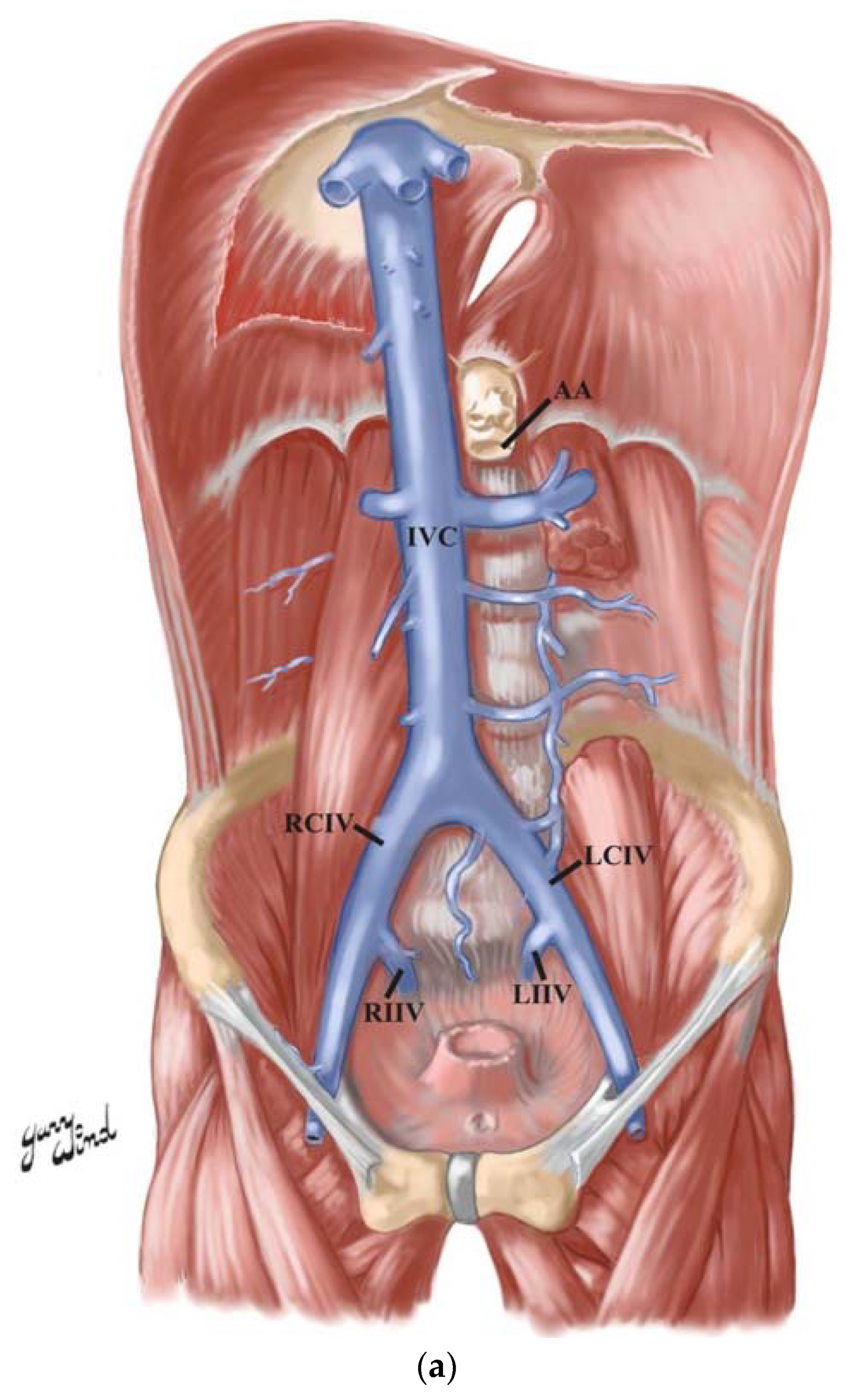
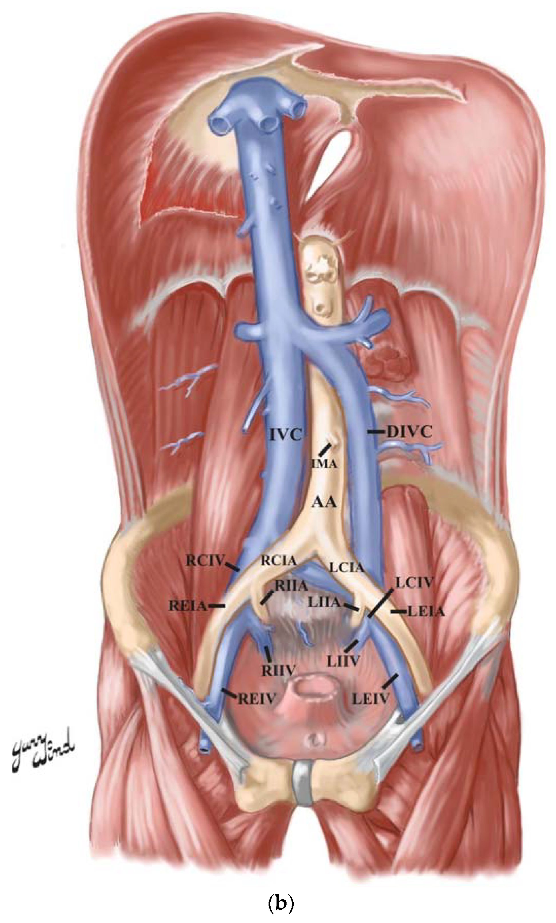
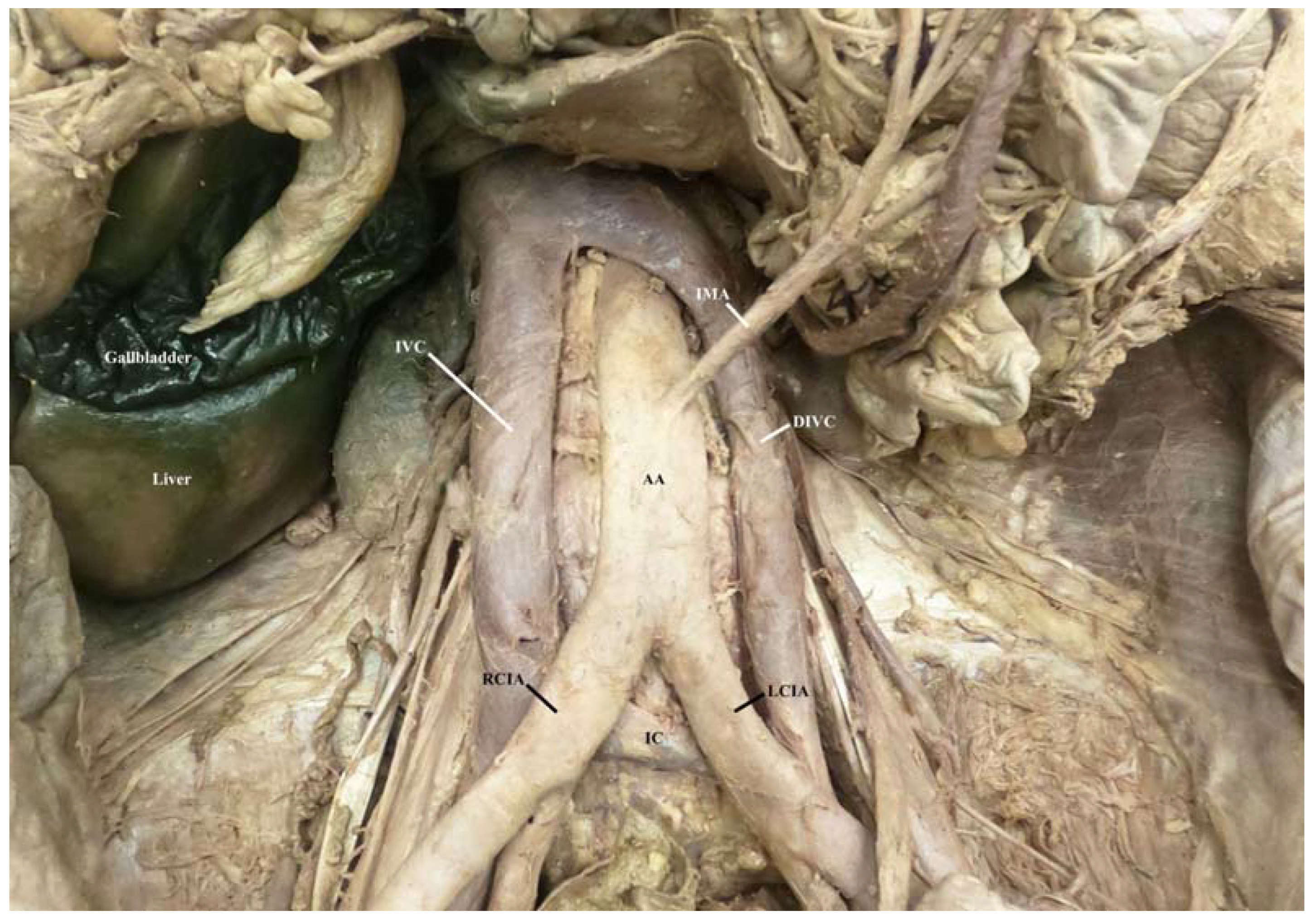
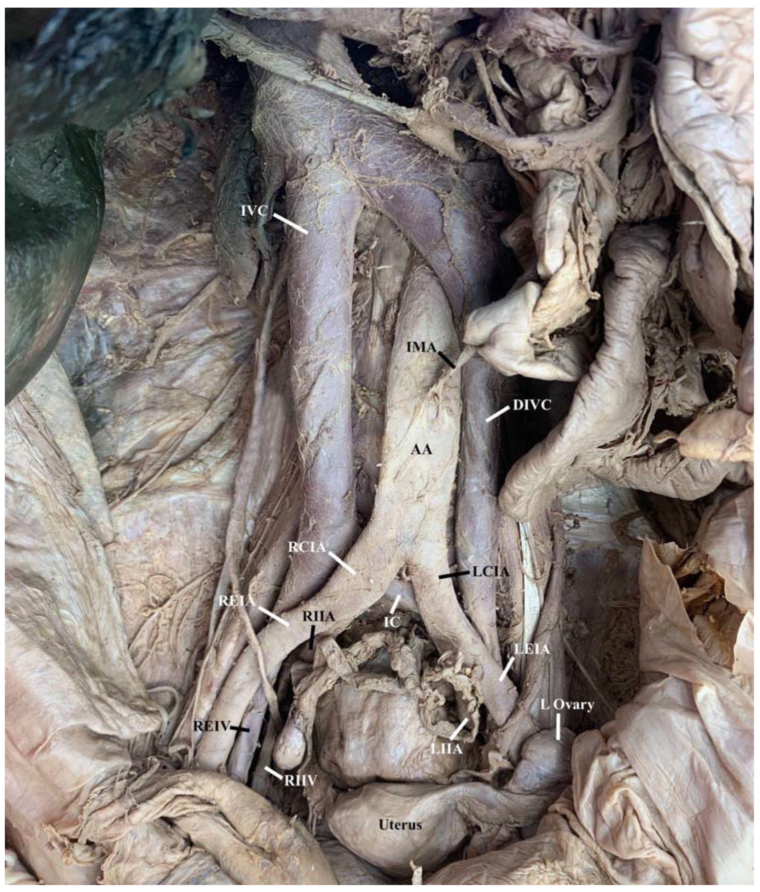
2. Case Description
3. Discussion
3.1. Duplicated or Double Inferior Vena Cava
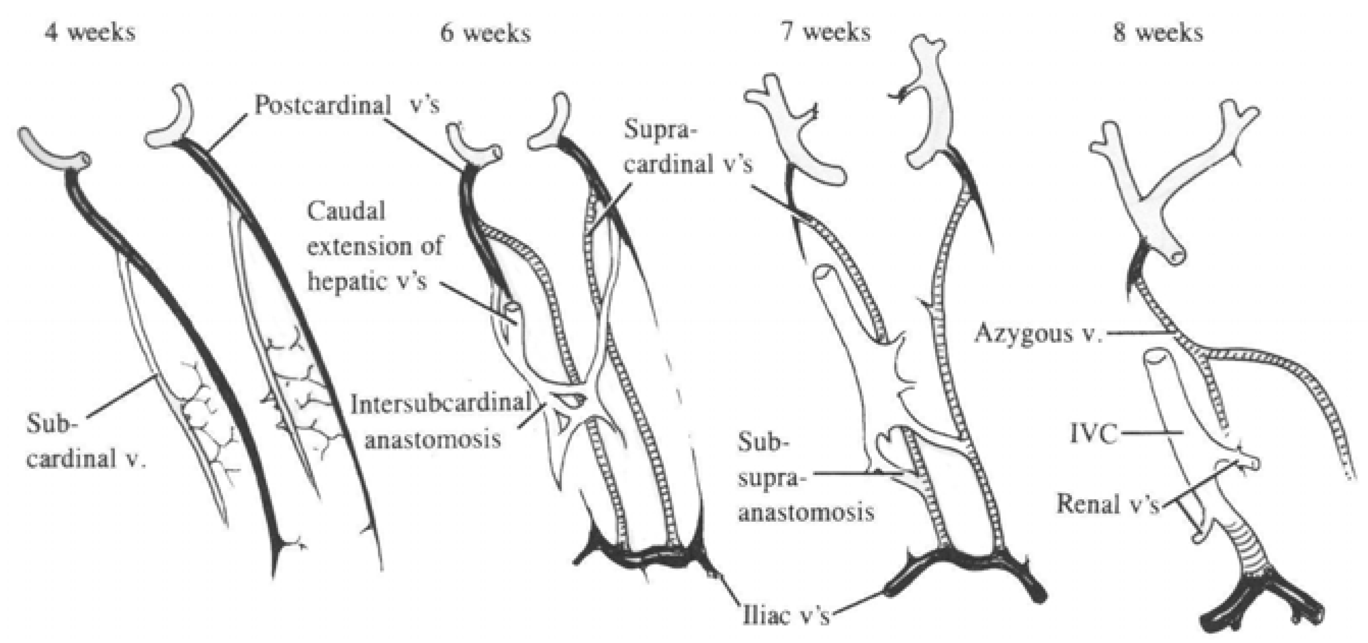
3.2. Embryonic Development
3.3. Associated Comorbidities
3.4. Clinical Significance
4. Conclusions
Author Contributions
Funding
Institutional Review Board Statement
Informed Consent Statement
Data Availability Statement
Acknowledgments
Conflicts of Interest
References
- Lee, B.B. Venous embryology: The key to understanding anomalous venous conditions. Phlebolymphology 2012, 19, 161–204. [Google Scholar]
- Sinkeet, S.; Mwachaka, P.; Muthoka, J.; Saidi, H. Branching Pattern of Inferior Mesenteric Artery in a Black African Population: A Dissection Study. ISRN Anat. 2013, 2013, 962904. [Google Scholar] [CrossRef] [PubMed] [Green Version]
- Aaditya, A.; Neelam, D.; Nageswar Rao, J.; Deepak, G. Congenital Anomalies of Inferior Vena Cava, Review of Embryogenesis, Presentation, Associated Congenital Anomalies and Surgical Importance. Cardiovasc. Thorac. Surg. 2018, 3, 1–6. [Google Scholar]
- Lesma, A.; Bocciardi, A.; Rigatti, P. Circumcaval Ureter: Embryology. Eur. Assoc. Urol. 2006, 5, 444–448. [Google Scholar] [CrossRef]
- Shaha, P.; Garg, A.; Sahoo, K.; Kothari, N.; Garg, P. Duplication of Inferior Vena Cava with Associated Anomalies: A Rare Case Report. J. Clin. Diagn. Res. 2016, 10, TD01. [Google Scholar] [CrossRef] [PubMed]
- Cohen, M.; Gore, R.M.; Vogelzang, R.L.; Rochester, D.; Neiman, H.L.; Crampton, A.R. Accessory Hemiazygos Continuation of Left Inferior Vena Cava: CT Demonstration. J. Comput. Assist. Tomogr. 1984, 8.4, 777–779. [Google Scholar] [CrossRef] [PubMed]
- Geley, T.; Unsinn, K.M.; Auckenthaler, T.M.; Fink, C.J.; Gassner, I. Azygos Continuation of the Inferior Vena Cava: Sonographic Demonstration of the Renal Artery Ventral to the Azygos Vein as a Clue to Diagnosis. AJR 1999, 172, 1659–1662. [Google Scholar] [CrossRef] [PubMed] [Green Version]
- Li, W.; Feng, H.; Jin, L.; Chen, X.M.; Zhang, Z.W. Duplication of the Inferior Vena Cava: A Series. J. Int. Med. Res. 2022, 50, 03000605221100771. [Google Scholar] [CrossRef] [PubMed]
- Rao, B.; Duran, C.; Steigner, M.L.; Rybicki, F.J. Inferior Vena Cava Filter—Associated Abnormalities: MDCT Findings. Am. Roentgen Ray Soc. 2012, 198, 605–610. [Google Scholar] [CrossRef] [PubMed]
- Effler, D.B.; Greer, A.E.; Sifers, E.C. Anomaly of the Vena Cava Inferior: Report of Fatality After Ligation. JAMA 1951, 146, 1321–1322. [Google Scholar] [CrossRef] [PubMed]


Disclaimer/Publisher’s Note: The statements, opinions and data contained in all publications are solely those of the individual author(s) and contributor(s) and not of MDPI and/or the editor(s). MDPI and/or the editor(s) disclaim responsibility for any injury to people or property resulting from any ideas, methods, instructions or products referred to in the content. |
© 2023 by the authors. Licensee MDPI, Basel, Switzerland. This article is an open access article distributed under the terms and conditions of the Creative Commons Attribution (CC BY) license (https://creativecommons.org/licenses/by/4.0/).
Share and Cite
Klansek, J.; Meshida, K.; Maynes, E.; Leighton, M.X.; Wind, G.; Granite, G. Duplicated Inferior Vena Cava in a 69-Year-Old White Female Donor. Anatomia 2023, 2, 117-123. https://doi.org/10.3390/anatomia2020011
Klansek J, Meshida K, Maynes E, Leighton MX, Wind G, Granite G. Duplicated Inferior Vena Cava in a 69-Year-Old White Female Donor. Anatomia. 2023; 2(2):117-123. https://doi.org/10.3390/anatomia2020011
Chicago/Turabian StyleKlansek, Joanna, Keiko Meshida, Elizabeth Maynes, Maria Ximena Leighton, Gary Wind, and Guinevere Granite. 2023. "Duplicated Inferior Vena Cava in a 69-Year-Old White Female Donor" Anatomia 2, no. 2: 117-123. https://doi.org/10.3390/anatomia2020011