Article

Using a Portable Autostereoscopic Screen to Improve Anatomy Teaching and Learning

Department of Biomedical Engineering, School of Biomedical Engineering and Imaging Sciences, King’s College London, St. Thomas’ Hospital, Westminster Bridge Road, London SE1 7EH, UK
*
Author to whom correspondence should be addressed.
Anatomia 2023, 2(1), 88-98; https://doi.org/10.3390/anatomia2010008
Submission received: 29 November 2022 / Revised: 25 January 2023 / Accepted: 6 February 2023 / Published: 14 February 2023
(This article belongs to the Special Issue Advances in Anatomy and Its History)

Abstract

:
Conventional anatomical models and cadaveric specimens can be time-consuming and resource intensive for any anatomical institute. In recent years, there has been a push for more flexible and varied approaches to teaching, including problem-based and computer-aided learning, which includes web-based anatomical models or the use of three-dimensional visualization technology. With advances in hardware, autostereoscopic (AS) 3D screens have become more affordable, portable, and accessible to individuals, not just institutes. At King’s College London (KCL), we developed the Virtual Anatomy and Histology (VAH) platform—an online resource which focuses on perspective volumetric 3D viewing of medical scan data and 3D models to facilitate the online teaching and learning of anatomy. This paper presents the features of VAH and details a study that was conducted in 2022, to evaluate the VAH 3D AS viewer configured with The Looking Glass Portrait (TLG) (Looking Glass, New York, NY, USA) 8-inch AS display. We tested the hypothesis that using an AS display can improve spatial understanding of cardiovascular anatomy. A cardiovascular 3D textured model was used from our gallery to carry out a spatial test. Twenty current healthcare students at King’s participated in the study and completed a structured questionnaire. Results showed that 47.6% and 52.4% of participants agreed and strongly agreed, respectively, that identifying anatomical structures was easier in 3D compared to 2D. Qualitative feedback was positive as most students found King’s VAH and TLG display “useful for people who need help with spatial understanding” and that “it was a good tool to test your anatomical knowledge”. In conclusion, based on the quantitative results and feedback, we are optimistic that King’s VAH and portable AS displays can be beneficial in anatomy education. With the increasing availability of such systems and competitive pricing, this technology is likely to have a significant impact in education in coming years.

1. Introduction

There is a consensus that knowledge of gross anatomy is fundamental in medical education and professions [1]. Understanding the relationship between the structure and function of the human body under normal and pathologic circumstances creates a solid foundation in all healthcare specialties. Moreover, with the growing prominence of medical imaging and minimally invasive therapy, the need to interpret images and anatomical pathways is increasingly important [2].
The long-standing format of many institutions’ anatomical education can be categorized into lectures, practical sessions, and assessments. However, with the rapid development of technology, and in turn medical imaging, there has been a significant paradigm shift in medical education from passive and informative lectures centered around the teacher to a more active and clinical-based student approach. Therefore, the demand for practical sessions has gained more precedence in anatomical education. Practical examinations were identified as the most recommended method of assessment in anatomical education in the Rowland et al. (2011) study by medical students (59.1%), trainees (all stages combined; 54.2%), and specialists (51.7%). This further supports the need for practical educational sessions [3].

1.1. Issues Facing Current Implementation of Practical Anatomy
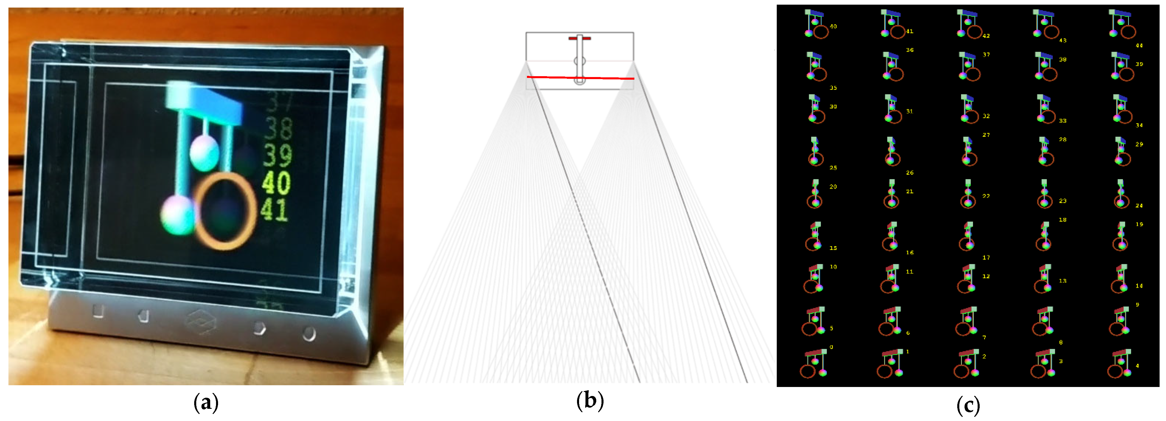
Historically, cadaveric dissection has been the most significant aid in teaching gross anatomy in conjunction with textbooks. A study conducted by Azer and Eizenberg (2007) found that some students feel at a disadvantage without dissection classes [4]. Their study also found that cadaveric dissection was twice as useful as a teaching resource for learning gross anatomy compared to textbooks alone. Cadaveric dissections are impractical at times due to their time constraints, because there is a set number of accessible hours in a dissection theater. Additionally, cadavers are in short supply. Both the UK and the US have been increasing medical student placements to match population demands, but donation numbers are not increasing proportionately [5,6,7]. Cadavers have high costs too; in the UK, cadavers are not freely bought or sold, but institutes must pay for transportation, embalming, and maintaining dissection labs that store the donated cadavers, which all increase cost. In the US, donated human bodies can be sold for around $3000 to $5000, though prices sometimes reach $10,000 [8,9,10].
The coronavirus (COVID-19) pandemic was an unprecedented emergency which had massive economic and social repercussions, and the education sector was not immune to its ramifications [11]. The implementation of social distancing and isolation protocols to curb rising COVID-19 infections within the population meant that more than 1.5 billion students worldwide were affected by closures of schools and universities [12]. Educational institutions around the world were faced with the challenge of teaching and learning in a distant manner.
For teaching, this had instant consequences, which included the cessation of or limited access to conventional anatomy education pedagogies, such as cadaveric dissection and access to non-digitized textbooks. Therefore, many medical institutions were forced to adopt a blended learning or a full e-learning approach. Blended learning involves the integration of online learning and face-to face teaching, which includes online lectures, pre-recorded dissection, and computer-based 3D platforms. While e-learning is fully online, where students interact with learning materials and receive feedback on an online platform, designing an online platform that replicates the visuo-spatial and social interaction of traditional teaching is a unique and growing challenge for educational developers and facilitators [13].
A recurring obstacle among some medical students is the capability to grasp the necessary spatial understanding of 3D anatomy when learning from static 2D illustrations in textbooks and medical imaging [14,15]. Visuo-spatial or spatial ability (SA) is the cognitive capacity to understand and perform mental manipulation, such as object rotation and folding to imagine different perspectives of objects and memorize a spatial representation of the object that includes the relationships among their parts and surroundings [16,17,18]. Preece et al. (2013) and Marks (2000) drew similar conclusions that a poor visuo-spatial ability can compromise students’ abilities to keep up with advancing medical imaging in clinical practice [19,20].
One way to reduce some of the visuo-spatial information lost when one is unable to perform in-person cadaveric practical dissection is to use 3D visualization technologies that have gained momentum in the technological boom in the last couple of decades.

1.2. 3D Displays

3D display technologies are split into two categories. First, there are stereoscopic display technologies that require special glasses or headwear to create the 3D sensation. Second are autostereoscopic 3D display technologies that are glasses-free 3D displays and can be viewed with the naked eye. Stereoscopic and autostereoscopic (AS) displays are groups of 3D displays that only use binocular disparity out of all of the possible physical depth cues.
Stereoscopic displays require 3D glasses to be worn, which can make them a more solitary learning style as they immerse but also isolate the viewer from the world around them. AS displays, on the other hand, can cater to other learning styles, such as social learning, and do not require a headset that can be uncomfortable to wear.
Some AS displays work by using a lenticular lens, such as The Looking Glass (TLG) (Looking Glass, New York, NY, USA) displays, shown in Figure 1a below. In these devices, an array of lenticular lenses is attached to the top of the screen. The lenticular array distributes the pixels of the display to multiple viewpoints. Each lens behaves like a tiny magnifying glass, inflating one of the sub-pixels and hiding the remaining ones. However, depending on the angle of view, each inflated sub-pixel is different. Since both eyes watch the screen from slightly offset angles, they both see different sub-pixels in any given location of the screen, creating binocular disparity, as shown in Figure 1b [21]. Figure 1c showcases the TLG’s simultaneous 45 viewpoints/images rendered to create its 3D visualization.
Dedicated holographic devices are markedly expensive. Depending on size of the display, a standard device used to price at a minimum of $3000. However, with recent advances in hardware capabilities and a growing market, now more affordable, portable, and accessible systems are available, such as TLG Portrait (displayed in Figure 2) which is $399, and the Leia Inc. (Leia Inc., Menlo Park, CA, USA) Lume pad tablet, which is $650 [23,24]. This accessibility opens up more opportunities for educators and students to use these devices in anatomical teaching and learning.

1.3. 3D Displays in Anatomical Education

There is some evidence in anatomical education literature showing the application of autostereoscopic displays in the medical field. In a few studies, AS has been used for training surgeons. Narita et al. (2014) studied twenty neurosurgeons and asked them to identify blood vessels using a 2D screen and an autostereoscopic screen. They found significantly more correct answers were attained using the 3D autostereoscopic screens, with 91.7% correct responses compared to 56.7% with the 2D screen [26]. This shows that AS gives the viewer a higher sense of realism and accuracy, therefore enhancing their performance in identifying key anatomical structures. Silvestri et al. (2011) also found that AS enhanced the viewers’ performance. They found that the fifteen participants, who were physicians, were able to carry out the five assigned tasks faster on the autostereoscopic screens compared to the 2D monitor [27].
The benefits of this system and the studies carried out on the training of surgeons mean that there is some promise in using AS to teach anatomy. Luursema et al. (2011) studied stereoscopic screens that utilized glasses and showed that students with low visuospatial ability benefitted more from making use of the screen than students with higher spatial abilities, as students were able to visualize abdominal structures more easily than with 2D models [28]. Although this study was conducted with a stereoscopic screen, the results are likely to be replicated with AS, as they are more user-friendly. Di Natale et al. (2020) have also identified that using immersive VR in teaching promoted experiential learning, intrinsic motivation, and engagement, and supported the transfer of knowledge [29]. Sinha et al. (2022) evaluated a 42-inch AS display for anatomical education and found that the AS display promoted 3D stereoscopic viewing in a group setting for collaborative learning that a single-user VR headset could not [30]. This type of social learning and the other promoted attributes mentioned are all key psychological factors that support deep learning and teaching. Therefore, in this study, we will explore the use of the portable 3D AS display we configured to be compatible with our online learning resource.

2. Materials and Methods

We proposed a 3D visualization system that incorporates volumetric 3D medical data and 3D models with the capabilities of displaying these on 3D displays.

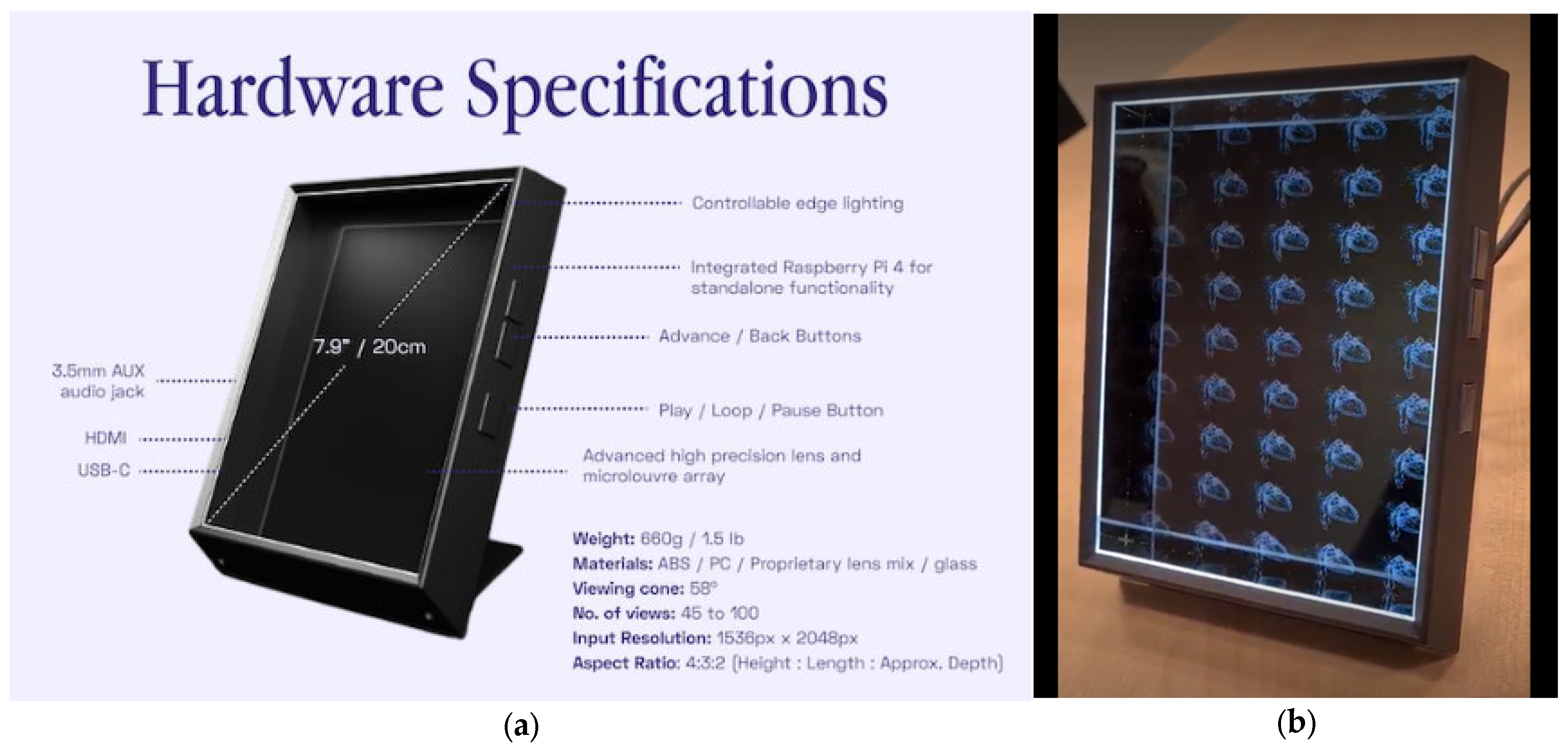
2.1. System Overview

Virtual Anatomy and Histology (VAH) is a university-based online learning web-based application, shown in Figure 3 below, and was developed to allow students to study and interact with anatomy through 3D models, histological images, and 3D imaging scans that the different King’s College London (KCL) departments and museums possessed. The departments and museums had numerous subscriptions for anatomical software. It was decided that an in-house all-in-one online anatomical learning platform or tool could reduce costs, as the new cost would go towards cloud and web app hosting. It also provides a more bespoke solution, for example, by allowing educators and students to design and save their own 3D annotated anatomical scenes for lessons and private study. VAH has volumetric datasets, 3D anatomical models, and histology slides from a cloud-based gallery which can be accessed by users to view and interact with.
The TLG Portrait 8-inch AS display was configured to be compatible with VAH’s model viewer. From this point onwards, we will be focusing on the VAH TLG viewer, which creates a popup window that can be dragged onto the TLG’s screen, to allow models to be viewed in 3D mode or 2D mode. The viewer can load 3D models from the site catalogue or from the user’s personal computer files. The VAH TLG viewer was now ready for usability testing.

2.2. Study Design

2.2.1. Participants

Ethics approval was obtained via the Institutional Review Board (or Ethics Committee) of the KCL Research Ethics Office (MRSP-20/21-21158). Participation in the study was anonymous and voluntary. Information sheets about the study were provided during recruitment via cohort mailing lists, as required by the ethics committee. As explained in the information sheet and discussed in person, students who completed and submitted the survey, had provided informed consent for their data to be used. Inclusion criteria included being a current healthcare student in the Faculty of Life Sciences and Medicine. Exclusion criteria included if the user had photosensitive epilepsy or cybersickness. Ultimately, twenty current healthcare students at KCL were recruited to participate in the study via this strategy. Their level of education ranged from 1st year undergraduates to Ph.D. candidates, with 10 undergraduates (UG) and 10 postgraduates (PG).

2.2.2. Procedure

The study was conducted in 2022, with the aim to assess the use of VAH in anatomical education and to test the hypothesis that visuospatial understanding of cardiovascular anatomy was improved by using TLG display. The cardiovascular 3D textured model from our gallery that was used in the spatial test can be seen in Figure 4a.
The assessment used a repeated measures study design, where participants were asked to describe the spatial relationship between two anatomical structures in a heart model in both 3D and 2D. The two anatomical structures were selected randomly when the spatial test button was pressed, with their names also on display. The rest of the model structures were textured with an “X-ray”-style shader to highlight the positions of the selected structures, as shown in Figure 4b. The spatial relationship had to be described using anatomical orientations and directional terms with the use of the reference diagram shown in Figure 4c. For example, in the case of Figure 4b, the papillary muscles and the aortic valve are the selected/highlighted test objects 1 and 2. Therefore, participants were asked to describe the relationship of object 2 with respect to object 1 with the diagram Figure 4c as a reference while they interact with the viewer through orbiting, rotating, translation, and zooming. There were 4 parts to an answer:
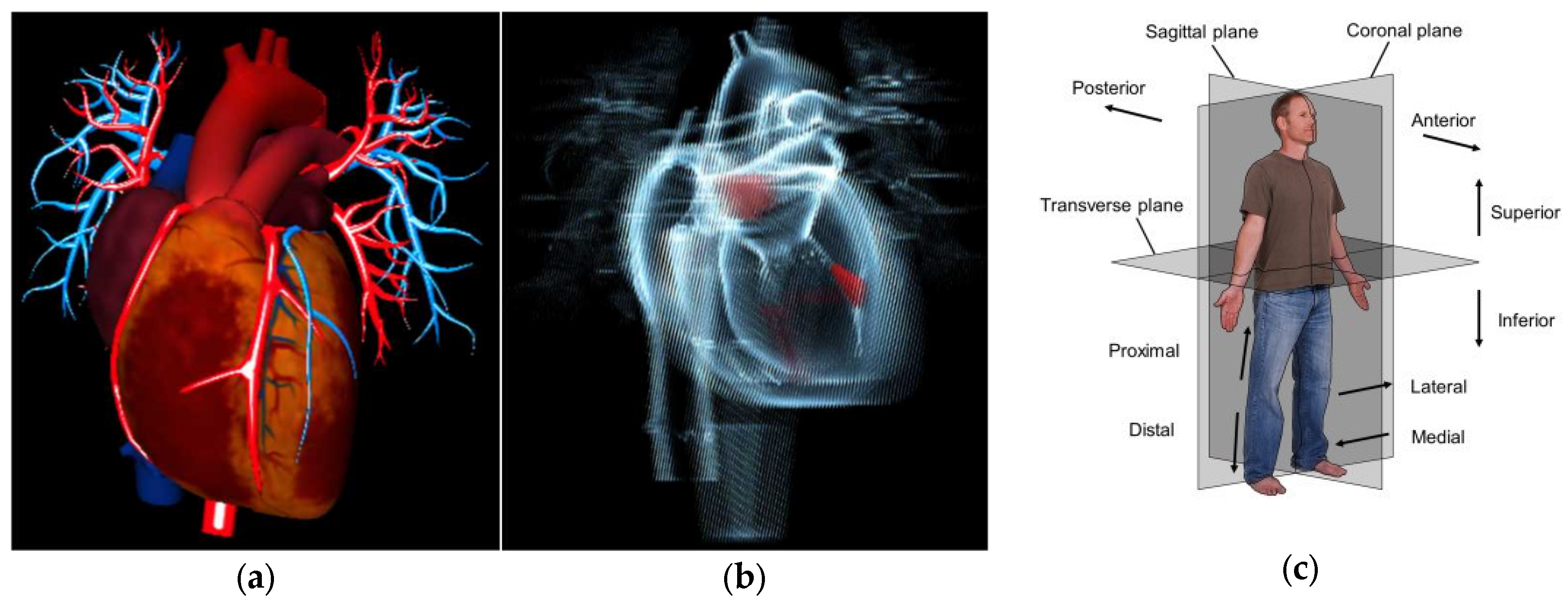
  • Superior or inferior or in the same transversal plane;
  • Anterior or posterior or in the same coronal plane;
  • Left or right or in the same sagittal plane;
  • Proximal and distal or lateral and medial or on the same medial line.
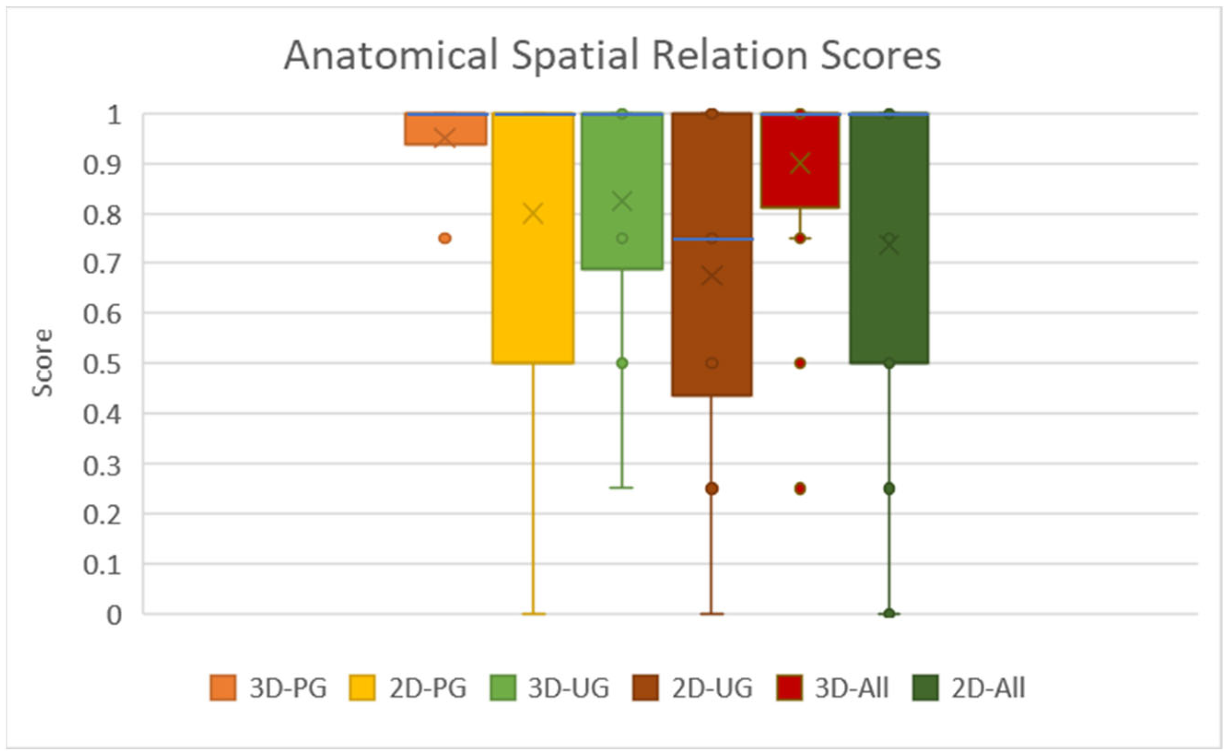
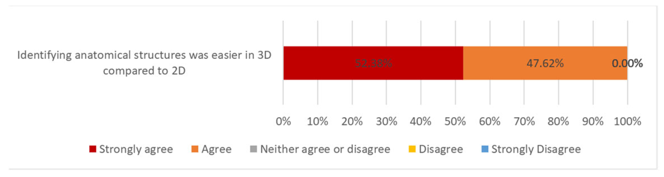
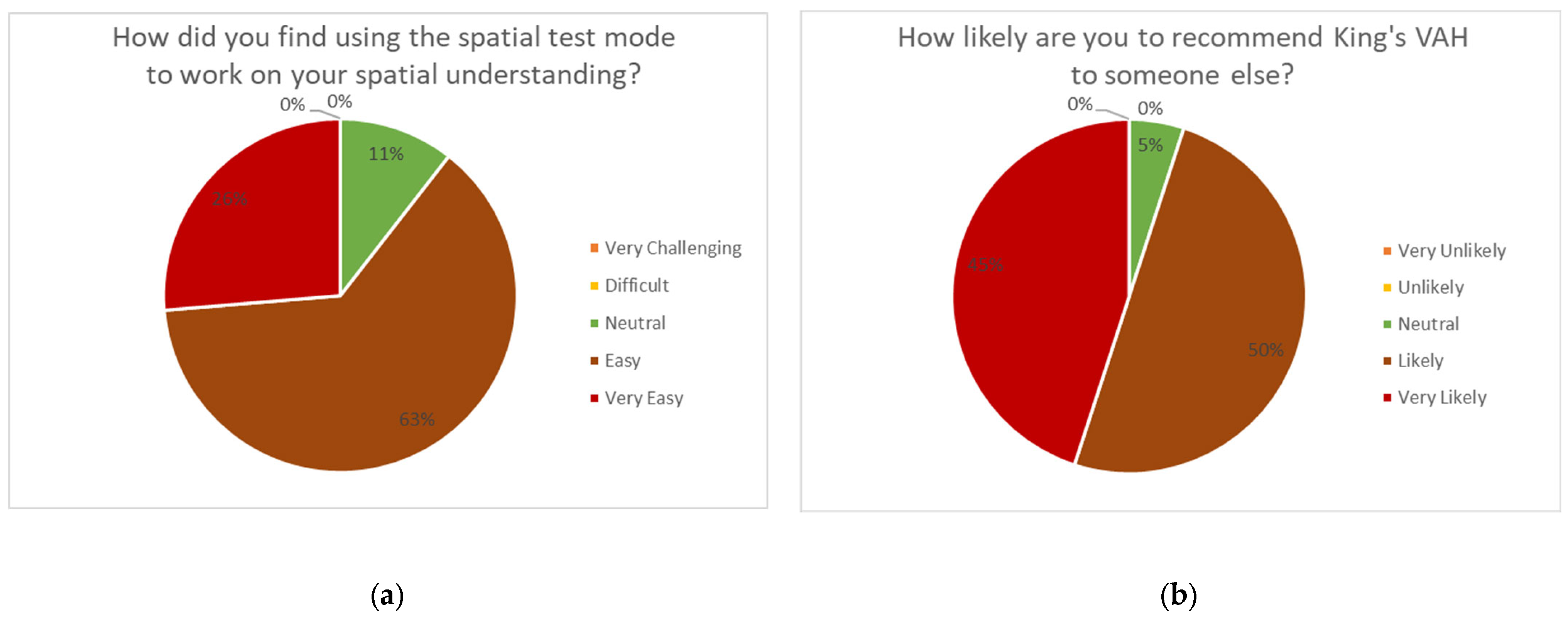
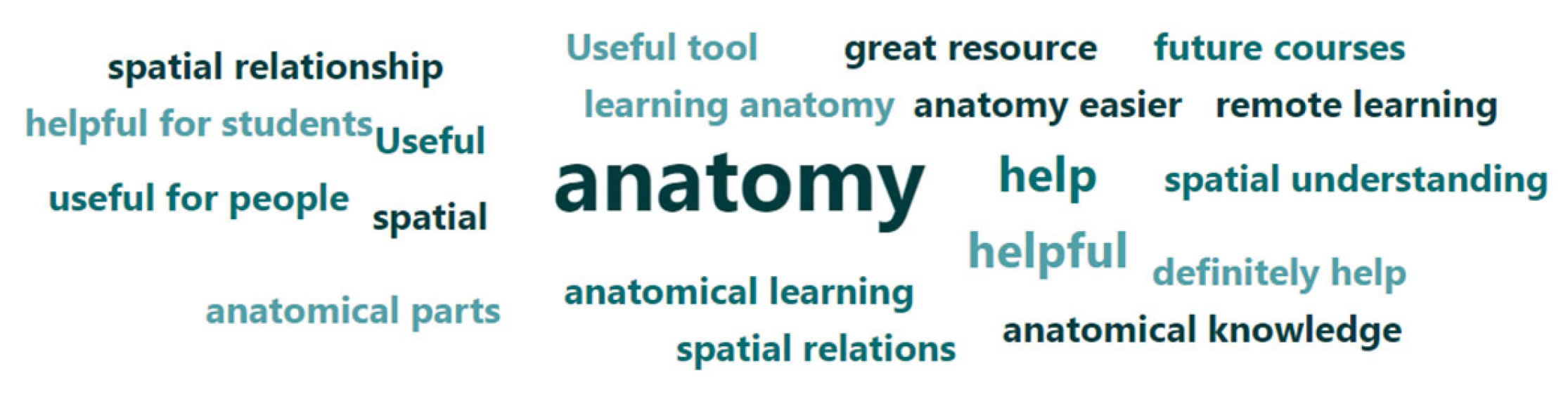
Each correct response was worth 0.25, amounting to a total mark of 1 for a perfect score. We rated the performance by comparing our version, which we made from analyzing the anatomical model we used in the assessment. We calculated the centroid and the bounding box of each of the objects and compared them in the different planes. Therefore, someone who scored 0 was interpreted as having little spatial understanding of the physical relationship between the anatomical objects when using 3D or 2D display mode. On the other hand, someone who scored 1 was interpreted as having a high understanding of the spatial relationship between the anatomical objects.
Figure 4. Shows some of the scenes throughout the experiment. (a) Shows the normal textured heart model used in the assessment through a 2D display. (b) Shows one of the randomized spatial test scenes in 3D mode. (c) Shows the anatomical orientation and the reference diagram participants used [31].
Anatomia 02 00008 g004
Volunteers performed the activity three times, with an initial attempt in 3D mode using the 3D display. The initial attempt was used as practice to get used to VAH and to confirm that they were able to perceive the 3D visuals of the screen, and to allow the users to adjust the settings of the viewer to best enhance the 3D visualizations (i.e., background color, directional lighting, model size, and model position). We allowed participants autonomy to choose the order they wanted to complete their assessed 2D and 3D tries, which were all conducted on the TLG display. All results were filled in and collected using an online Microsoft form (Microsoft, Redmond, WA, USA), for the possibility of remote participants. However, in the end, all the 3D TLG assessments ended up being completed in person on campus in a classroom, and not remotely. Consequently, there was an additional in-person supervisor who provided initial assistance when participants used the 3D screen for the first time. A paired t-test analysis was performed comparing the overall 2D mode (“before”) vs 3D mode (“after”) test scores, to establish statistical significance at p = 0.05. Participants were also asked to complete a questionnaire on the form containing a series of Likert scale user experience questions and statements such as “How likely are you to recommend King’s VAH to someone else?”, or grading their agreement with the statement “Identifying anatomical structures was easier in 3D compared to 2D”. There were also some additional open-ended questions, such as “Based on today’s session, to what extent do you think King’s VAH might impact your future anatomical learning?” and “Do you have any final feedback regarding the King’s VAH website?”. All these helped to collect feedback about the site, the 3D display, and the tools which the volunteers found particularly useful.

3. Results

Figure 5 shows the anatomical spatial test scores for 3D and 2D viewing.
The 3D-All score had a first quartile (Q1 = 0.875) and a third quartile (Q3 = 1) with an interquartile range (IQR = 0.125) and a median = 1. While the 2D-All score had a first quartile (Q1 = 0.5) and a third quartile (Q3 = 1) with an interquartile range (IQR = 0.5) and a median = 1. In terms of the mean (M) and standard deviation (SD), the 2D display had a score of (M = 0.73, SD = 0.34) and the 3D display one of (M = 0.90, SD = 0.20). For the paired t-test, t and p-values were t(19) = 2.37 and p = 0.028, which is less than a = 0.05. The 3D-All mean score was higher than the mean score of the 2D-All, and the results of the paired t-test indicated there was a significant difference between the two groups. If we separate the groups into level of education, UG and PG scores, we still see similar trends between 2D and 3D groups. The 3D groups have lower IGRs than 2D, 3D-UG = 0.313 and 3D-PG = 0.062 vs. 2D-UG = 0.563 and 2D-PG = 0.500. In terms of medians, both 3D-PG = 1 and 2D-PG = 1 were the same, but for undergraduates, 3D-UG = 1 had a higher median than 2D-UG = 0.5. The 2D-UG score had the most evenly distributed test scores, while 3D-UG had a more positively skewed score.
Figure 6 shows the agreement level of participants based on the Likert scale for the statement “Identifying anatomical structures was easier in 3D compared to 2D”.
From Figure 6, we can see that 47.6% and 52.4% of participants agreed and strongly agreed, respectively, that identifying anatomical structures was easier in 3D compared to 2D. The results show an extreme positive skew.
Figure 7 displays participants’ responses to two further questions asked in the questionnaire. Both results are skewed to more positive responses.

Qualitative Results

The most common themes that appeared in the responses to the question “Based on today’s session, to what extent do you think King’s VAH might impact your future anatomical learning?” were ‘helpful’ or ‘useful’. Visualized in a word cloud in Figure 8, the words helpful or useful appeared in 45% of people’s responses.
When asked the question “Do you have any final feedback regarding the King’s VAH website?”, 30% of users suggested they would have liked an anatomical position reset button in the spatial test.

4. Discussion and Conclusions

Most participants that used the 3D visualization system with the 3D display performed better than in the 2D display. Both the 2D and 3D test scores had a median of 1, but the interquartile range of 0.5 for the 2D was 4× larger than the 0.125 of the 3D, shown in Figure 5. This shows that both sets of data are left skewed, but 2D has a higher variability. The positive skew of both datasets could be related to the freedom of VAH as an interactive viewer to allow for explorative spatial understanding, through orbiting the model. From the less variability of the 3D results, we can infer that the AS screen further enhances anatomical spatial understanding through the visualization of depth information in a natural way for participants. This is further reinforced by looking at UG scores alone, as 2D-UG had the most normal distribution. However, when participants used the 3D display, 3D-UG scores were more positively skewed. We can also analyze the different group means, where 2D-All had a lower mean (M = 0.73, SD = 0.34) than 3D-All (M = 0.90, SD = 0.20). In addition, the paired t-test results indicate that the mean difference between 3D and 2D datasets were different enough to support the hypothesis that “visuospatial understanding of cardiovascular anatomy was improved by using the TLG display”. The hypothesis is further supported by the fact that 47.6% and 52.4% of participants agree and strongly agree, respectively, that identifying anatomical structures was easier in 3D compared to 2D. This is particularly positive, because it suggests that portable and affordable AS screens can provide the necessary stereo depth to assist with spatial understanding, which could enhance remote learning. However, n = 20 is a small sample size for an educational assessment and in future work we would like to increase the cohort size.
An observation throughout the study was that all participants chose to complete the 3D assessment first. A probable reason may have been the excitement related to a new technology. As all participants ended up choosing 2D mode display last, we can ask whether there was any brain fatigue that affected the scores. It may have been better to have randomized the order of the tests.
A limitation of the spatial test was that pairing selection of the organ structures were chosen at random and therefore, a few combinations of test objects had easier spatial relationships to describe than others. The supervisors during the experiment did allow for additional randomizer selection tries, if the relationship was too easy, to balance out the difficulty. However, anatomical names generally do provide some prior information of the anatomical position of an object, for example, middle cardiac vein. Therefore, part of the spatial relationship between structures could be deciphered before visualization. Nevertheless, as the focus was on learning, we still provided the names of the anatomical parts, to help them match names and characteristics of model structures, such as positional information, which is good for learning and is one of our aims.
One of the main critiques and feedback provided was the need for an anatomical reset button. This is very reasonable as the 3D scene itself is rotatable because we can orbit around the model even in 2D. We anticipated that if resolved, this would reduce the time taken to manually reset the orientation. Since the writing of this paper, the button has been added. In addition, the toolbar was also mentioned as something to improve.
Helpful or useful appeared in 45% of people’s responses when describing the future impact of VAH in anatomical learning. We can infer that VAH has a place in the future of anatomical learning, especially as 95% of participants would likely or very likely recommend VAH to someone else.
In conclusion, based on the quantitative and qualitative results and feedback, we are optimistic that King’s VAH and small portable AS displays can be beneficial in anatomical education. There are improvements to be made to the controls and additional features. With the increasing availability of such systems and reducing costs, this technology is likely to have a significant impact in education in coming years.

Author Contributions

Conceptualization, K.R.; Investigation, O.W. and E.-M.A.O.; Methodology, E.-M.A.O.; Software, E.-M.A.O.; Validation, H.L. and E.-M.A.O.; Supervision, K.R. All authors have read and agreed to the published version of the manuscript.

Funding

This work is funded by the King’s College London and Imperial College London EPSRC Centre for Doctoral Training in Medical Imaging (EP/L015226/1) and EPSRC National Productivity Investment Fund (NPIF) (EP/R512552/1). This research was supported by the National Institute for Health Research (NIHR) Biomedical Research Centre award to Guy’s and St Thomas’ NHS Foundation Trust in partnership with King’s College London, and by the NIHR Healthcare Technology Co-operative for Cardiovascular Disease at Guy’s and St Thomas’ NHS Foundation Trust, Project 651120.

Institutional Review Board Statement

The study was conducted in accordance with the Declaration of Helsinki, and approved by the Institutional Review Board (or Ethics Committee) of KCL Research Ethics Office (MRSP-20/21-21158).

Informed Consent Statement

Informed consent was obtained from all subjects involved in the study.

Data Availability Statement

Not applicable.

Acknowledgments

The Heart texture model was provided by YingLiang Ma.

Conflicts of Interest

The authors declare no conflict of interest.

References

  1. Böckers, A.; Jerg-Bretzke, L.; Lamp, C.; Brinkmann, A.; Traue, H.C.; Böckers, T.M. The Gross Anatomy Course: An Analysis of Its Importance. Anat. Sci. Educ. 2010, 3, 3–11. [Google Scholar] [CrossRef] [PubMed]
  2. McCuskey, R.S.; Carmichael, S.W.; Kirch, D.G. The Importance of Anatomy in Health Professions Education and the Shortage of Qualified Educators. Acad. Med. 2005, 80, 349–351. [Google Scholar] [CrossRef] [PubMed]
  3. Rowland, S.; Ahmed, K.; Davies, D.C.; Ashrafian, H.; Patel, V.; Darzi, A.; Paraskeva, P.A.; Athanasiou, T. Assessment of Anatomical Knowledge for Clinical Practice: Perceptions of Clinicians and Students. Surg. Radiol. Anat. 2011, 33, 263–269. [Google Scholar] [CrossRef] [PubMed]
  4. Azer, S.A.; Eizenberg, N. Do We Need Dissection in an Integrated Problem-Based Learning Medical Course? Perceptions of First- and Second-Year Students. Surg. Radiol. Anat. 2007, 29, 173–180. [Google Scholar] [CrossRef] [PubMed]
  5. The Expansion of Medical Student Numbers in the United Kingdom Medical Schools Council Position Paper; Medical Schools Council: London, UK, 2021; Available online: https://www.medschools.ac.uk/media/2899/the-expansion-of-medical-student-numbers-in-the-united-kingdom-msc-position-paper-october-2021.pdf (accessed on 29 November 2022).
  6. AAMC. U.S. Medical School Enrollment Rises 30%. Available online: https://www.aamc.org/news-insights/us-medical-school-enrollment-rises-30 (accessed on 12 January 2023).
  7. Habicht, J.L.; Kiessling, C.; Winkelmann, A. Bodies for Anatomy Education in Medical Schools: An Overview of the Sources of Cadavers Worldwide. Acad. Med. 2018, 93, 1293–1300. [Google Scholar] [CrossRef] [PubMed]
  8. Singh, V.; Kharb, P. A Paradigm Shift from Teaching to Learning Gross Anatomy: Meta-Analysis of Implications for Instructional Methods. J. Anat. Soc. India 2013, 62, 84–89. [Google Scholar] [CrossRef]
  9. In the U.S. Market for Human Bodies, Anyone Can Sell the Donated Dead. Available online: https://www.reuters.com/investigates/special-report/usa-bodies-brokers/ (accessed on 12 January 2023).
  10. Heizenrader. Why Virtual Cadavers Are a Wise Investment for Medical Schools. Available online: https://heizenrader.com/why-virtual-cadavers-are-a-wise-investment-for-medical-schools/ (accessed on 12 January 2023).
  11. Ayittey, F.K.; Ayittey, M.K.; Chiwero, N.B.; Kamasah, J.S.; Dzuvor, C. Economic Impacts of Wuhan 2019-NCoV on China and the World. J. Med. Virol. 2020, 92, 473–475. [Google Scholar] [CrossRef] [PubMed]
  12. UNESCO’s Education Response to COVID-19. Available online: https://en.unesco.org/covid19/educationresponse/support (accessed on 8 August 2022).
  13. Tóth, Á.; Pentelényi, P.; Tóth, P. Virtual Learning Aspects of Curriculum Development in Technical Teacher Training. In Proceedings of the 2006 International Conference on Intelligent Engineering Systems, London, UK, 26–28 June 2006; pp. 308–313. [Google Scholar] [CrossRef]
  14. Berney, S.; Bétrancourt, M.; Molinari, G.; Hoyek, N. How Spatial Abilities and Dynamic Visualizations Interplay When Learning Functional Anatomy with 3D Anatomical Models. Anat. Sci. Educ. 2015, 8, 452–462. [Google Scholar] [CrossRef] [PubMed]
  15. Pedersen, K. Supporting Students with Varied Spatial Reasoning Abilities in the Anatomy Classroom. Teach. Innov. Proj. 2012, 2. Available online: https://ojs.lib.uwo.ca/index.php/tips/article/view/3576 (accessed on 29 November 2022).
  16. Bogomolova, K.; Hierck, B.P.; van der Hage, J.A.; Hovius, S.E.R. Anatomy Dissection Course Improves the Initially Lower Levels of Visual-Spatial Abilities of Medical Undergraduates. Anat. Sci. Educ. 2020, 13, 333–342. [Google Scholar] [CrossRef] [PubMed]
  17. Gonzales, R.A.; Ferns, G.; Vorstenbosch, M.A.T.M.; Smith, C.F. Does Spatial Awareness Training Affect Anatomy Learning in Medical Students? Anat. Sci. Educ. 2020, 13, 707–720. [Google Scholar] [CrossRef] [PubMed]
  18. Hegarty, M.; Waller, D. A Dissociation between Mental Rotation and Perspective-Taking Spatial Abilities. Intelligence 2004, 32, 175–191. [Google Scholar] [CrossRef]
  19. Preece, D.; Williams, S.B.; Lam, R.; Weller, R. “Let’s Get Physical”: Advantages of a Physical Model over 3D Computer Models and Textbooks in Learning Imaging Anatomy. Anat. Sci. Educ. 2013, 6, 216–224. [Google Scholar] [CrossRef] [PubMed]
  20. Marks, S.C., Jr. The Role of Three-Dimensional Information in Health Care and Medical Education: The Implications for Anatomy and Dissection. Clin. Anat. 2000, 13, 448–452. [Google Scholar] [CrossRef] [PubMed]
  21. Alioscopy. How Does It Work? Available online: https://www.alioscopy.com/en/principles.php (accessed on 30 September 2022).
  22. Looking Glass Documentation. How the Looking Glass Works. Available online: https://docs.lookingglassfactory.com/keyconcepts/how-it-works (accessed on 30 September 2022).
  23. Looking Glass Portrait. Available online: https://lookingglassfactory.com/looking-glass-portrait (accessed on 4 November 2022).
  24. CNET. Lume Pad Brings Back Glasses-Free 3D—This Time on an Android Tablet. Available online: https://www.cnet.com/tech/computing/lume-pad-brings-glasses-free-3d-back-again-on-an-android-tablet/ (accessed on 4 November 2022).
  25. Kickstarter. Looking Glass Portrait by Looking Glass. Available online: https://www.kickstarter.com/projects/lookingglass/looking-glass-portrait/description (accessed on 30 September 2022).
  26. Narita, Y.; Tsukagoshi, S.; Suzuki, M.; Miyakita, Y.; Ohno, M.; Arita, H.; Saito, Y.; Kokojima, Y.; Watanabe, N.; Moriyama, N.; et al. Usefulness of a Glass-Free Medical Three-Dimensional Autostereoscopic Display in Neurosurgery. Int. J. Comput. Assist. Radiol. Surg. 2014, 9, 905–911. [Google Scholar] [CrossRef] [PubMed]
  27. Silvestri, M.; Ranzani, T.; Argiolas, A.; Vatteroni, M.; Menciassi, A. A Multi-Point of View 3D Camera System for Minimally Invasive Surgery. Procedia Eng. 2012, 47, 1211–1214. [Google Scholar] [CrossRef]
  28. Luursema, J.-M.; Verwey, W.B.; Editor, A.; Kumar Tripathi, A. The Contribution of Dynamic Exploration to Virtual Anatomical Learning. Adv. Hum. Comput. Interact. 2011, 2011, 965342. [Google Scholar] [CrossRef]
  29. Di Natale, A.F.; Repetto, C.; Riva, G.; Villani, D. Immersive Virtual Reality in K-12 and Higher Education: A 10-Year Systematic Review of Empirical Research. Br. J. Educ. Technol. 2020, 51, 2006–2033. [Google Scholar] [CrossRef]
  30. Sinha, S.; DeYoung, V.; Chan, S.; Ives, R.; Lohit, S.; Reis, I.; Touliopoulos, E.; Nehru, A.; Mitchell, J.P.; Brewer-Deluce, D.; et al. Evaluating Autostereoscopy (AlioscopyTM) Use for Anatomy Education. FASEB J. 2022, 36. [Google Scholar] [CrossRef]
  31. Quizlet. Directional Terms Anatomy and Physiology Diagram. Available online: https://quizlet.com/424696628/directional-terms-anatomy-and-physiology-diagram/ (accessed on 27 November 2022).
Figure 1. Diagrams demonstrating how the TLG display works: (a) is an image that shows what is viewed on the TLG display at different positions. The numbers in the scene showcase the different views/perspective of the scene as the viewer moves from one edge to the other [22]. (b) is a top-down ray diagram of the parallax used to create the 3D visualization at different positions. The bold lines show the eye positions needed to see the viewpoint shown in (a) [22]. It also shows the zero-parallax plane (the thin red line) where objects are the sharpest in the scene. (c) is known as the ‘quilt’, which displays the 45 views simultaneously on TLG [22].
Anatomia 02 00008 g001
Figure 2. (a) The hardware specification of TLG portrait [25]. (b) An image of TLG portrait in quilt mode showing an X-ray textured model [23].
Anatomia 02 00008 g002
Figure 3. Shows King’s VAH Home Page with its different features, such as Histology (slide) viewer, 3D model viewer, 3D scans (volumetric) viewer, augmented reality (AR) viewer, and TLG Viewer.
Anatomia 02 00008 g003
Figure 5. Shows box and whisker plots of the anatomical spatial relation scores different level of education (undergraduates—UG, postgraduates—PG and All). The cross indicates the mean, and the medium line has been highlighted blue.
Anatomia 02 00008 g005
Figure 6. A stacked bar graph showing the Likert scale results for participant responses to the statement comparing 2D and 3D.
Anatomia 02 00008 g006
Figure 7. Pie charts of two user experience Likert scale results. (a) The figure shows a pie chart of the results to the question “How did you find using the spatial test mode to work on your spatial understanding?”. (b) The figure shows a pie chart of the results to the question “How likely are you to recommend King’s VAH to someone else?”.
Anatomia 02 00008 g007
Figure 8. Shows a collection of responses to the question “Based on today’s session, to what extent do you think King’s VAH might impact your future anatomical learning?”.
Anatomia 02 00008 g008
Disclaimer/Publisher’s Note: The statements, opinions and data contained in all publications are solely those of the individual author(s) and contributor(s) and not of MDPI and/or the editor(s). MDPI and/or the editor(s) disclaim responsibility for any injury to people or property resulting from any ideas, methods, instructions or products referred to in the content.

