
Advances in Neuroanatomy through Brain Atlasing
Nowinski Brain Foundation, Warsaw West County, P.O. Box 56, 05-092 Lomianki, Poland
Anatomia 2023, 2(1), 28-42; https://doi.org/10.3390/anatomia2010004
Submission received: 11 November 2022
/
Revised: 4 January 2023
/
Accepted: 14 January 2023
/
Published: 19 January 2023
(This article belongs to the Special Issue Advances in Anatomy and Its History)
Abstract
:Human brain atlases are tools to gather, present, use, and discover knowledge about the human brain. The developments in brain atlases parallel the advances in neuroanatomy. The brain atlas evolution has been from hand-drawn cortical maps to print atlases to digital platforms which, thanks to tremendous advancements in acquisition techniques and computing, has enabled progress in neuroanatomy from gross (macro) to meso-, micro-, and nano-neuroanatomy. Advances in neuroanatomy have been feasible because of introducing new modalities, from the initial cadaveric dissections, morphology, light microscopy imaging and neuroelectrophysiology to non-invasive in vivo imaging, connectivity, electron microscopy imaging, genomics, proteomics, transcriptomics, and epigenomics. Presently, large and long-term brain projects along with big data drive the development in micro- and nano-neuroanatomy. The goal of this work is to address the relationship between neuroanatomy and human brain atlases and, particularly, the impact of these atlases on the understanding, presentation, and advancement of neuroanatomy. To better illustrate this relationship, a brief outline on the evolution of the human brain atlas concept, creation of brain atlases, atlas-based applications, and future brain-related developments is also presented. In conclusion, human brain atlases are excellent means to represent, present, disseminate, and support neuroanatomy.
1. Introduction
For centuries, the human brain has been an enormous challenge for scientists and an abundant inspiration for artists. However, the great importance of the brain has not always been fully understood. In Ancient Egypt, for instance, the brain was considered a rather useless organ with no need to be mummified. In Ancient Greece, Herodotus advising on the mummification process recommended removing as much of the brain as possible and mixing any remains of it with drugs, implying the brain was toxic. One of the greatest philosophers of Antiquity, Aristotle, who also substantially contributed to natural sciences, viewed the brain as a cooling mechanism for blood, while the heart was the seat of intelligence. Toward the end of Antiquity, St. Augustine, considered the father of psychology, demonstrated a better understanding of the brain by dividing it into three compartments, the environment with the senses, the movement environment, and the seat of memory. Then, after one thousand years of stagnation, Leonardo da Vinci created beautiful images, though not always anatomically correct, of the brain capturing its anatomy, by bridging art and science. It was however Vesalius, universally considered to be the most important anatomist and the founder of modern anatomy, who started a new era of anatomical investigation ending its dependence on Greek and Arabic authorities, often erroneous and based upon animal rather than human studies [1]. Vesalius also made a substantial contribution to neuroanatomy by providing the first description of the human corpus callosum linking two halves of the brain, putamen, globus pallidus, caudate nucleus, pulvinar, midbrain, pineal body, and internal capsule, among others. Willis introduced a new level of neuroanatomical accuracy and reclassified the cranial nerves. Neuroanatomy advancements through brain gross dissections were accomplished by 19th-century neuroanatomists including Arnold, Burdach, Foville, Gratiolet, Mayo, and Reil as it was illustrated and reviewed by Schmahmann and Pandya [2]. One of the first maps of the human cortical surface based on cytoarchitectonics was created in 1909 by a German neurologist named Korbinian Brodmann [3]. Brodmann postulated that areas differing in structure perform different functions. Brodmann’s areas are still in use today in neuroeducation and research.
Since then, there has been a tremendous development of human brain maps and atlases in terms of concept, content, functionality, applications, and availability. I have earlier distinguished four generations of brain atlases: early cortical maps, print stereotactic atlases, early digital atlases, and advanced brain atlas platforms [4].
Neuroanatomy, as the study of the structure and organization of the nervous system, and human brain atlases, as tools to gather, present, use, and discover knowledge about the human brain, are obviously linked. The goal of this work is to address the relationship between neuroanatomy and human brain atlases and, particularly, the impact of these atlases on the understanding, presentation, and advancement of neuroanatomy. To better illustrate this impact, a brief outline about the evolution of the human brain atlas concept, creation of brain atlases, atlas-based applications, and future brain-related developments is also presented.
2. Evolution of Brain Atlas Concept
The concept of the brain atlas has been evolving together with the tremendous progress in neuroanatomy thanks to imaging and computing. It should be noted that various authors consider or define the brain atlas differently as briefly overviewed below. Traditionally, the brain atlas is considered a collection of brain maps or a database. Here, there are a few examples. Roland and Zilles define brain atlases as collections of micrographs or schematic drawings of brain sections with identified anatomic structures [5]. Evans et al. treat brain atlases as large-scale neuroimaging databases providing the mean and variance in the population [6]. Mori et al. consider the brain atlas a tool for image structurization via atlas-based image subdivision to exploit a great amount of imaging information offered by medical systems [7]. Amunts et al. regard brain atlases as central for integrating diversified information about various aspects of the brain [8]. Kuan et al. consider the brain atlas a tool aiming to integrate diverse information, understand complex brain anatomy, localize experimental data, and plan experiments [9]. Costa et al. consider the atlases the means able to produce specific, testable hypotheses about circuit organization and connectivity [10]. Chon et al. find anatomical atlases in standard coordinates to be necessary for the interpretation and integration of research findings in a common spatial context [11]. Hence, despite some minor differences, what is common for all these approaches is that they mainly reflect a research usefulness of brain atlases in human and/or animal studies.
I proposed a different concept of the human brain atlas by extending its standard imaging content with a knowledge database, tools for content processing and analysis, and means to broaden this content with the user’s data [12]. This concept has been customized to stereotactic and functional neurosurgery as a population-based, self-growing, and structural-functional multi-atlas. Subsequently, based on the atlas evolution review [4] and considering various perspectives and applications, my latest definition of the human brain atlas has evolved as follows: “the reference human brain atlas is a vehicle to gather, present, use, and discover knowledge about the human brain with a highly organized content, tools enabling a wide range of its applications, massive and heterogeneous knowledge database, and means for content and knowledge updating and growing by its user” [13]. Correspondingly, an architecture embodying such a brain atlas is proposed along with a method of its implementation [13].
3. Creation of Human Brain Maps and Atlases
The evolution of brain fixation techniques combined with optical microscopy enabled neuroanatomy advancement beyond gross anatomy toward microanatomy. Several early cortical maps were created from microscopy in the first three decades of the 20th century encapsulating new knowledge about the human brain. Early brain mappers include Brodmann [3], Campbell [14], Flechsig [15], Vogt and Vogt [16], and Von Economo and Koskinas [17]. Their maps were made for a single modality, cytoarchitectonics [3,17] or myeloarchitectonics [15,16], and varied in the number of parcellated cortical areas. This development was a substantial step forward in comparison to examining gross neuroanatomy from cadaveric studies.
To localize cerebral structures in neurosurgery in the pre-tomographic imaging era, stereotactic brain atlases were developed. These, initially print, atlases represented a significant step forward in atlas development both in terms of atlas content and concept. In the 1950s, stereotactic brain atlases were created by Speigel and Wycis in 1952 [18], Talairach et al. in 1957 [19], and Schaltenbrand and Bailey in 1959 [20], followed by Andrew and Watkins in 1969 [21], Van Buren and Borke in 1972 [22], Schaltenbrand and Wahren in 1977 [23], Afshar et al. in 1978 [24], and Talairach and Tournoux in 1988 [25] and 1993 [26]. The contents of these atlases vary covering deep gray nuclei (by Talairach et al., 1957), the thalamus and adjacent structures (by Andrew and Watkins, 1969), variations and connections of the thalamus (by Van Buren and Borke, 1972), deep structures and the whole brain (by Schaltenbrand and Wahren, 1977), the brainstem and cerebellar nuclei (by Afshar et al., 1978), the whole brain (by Talairach and Tournoux (1988), and brain connections (by Talairach and Tournoux, 1993).
Besides stereotactic, other print atlases were published for neuroradiology, neurosurgery, neuroscience, and neuroeducation, including a brain atlas for computed tomography [27], an atlas of the hippocampus [28], an atlas of the cerebral sulci [29], an atlas of brain function [30], an atlas of the brainstem and cerebellum [31], an atlas of morphology and functional neuroanatomy [32], an atlas of the brainstem and cerebellum with magnetic resonance 9.4 Tesla (T) images [33], and the Netter’s atlas of neuroscience [34].
As print atlases had several limitations, including static content, sparseness of image plates, limited functionality, and difficulty in mapping into patients’ scans, electronic and interactive brain atlases have been developed. Initially, these were digitalized versions of the stereotactic print atlases followed by their enhancements and extensions as reviewed in [4,35].
In particular, two stereotactic brain atlases are of great importance, “Atlas of Stereotaxy of the Human Brain” by Schaltenbrand and Wahren [23] and “Co-Planar Stereotactic Atlas of the Human Brain” by Talairach and Tournoux” [25]. The Schaltenbrand and Wahren atlas is based on 111 brains and comprises photographic plates of macroscopic and microscopic sections through the hemispheres and the brainstem. The macroscopic plates provide the extent of variation in the brain structures. The microscopic myelin-stained sections demonstrate in great detail cerebral deep structures which usually are not well visible on brain scans. This atlas is available in most surgical workstations. The Talairach and Tournoux atlas presents the cerebral structures as colored drawings through axial, coronal, and sagittal sections of a single, normal brain specimen. It is applied in neurosurgery and brain research reaching over 22,000 citations.
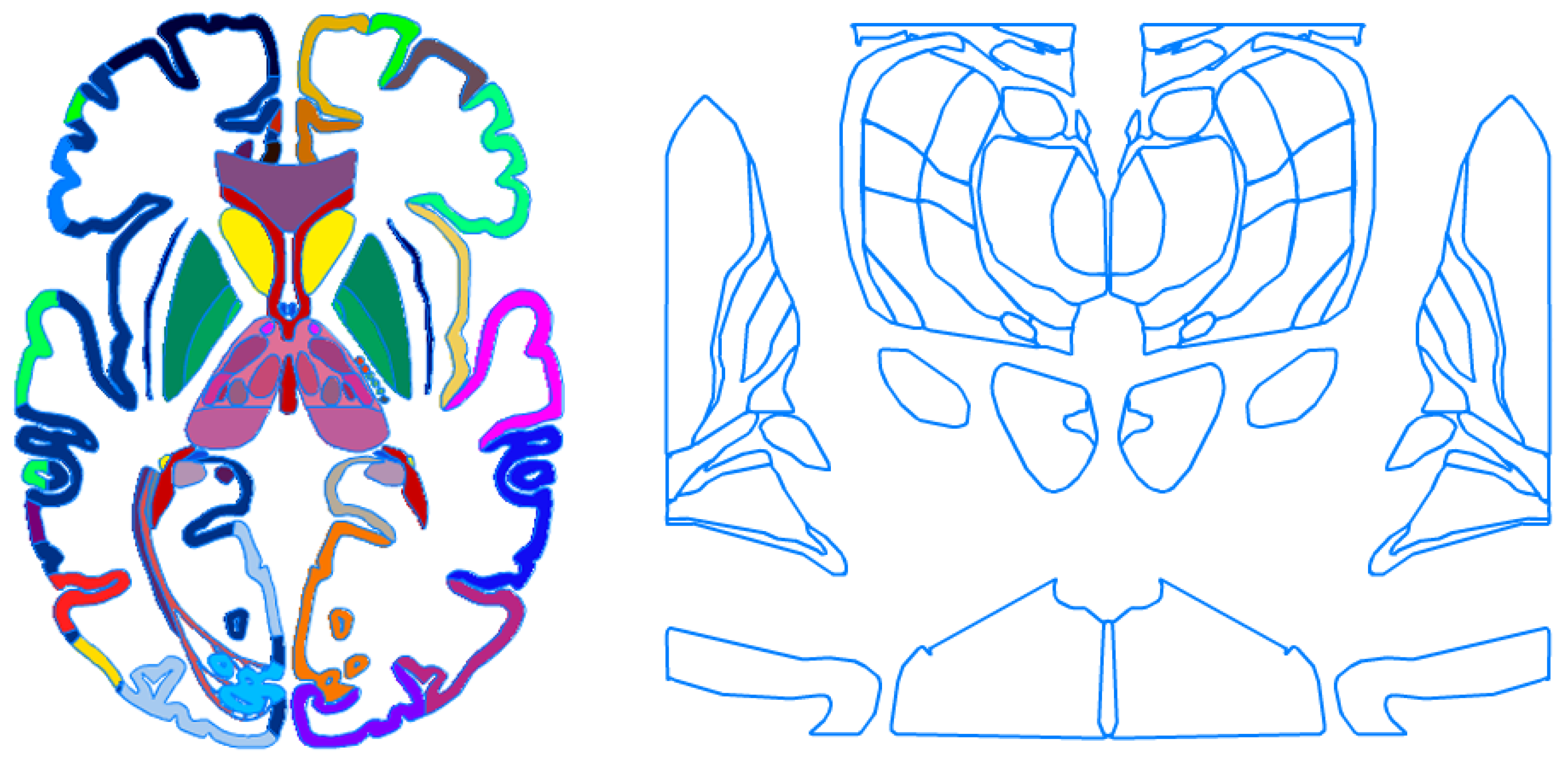
Because of the importance of these two brain atlases, we have developed their enhanced and extended electronic versions, and the applied processing was explained in detail in [36]. These electronic atlases are fully parcellated which enables their automatic labeling. This parcellation is by unique coloring and closed contouring (a contour representation is additionally useful for atlas-to-data registration as the contours do not block the actual patient data); see Figure 1. These electronic atlases have been embedded into atlas-assisted stand-alone applications [37,38,39,40] and plug-in libraries licensed to 13 companies and integrated with major surgical workstations [41].
Enormous advancements in imaging, brain mapping, and computing drive the development of human brain atlas platforms. I have specified 23 directions in the evolution of brain atlas content development grouped into eight categories by employing various criteria, including scope, parcellation, plurality, modality, scale, ab/normality, ethnicity, and a combination of them [4]. I briefly overview these brain atlas categories and provide some examples of brain atlases from numerous centers.
The scope of brain atlases ranges from structural neuroanatomy [42,43,44,45] to connectional neuroanatomy [46,47,48,49,50,51] to vascular neuroanatomy [52,53] including cerebral variants [54] to cranial nerves and nuclei [55] to gene expression [56] including gene expression in brain development [57].
In general, the human brain can be parcellated into numerous anatomically and/or functionally distinct cortical regions and subcortical structures based on macrostructural, microstructural, functional, and/or connectional features. The parcellation category represents novel and/or finer parcellations of brain structures and surfaces based on various modalities and approaches. The developments here are from classic gross anatomy, cytoarchitecture, and myeloarchitecture to functional magnetic resonance imaging (fMRI) exploiting resting-state and task-based sequences [58], chemoarchitecture [59], vascular territories [60], anatomic connectivity based on diffusion tensor imaging [48] and diffusion spectrum imaging [61], anatomic-functional connectivity based on diffusion and resting-state MRI [62], electroencephalography [63], (multi)receptor architecture [64], and/or multiplicity of them [50,65]. Both the size and the number of the parcellated regions can be variable; for instance, a multi-modal MRI-based parcellation of the cerebral cortex results in 180 variable-size areas per hemisphere [65], the Brainnetome atlas is parcellated into 210 various cortical areas and 36 subcortical regions [62], and the Yale Brain Atlas consists of 690 same-size one-square centimeter parcels [63].
Parcellation not only introduces subdivision but also enables systematization, localization, and comparison, ideally making the brain “addressable”. Parcellated regions can be named based on some existing nomenclatures, such as Terminologia Anatomica [66] which is an international standard for the whole body or Terminologia Neuroanatomica targeting the central nervous system, peripheral nervous system, and sensory organs [67]. Several nomenclatures have been introduced for research applications, such as NeuroNames supporting synonyms and multiple languages [68], Uberon [69] supporting single- and cross-species queries, Foundation Model of Anatomy (FMA) providing a structure-based template from the molecular to the macroscopic levels for representing biological functions of the human body [70], and Common Coordinate Framework (CCF) ontology to define positions in the body down to individual cells [71]. Alternatively, parcellation-related identifiers are used, such as numbers in naming Brodmann’s areas [3] or parcel unique names with a gyrus code and a letter indicating the parcel position within the gyrus in the Yale Brain Atlas [63].
Within the plurality category, probabilistic brain atlases provide novel neuroanatomical information in terms of statistical distributions of the studied entities. For instance, these atlases may contain the mean values, standard deviations, moments, and other quantifiers of volumes (e.g., for the entire brain [72], white matter [73], cerebellum [74], or subcortical structures [75]), areas (such as cortical surface regions [76]) or distances (e.g., the thickness of the cortical mantle). Multi-atlases can illustrate neuroanatomy over the lifespan. For instance, a mega multi-atlas [77] comprises 90 component brain atlases with the brain specimens ranging from 4 to 82 years of age.
In the modality category, the major advancement has been from postmortem to in vivo data enabled by neuroimaging allowing to accomplish a “living neuroanatomy”. Furthermore, more detailed neuroanatomical images with better quality are feasible in brain atlasing due to the increased teslage of the acquired MRI neuroimages, namely, from 1.5T [78] to 3T [45,53] to 7T [52,79,80,81,82,83] to 9.4T [84].
The scale category includes brain atlases with various temporal, spatial, and combined spatiotemporal scales. Several temporal scale-related brain atlases aggregate age-dependent neuroanatomical changes ranging from pediatric to geriatric populations [85,86,87]. Other relevant works include a dynamic 4D atlas of the developing brain [88] and a temporal cell atlas of gene expression in brain development [57].
The spatial scale of brain atlases ranges from macro- to meso- to micro- to nano-scale, including the integration of atlas data across multiple scales. The developments in this area include the BigBrain with a 20-micrometer resolution [89], a comprehensive cellular-resolution (of 1 μm/pixel) atlas linking macroscopic anatomical and microscopic cytoarchitectural parcellations [90], a whole-brain cell atlas integrating anatomical, physiological and molecular annotations for a complete characterization of neuronal cell types, their distributions, and patterns of connectivity [91], a genomics brain atlas [56], an atlas of brain transcriptome [92], an atlas of serotonin [93], and a proteomic brain atlas [94].
Several disease-specific atlases have been created, e.g., for Alzheimer’s disease [95], dementia [96], stroke [97,98], brain tumors [99], and epilepsy [100]. Some of them enable the quantification of brain structural deficits in epilepsy, depression, schizophrenia, Alzheimer’s disease, autism, and bipolar disorders [101]; others include the Probabilistic Stroke Atlas [98] which facilitates outcome prediction, the Virtual Epileptic Patient atlas which provides an automated brain region parcellation and labeling for epileptology and functional neurosurgery [100], and the Probabilistic Atlas of Diffuse WHO Grade II Glioma Locations which identifies the preferential locations of these gliomas in the brain [99]. A different way of atlas use is presented in [102] to investigate genetic correlations between brain phenotypes (attained as cortical surface area and thickness) and psychiatric/neurological disorders by means of genetically informed brain atlases. This study revealed the association between global surface and fronto-parietal thickness with attention-deficit hyperactivity disorder, temporal area with schizophrenia and autism spectrum disorder, and fronto-occipital morphology with neurological disorders.
Ethnicity-based brain atlases enable comparison of neuroanatomy between various populations, such as Chinese and Caucasian [103] and Indian with Chinese and Caucasian [104].
The design, development, and validation of a human brain atlas is a painstaking and time-consuming process that requires high attention to detail. The design principles of a holistic and reference brain atlas are formulated in [105], computational methods employed in brain atlas development are addressed in [106], visualization and interaction are discussed in [107], and a user-centric and application-balanced architecture cum implementation of a reference human brain atlas is proposed in [13].
4. Brain Atlas-Assisted Applications
The human brain atlases are employed across education, research, and clinics [4]. In neuroeducation, the brain atlas assists students and educators as a visual and interactive tool with parcellated and labeled virtual brain models, equipped with an intuitive and friendly user interface, able to communicate cerebral complexity in a more convenient and comprehensible manner. In research, brain atlases focus predominantly on how to integrate and openly share massive amounts of heterogeneous experimental data in a common reference atlas space and to relate these data across scales. In clinics, brain atlases are valuable computer-aided tools to support and enhance screening, diagnosis, treatment, and prediction.
4.1. Education
The history of neuroanatomy over the centuries has been linked to the teaching methods employed, including cadaveric dissection, plastination, observation of live models, live surgery, animal dissection, synthetic models, bibliographic sources, radiology, and audiovisual virtual reality including stereoscopy [108]. Electronic and interactive brain atlases may be embedded in synthetic models, radiology, audiovisual virtual reality, and computer-aided live surgery.
Several standard neuroeducational brain atlases have been developed, such as Digital Anatomist [109], A.D.A.M. [110], The Electronic Clinical Brain Atlas [37], Voxel-man [78], The Cerefy Atlas of Brain Anatomy [39], Primal’s Interactive Head and Neck [111], and The Cerefy Clinical Brain Atlas [40].
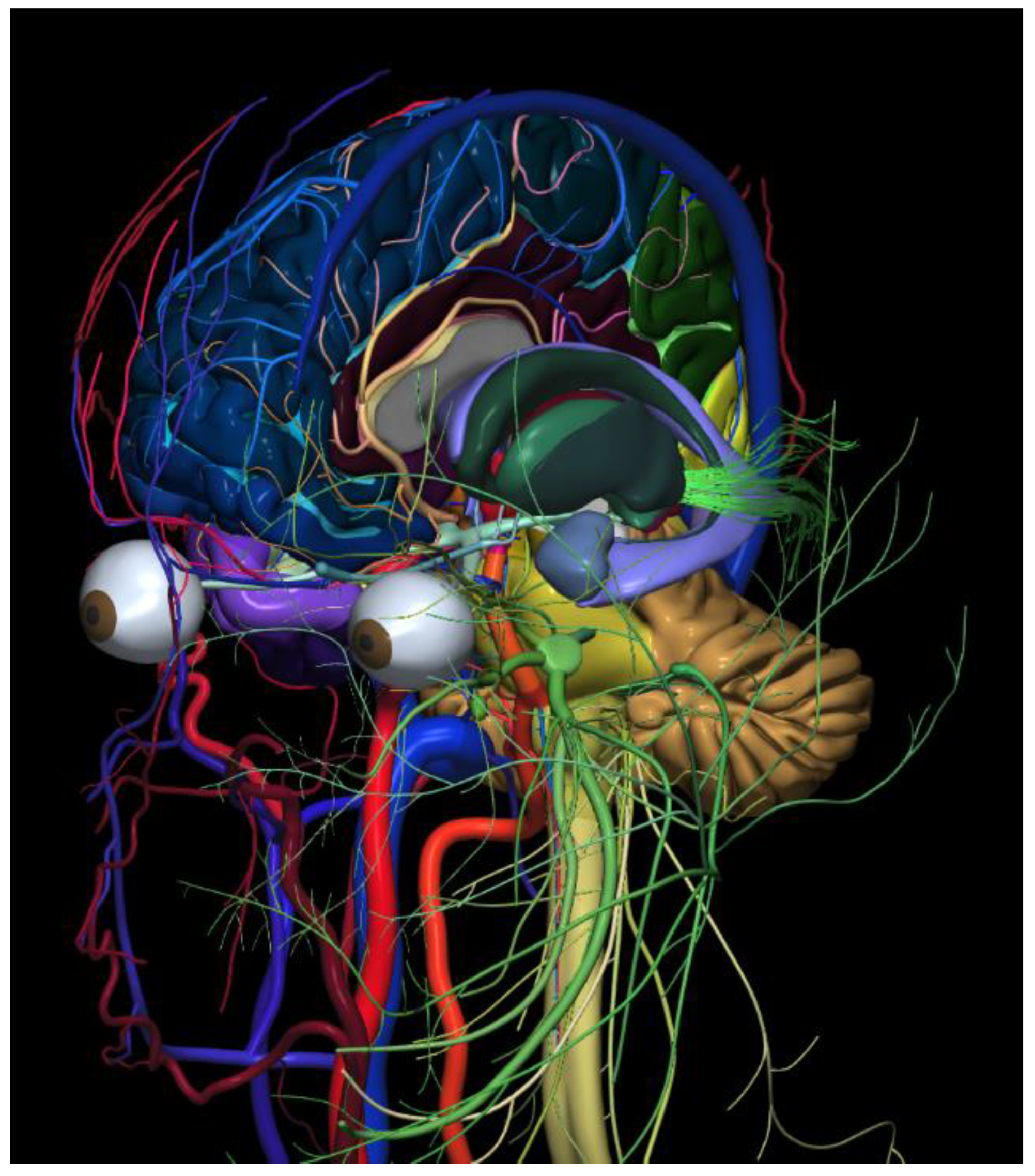
In comparison to the standard brain atlases, advanced atlases provide novel features in neuroeducation facilitating brain exploration and understanding. Examples of such atlases are The Cerefy Atlas of Cerebral Vasculature [53], The Human Brain in 1492 Pieces [43], The Human Brain in 1969 Pieces: Structure, Vasculature, Tracts, Cranial Nerves, Systems, Head Muscles, and Glands [44], and The Human Brain, Head and Neck in 2953 Pieces [81]. These novel features include continuous navigation and exploration, free composing and decomposing of a 3D explorable scene (see Figure 2), joint surface and sectional anatomy, presentation in context, correlation of anatomy and terminology, simultaneous presentation of multiple systems, wide scope of presentations (from local to global neuroanatomy), virtual dissections, quantification, and generation of teaching materials [112,113] as well as automatic testing and assessment of neuroanatomy knowledge [114] available, e.g., in The Cerefy Atlas of Cerebral Vasculature [53].
Technology advancements open new avenues in brain atlasing, although on the other hand, they may cause an increased cost and decreased accessibility of brain atlas applications, especially for users in less privileged countries. To address this issue, I have created the NOWinBRAIN 3D neuroimage public repository at www.nowinbrain.org. NOWinBRAIN is a large (the largest so far), systematic, comprehensive, extendable, spatially consistent, easy to use, long-lasting, and beautiful repository of 3D reconstructed images of a living human brain extended to the head and neck populated with over 7800 images (version 3.1) organized in 10 galleries. The design, development, and content of the primary and multi-tissue galleries are addressed in [115], the combined planar–surface gallery in [116], the dissection gallery in [117]; and the gallery of dual white matter–cortical surfaces with the cerebral sulci in [118]. Note that despite the tremendous development of various brain-related resources, such a repository is not yet available. This systematically designed repository is empowered with many novel features, such as multi-tissue galleries, the use of various spatially co-registered image sequences, and unique image-naming syntax. It is freely available and easily accessible as a web resource without any password or registration. These features make NOWinBRAIN valuable for neuroeducators, medical students, neuroscientists, and clinicians, especially, in less privileged countries. The current users are from over 75 countries on six continents. Most users are from Europe and the United States including the technologically advanced Silicon Valley. Frequent users are from India, China, and Egypt. There are also visitors from Nepal, Afghanistan, Sudan, Tanzania, Brazil, Argentine, and Peru.
4.2. Research
Brain atlases are widely applied in research for various purposes and play a key role in modern neuroimage analysis [119]. One of the main areas of brain atlas applications is human brain mapping. Then, the brain atlases, such as the BrainMap [120] or the Brain Atlas for Functional Imaging [38], provide the underlying neuroanatomy enabling the activation loci in functional images to be automatically labeled with cortical areas and stereotactic coordinates. Brain atlases are widely applicable for fast, automatic, and robust segmentation of neuroimages [121,122,123,124,125,126]. Brain atlases are central tools for data integration [127] enabling combining various brain-related information, such as micro- and macrostructural parcellation, connectivity, temporal dynamics, and regional functional specialization [8]. The brain atlas also serves as a tool for localizing experimental data and planning experiments [9] as well as to generate hypotheses about brain organization [10]. In addition, brain atlases enable knowledge discovery; for instance, Makowski et al. employed genetically informed brain atlases to determine the impact of genetic variants on the brain in genome-wide association studies of regional cortical surface area and thickness in about 40,000 adults and 9000 children [102]. These studies uncovered 440 genome-wide significant loci (largely acquired in childhood) related to early neurodevelopment and associated with neuropsychiatric risk.
4.3. Clinics
The first clinical application of human brain atlases has been stereotactic and functional neurosurgery. Initially, a digital atlas, such as The Electronic Clinical Brain Atlas [37], was employed offline in the operating room to aid neurosurgery. Subsequently, the brain atlas libraries derived from our brain atlas database [36] were directly incorporated into several surgical workstations, including the StealthStation (Medtronic) [41], to assist neurosurgery. In general, the brain atlas provides pre-, intra-, and post-operative support [128]. Pre-operatively, the atlas assists to plan the target and trajectory as well as provides a list of structures intersected by the trajectory. The usage of multiple brain atlases improves the planning quality and surgeon’s confidence [129,130]. Intra-operatively, the brain atlas specifies the structures already traversed by the electrode, identifies the actual structure where the electrode tip is located, measures distances to important structures, and provides the neuroanatomic and vascular context [130]. Post-operatively, the atlas enables the examination of the precision of placement of the stimulating electrode or a permanent lesion. Other atlas-assisted applications in neurosurgery include atlas-guided do-it-yourself neurosurgery [41] and an atlas-enhanced operating room for the future [131].
Several brain atlas-aided proofs of concepts (prototypes) have been developed in some other areas. Namely, in neuroradiology, brain atlases can assist in neuroimage interpretation by segmenting and labeling brain scans including pathological, template-based reporting, dealing with data explosion by facilitating processing multi-detector (especially 320-raw computed tomography) scans, and communication for both doctor-to-doctor and especially doctor-to-patient [132]. Multiple brain atlases have the potential in stroke management including prediction, diagnosis, and treatment by providing automated processes ensuring fast decisions [60,98,133]. In neurology, the 3D Atlas of Neurologic Disorders [134] demonstrates various locations of brain damage, including local neuroanatomy, cranial nerves, and cerebrovasculature, along with the resulting neurologic deficits, bridging in this way neuroanatomy, neuroradiology, and neurology [135]. Finally in psychiatry, a brain atlas allows for the automatic generation of neuroanatomic volumes of interest for statistical analysis, e.g., to study schizophrenic patients and controls [136].
5. Future Developments
There has been an enormous explosion of human brain-related endeavors in the last few years. These are advanced, big, government-led, and/or well-funded projects, initiatives, and/or national brain programs, such as The Human Connectome Project to map structural and functional connections to investigate the relationship between brain circuits and behavior [51]; The Allen Brain Atlas to map gene expression [56]; The Big Brain to acquire ultra-high resolution neuroimages [89]; The CONNECT project combining macro- and micro-structure [137]; the Brainnetome project to understand the brain and its disorders, develop methods for multi-scale brain network analysis, and create the Brainnetome atlas [138]; The BRAIN Initiative (Brain Research through Advancing Innovate Neurotechnologies) [139] to develop technology to advance neuroscience discovery [140]; The Blue Brain Project to simulate neocortical micro-circuitry [141]; The Human Brain Project to create a research infrastructure to decipher the human brain, reconstruct its multiscale organization, and develop brain-inspired information technology [142]; the Chinese Color Nest Project to study human connectomics across the life span [87]; the Japanese Brain/MINDS (Brain Mapping by Integrating Neurotechnologies for Disease Studies) project to better understand the human brain and neuropsychiatric disorders through ‘‘translatable’’ biomarkers [143]; and SYNAPSE (Synchrotron for Neuroscience—an Asia-Pacific Strategic Enterprise) to map the entire human brain at sub-cellular level by employing synchrotron tomography [144]—a proposal of how to build a corresponding human brain atlas I have recently presented at the SYNAPSE 2022 meeting; https://www.slri.or.th/th/index.php?option=com_attachments&task=download&id=4493 (28 December 2022).
These and other efforts have resulted in the acquisition of big data and the development of diverse brain-related databases, such as BigBrain, Allen Brain Atlas, HCP (Human Connectome Project) database and HCP Young Adult Data, BIRN (Biomedical Informatics Research Network) MRI and fMRI data, OpenNEURO, OASIS (Open Access Series of Imaging Studies) Brains Project, ABCD (Adolescent Brain Cognitive Development) Data Repository, BCP (Baby Connectome Project) database, BP (bipolar disorder) neuroimaging database, and the Alzheimer’s Disease Neuroimaging Initiative (ADNI) as overviewed in [145]. Moreover, the BRAIN Initiative resulted in the development of the Neuroscience Multi-Omic Archive repository containing transcriptomic and epigenomic data from over 50 million brain cells [146]. In addition, the online community repository NeuroMorpho.Org contains more than 140,000 neural reconstructions (including glia) consisting of 3D representations of branch geometry and connectivity in a standardized format, and for each reconstruction, a set of morphometric features is extracted [147].
The abovementioned large-scale endeavors and big data empowered with high-performance computing at peta- and exascale will enormously increase our knowledge and understanding of the human brain at various scales and will propel the development of novel and more powerful brain atlases.
6. Summary and Conclusions
Neuroanatomy, as the study of the structure and organization of the nervous system, and human electronic brain atlases, as tools to gather, present, use, and discover knowledge about the human brain, are naturally linked. Consequently, this work addresses this human brain atlas–neuroanatomy mutual relationship.
Brain atlasing has progressed from the initial brain drawings and hand-drawn cortical maps to advanced brain atlas platforms. Presently, human electronic brain atlases have been advancing tremendously in terms of content, functionality, and applications. The advancement is empowered by software engineering methods and tools, such as databases, image processing, computer graphics, and virtual and augmented reality. This advancement spreads in multiple directions which can be grouped with respect to scope, parcellation, plurality, modality, scale, ab/normality, ethnicity, and combination of them.
Neuroanatomy has also been transformed enormously. From gross neuroanatomy facilitated by cadaveric dissections to micro-neuroanatomy enabled by brain fixation techniques combined with optical microscopy to nano-neuroanatomy empowered by modern electron microscopy, genomics, proteomics, transcriptomics, and epigenomics, and also from cadaveric neuroanatomy to living neuroanatomy enabled by modern imaging of structure, function, vasculature, structural and functional connectivity, and molecular processes. Moreover, imaging offers new acquisition methods, ever-increasing spatial and temporal resolutions, a better quality of images, and shorter acquisition times, all supported by artificial intelligence.
This ever-growing neuroanatomical knowledge enables the creation of human electronic brain atlases. These atlases mirror the advances in neuroanatomy capturing the dramatically increasing knowledge about the human brain in health and disease. Numerous centers contribute to neuroanatomy and brain atlasing advancements from various perspectives as briefly outlined here.
Furthermore, reciprocally, the developments in brain atlasing impact neuroanatomy enabling the use, presentation, mining, dissemination, and growth of this knowledge as well as facilitating learning, understanding, exploring, researching, diagnosing, screening, decision making, outcome prediction, and treatment of the human brain. In addition, because of remarkable progress in brain atlasing, these atlases are able to more accurately, realistically, and completely represent and present this neuroanatomical knowledge and better disseminate and use it. In my opinion, human brain atlases are the best means to represent, present, disseminate, and support neuroanatomy.
Finally, the impact on neuroanatomy and brain atlasing by the ongoing large brain projects and acquired big data may be expected to be enormous.
Funding
This research received no external funding.
Institutional Review Board Statement
Not applicable.
Informed Consent Statement
Not applicable.
Data Availability Statement
NOWinBRAIN 3D neuroimage repository is publically available at www.nowinbrain.org.
Conflicts of Interest
The author declares no conflict of interest.
References
- Polyak, S. The Vertebrate Visual System; Kluever, K., Ed.; University of Chicago Press: Chicago, IL, USA, 1957. [Google Scholar]
- Schmahmann, J.D.; Pandya, D.N. Fiber Pathways of the Brain; Oxford University Press: Oxford, UK, 2006. [Google Scholar]
- Brodmann, K. Vergleichende Lokalisationslehre der Grosshirnrinde in ihren Prinzipien dargestellt auf Grund des Zellenbaues; Barth JA: Leipzig, Germany, 1909. [Google Scholar]
- Nowinski, W.L. Evolution of human brain atlases in terms of content, applications, functionality, and availability. Neuroinformatics 2021, 19, 1–22. [Google Scholar] [CrossRef] [PubMed]
- Roland, P.E.; Zilles, K. Brain atlases–a new research tool. Trends Neurosci. 1994, 17, 458–467. [Google Scholar] [CrossRef] [PubMed]
- Evans, A.C.; Janke, A.L.; Collins, D.L.; Baillet, S. Brain templates and atlases. Neuroimage 2012, 62, 911–922. [Google Scholar] [CrossRef] [PubMed]
- Mori, S.; Oishi, K.; Faria, A.V.; Miller, M.I. Atlas-based neuroinformatics via MRI: Harnessing information from past clinical cases and quantitative image analysis for patient care. Annu. Rev. Biomed. Eng. 2013, 15, 71–92. [Google Scholar] [CrossRef] [PubMed] [Green Version]
- Amunts, K.; Hawrylycz, M.J.; Van Essen, D.C.; Van Horn, J.D.; Harel, N.; Poline, J.B.; De Martino, F.; Bjaalie, J.G.; Dehaene-Lambertz, G.; Dehaene, S.; et al. Interoperable atlases of the human brain. Neuroimage 2014, 99, 525–532. [Google Scholar] [CrossRef] [PubMed] [Green Version]
- Kuan, L.; Li, Y.; Lau, C.; Feng, D.; Bernard, A.; Sunkin, S.M.; Zeng, H.; Dang, C.; Hawrylycz, M.; Ng, L. Neuroinformatics of the Allen Mouse Brain Connectivity Atlas. Methods 2015, 73, 4–17. [Google Scholar] [CrossRef] [PubMed]
- Costa, M.; Manton, J.D.; Ostrovsky, A.D.; Prohaska, S.; Jefferis, G.S. NBLAST: Rapid, sensitive comparison of neuronal structure and construction of neuron family databases. Neuron 2016, 91, 293–311. [Google Scholar] [CrossRef] [PubMed] [Green Version]
- Chon, U.; Vanselow, D.J.; Cheng, K.C.; Kim, Y. Enhanced and unified anatomical labeling for a common mouse brain atlas. Nat. Commun. 2019, 10, 5067. [Google Scholar] [CrossRef] [Green Version]
- Nowinski, W.L. Towards constructing an ideal stereotactic brain atlas. Acta Neurochir. 2008, 150, 1–14. [Google Scholar] [CrossRef]
- Nowinski, W.L. Towards an architecture of a multi-purpose, user-extendable reference human brain atlas. Neuroinformatics 2022, 20, 405–426. [Google Scholar] [CrossRef]
- Campbell, A.W. Histological Studies on the Localisation of Cerebral Function; Cambridge University Press: Cambridge, UK, 1905. [Google Scholar]
- Flechsig, P. Anatomie des Menschlichen Gehirns und Rückenmarks auf Myelogenetischer Grundlage; Thieme: Leipzig, Germany, 1920. [Google Scholar]
- Vogt, C.; Vogt, O. Allgemeinere Ergebnisse unserer Hirnforschung (English Translation: Results of our brain research in a broader context). J. Psychol. Neurol. 1919, 25, 292–398. [Google Scholar]
- Von Economo, C.; Koskinas, G.N. Die Cytoarchitektonik der Hirnrinde des Erwachsenen Menschen; Springer: Berlin, Germany, 1925. [Google Scholar]
- Speigel, E.A.; Wycis, H.T. Stereoencephalotomy: Part I. Methods and Stereotactic Atlas of the Human Brain; Grune and Stratton: New York, NY, USA, 1952. [Google Scholar]
- Talairach, J.; David, M.; Tournoux, P. Atlas d’Anatomie Stereotaxique des Noyaux Gris Centraux; Masson: Paris, France, 1957. [Google Scholar]
- Schaltenbrand, G.; Bailey, W. Atlas of Stereotaxy of the Human Brain; Georg Thieme Verlag: Stuttgart, Germany, 1959. [Google Scholar]
- Andrew, J.; Watkins, E.S. A Stereotaxic Atlas of the Human Thalamus and Adjacent Structures. A Variability Study; Williams and Wilkins: Baltimore, MD, USA, 1969. [Google Scholar]
- Van Buren, J.M.; Borke, R.C. Variations and Connections of the Human Thalamus; Springer: Berlin, Germany, 1972. [Google Scholar]
- Schaltenbrand, G.; Wahren, W. Atlas of Stereotaxy of the Human Brain; Georg Thieme Verlag: Stuttgart, Germany, 1977. [Google Scholar]
- Afshar, E.; Watkins, E.S.; Yap, J.C. Stereotactic Atlas of the Human Brainstem and Cerebellar Nuclei; Raven Press: New York, NY, USA, 1978. [Google Scholar]
- Talairach, J.; Tournoux, P. Co-Planar Stereotactic Atlas of the Human Brain; Thieme: Stuttgart, Germany; New York, NY, USA, 1988. [Google Scholar]
- Talairach, J.; Tournoux, P. Referentially Oriented Cerebral MRI Anatomy: Atlas of Stereotaxic Anatomical Correlations for Gray and White Matter; Thieme: Stuttgart, Germany, 1993. [Google Scholar]
- Takayoshi, M.; Hirano, A. Atlas of the Human Brain for Computerized Tomography; Igaku Shoin Medical Publishers: New York, NY, USA, 1978. [Google Scholar]
- Duvernoy, H.M. The Human Hippocampus: Atlas of Applied Anatomy; Bergman: Munich, Germany, 1988. [Google Scholar]
- Ono, M.; Kubik, S.; Abernathey, C.D. Atlas of the Cerebral Sulci; Georg Thieme Verlag/Thieme Medical Publishers: Stuttgart, Germany; New York, NY, USA, 1990. [Google Scholar]
- Orrison, W.W., Jr. Atlas of Brain Function; Thieme: New York, NY, USA, 1995. [Google Scholar]
- Duvernoy, H.M. The Human Brain Stem and Cerebellum. Surface, Structure, Vascularization, and Three-Dimensional Sectional Anatomy, with MRI; Springer: Wien, Austria; New York, NY, USA, 1995. [Google Scholar]
- Scarabino, T.; Salvolini, U.; DiSalle, F.; Duvernoy, H.; Rabischong, P. (Eds.) Atlas of Morphology and Functional Anatomy of the Brain; Springer: Berlin, Germany, 2006. [Google Scholar]
- Naidich, T.h.P.; Duvernoy, H.M.; Delman, B.N.; Sorensen, A.G.; Kollias, S.S.; Haacke, E.M. Duvernoy’s Atlas of the Human Brain Stem and Cerebellum; Springer: Wien, Austria; New York, NY, USA, 2009. [Google Scholar]
- Felten, D.L.; O’Banion, M.K.; Maida, M.E. Netter’s Atlas of Neuroscience, 3rd ed.; Elsevier: Amsterdam, The Netherlands, 2015. [Google Scholar]
- Alho, E.J.L.; Grinberg, L.; Heinsen, H. Review of printed and electronic stereotactic atlases of the human brain. In Neuroimaging for Clinicians: Combining Research and Practice; Peres, J.F.P., Ed.; InTech: Rijeka, Croatia, 2011; pp. 145–172. [Google Scholar]
- Nowinski, W.L.; Fang, A.; Nguyen, B.T.; Raphel, J.K.; Jagannathan, L.; Raghavan, R.; Bryan, R.N.; Miller, G. Multiple brain atlas database and atlas-based neuroimaging system. Comput. Aided Surg. 1997, 2, 42–66. [Google Scholar] [CrossRef] [PubMed]
- Nowinski, W.L.; Bryan, R.N.; Raghavan, R. The Electronic Clinical Brain Atlas. Multiplanar Navigation of the Human Brain; Thieme: New York, NY, USA, 1997. [Google Scholar]
- Nowinski, W.L.; Thirunavuukarasuu, A.; Kennedy, D.N. Brain Atlas for Functional Imaging. Clinical and Research Applications; Thieme: New York, NY, USA, 2000. [Google Scholar]
- Nowinski, W.L.; Thirunavuukarasuu, A.; Bryan, R.N. The Cerefy Atlas of Brain Anatomy. An Introduction to Reading Radiological Scans for Students, Teachers, and Researchers; Thieme: New York, NY, USA, 2002. [Google Scholar]
- Nowinski, W.L.; Thirunavuukarasuu, A. The Cerefy Clinical Brain Atlas on CD-ROM; Thieme: New York, NY, USA, 2004. [Google Scholar]
- Nowinski, W.L. Anatomical and probabilistic functional atlases in stereotactic and functional neurosurgery. In Textbook of Stereotactic and Functional Neurosurgery, 2nd ed.; Lozano, A., Gildenberg, P., Tasker, R., Eds.; Springer: Berlin, Germany, 2009; pp. 395–441. [Google Scholar]
- Mandal, P.K.; Mahajan, R.; Dinov, I.D. Structural brain atlases: Design, rationale, and applications in normal and pathological cohorts. J. Alzheimers Dis. 2012, 31 (Suppl. 3), S169–S188. [Google Scholar] [CrossRef] [PubMed] [Green Version]
- Nowinski, W.L.; Chua, B.C.; Qian, G.Y.; Marchenko, Y.; Puspitasari, F.; Nowinska, N.G.; Knopp, M.V. The Human Brain in 1492 Pieces: Structure, Vasculature, and Tracts; Thieme: New York, NY, USA, 2011. [Google Scholar]
- Nowinski, W.L.; Chua, B.C. The Human Brain in 1969 Pieces: Structure, Vasculature, Tracts, Cranial Nerves, Systems, Head Muscles, and Glands (Version 2.0); Thieme: New York, NY, USA, 2014. [Google Scholar]
- Rohlfing, T.; Zahr, N.M.; Sullivan, E.V.; Pfefferbaum, A. The SRI24 multichannel atlas of normal adult human brain structure. Hum. Brain Mapp. 2010, 31, 798–819. [Google Scholar] [CrossRef] [Green Version]
- Baker, C.M.; Burks, J.D.; Briggs, R.G.; Conner, A.K.; Glenn, C.A.; Sali, G.; McCoy, T.M.; Battiste, J.D.; O’Donoghue, D.L.; Sughrue, M.E. A connectomic atlas of the human cerebrum chapter 1, introduction, methods, and significance. Oper. Neurosurg. 2018, 15, S1–S9. [Google Scholar] [CrossRef]
- Briggs, R.G.; Conner, A.K.; Baker, C.M.; Burks, J.D.; Glenn, C.A.; Sali, G.; Battiste, J.D.; O’Donoghue, D.L.; Sughrue, M.E. A connectomic atlas of the human cerebrum-Chapter 18, The Connectional Anatomy of Human Brain Networks. Oper. Neurosurg. 2018, 15 (Suppl. 1), S470–S480. [Google Scholar] [CrossRef]
- Mori, S.; Wakana, S.; Nagae-Poetscher, L.M.; van Zijl, P.C. MRI Atlas of Human White Matter; Elsevier: Amsterdam, The Netherlands.
- Nowinski, W.L.; Chua, B.C.; Yang, G.L.; Qian, G.Y. Three-dimensional interactive human brain atlas of white matter tracts. Neuroinformatics 2012, 10, 33–55. [Google Scholar] [CrossRef] [PubMed]
- Van Essen, D.C. Cartography and connectomes. Neuron 2013, 80, 775–790. [Google Scholar] [CrossRef] [Green Version]
- Van Essen, D.C.; Smith, S.M.; Barch, D.M.; Behrens, T.E.J.; Yacoub, E.; Ugurbil, K. The WU-Minn Human Connectome Project: An overview. NeuroImage 2013, 80, 62–79. [Google Scholar] [CrossRef] [Green Version]
- Huck, J.; Wanner, Y.; Fan, A.P.; Jäger, A.T.; Grahl, S.; Schneider, U.; Villringer, A.; Steele, C.J.; Tardif, C.L.; Bazin, P.L.; et al. High resolution atlas of the venous brain vasculature from 7 T quantitative susceptibility maps. Brain Struct. Funct. 2019, 224, 2467–2485. [Google Scholar] [CrossRef]
- Nowinski, W.L.; Thirunavuukarasuu, A.; Volkau, I.; Marchenko, Y.; Runge, V.M. The Cerefy Atlas of Cerebral Vasculature; Thieme: New York, NY, USA, 2009. [Google Scholar]
- Nowinski WL, A. Thirunnavuukarasuu, Volkau I, Marchenko Y, Aminah B, Puspitasaari F, Runge VM. A three-dimensional interactive atlas of cerebral arterial variants. Neuroinformatics 2009, 7, 255–264. [Google Scholar] [CrossRef] [PubMed]
- Nowinski, W.L.; Johnson, A.; Chua, B.C.; Nowinska, N.G. Three-dimensional interactive and stereotactic atlas of cranial nerves and nuclei correlated with surface neuroanatomy, vasculature and magnetic resonance imaging. J. Neurosci. Methods 2012, 206, 205–216. [Google Scholar] [CrossRef] [PubMed]
- Sunkin, S.M.; Ng, L.; Lau, C.; Dolbeare, T.; Gilbert, T.L.; Thompson, C.L.; Hawrylycz, M.; Dang, C. Allen Brain Atlas: An integrated spatio-temporal portal for exploring the central nervous system. Nucleic Acids Res. 2013, 41, D996–D1008. [Google Scholar] [CrossRef] [PubMed] [Green Version]
- Kanton, S.; Boyle, M.J.; He, Z.; Santel, M.; Weigert, A.; Sanchís-Calleja, F.; Guijarro, P.; Sidow, L.; Fleck, J.S.; Han, D.; et al. Organoid single-cell genomic atlas uncovers human-specific features of brain development. Nature 2019, 574, 418–422. [Google Scholar] [CrossRef]
- James, G.A.; Hazaroglu, O.; Bush, K.A. A human brain atlas derived via n-cut parcellation of resting-state and task-based fMRI data. Magn. Reason. Imaging 2016, 34, 209–218. [Google Scholar] [CrossRef] [Green Version]
- Yelnik, J.; Bardinet, E.; Dormont, D.; Malandain, G.; Ourselin, S.; Tandé, D.; Karachi, C.; Ayache, N.; Cornu, P.; Agid, Y. A three-dimensional, histological and deformable atlas of human basal ganglia. I. Atlas construction based on immunohistochemical and MRI data. NeuroImage 2007, 34, 618–638. [Google Scholar] [CrossRef]
- Nowinski, W.L.; Qian, G.; Bhanu Prakash, K.N.; Thirunavuukarasuu, A.; Hu, Q.M.; Ivanov, N.; Parimal, A.S.; Runge, V.M.; Beauchamp, N.J. Analysis of ischemic stroke MR images by means of brain atlases of anatomy and blood supply territories. Acad. Radiol. 2006, 13, 1025–1034. [Google Scholar] [CrossRef]
- Arsiwalla, X.D.; Zucca, R.; Betella, A.; Martinez, E.; Dalmazzo, D.; Omedas, P.; Deco, G.; Verschure, P.F. Network dynamics with BrainX(3): A large-scale simulation of the human brain network with real-time interaction. Front. Neuroinform. 2015, 9, 2. [Google Scholar] [CrossRef]
- Fan, L.; Li, H.; Zhuo, J.; Zhang, Y.; Wang, J.; Chen, L.; Yang, Z.; Chu, C.; Xie, S.; Laird, A.R.; et al. The human brainnetome atlas: A new brain atlas based on connectional architecture. Cereb. Cortex 2016, 26, 3508–3526. [Google Scholar] [CrossRef] [Green Version]
- McGrath, H.; Zaveri, H.P.; Collins, E.; Jafar, T.; Chishti, O.; Obaid, S.; Ksendzovsky, A.; Wu, K.; Papademetris, X.; Spencer, D.D. High-resolution cortical parcellation based on conserved brain landmarks for localization of multimodal data to the nearest centimeter. Sci. Rep. 2022, 12, 18778. [Google Scholar] [CrossRef]
- Amunts, K.; Lenzen, M.; Friederici, A.D.; Schleicher, A.; Morosan, P.; Palomero-Gallagher, N.; Zilles, K. Broca’s region: Novel organizational principles and multiple receptor mapping. PLoS Biol. 2010, 8, e1000489. [Google Scholar] [CrossRef] [PubMed]
- Glasser, M.F.; Coalson, T.S.; Robinson, E.C.; Hacker, C.D.; Harwell, J.; Yacoub, E.; Ugurbil, K.; Andersson, J.; Beckmann, C.F.; Jenkinson, M.; et al. A multi-modal parcellation of human cerebral cortex. Nature 2016, 536, 171–178. [Google Scholar] [CrossRef] [PubMed] [Green Version]
- Federative Committee on Anatomical Terminology (FCAT). Terminologia Anatomica, International Anatomical Terminology; Thieme: Stuttgart, Germany; New York, NY, USA, 1988. [Google Scholar]
- Federative International Programme for Anatomical Terminology. Terminologia Neuroanatomica. 2017. Available online: https://fipat.library.dal.ca (accessed on 28 December 2022).
- Bowden, D.M.; Song, E.; Kosheleva, J.; Dubach, M.F. NeuroNames: An ontology for the BrainInfo portal to neuroscience on the web. Neuroinformatics 2012, 10, 97–114. [Google Scholar] [CrossRef] [PubMed] [Green Version]
- Haendel, M.A.; Balhoff, J.P.; Bastian, F.B.; Blackburn, D.C.; Blake, J.A.; Bradford, Y.; Comte, A.; Dahdul, W.M.; Dececchi, T.A.; Druzinsky, R.E.; et al. Unification of multi-species vertebrate anatomy ontologies for comparative biology in Uberon. J. Biomed. Semant. 2014, 5, 21. [Google Scholar] [CrossRef] [Green Version]
- Rosse, C.; Mejino, J.L., Jr. A reference ontology for biomedical informatics: The Foundational Model of Anatomy. J. Biomed. Inform. 2003, 36, 478–500. [Google Scholar] [CrossRef] [Green Version]
- Börner, K.; Teichmann, S.A.; Quardokus, E.M.; Gee, J.C.; Browne, K.; Osumi-Sutherland, D.; Herr BW 2nd Bueckle, A.; Paul, H.; Haniffa, M.; Jardine, L.; et al. Anatomical structures, cell types and biomarkers of the Human Reference Atlas. Nat. Cell Biol. 2021, 23, 1117–1128. [Google Scholar] [CrossRef]
- Liang, P.; Shi, L.; Chen, N.; Luo, Y.; Wang, X.; Liu, K.; Mok, V.C.; Chu, W.C.; Wang, D.; Li, K. Construction of brain atlases based on a multi-center MRI dataset of 2020 Chinese adults. Sci. Rep. 2015, 5, 18216. [Google Scholar] [CrossRef] [Green Version]
- Figley, T.D.; Mortazavi Moghadam, B.; Bhullar, N.; Kornelsen, J.; Courtney, S.M.; Figley, C.R. Probabilistic white matter atlases of human auditory, basal ganglia, language, precuneus, sensorimotor, visual and visuospatial networks. Front. Hum. Neurosci. 2017, 11, 306. [Google Scholar] [CrossRef]
- Diedrichsen, J.; Balsters, J.H.; Flavell, J.; Cussans, E.; Ramnani, N. A probabilistic MR atlas of the human cerebellum. NeuroImage 2009, 46, 39–46. [Google Scholar] [CrossRef]
- Pauli, W.M.; Nili, A.N.; Tyszka, J.M. A high-resolution probabilistic in vivo atlas of human subcortical brain nuclei. Sci. Data 2018, 5, 180063. [Google Scholar] [CrossRef] [Green Version]
- Shattuck, D.W.; Mirza, M.; Adisetiyo, V.; Hojatkashani, C.; Salamon, G.; Narr, K.L.; Poldrack, R.A.; Bilder, R.M. Toga AW Construction of a 3D probabilistic atlas of human cortical structures. NeuroImage 2008, 39, 1064–1080. [Google Scholar] [CrossRef] [PubMed]
- Wu, D.; Ma, T.; Ceritoglu, C.; Li, Y.; Chotiyanonta, J.; Hou, Z.; Hsu, J.; Xu, X.; Brown, T.; Miller, M.I.; et al. Resource atlases for multi-atlas brain segmentations with multiple ontology levels based on T1-weighted MRI. Neuroimage 2016, 125, 120–130. [Google Scholar] [CrossRef] [PubMed] [Green Version]
- Hoehne, K.H. VOXEL-MAN, Part 1, Brain and Skull; Version 2.0.; Springer: Berlin/Heidelberg, Germany, 2001. [Google Scholar]
- Cho, Z.H.; Kim, Y.B.; Han, J.Y.; Min, H.K.; Kim, K.N.; Choi, S.H.; Veklerov, E.; Shepp, L.A. New brain atlas—Mapping the human brain in vivo with 7.0 T MRI and comparison with postmortem histology: Will these images change modern medicine? Int. J. Imaging Syst. Technol. 2008, 18, 2–8. [Google Scholar] [CrossRef]
- Liu, Y.; D’Haese, P.F.; Newton, A.T.; Dawant, B.M. Generation of human thalamus atlases from 7 T data and application to intrathalamic nuclei segmentation in clinical 3 T T1-weighted images. Magn. Reason. Imaging 2020, 65, 114–128. [Google Scholar] [CrossRef]
- Nowinski, W.L.; Chua, B.C.; Thaung, T.S.L.; Wut Yi, S.H. The Human Brain, Head and Neck in 2953 Pieces; Thieme: New York, NY, USA, 2015; Available online: http://www.thieme.com/nowinski/ (accessed on 2 January 2023).
- Saygin, Z.M.; Kliemann, D.; Iglesias, J.E.; van der Kouwe, A.J.W.; Boyd, E.; Reuter, M.; Stevens, A.; Van Leemput, K.; McKee, A.; Frosch, M.P.; et al. High-resolution magnetic resonance imaging reveals nuclei of the human amygdala: Manual segmentation to automatic atlas. Neuroimage 2017, 155, 370–382. [Google Scholar] [CrossRef] [PubMed]
- Schira, M.M.; Isherwood, Z.J.; Kassem, M.; Barth, M.; Shaw, T.B.; Roberts, M.M.; Paxinos, G. HumanBrainAtlas: An in vivo MRI dataset for detailed segmentations. bioRxiv 2022. [Google Scholar] [CrossRef]
- Yushkevich, P.A.; Avants, B.B.; Pluta, J.; Das, S.; Minkoff, D.; Mechanic-Hamilton, D.; Glynn, S.; Pickup, S.; Liu, W.; Gee, J.C.; et al. A high-resolution computational atlas of the human hippocampus from postmortem magnetic resonance imaging at 9.4. T. NeuroImage 2009, 44, 385–398. [Google Scholar] [CrossRef]
- Oishi, K.; Linda Chang, L.; Huang, H. Baby brain atlases. NeuroImage 2019, 185, 865–880. [Google Scholar] [CrossRef]
- Zhang, Y.; Wei, H.; Cronin, M.J.; He, N.; Yan, F.; Liu, C. Longitudinal atlas for normative human brain development and aging over the lifespan using quantitative susceptibility mapping. Neuroimage 2018, 171, 176–189. [Google Scholar] [CrossRef]
- Zuo, X.N.; He, Y.; Betzel, R.F.; Colcombe, S.; Sporns, O.; Milham, M.P. Human connectomics across the life span. Trends Cogn. Sci. 2017, 21, 32–45. [Google Scholar] [CrossRef]
- Kuklisova-Murgasova, M.; Aljabar, P.; Srinivasan, L.; Counsell, S.J.; Doria, V.; Serag, A.; Gousias, I.S.; Boardman, J.P.; Rutherford, M.A.; Edwards, A.D.; et al. A dynamic 4D probabilistic atlas of the developing brain. Neuroimage 2011, 54, 2750–2763. [Google Scholar] [CrossRef] [PubMed]
- Amunts, K.; Lepage, C.; Borgeat, L.; Mohlberg, H.; Dickscheid, T.; Rousseau, M.É.; Bludau, S.; Bazin, P.L.; Lewis, L.B.; Oros-Peusquens, A.M.; et al. Bigbrain: An ultrahigh-resolution 3D human brain model. Science 2013, 340, 1472–1475. [Google Scholar] [CrossRef] [PubMed] [Green Version]
- Ding, S.L.; Royall, J.J.; Sunkin, S.M.; Ng, L.; Facer, B.A.; Lesnar, P.; Guillozet-Bongaarts, A.; McMurray, B.; Szafer, A.; Dolbeare, T.A.; et al. Comprehensive cellular-resolution atlas of the adult human brain. J. Comp. Neurol. 2016, 524, 3127–3481. [Google Scholar] [CrossRef] [PubMed] [Green Version]
- Ecker, J.R.; Geschwind, D.H.; Kriegstein, A.R.; Ngai, J.; Osten, P.; Polioudakis, D.; Regev, A.; Sestan, N.; Wickersham, I.R.; Zeng, H. The BRAIN Initiative Cell Census Consortium: Lessons learned toward generating a Comprehensive Brain Cell Atlas. Neuron 2017, 96, 542–557. [Google Scholar] [CrossRef] [PubMed]
- Hawrylycz, M.J.; Lein, E.S.; Guillozet-Bongaarts, A.L.; Shen, E.H.; Ng, L.; Miller, J.A.; van de Lagemaat, L.N.; Smith, K.A.; Ebbert, A.; Riley, Z.L.; et al. An anatomically comprehensive atlas of the adult human brain transcriptome. Nature 2012, 489, 391–399. [Google Scholar] [CrossRef] [Green Version]
- Beliveau, V.; Ganz, M.; Feng, L.; Ozenne, B.; Højgaard, L.; Fisher, P.M.; Svarer, C.; Greve, D.N.; Knudsen, G.M. A high-resolution in vivo atlas of the human brain’s serotonin system. J. Neurosci. 2017, 37, 120–128. [Google Scholar] [CrossRef]
- McKetney, J.; Runde, R.M.; Hebert, A.S.; Salamat, S.; Roy, S.; Coon, J.J. Proteomic atlas of the human brain in Alzheimer’s Disease. J. Proteome Res. 2019, 18, 1380–1391. [Google Scholar] [CrossRef]
- Thompson, P.M.; Mega, M.S.; Woods, R.P.; Zoumalan, C.I.; Lindshield, C.J.; Blanton, R.E.; Moussai, J.; Holmes, C.J.; Cummings, J.L.; Toga, A.W. Cortical change in Alzheimer’s disease detected with a disease-specific population-based brain atlas. Cereb. Cortex 2001, 11, 1–16. [Google Scholar] [CrossRef]
- Mega, M.S.; Dinov, I.D.; Mazziotta, J.C.; Manese, M.; Thompson, P.M.; Lindshield, C.; Moussai, J.; Tran, N.; Olsen, K.; Zoumalan, C.I.; et al. Automated brain tissue assessment in the elderly and demented population: Construction and validation of a sub-volume probabilistic brain atlas. NeuroImage 2005, 26, 1009–1018. [Google Scholar] [CrossRef]
- de Haan, B.; Karnath, H.O. ‘Whose atlas I use, his song I sing?’–The impact of anatomical atlases on fiber tract contributions to cognitive deficits after stroke. Neuroimage 2017, 163, 301–309. [Google Scholar] [CrossRef] [Green Version]
- Nowinski, W.L.; Gupta, V.; Qian, G.; Ambrosius, W.; Kazmierski, R. Population-based stroke atlas for outcome prediction: Method and preliminary results for ischemic stroke from CT. PLoS ONE 2014, 9, e102048. [Google Scholar] [CrossRef] [PubMed]
- Parisot, S.; Darlix, A.; Baumann, C.; Zouaoui, S.; Yordanova, Y.; Blonski, M.; Rigau, V.; Chemouny, S.; Taillandier, L.; Bauchet, L.; et al. A Probabilistic Atlas of Diffuse WHO Grade II Glioma Locations in the Brain. PLoS ONE 2021, 11, e0144200. [Google Scholar] [CrossRef] [PubMed] [Green Version]
- Wang, H.E.; Scholly, J.; Triebkorn, P.; Sip, V.; Medina Villalon, S.; Woodman, M.M.; Le Troter, A.; Guye, M.; Bartolomei, F.; Jirsa, V. VEP atlas: An anatomic and functional human brain atlas dedicated to epilepsy patients. J. Neurosci. Methods 2021, 348, 108983. [Google Scholar] [CrossRef] [PubMed]
- Toga, A.W.; Thompson, P.M. Brain atlases of normal and diseased populations. Int. Rev. Neurobiol. 2005, 66, 1–54. [Google Scholar] [PubMed]
- Makowski, C.; van der Meer, D.; Dong, W.; Wang, H.; Wu, Y.; Zou, J.; Liu, C.; Rosenthal, S.B.; Hagler, D.J., Jr.; Fan, C.C.; et al. Discovery of genomic loci of the human cerebral cortex using genetically informed brain atlases. Science 2022, 375, 522–528. [Google Scholar] [CrossRef]
- Tang, Y.; Hojatkashani, C.; Dinov, I.D.; Sun, B.; Fan, L.; Lin, X.; Qi, H.; Hua, X.; Liu, S.; Toga, A.W. The construction of a Chinese MRI brain atlas: A morphometric comparison study between Chinese and Caucasian cohorts. Neuroimage 2010, 51, 33–41. [Google Scholar] [CrossRef] [Green Version]
- Bhalerao, G.V.; Parlikar, R.; Agrawal, R.; Shivakumar, V.; Kalmady, S.V.; Rao, N.P.; Agarwal, S.M.; Narayanaswamy, J.C.; Reddy, Y.C.J.; Venkatasubramanian, G. Construction of population-specific Indian MRI brain template: Morphometric comparison with Chinese and Caucasian templates. Asian J. Psychiatr. 2018, 35, 93–100. [Google Scholar] [CrossRef]
- Nowinski, W.L. Towards the holistic, reference and extendable atlas of the human brain, head and neck. Brain Inform. 2015, 2, 65–76. [Google Scholar] [CrossRef] [Green Version]
- Nowinski, W.L. Computational and mathematical methods in brain atlasing. Neuroradiol. J. 2017, 30, 520–534. [Google Scholar] [CrossRef]
- Nowinski, W.L. Visualization and interaction in the atlas of the human brain, head and neck. Mach. Graph. Vis. 2014, 23, 3–10. [Google Scholar] [CrossRef]
- Abarca-Olivas, J.; González-López, P.; Fernández-Cornejo, V.; Verdú-Martínez, I.; Martorell-Llobregat, C.; Baldoncini, M.; Campero, A. 3D stereoscopic view in neurosurgical anatomy: Compilation of basic methods. World Neurosurg. 2022, 163, e593–e609. [Google Scholar] [CrossRef] [PubMed]
- Sundsten, J.W.; Brinkley, J.F.; Eno, K.; Prothero, J. The Digital Anatomist. Interactive Brain Atlas. CD ROM for the Macintosh; University of Washington: Seattle, WA, USA, 1994. [Google Scholar]
- A.D.A.M. A.D.A.M Animated Dissection of Anatomy for Medicine. User’s Guide; A.D.A.M. Inc.: Atlanta, GA, USA, 1996. [Google Scholar]
- Berkovitz, B.; Kirsch, C.; Moxham, B.; Alusi, G.; Cheeseman, T. Interactive Head & Neck.; Primal Pictures Ltd.: London, UK, 2003. [Google Scholar]
- Nowinski, W.L. 3D atlas of the brain, head and neck in 2953 pieces. Neuroinformatics 2017, 15, 395–400. [Google Scholar] [CrossRef] [PubMed]
- Nowinski, W.L.; Chua, B.C.; Qian, G.Y.; Nowinska, N.G. The human brain in 1700 pieces: Design and development of a three-dimensional, interactive and reference atlas. J. Neurosci. Methods 2012, 204, 44–60. [Google Scholar] [CrossRef] [PubMed]
- Nowinski, W.L.; AThirunavuukarasuu, A.; Ananthasubramaniam, A.; Chua, B.C.; Qian, G.; Nowinska, N.G.; Marchenko, Y.; Volkau, I. Automatic testing and assessment of neuroanatomy using a digital brain atlas: Method and development of computer- and mobile-based applications. Anat. Sci. Educ. 2009, 2, 244–252. [Google Scholar] [CrossRef]
- Nowinski, W.L. NOWinBRAIN: A large, systematic, and extendable repository of 3D reconstructed images of a living human brain cum head and neck. J. Digit. Imaging 2022, 35, 98–114. [Google Scholar] [CrossRef]
- Nowinski, W.L. Bridging neuroradiology and neuroanatomy: NOWinBRAIN–a repository with sequences of correlated and labeled planar-surface neuroimages. Neuroradiol. J. 2022. Available online: https://pubmed.ncbi.nlm.nih.gov/35702757/ (accessed on 2 January 2023). [CrossRef]
- Nowinski, W.L. NOWinBRAIN 3D neuroimage repository: Exploring the human brain via systematic and stereotactic dissections. Neurosci. Inform. 2022, 2, 100085. [Google Scholar] [CrossRef]
- Nowinski, W.L. On the definition, construction, and presentation of the human cerebral sulci: A morphology-based approach. J. Anat. 2022. [Google Scholar] [CrossRef]
- Hess, A.; Hinz, R.; Keliris, G.A.; Boehm-Sturm, P. On the usage of brain atlases in neuroimaging research. Mol. Imaging Biol. 2018, 20, 742–749. [Google Scholar] [CrossRef]
- Lancaster, J.L.; Woldorff, M.G.; Parsons, L.M.; Liotti, M.; Freitas, C.S.; Rainey, L.; Kochunov, P.V.; Nickerson, D.; Mikiten, S.A.; Fox, P.T. Automated Talairach atlas labels for functional brain mapping. Hum. Brain Mapp. 2000, 10, 120–131. [Google Scholar] [CrossRef]
- Aljabar, P.; Heckemann, R.A.; Hammers, A.; Hajnal, J.V.; Rueckert, D. Multi-atlas based segmentation of brain images: Atlas selection and its effect on accuracy. Neuroimage 2009, 46, 726–738. [Google Scholar] [CrossRef] [PubMed]
- Artaechevarria, X.; Munoz-Barrutia, A.; Ortiz-de Solorzano, C. Combination strategies in multi-atlas image segmentation: Application to brain MR data. IEEE Trans. Med. Imaging 2009, 28, 1266–1277. [Google Scholar] [CrossRef] [PubMed]
- Labra, N.; Guevara, P.; Duclap, D.; Houenou, J.; Poupon, C.; Mangin, J.F.; Figueroa, M. Fast automatic segmentation of white matter streamlines based on a multi-subject bundle atlas. Neuroinformatics 2017, 15, 71–86. [Google Scholar] [CrossRef] [PubMed]
- Li, X.; Chen, L.; Kutten, K.; Ceritoglu, C.; Li, Y.; Kang, N.; Hsu, J.T.; Qiao, Y.; Wei, H.; Liu, C.; et al. Multi-atlas tool for automated segmentation of brain gray matter nuclei and quantification of their magnetic susceptibility. Neuroimage 2019, 191, 337–349. [Google Scholar] [CrossRef] [PubMed]
- Lötjönen, J.M.; Wolz, R.; Koikkalainen, J.R.; Thurfjell, L.; Waldemar, G.; Soininen, H.; Rueckert, D.; Alzheimer’s Disease Neuroimaging Initiative. Fast and robust multi-atlas segmentation of brain magnetic resonance images. NeuroImage 2010, 49, 2352–2365. [Google Scholar] [CrossRef]
- Zaffino, P.; Ciardo, D.; Raudaschl, P.; Fritscher, K.; Ricotti, R.; Alterio, D.; Marvaso, G.; Fodor, C.; Baroni, G.; Amato, F.; et al. Multi atlas based segmentation: Should we prefer the best atlas group over the group of best atlases? Phys. Med. Biol. 2018, 63, 12NT01. [Google Scholar] [CrossRef]
- Bjerke, I.E.; Øvsthus, M.; Papp, E.A.; Yates, S.C.; Silvestri, L.; Fiorilli, J.; Pennartz, C.M.A.; Pavone, F.S.; Puchades, M.A.; Leergaard, T.B.; et al. Data integration through brain atlasing: Human Brain Project tools and strategies. Eur. Psychiatry 2018, 50, 70–76. [Google Scholar] [CrossRef]
- Nowinski, W.L. Computerized brain atlases for surgery of movement disorders. Semin. Neurosurg. 2001, 12, 183–194. [Google Scholar] [CrossRef]
- Nowinski, W.L.; Yang, G.L.; Yeo, T.T. Computer-aided stereotactic functional neurosurgery enhanced by the use of the multiple brain atlas database. IEEE Trans. Med. Imaging 2000, 19, 62–69. [Google Scholar] [CrossRef]
- Nowinski, W.L.; Chua, B.C.; Volkau, I.; Puspitasari, F.; Marchenko, Y.; Runge, V.M.; Knopp, M.V. Simulation and assessment of cerebrovascular damage in deep brain stimulation using a stereotactic atlas of vasculature and structure derived from multiple 3T and 7T scans. J. Neurosurg. 2010, 113, 1234–1241. [Google Scholar] [CrossRef]
- Benabid, A.L.; Nowinski, W.L. Intraoperative robotics for the practice of neurosurgery: A surgeon’s perspective. In The Operating Room for the 21th Century; Apuzzo, M.L., Ed.; American Association of Neurological Surgeons: Rolling Meadows, IL, USA, 2003; pp. 103–118. [Google Scholar]
- Nowinski, W.L. Usefulness of brain atlases in neuroradiology: Current status and future potential. Neuroradiol. J. 2016, 29, 260–268. [Google Scholar] [CrossRef] [PubMed]
- Nowinski, W.L. Human brain atlases in stroke management. Neuroinformatics 2020, 18, 549–567. [Google Scholar] [CrossRef] [Green Version]
- Nowinski, W.L.; Chua, B.C.; Wut Yi, S.H. 3D Atlas of Neurologic Disorders; Thieme: New York, NY, USA, 2014. [Google Scholar]
- Nowinski, W.L.; Chua, B.C. Bridging neuroanatomy, neuroradiology and neurology: Three-dimensional interactive atlas of neurological disorders. Neuroradiol. J. 2013, 26, 252–262. [Google Scholar] [CrossRef] [Green Version]
- Sim, K.; Yang, G.L.; Loh, D.; Poon, L.Y.; Sitoh, Y.Y.; Verma, S.; Keefe, R.; Collinson, S.; Chong, S.A.; Heckers, S.; et al. White matter abnormalities and neurocognitive deficits associated with the passivity phenomenon in schizophrenia: A diffusion tensor imaging study. Psychiatry Res. 2009, 172, 121–127. [Google Scholar] [CrossRef]
- Assaf, Y.; Alexander, D.C.; Jones, D.K.; Bizzi, A.; Behrens, T.E.; Clark, C.A.; Cohen, Y.; Dyrby, T.B.; Huppi, P.S.; Knoesche, T.R.; et al. The CONNECT project: Combining macro- and micro-structure. Neuroimage 2013, 80, 273–282. [Google Scholar] [CrossRef]
- Jiang, T. Brainnetome: A new -ome to understand the brain and its disorders. Neuroimage 2013, 80, 263–272. [Google Scholar] [CrossRef]
- BRAIN Working Group. 2014. BRAIN 2025. A Scientific Vision. NIH. Available online: https://www.braininitiative.nih.gov/pdf/BRAIN2025_508C.pdf (accessed on 2 January 2023).
- Jorgenson, L.A.; Newsome, W.T.; Anderson, D.J.; Bargmann, C.I.; Brown, E.N.; Deisseroth, K.; Donoghue, J.P.; Hudson, K.L.; Ling, G.S.; MacLeish, P.R.; et al. The BRAINInitiative: Developing technology to catalyse neuroscience discovery. Philos. Trans. R. Soc. B Biol. Sci. 2015, 370, 20140164. [Google Scholar] [CrossRef]
- Markram, H.; Muller, E.; Ramaswamy, S.; Reimann, M.W.; Abdellah, M.; Sanchez, C.A.; Ailamaki, A.; Alonso-Nanclares, L.; Antille, N.; Arsever, S.; et al. Reconstruction and simulation of neocortical microcircuitry. Cell 2015, 163, 456–492. [Google Scholar] [CrossRef]
- Amunts, K.; Ebell, C.; Muller, J.; Telefont, M.; Knoll, A.; Lippert, T. The Human Brain Project: Creating a European research infrastructure to decode the human brain. Neuron 2016, 92, 574–581. [Google Scholar] [CrossRef] [Green Version]
- Sadato, N.; Morita, K.; Kasai, K.; Fukushi, T.; Nakamura, K.; Nakazawa, E.; Okano, H.; Okabe, S. Neuroethical issues of the Brain/MINDS Project of Japan. Neuron 2019, 101, 385–389. [Google Scholar] [CrossRef] [Green Version]
- Chin, A.L.; Yang, S.M.; Chen, H.H.; Li, M.T.; Lee, T.T.; Chen, Y.J.; Lee, T.K.; Petibois, C.; Cai, X.; Low, C.M.; et al. A synchrotron X-ray imaging strategy to map large animal brains. Chin. J. Phys. 2020, 65, 24–32. [Google Scholar] [CrossRef]
- Chen, S.; He, Z.; Han, X.; He, X.; Li, R.; Zhu, H.; Zhao, D.; Dai, C.; Zhang, Y.; Lu, Z.; et al. How big data and high-performance computing drive brain science. Genom. Proteom. Bioinform. 2019, 17, 381–392. [Google Scholar] [CrossRef] [PubMed]
- Ament, S.A.; Adkins, R.S.; Carter, R.; Chrysostomou, E.; Colantuoni, C.; Crabtree, J.; Creasy, H.H.; Degatano, K.; Felix, V.; Gandt, P.; et al. The Neuroscience Multi-Omic Archive: A BRAIN Initiative resource for single-cell transcriptomic and epigenomic data from the mammalian brain. Nucleic Acids Res. 2022, 9, 2022. [Google Scholar] [CrossRef] [PubMed]
- Akram, M.A.; Ljungquist, B.; Ascoli, G.A. Efficient metadata mining of web-accessible neural morphologies. Prog. Biophys. Mol. Biol. 2022, 168, 94–102. [Google Scholar] [CrossRef] [PubMed]
Figure 1.
Electronic brain atlases: (left) Talairach and Tournoux axial fully color-coded plate 4 mm above the intercommissural plane; (right) Schaltenbrand and Wahren coronal microscopic plate in contour representation 4 mm behind the posterior commissure (note that all the contours are closed).
Figure 1.
Electronic brain atlases: (left) Talairach and Tournoux axial fully color-coded plate 4 mm above the intercommissural plane; (right) Schaltenbrand and Wahren coronal microscopic plate in contour representation 4 mm behind the posterior commissure (note that all the contours are closed).

Figure 2.
Neuroanatomy composed of 3D pieces (such as Lego blocks) and parcellated by unique color coding. The composed 3D scene contains the brain with the left hemisphere removed and the right hemisphere parcellated into gyri and sulci, cervical spine, deep gray nuclei, cerebral ventricles, intracranial and extracranial vasculature on the right, cranial nerves on the left, and the visual system (an antero-left lateral view).

Disclaimer/Publisher’s Note: The statements, opinions and data contained in all publications are solely those of the individual author(s) and contributor(s) and not of MDPI and/or the editor(s). MDPI and/or the editor(s) disclaim responsibility for any injury to people or property resulting from any ideas, methods, instructions or products referred to in the content. |
© 2023 by the author. Licensee MDPI, Basel, Switzerland. This article is an open access article distributed under the terms and conditions of the Creative Commons Attribution (CC BY) license (https://creativecommons.org/licenses/by/4.0/).
Share and Cite
MDPI and ACS Style
Nowinski, W.L. Advances in Neuroanatomy through Brain Atlasing. Anatomia 2023, 2, 28-42. https://doi.org/10.3390/anatomia2010004
AMA Style
Nowinski WL. Advances in Neuroanatomy through Brain Atlasing. Anatomia. 2023; 2(1):28-42. https://doi.org/10.3390/anatomia2010004
Chicago/Turabian StyleNowinski, Wieslaw L. 2023. "Advances in Neuroanatomy through Brain Atlasing" Anatomia 2, no. 1: 28-42. https://doi.org/10.3390/anatomia2010004