1. Introduction
Flaminio Rota (1555–1611) was a 16th century anatomist and medical figure at the University of Bologna. Rota’s career was influenced by the fame of his father, Giovanni Francesco Rota (1520–1558). From 1546–1547, Giovanni Francesco graduated in Philosophy and Medicine at the University of Bologna, becoming a professor of anatomy and surgery. His works were particularly appreciated, including his most important book,
De bellicorum tormentorum vulnerum natura et curatione liber (
A Book on the Nature and Treatment of Artillery Wounds in War), written on the basis of his experience as a military surgeon, describing the effects of contusions and shell explosions. Gunshot wounds were a very new field of research at that time. A bust was placed inside the famous anatomical theater of the Archiginnasio of Bologna in his memory [
1].
Flaminio followed the career of his father. He graduated in Philosophy and Medicine at the University of Bologna on 8 March 1577, and he enrolled in all of the most important medical institutions in the city. Since 1579, he was a lecturer (Ad lecturam chirurgiae), and from 1589–1590, he also taught anatomy (Ad anathomiam), until his death (16 January 1611). Although the chairs of anatomy and surgery were separated at the behest of Aranzio in 1570, there was certainly still a strong affinity between the two disciplines [
2,
3,
4,
5]. Lectures in surgery were based on Galen’s tripartite work: (1)
De tumoribus praeter naturam (
Unnatural Tumors); (2)
De ulceribus (
Ulcers); and (3)
De vulneribus (
Wounds) [
6]. Rota’s famous colleagues were Giulio Cesare Aranzio (1530–1589), pupil of the famous Flemish anatomist Andreas Vesalius (Andreas van Wesel; 1514–1564), Gaspare Tagliacozzi (1545–1599), a pioneer in plastic surgery, Girolamo Mercuriale (1530–1606), who renewed the therapy of syphilis, and Giovanni Battista Cortesi (c. 1553–1634), known for his pharmacopeia and anatomical studies on the central nervous system [
1]. In 1595, Rota enrolled in the prestigious
Collegio di Filosofia e Medicina, an important status symbol in Bologna, which allowed special contact between political and economic circles [
7].
Rota was also an appreciated surgeon and physician. From 1576 to 1580, he was a student assistant (a so-called astante) at the Hospital of
Santa Maria della Morte in Bologna. The student assistant fed the sick and poor according to the dietetic prescriptions indicated by the physicians. Rota also needed to visit the sick several times a day, checking medications and reporting on patient pain levels. From 1578 until his death, Rota was active as a surgeon at the Hospital of Saint Job of the Incurable, one of the first hospitals specialized in venereal diseases. In 1585, he was named “supernumerary surgeon” at the Hospital of
Santa Maria della Vita [
6,
8].
Medici [
3] evidenced that some historians neglected Rota’s memory, very likely because he never published his scientific research. In the past centuries, the modern aphorism, “
publish or perish,” was not so compulsory and determinant. In the time of Rota, only the lower-status learned surgeons were really moved to publish their anatomical and surgical practical activities to gain advancement and to establish their names. An example of such a situation can be found at the University of Bologna, where five surgeons had different backgrounds. Giulio Cesare Aranzio and Gaspare Tagliacozzi came from the artisanal class, whereas Giovanni Battista Cortesi was born into poor conditions. On the contrary, Rota and Angelo Michele Sacchi (1538–1611) were part of medical families of collegiate physicians. Therefore, not surprisingly, both Rota and Sacchi were the only ones to not publish. As a matter of fact, Rota competed with scientific activities in different anatomical arguments, but he did not publish any important research. Nevertheless, we know the principal results of his scientific activity because indirect information can be found in other publications. Some of his studies were admirably mentioned by his contemporary colleagues, including Ulisse Aldrovandi (1522–1605), Vincenzo Alsario dalla Croce (1576 ca–1632), Giovanni Battista Codronchi (1547–1628), Daniel Sennert (1572–1637), and Giovanni Zecchi (1533–1601) [
1]. In
Memoriae medicorum nostri seculi clarissimorum renovatae decas prima (
Memoirs of the Most Famous Physicians of our Century, Renewed in the First Decade), Henning Witte (1634–1696) even mentioned Rota as a very famous Italian medical figure, together with Galileo Galilei (1564–1642) and Santorio Santorio (1561–1636) [
9].
The memory of Flaminio Rota was guaranteed by his teaching ability. Thus, “Rota built his career upon his ability as a teacher, both in private and in public, and by exploiting the social prestige he inherited from his family tradition” [
8]. Indeed, Rota was undoubtedly a praised teacher. The Palace of Archiginnasio in Bologna preserves the best evidence of the recognition of his teachings [
3,
10,
11]. This palace was the seat of Bologna University, where today, visitors can still appreciate a remarkable collection of coats of arms referring to the different colleges, as well as marble epigraphs, in memory of professors particularly praised by students. In this context, when alive, Rota was celebrated with six memorial epigraphs. To have an idea of the value of this recognition, the previously mentioned anatomist, Aranzio, deserved eight epigraphs. Rota’s success was due to the radical innovation he provided in anatomical teaching, with interactive lectures and open discussions, and, in the course of public anatomy lectures, he was quick to provide solutions to “the very difficult objections raised extemporaneously by distinguished scholars” [
11]. It is solely for this reason that he is regarded as a celebrated anatomist [
1].
Other than his teaching ability, what do we know about Rota? In this paper, we try to theorize regarding his scientific thinking by analyzing the pictorial cycle he commissioned in his Bolognese country house.
2. Rota’s Life on the Walls
Although he never published his scientific works, Flaminio Rota’s fame has become known to us thanks to the epigraphs that his students left on the walls of the Archiginnasio, which are still visible to everyone, to celebrate his teaching skills. Less visible is the direct testimony from Rota that would likely represent his scientific thoughts.
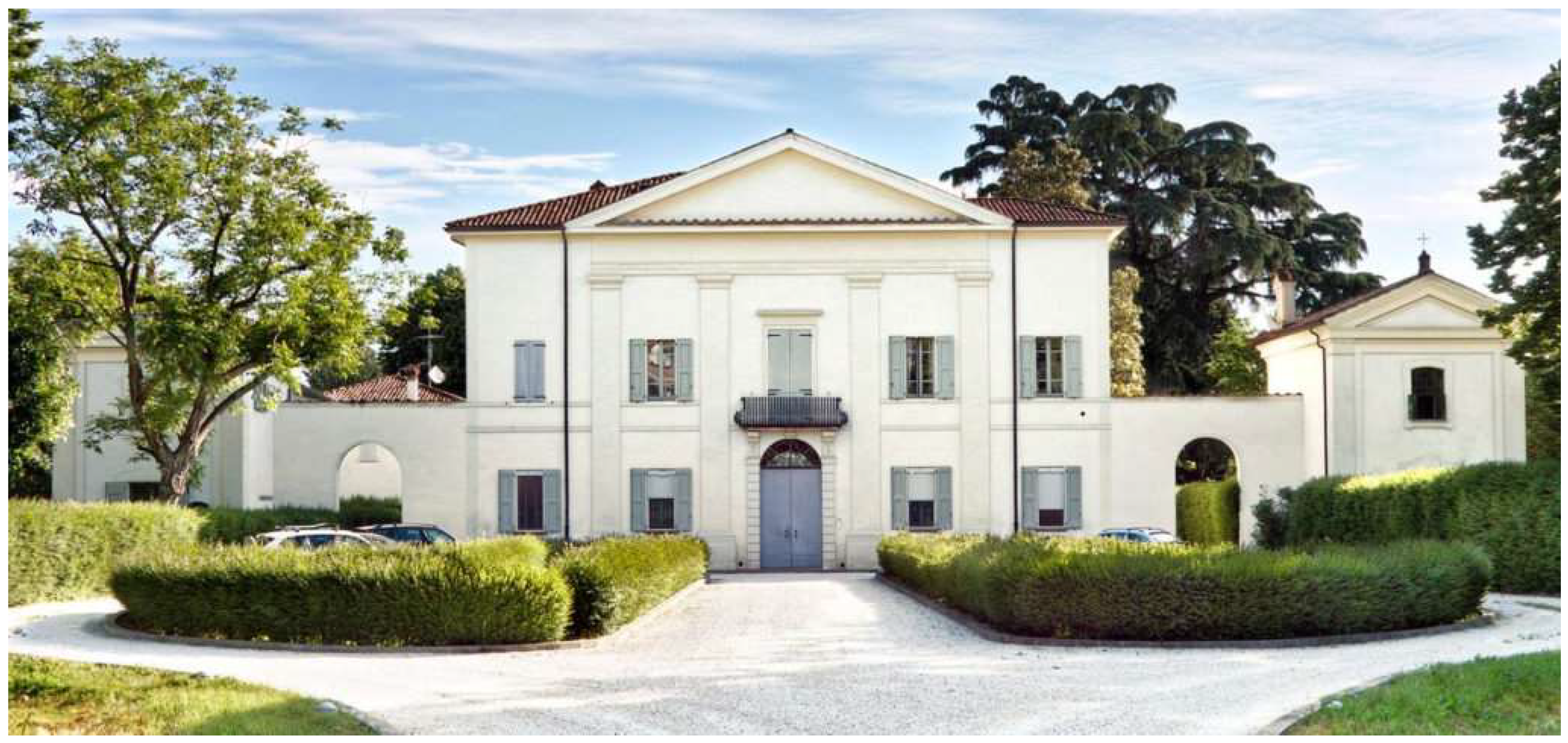
In the south-eastern outskirts of Bologna, there is an 18th century villa realized by the architect Angelo Venturoli (1749–1821). It includes a more ancient annex that is likely to be the country estate of Flaminio Rota—the presence of his heraldic crest makes this hypothesis very plausible—whose intended use has not yet been recognized with certainty. After many years of neglect, the villa, known as Villa Rivalta or Villa Negri, was restored in the 2000s and is currently a privately inhabited property (
Figure 1).
Inside the annex, the upper parts of the walls and the ceiling are decorated with a pictorial cycle illustrating medical scenes. Here, archives witness that Rota hosted students, as it was the custom at that time. For example, the official biographies of Cesare Magati claim this [
3,
12]. Thus, it is likely that the building was intended both for professional and educational activity. The frescoes are attributable to the school of Cesare Baglione (1550–1615), who was very active in Bologna at the end of the 16th century. The pictorial cycle may have been made between 1580 and 1610. A stone architrave placed under the frescoes, engraved with MDCIX (1609), suggests that this year corresponds with the pictorial realization. The work was likely commissioned before Rota’s second marriage in 1610 [
13].
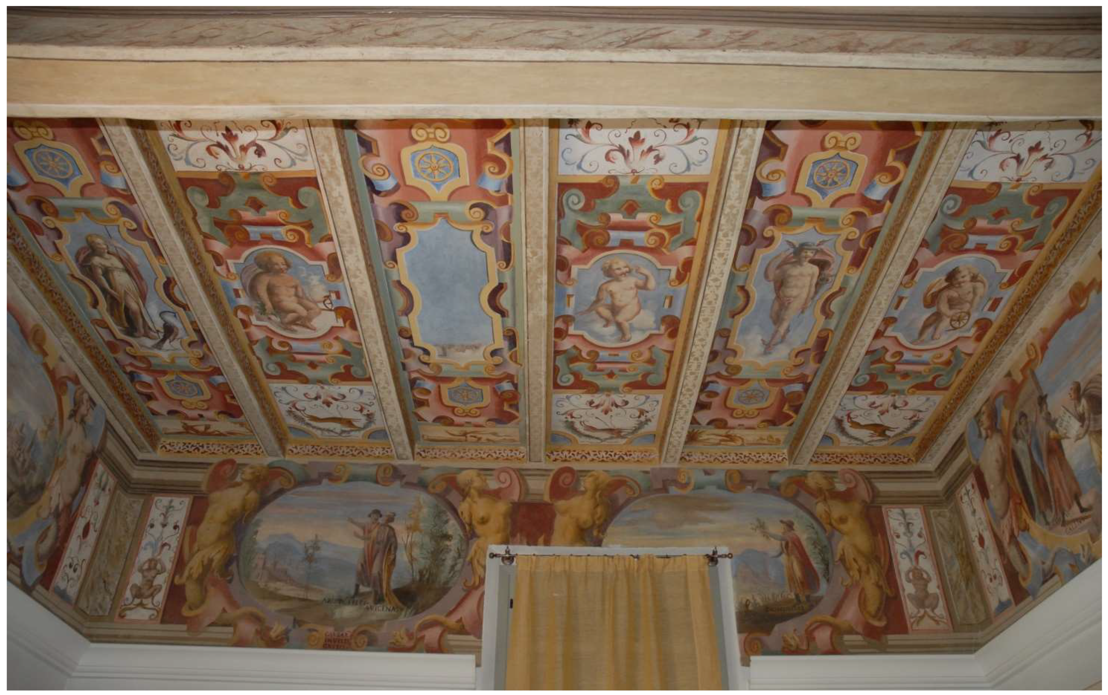
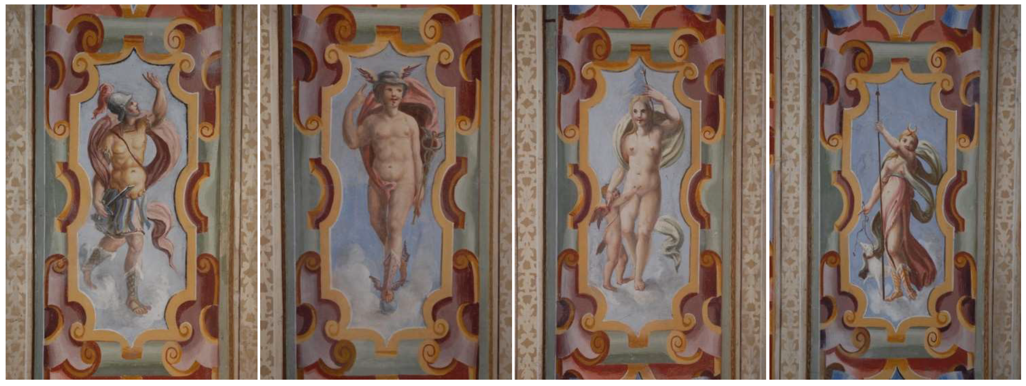
The decoration of the ceiling (
Figure 2), implanted on very curled cartouches and grotesques, with figures of birds, presents a human figure at the center of each background, delimited by the joists. Among these, pagan divinities (Mars, Venus, Diana, Minerva, Mercury, and another disappeared due to the collapse of a portion of the canopy that supports the painted surface) (
Figure 3) alternate with putti. The heraldic symbol of the wheel (in old-fashion Italian, Rota means wheel) appears in badges that accompany the mythological figures, and it is also found in the hand of one of the putti, who is playing with it.
For the purposes of this study, the most interesting images are undoubtedly those painted on the upper parts of the walls of the room. In fact, the room is frescoed with a series of oval paintings, two on each wall, which depict scenes that recall the fundamental disciplines of medical practice and well-known characters in the history of medicine. This was likely to be considered the most relevant by the client. In each oval painting, explicit writings identify the discipline and the characters depicted, leaving no interpretative doubts.
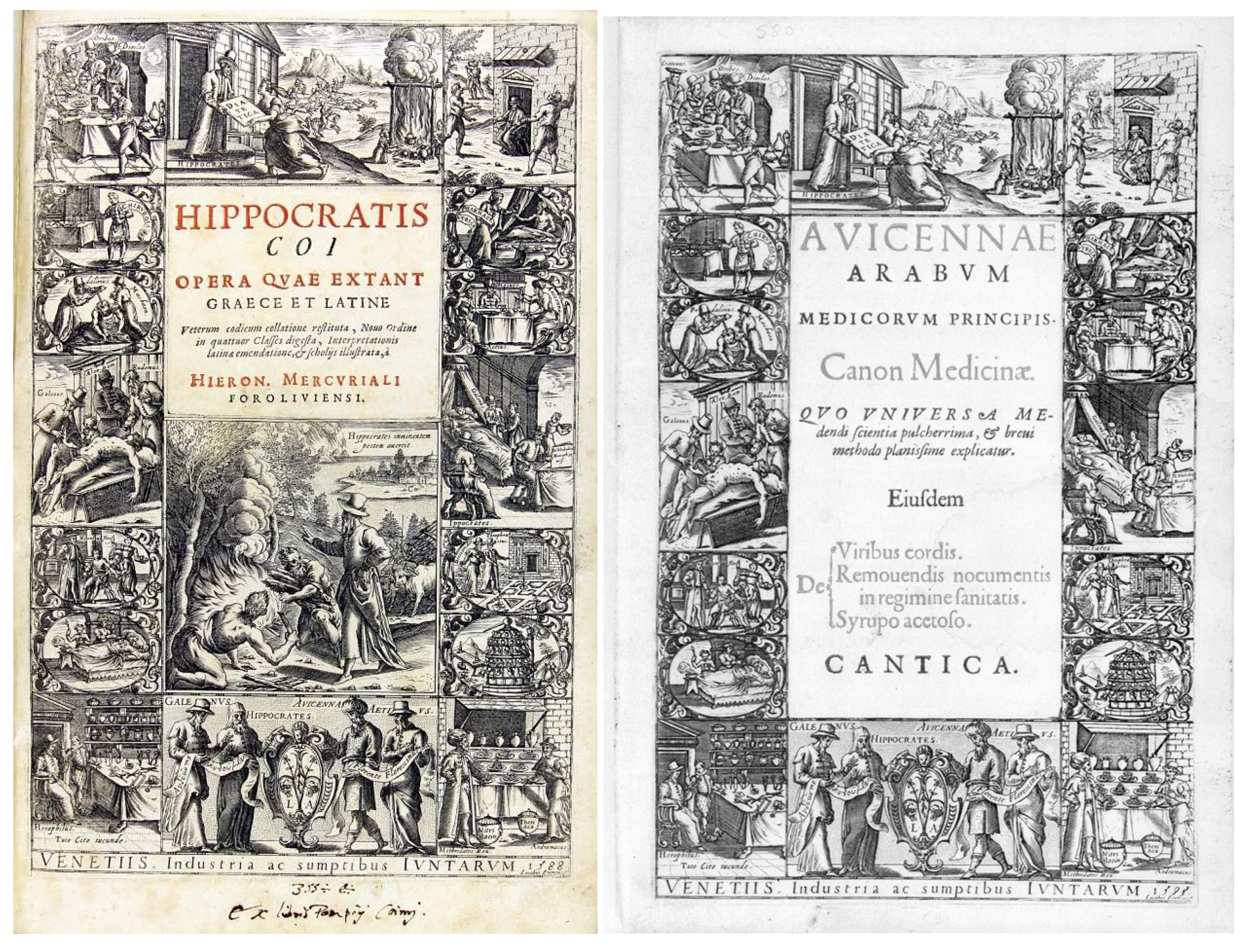
It is very likely that the pictorial cycle was inspired by the title page of the
Hippocratis Coi opera, that is, the publication of the writings of Hippocrates in Venice by Giunti printers in 1588 and supervised by Girolamo Mercuriale [
14]. It was engraved with a burin by the Italian engraver and publisher by Giacomo Franco (1550–1620), as attested by the signature “Iacobus Francus f.” affixed to the lower right corner. It was later used until 1609 by the same publishers for the editions of scientific works in folio format (for example, Avicenna’s
Canon) [
15] (
Figure 4).
Mercuriale is remembered as one of the fathers of dermatology. His De morbis cutaneis et omnibus corporis humani excrementis (Diseases of the Skin, and All the Wastes of the Human Body) in five volumes, collected around 1572, is considered the first treatise on dermatology. In 1587, Mercuriale left the chair of Padua to move to Bologna. At that time, Rota was already a lecturer in anatomy and surgery and a surgeon at the Hospital of Saint Job, set up for the treatment of syphilitics. It is conceivable to think that the two physicians met and confronted each other on scientific issues.
3. About the Frescoes
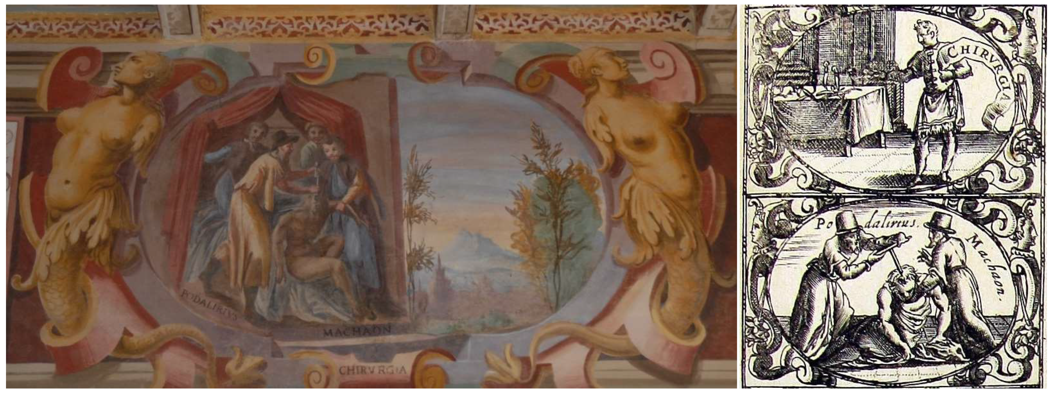
On the eastern wall, the first oval painting is dedicated to CHIRVRGIA (surgery), where a scene of the trephination of the skull is depicted. The operation is being performed by Podalirius and Machaon (the names of the two surgeons appear near their feet as “PODALIRIVS” and “MACHAON”), who are supported by two assistants. Podalirius and Machaon were doctors from Greek mythology. Tradition has it that they were sons of Asclepius (god of medicine) and Epione (princess of Kos). They learned their healing arts from their father and the centaur Chiron [
16,
17]. This image is also found in the title page of Giunti’s edition, supervised by Mercuriale (
Figure 5).
The next oval painting represents the “distillery” (at the bottom, there is the inscription DESTILATORIA), where a figure with the explanatory inscription MESVES approaches a colossal support full of stills. The distillery also appears in the frontispiece but with no characters nearby (
Figure 6).
Distillery, and in particular, the use of alcohol, is attributed to the Arab tradition. However, it is curious to note that to represent this practice, Rota did not use the much better-known Avicenna (represented in another oval painting) or Rhazes, remembered in the history of medicine for his pioneering alchemical studies based on distillation. Mesue is known as Mesue senior, that is, Yuhanna Ibn Masawaih (777–857). He became a knowledgeable physician and anatomist, and he was appointed as director of a hospital in Baghdad. He was the personal physician to four caliphs. He composed medical treatises on a number of topics, including ophthalmology, fevers, leprosy, headache, melancholia, dietetics, and medical aphorisms. One of Mesue’s treatises concerns aromatics, entitled
On Simple Aromatic Substances. It was reported that Mesue regularly held an assembly of some sort, where he consulted with patients and discussed subjects with pupils. He apparently attracted considerable audiences, having acquired a reputation for repartee. He translated various Greek medical works into Syriac, but wrote his own works in Arabic. Apes were supplied to him by the caliph al-Mu’tasim for dissection. Many anatomical and medical writings are credited to him, notably the
Disorder of the Eye (Daghal al-’ain), which is the earliest systematic treatise on ophthalmology extant in Arabic, and
The Aphorisms, the Latin translation of which became very popular in the Middle Ages [
18]. Mesue was representative of a transition period in Arabic medicine, when physicians no longer limited themselves to translations but also began to develop original medical studies and practices [
19]. In his works, there were many innovations that would provide the basis for the theory and practice of pharmacy for centuries and arguably formed part of the artisanal epistemological influence on the scientific revolution [
20].
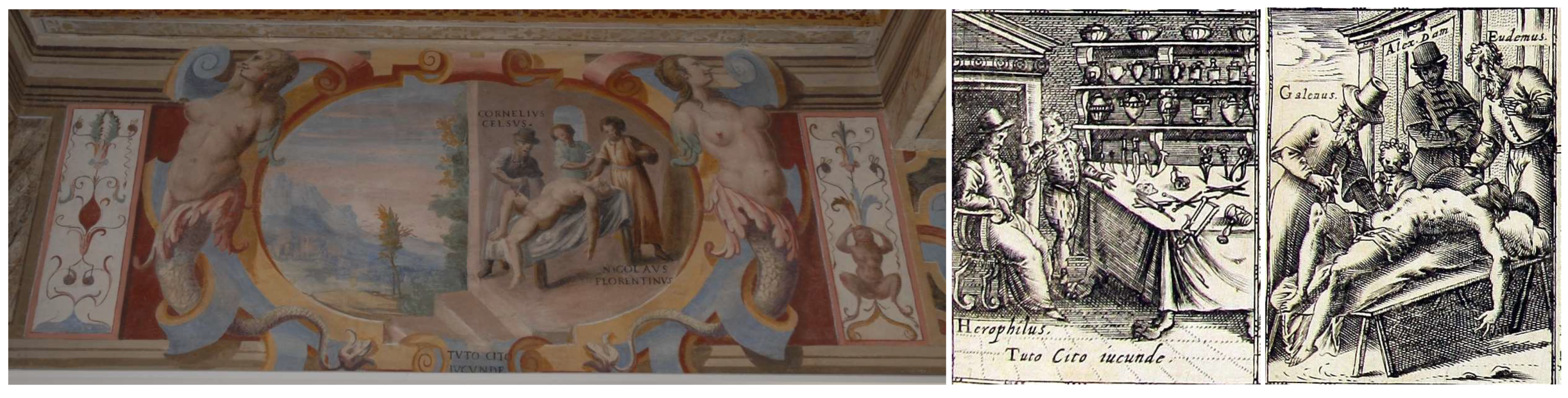
In the first oval painting of the southern wall, there is a scene where a sick person, naked on a bed, is visited by Aulus Cornelius Celsus and Nicola Fiorentino (as reported in the captions CORNELIVS CELSVS and NICOLAVS FLORENTINVS). At the bottom, there is the inscription TUTO CITO IVCVNDE (
Figure 7). Cito tuto iucunde (meaning to treat patients “swiftly, safely, and sweetly”) was a motto of Asclepiades of Bithynia (c. 130–40 BC). He opposed the humoral conception of Hippocrates and affirmed the atomistic doctrine, believing that all the phenomena of life must be attributed to mechanical laws, considering that diseases reside in the atoms, which live in continuous movement among them and are joined by channels, the so-called pores. As a skilled surgeon, he was certainly the first doctor who came to Rome to practice medicine based on scientific concepts [
17,
21].
However, this fresco can raise doubts concerning its interpretation [
13]. Indeed, in Giunti’s edition, the motto is associated with another scene, which depicts the anatomist, Herophilos, seated in front of a table with dissection instruments. It is evident that the artist’s intentions were to depict an anatomical dissection exercise. In fact, on the frontispiece, this vignette is in the right column, where surgery scenes are depicted. Furthermore, Giunti’s frontispiece features Galen (ca. 129–ca. 200) intent on dissecting a corpse under the gazes of the philosopher Eudemus, who benefited from his care, and Alessandro Damasceno, who, according to the tradition, was skeptical of the experiments carried out by Galen [
22,
23]. On the other hand, in the oval painting, the three figures were reinterpreted to feature Aulus Cornelius Celsus (c. 25 BC–c. 50 AD) and Nicolò Falcucci (also known as Nicolò Nicolai), who was a Florentine doctor who died in 1412, while the figure in the middle is just an unnamed bystander.
The Roman Celsus, a profound connoisseur of Hippocrates, certainly had contacts with Alexandrian medicine and with some Greek doctors transferred to Rome. He wrote a general encyclopedia (
De Artibus), including some medical arguments (
De Medicina), and two books of surgery. Following Hippocrates’ tradition, it can be considered the most significant medical work, and it had a great diffusion in Roman medical teachings until the arrival of Galen. Forgotten for several centuries, it was rediscovered in 1443 by Pope Nicolas V, and it was the first medical and surgical book to be printed. It is a matter of debate as to whether Celsus actually practiced as a surgeon or simply collected the medical knowledge available at that time [
24].
Even the fame of Nicolò Falcucci, more than to the practice of the medical profession, is linked to his remarkable work (
Sermones medicinales), which, according to many historians, was for several decades an obligatory point of reference for students in medicine. This was a handbook and a summary of the medical science of the time. He provided detailed indications on how medicine could be taught to medical students, and the book was to be an educational tool to improve the skills and knowledge of physicians. In this formation, theoretical and doctrinal competence and operative skill must support each other. This peculiar feature of medicine implied that the education of a physician must follow a unified course of study but according to a composite pattern of competence, in which “to know” and “to do” are integrated. In his book, Falcucci also introduced the good student and the good teacher of medicine. He prescribed that the student of medicine must be willing to learn (disciplinabilis), must not show off, and should attend the Studia. Above all, the pupil must love his teacher as a father, praise him, and defend his fame. To the master, on the contrary, he assigns more specific tasks. He affirmed, indeed, that the physician who is doctoratus must transform himself into a teacher and transmit whatever he has learned. We recognize here, translated in terms of educational relations and forms of writing, the two main divisions of medical knowledge: the theoretic part, with the techniques of transmission of commentary, and the practical part, which is better transmitted in the form of compendia and collections. We can also recognize three institutional characteristics imposed on medicine, as on every other discipline, by university structure: 1) The importance of the written text; 2) the role of authoritative texts in defining the scope and the identity of a discipline; and 3) the general need that what has been acquired by experience should be checked by comparison with authoritative texts and should be transformed by means of writing into doctrine that is thus rendered valid and transmissible [
25].
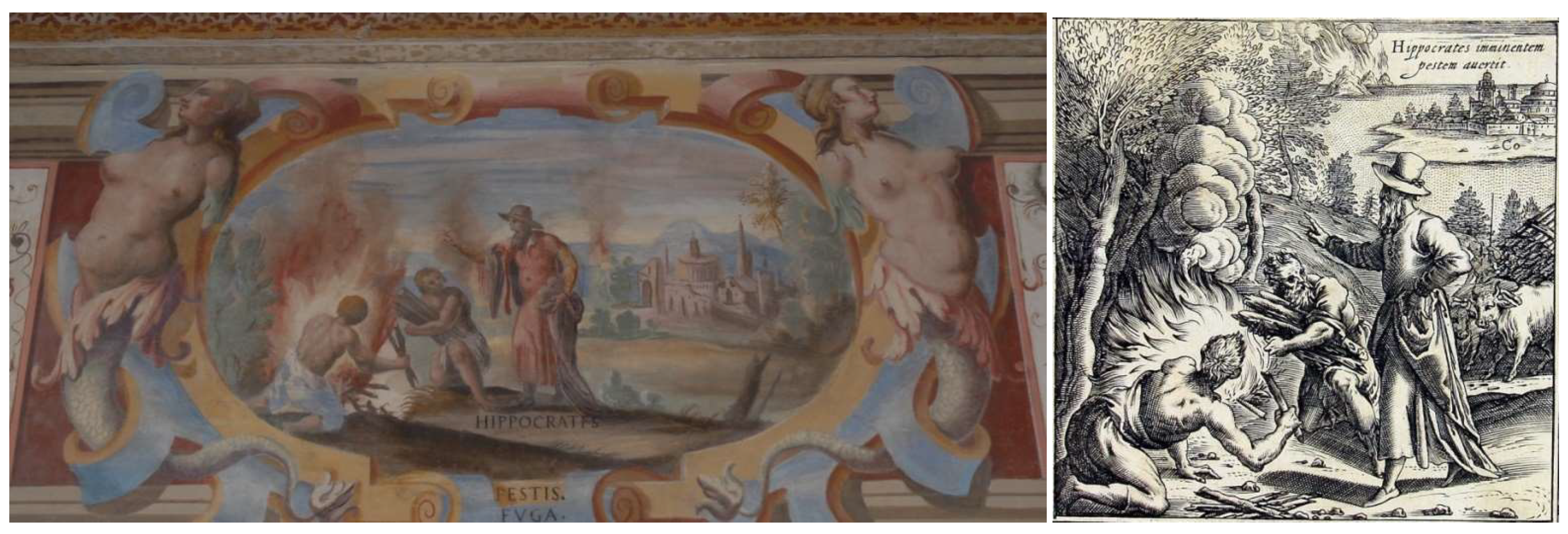
The second oval painting of the southern wall depicts a scene that takes place completely outdoors: in a countryside not far from a city, two men are burning infected materials according to the directives of a doctor, whose name can be read below: HIPPOCRATES. In the cartouche below, the inscription PESTIS, FVGA clearly explains the meaning of the representation. It should be remembered that Hippocrates acquired great fame in ancient times by eradicating the great plague of Athens in 429 BC [
26]. In Giunti’s frontispiece, there is a similar vignette to that of the oval painting just described (
Figure 8).
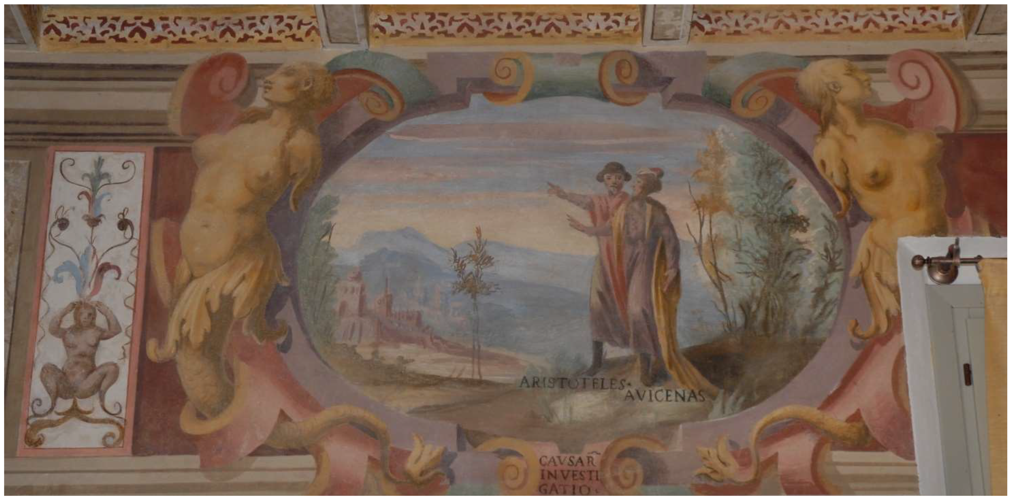
On the western wall, the first painting shows the two figures of ARISTOTELES and AVICENAS in the act of scrutinizing the skies, the air, and the countryside. The caption below reports CAVSARVM INVESTIGATIO (
Figure 9). In Giunti’s frontispiece, there are no vignettes similar to that of the oval painting just described. Rota entrusted the search for the causes of the diseases to two philosophers, Aristotle and Avicenna, respectively, exponents of Greek and Arab culture.
Avicenna reworked and updated all the Aristotelian sciences (with the addition of mathematics) to conform them to the criteria of demonstration that Aristotle outlined in the
Posterior Analytics. In the Avicenna’s system of science, metaphysics has to provide the foundation of all special sciences [
27].
The next depiction of the western wall is incomplete, due to a window being open on the wall after the completion of the fresco. The oval painting shows a man holding a bunch of freshly picked medicinal herbs. At the man’s feet, there is the following inscription: DIOSCORIDES. In Giunti’s frontispiece, there is a similar scene, where Dioscorides is in the company of Theophrastus in a botanical garden (
Figure 10). Pedanius Dioscorides (c. 40–90 AD) was a Greek surgeon and military physician for the emperor Nero; he was also a pharmacologist, botanist, and the author of
De materia medica, an encyclopedia about herbal medicine and related medicinal substances. More than 600 plants and 1000 drugs were described in this pharmacopeia, which remained the standard medical text until the 17th century. His notes on the plants include their habitats, methods of preparation, and medicinal uses of the drugs they contained [
17,
28]. With regard to Western materia medica, through the early modern period, Dioscorides’ text eclipsed the
Hippocratic corpus.
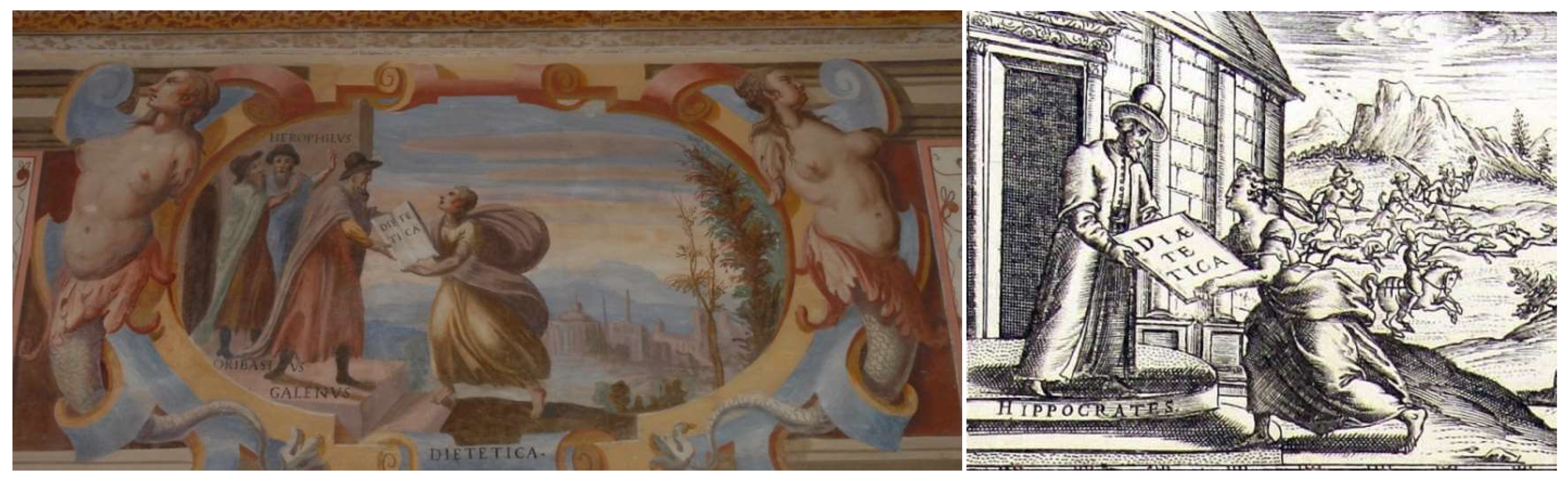
On the fourth and last wall, the northern one, the first oval painting is dedicated to dietetics: a messenger wrapped in a large cloak receives a book entitled DIETETICA from the hands of GALENVS, while HEROPHILVS and ORIBASIVS are present behind him. It is interesting to note that a very similar image is present in Giunti’s frontispiece, but the book is in the hands of Hippocrates (
Figure 11).
Hippocrates (5th century BC), the father of medicine, emphasized the concept that the diet is the best way to treat a disease. In the following centuries, Herophilos (3rd century BC), Galen (2nd century AD), and Oribasius (4th century AD) produced many writings on dietetics [
17,
29].
Herophilos (335–280 BC) was a Greek physician regarded as one of the earliest anatomists. He was the founder of the great medical school of Alexandria in Egypt and the first scientist to systematically perform scientific dissections of human cadavers. Herophilos believed that exercise and a healthy diet were integral to an individual’s quality of life [
17,
30].
The aforementioned Galen was a Greek physician, surgeon, and philosopher in the Roman Empire. He is considered one of the most accomplished of all medical researchers of antiquity, and his doctrine in medicine and science became law for the following twelve centuries. He was one of the believers in diet, stating that health depended chiefly on the choice of food [
17,
31].
Oribasius (c. 320–403) was a medical writer and the personal physician of the Roman emperor Julian. He was the most famous doctor of Byzantium. In his works, he discussed hygiene and dietetics. According to Oribasius, the physician must pave the way for new research by drawing lessons from the clinical observations and experimental methods of the great masters of the classical era. This figure also represents an important step on the path of Galenism, since he was the first to attribute a relevant part to the work of Galen, considering it as fundamental for the progress of medicine [
17,
32].
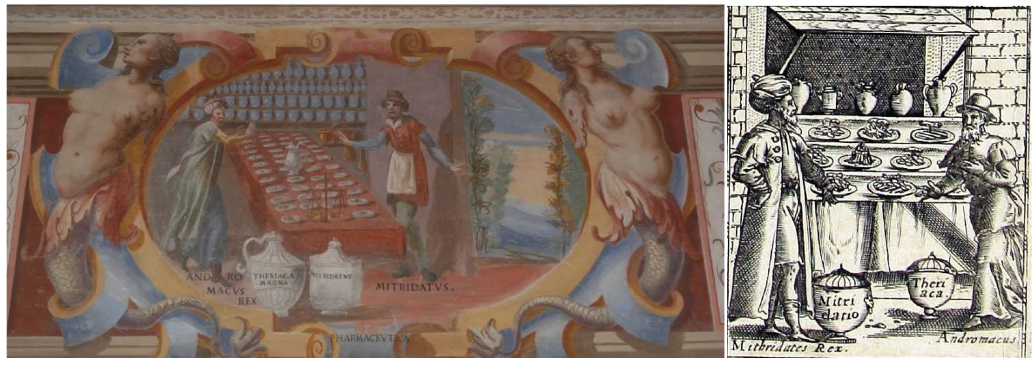
The second oval painting of the northern wall, the last of the series, depicts the interior of a pharmaceutical workshop with a large number of jars of various shapes and sizes, a mortar, a scale, and other objects; in the foreground, two large vessels bear the name of the medicaments they contain: THERIACA MAGNA and MITRIDATVS. Near these jars, on the sides of a counter cluttered with preparations, are the inventors of the two remedies. In this scene, however, the painter was the victim of a misunderstanding because under the figure of Mithridates, clearly recognizable by the dress and the crown he wears, he wrote ANDROMACVS REX, and under the figure of Andromachus, he wrote MITRIDATVS, thus exchanging one character with another. Below is the title of the representation: PHARMACEVTICA. A very similar scene is depicted in Giunti’s frontispiece (
Figure 12).
Mithridates, king of Pontus in the first century BC, was famous for taking poisons in small doses to be immunized against their effects, and the property of a universal antidote against poisoning was attributed to the drug that bears his name [
17].
Andromachus lived during the first century and was Nero’s physician. He came from the island of Crete, where herbs were gathered by “botanical persons” in the service of the emperor and placed in knitted pots, which were sent not only to Rome but also to other countries. The vast knowledge of botany by Andromachus helped him “provide the requisite medicines for mankind” [
33]. Andromachus took a great deal of Mithridates’ ingredients and produced a new antidote. The most important alteration of Andromachus to the Mithridatium antidote was the replacement of an African lizard, called skink, with the viper, which created a new antidote: the Theriac. This remedy continued to be used in Western medicine until the 17th century [
17,
33]. The composition of the Theriac was a controversial topic in 16th century Bologna, and the college of doctors was also consulted a few decades before Rota joined [
34].
4. Conclusions
This paper examined the medical pictorial cycle realized in the annex of the villa of Flaminio Rota and revealed a very interesting cultural aspect that added new information to a previously published article dealing with the biographical knowledge and the academic life of this intriguing 16th-century anatomist [
1]. In detail, this additional research allowed us to reach some considerations.
The first consideration is of an artistic nature. It is clear that the pictorial cycle attributed to the school of Cesare Baglione painted in the annex that belonged to Rota was inspired by the frontispieces of Hippocratis Coi and Avicenna’s Canon published by Giunti and not vice versa.
By analyzing the differences rather than the similarities, between the pictorial cycle and Giunti’s editions, it is possible to outline the intellectual and scientific profile of the Bolognese anatomist. In fact, it is assumed that the differences are the result of the client’s preferences.
For example, in the frescoes, there are no scenes dealing with medical treatments (present in Giunti’s editions), an activity not practiced by Rota. In the oval painting representing dietetics, Rota preferred Galen surrounded by the greatest exponents of the Alexandrian school (Herophilos and the Byzantine Oribasius), rather than Hippocrates. Galen paved the way for progress in medicine by combining the anatomo–physiological method of the school of Alexandria in Egypt with the Hippocratic tradition. Similarly, in another oval painting, Rota decided to support Aristotle with Avicenna to represent the passing of the baton of medical knowledge directed towards progress [
35].
The scene dedicated to anatomy from the analysis is perhaps the most significant, considering Rota’s career. At the dissecting table, he decided to put Celsus and Nicolò Falcucci. They are not famous anatomists, such as Herophilos or Galen, but didactic innovators or, better said, clarifiers and interpreters of already existing theories. Furthermore, in the figure of Celsus, there is the intention to honor the memory of his father, Giovanni Francesco Rota. Celsus attended valetudinarians, was a classifier of skin pathologies, and became appreciated as an elegant writer. Giovanni Francesco was a doctor of the papal army, professor of anatomy, a scholar of gunshot burns, and was remembered as an elegant writer. Even the choice of enhancing the figure of Mesue is perhaps attributable to his skills as an anatomist and teacher.
In summary, from the pictorial cycle, it is evident that Flaminio Rota was a doctor open to scientific progress and a supporter of experimental research based on the study of anatomy, which is capable of improving knowledge of Hippocratic origin. From the university point of view, it can be thought that Rota was attentive when teaching, which he carried out in a clear and concise but, at the same time, exhaustive and elegant way. Furthermore, his tendency to combine academic teaching with practical teaching clearly emerged. This tendency can be confirmed by the documentary biographical information known so far.
At the same time, thanks to the biographical information we know about Rota, it is possible to recognize him as the client of the pictorial cycle and to consolidate the hypothesis that the annex belonged to him.
Finally, regardless of the artistic value of the pictorial cycle and the biography of Flaminio Rota, questioning the meaning of the proposed images allows the reader to have an overview of the medical knowledge in the late 16th century.


