1. Introduction
Schwannoma is a benign peripheral nerve sheath tumor that is identified by a biphasic histologic appearance composed of antoni A and antoni B areas [
1]. Several histologic variants of schwannoma including “ancient”, plexiform, epithelioid, and neuroblastoma-like schwannoma have been identified [
2]. Neuroblastoma-like schwannoma is a rare variant of this entity. It was first described by Goldblum JR et al. [
3] in 1994, and by 2019, 24 cases of neuroblastoma-like schwannoma had been reported [
4]. This tumor is characterized by a typical immunophenotyping profile accompanied by areas that resemble neuroblastoma [
5]. Neuroblastoma is predominantly composed of small round hyperchromatic Schwann cells surrounding perivascular and giant rosettes [
6]. Herein, we report a case of neuroblastoma-like schwannoma that was presented as a lesion on the dorsal side of the left hand.
2. Case Report
A 39-year-old woman presented with a 3-year history of a slowly growing painless lesion of 2 cm in size, located on the dorsal side of the left hand. No accompanying symptoms were reported. There was no family history of schwannoma or any kind of cutaneous malignancy. The patient did not report a history of smoking and comorbidity.
The lesion was completely excised by an expert dermatologist at the hospital. Microscopically, an encapsulated creamy whitish round mass with a size of 2 × 2 cm, homogenous on cutting, without hemorrhage or necrosis, was observed.
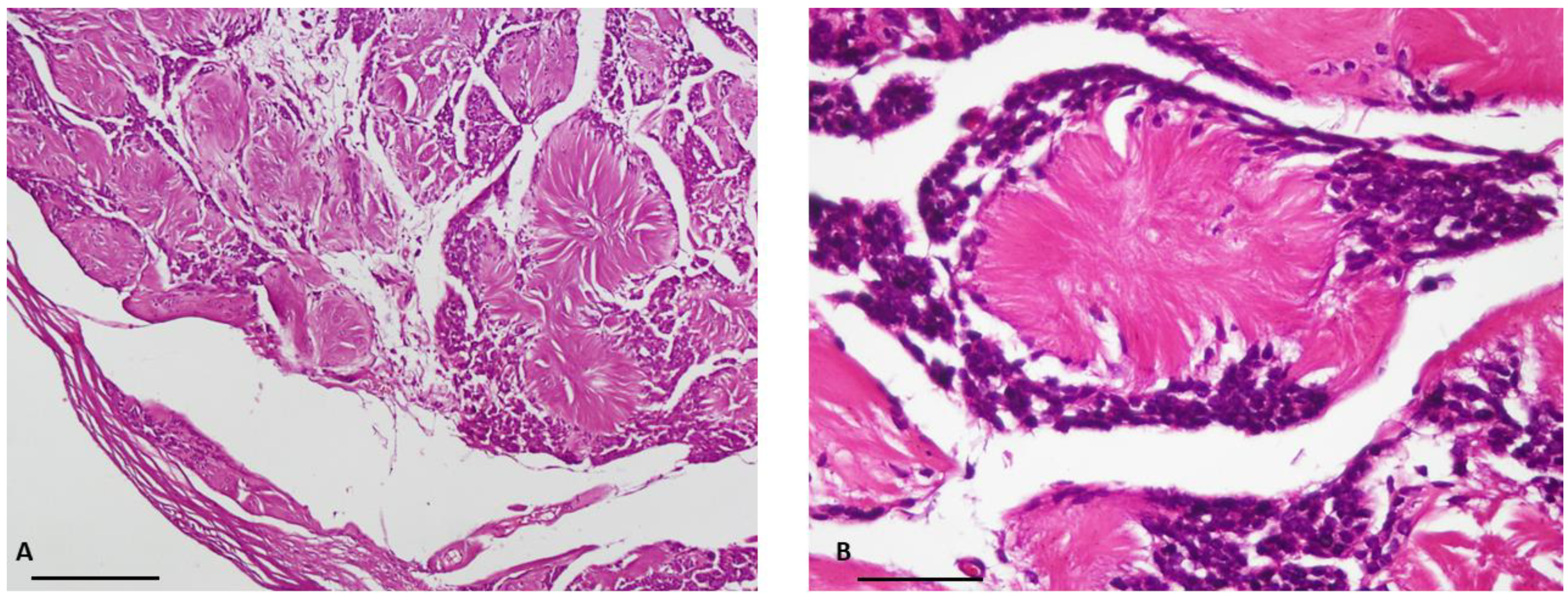
Histopathological investigation revealed a neoplastic tissue surrounded by a thin fibrous capsule and consisting of uniform small lymphocyte-like round cells with scant cytoplasm and small hyperchromatic nuclei. The cells were radially arranged around eosinophilic collagenous-core-forming giant rosettes. No cellular atypia, necrosis or mitosis was evident in this specimen. The classic pattern of schwannoma was notably absent in all representative sections.
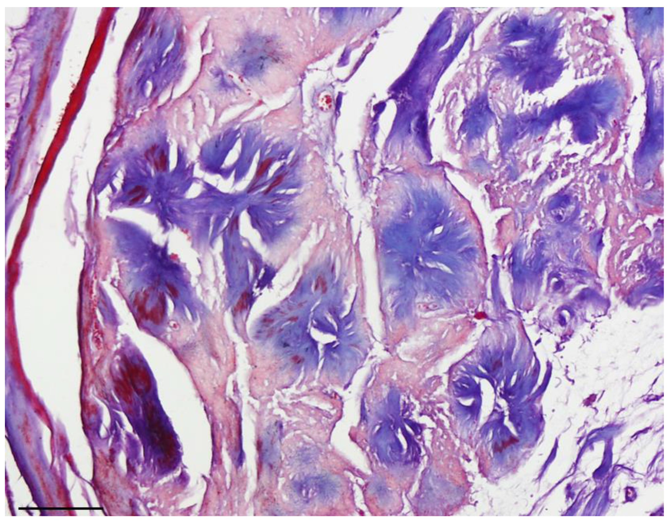
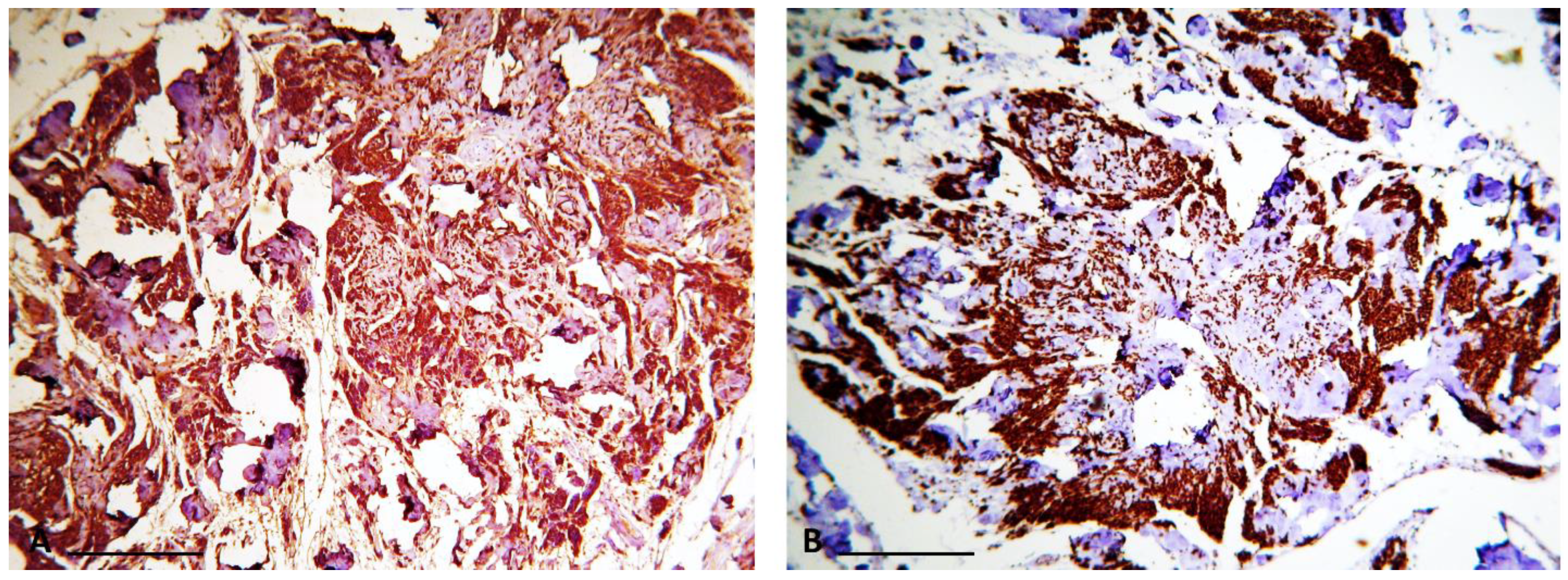
Masson’s trichrome staining confirmed that the central core of the rosettes was composed of collagen fibers. Subsequent immunohistochemical study revealed diffuse expression of S100 protein (as the most commonly used marker to detect tumoral cells) [
6] in the tumoral cells, confirming the Schwann cell origin of the tumor (
Figure 1 and
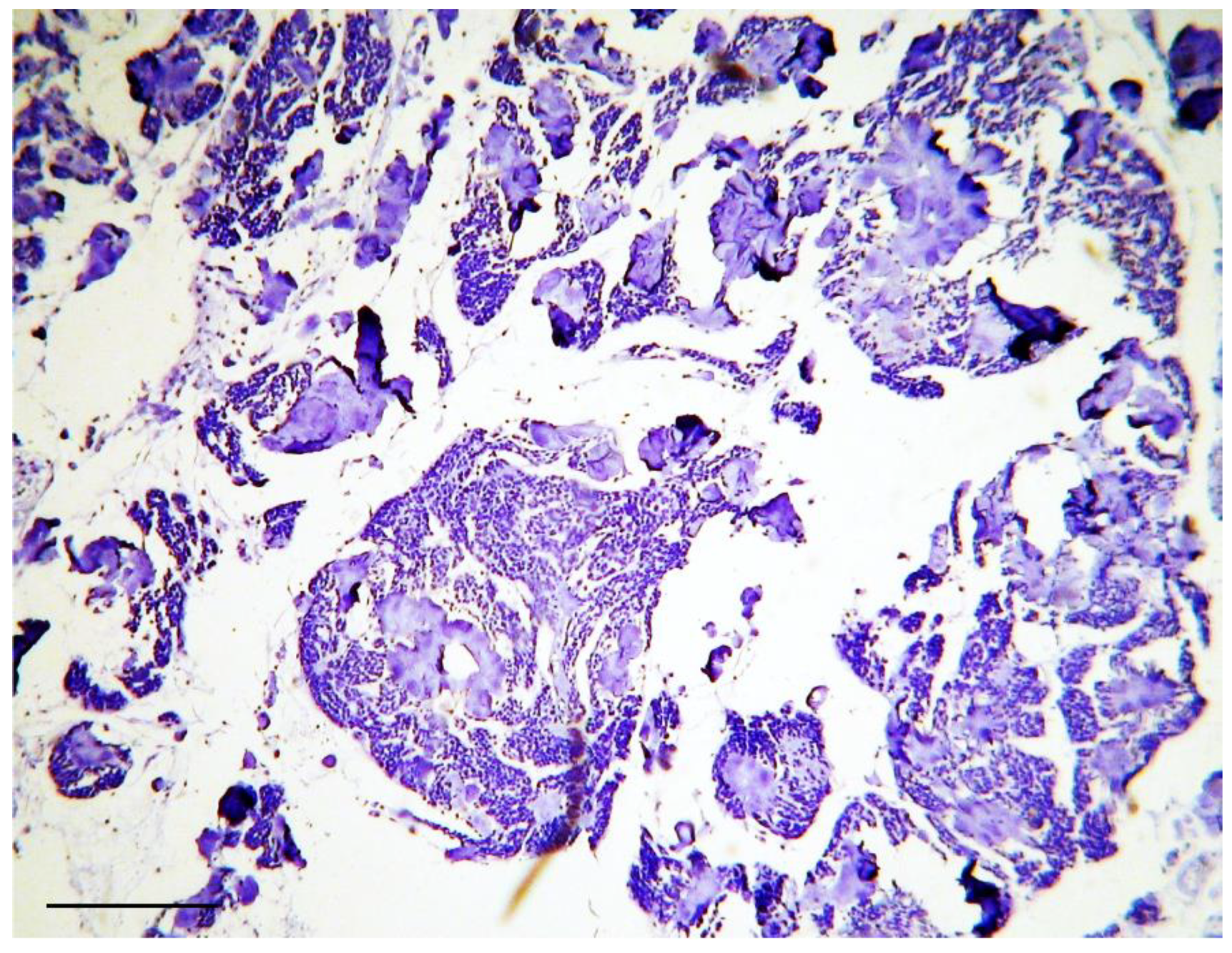
Figure 2). Additionally, staining for the neural markers showed focal positive expression for Neuron Specific Enolase (NSE) and negative for synaptophysin which confirmed our diagnosis (
Figure 3 and
Figure 4).
3. Discussion
Neuroblastoma-like schwannoma is a benign nerve sheath tumor as it grows slowly and usually does not represent an immediate risk to health. To date, a limited number of cases with a diagnosis of neuroblastoma-like schwannoma, as an exceptional case of schwannoma, have been described in the literature. Regarding the location of lesion, it has been shown that neuroblastoma-like schwannoma lesions are mostly located on the neck, trunk and extremities [
7]. However, lumbar spine nerve root [
8], lower labial mucosa [
9], orbit [
10] and pleura [
11] were also reported as sites of neuroblastoma-like schwannoma lesions. Interestingly, the location of lesion in our case was on the dorsal side of the left hand and that has not been reported previously.
Prevalence of neuroblastoma-like schwannoma is known to be higher in women than men and is usually reported as a painful mass [
12,
13]. Our patient was also a woman, however had a painless lesion. Higher prevalence has been suggested as a reason for an increased clinicopathological variation of schwannoma in women compared to men [
13].
Microscopically, neuroblastoma-like schwannoma can mimic other small round cell tumors, as originally described by Goldblum JR et al., and the presence of these small round cells along with rosette formation may cause confusion with primitive neuroectodermal tumors (PNETs) and neuroblastoma. Zachary T. Lewis believes that the name “neuroblastoma-like schwannoma” may overestimate these similarities [
13]. Neuroblastoma-like schwannoma can be differentiated from mentioned entities based on the presence of areas of conventional schwannoma, encapsulation, lack of mitoses and atypia, along with diffuse positivity for S100 protein [
2].
Difference in the size of rosettes is considered an important histological criterion to distinguish neuroblastoma from neuroblastoma-like schwannoma [
2,
5]. In agreement with previously reported observations, rosettes in our neuroblastoma-like schwannoma case were trichrome positive [
12].
Dendritic cell neurofibroma is another dermal lesion with pseudorosettes that might be confused in diagnosis with neuroblastoma-like schwannoma. The differentiation between dendritic cell neurofibroma and neuroblastoma-like schwannoma is based on the smaller size of the pseudorosettes, with a histopathological characterization including lagers cells with pale eosinophilic cytoplasm and vesicular nuclei, and strong CD57 positivity [
14,
15]. The other distinguishing feature between dendritic cell neurofibroma and neuroblastoma-like schwannoma is positive staining with S100 protein and positive/absent CD34 staining [
14].
In our case, the histopathological investigation on Hematoxylin and Eosin (H&E) stained sections revealed a small cell tumor with the cellular characteristics of neuroblastoma described earlier, along with areas of conventional schwannoma. Moreover, no cellular atypia, necrosis or mitosis was evident in the specimen of our patient. These findings are consistent with the findings of previous neuroblastoma-like schwannoma case reports [
16].
Immunohistochemical methods helped us to reach a definite diagnosis. Indeed, in our case, the tumor cells were strongly and diffusely positive for S100 protein and focally positive for NSE. Neuroblastoma-like schwannoma showed focal areas with typical immunophenotyping of conventional schwannoma and all tumor cells were strongly and diffusely positive for S100 protein, but typically negative for other markers of neural differentiation, such as synaptophysin [
3]. However, there are reports in which the tumor cells show positivity for other neural markers such as NSE [
9].
4. Conclusions
Neuroblastoma-like schwannoma is an unusual variant of schwannoma which can be misinterpreted as malignancy. Therefore, performing more specific histopathological characterization, such as examining the expression of S100 protein in tumoral cells, in combination with histological investigation, including H&E staining, is of fundamental importance for the pathologists.
Author Contributions
Conceptualization, S.M., K.K.H., A.K.; investigation, S.M.; resources, S.M., K.K.H., A.K., M.A.; writing—original draft preparation S.M., K.K.H., A.K.; writing—review and editing, M.A., S.M. All authors have read and agreed to the published version of the manuscript.
Funding
This research received no external funding.
Institutional Review Board Statement
The study was conducted in accordance with the Declaration of Helsinki, and approved by This research was approved by the ethics committee of Tehran University of Medical Sciences (2017/675, March 2017).
Informed Consent Statement
Informed consent was obtained from subject involved in the study.
Data Availability Statement
Not applicable.
Conflicts of Interest
The authors declare no conflict of interest.
References
- Rodriguez, F.J.; Folpe, A.L.; Giannini, C.; Perry, A. Pathology of peripheral nerve sheath tumors: Diagnostic overview and update on selected diagnostic problems. Acta Neuropathol. 2012, 123, 295–319. [Google Scholar] [CrossRef] [PubMed] [Green Version]
- Somerhausen, N.D.S.A.; Valaeys, V.; Geerts, M.; André, J. Neuroblastoma-like schwannoma: A case report and review of the literature. Am. J. Dermatopathol. 2003, 25, 32–34. [Google Scholar] [CrossRef] [PubMed]
- Goldblum, J.R.; Beals, T.F.; Weiss, S.W. Neuroblastoma-like neurilemoma. Am. J. Surg. Pathol. 1994, 18, 266–273. [Google Scholar] [CrossRef] [PubMed]
- Mahjoub, W.K.; Jouini, R.; Khanchel, F.; Ben Brahim, E.; Llamas-Velasco, M.; Helel, I.; Khayat, O.; Chadli, A.; Badri, T.; Mentzel, T. Neuroblastoma-like schwannoma with giant rosette: A potential diagnostic pitfall for hyalinizing spindle cell tumor. J. Cutan. Pathol. 2019, 46, 234–237. [Google Scholar] [CrossRef] [PubMed]
- Sharma, P.; Chatterjee, D.; Das, A. Neuroblastoma like schwannoma: A diagnostic challenge. BMJ Case Rep. 2017. [Google Scholar] [CrossRef] [PubMed] [Green Version]
- Sulhyan, K.R.; Deshmukh, B.D.; Gosavi, A.V.; Ramteerthakar, N.A. Neuroblastoma-like schwannoma in a case of schwannomatosis: Report of a rare case. Int. J. Health Sci. 2015, 9, 478–481. [Google Scholar] [CrossRef]
- Suchak, R.; Luzar, B.; Bacchi, C.E.; Maguire, B.; Calonje, E. Cutaneous neuroblastoma-like schwannoma: A report of two cases, one with a plexiform pattern, and a review of the literature. J. Cutan. Pathol. 2010, 37, 997–1001. [Google Scholar] [CrossRef]
- Sharma, G.K.; Eschbacher, J.M.; Uschold, T.D.; Theodore, N. Neuroblastoma-like schwannoma of lumbar spinal nerve root. J. Neurosurg. Spine 2010, 13, 82–86. [Google Scholar] [CrossRef]
- Sedassari, B.T.; Lascane, N.A.D.S.; Gallottini, M.H.C.; de Sousa, S.C.O.M.; Júnior, D.D.S.P. Neuroblastoma-like schwannoma of the lower labial mucosa: A rare morphologic variant of peripheral nerve sheath tumor. Oral. Surg. Oral. Med. Oral. Pathol. Oral. Radiol. 2014, 118, 579–582. [Google Scholar] [CrossRef] [PubMed]
- Kukreja, M.; Gupta, R.; Julka, A.; Sharma, M.C. Neuroblastoma-like schwannoma as a rare cause of proptosis: A case report. Can. J. Ophthalmol. 2007, 42, 624–625. [Google Scholar] [CrossRef]
- Adams, K.; Liu, X.S.; Akhtar, I.; Flowers, R.; Baliga, M. Pleural-based neuroblastoma-like schwannoma: A case report with cytologic findings and review of literature. Diagn. Cytopathol. 2015, 43, 650–653. [Google Scholar] [CrossRef]
- Bhatnagar, S.; Banerjee, S.S.; Mene, A.R.; Prescott, R.J.; Eyden, B.P. Schwannoma with features mimicking neuroblastoma: Report of two cases with immunohistochemical and ultrastructural findings. J. Clin. Pathol. 1998, 51, 842–845. [Google Scholar] [CrossRef] [Green Version]
- Lewis, Z.T.; Geisinger, K.R.; Pichardo, R.; Sangueza, O.P. Schwannoma with neuroblastoma-like rosettes: An unusual morphologic variant. Am. J. Dermatopathol. 2005, 27, 243–246. [Google Scholar] [CrossRef]
- Michal, M.; Fanburg–Smith, J.C.; Mentzel, T.; Kutzner, H.; Requena, L.; Zamecnik, M.; Miettinen, M. Dendritic cell neurofibroma with pseudorosettes: A report of 18 cases of a distinct and hitherto unrecognized neurofibroma variant. Am. J. Surg. Pathol. 2001, 25, 587–594. [Google Scholar] [CrossRef]
- Lerman, M.A.; Li, C.-C.; Woo, S.-B. Dendritic cell neurofibroma with pseudorosettes: A clinicopathologic and immunohistochemical study of 5 intraoral cases. Oral Surg. Oral Med. Oral Pathol. Oral Radiol. 2014, 117, 221–226. [Google Scholar] [CrossRef]
- Van Nguyen, A.; Argenyi, Z.B. Cutaneous neuroblastoma. Peripheral neuroblastoma. Am. J. Dermatopathol. 1993, 15, 7–14. [Google Scholar] [CrossRef] [PubMed]
| Publisher’s Note: MDPI stays neutral with regard to jurisdictional claims in published maps and institutional affiliations. |
© 2022 by the authors. Licensee MDPI, Basel, Switzerland. This article is an open access article distributed under the terms and conditions of the Creative Commons Attribution (CC BY) license (https://creativecommons.org/licenses/by/4.0/).


