1. Introduction
Surgical procedures and clinical diagnoses require a deep understanding of the common and less common anatomical variations of structures located in the surgical field, particularly in relation to blood vessels. The concept of “normal anatomy” is based on repeated observations [
1]. Detailed knowledge is now more important than ever because of present and future improvements in surgical procedures. Dissection—although not a surgical act—is essential and preparatory for an oral and maxillofacial surgeon, whose aim is to deepen the knowledge of anatomical organization beyond the limits of the operating field, thus guaranteeing greater safety and a less traumatic surgery. Thus, in situations where normal anatomical variation could be confusing, a deep understanding of vessel anatomy is mandatory. During radical neck surgery, the carotid bifurcation (CB) is a significant anatomical landmark, which helps to define the plane of dissection [
2], and neck surgery must be planned according to the anatomical pattern of the CB in order to minimize complications [
3].
2. Case Report
A rare configuration of the origin of the carotid artery anterior branch, which cannot be included in current classifications, was found on the left side of the neck of a 72-year-old man during a routine didactical neck dissection. The educational focus was on vessels supplying the oral cavity for students at the Sapienza University of Rome, and the dissection was performed at the ICLO Teaching and Research Center of Verona, Italy.
The cadaver specimen was treated using the “fresh frozen” technique (−20 °C). No formalin fixation solution was used in order to preserve the tissue features for the in vivo presentation. A vascular injection fluid (latex) was used to achieve vascular patterns.
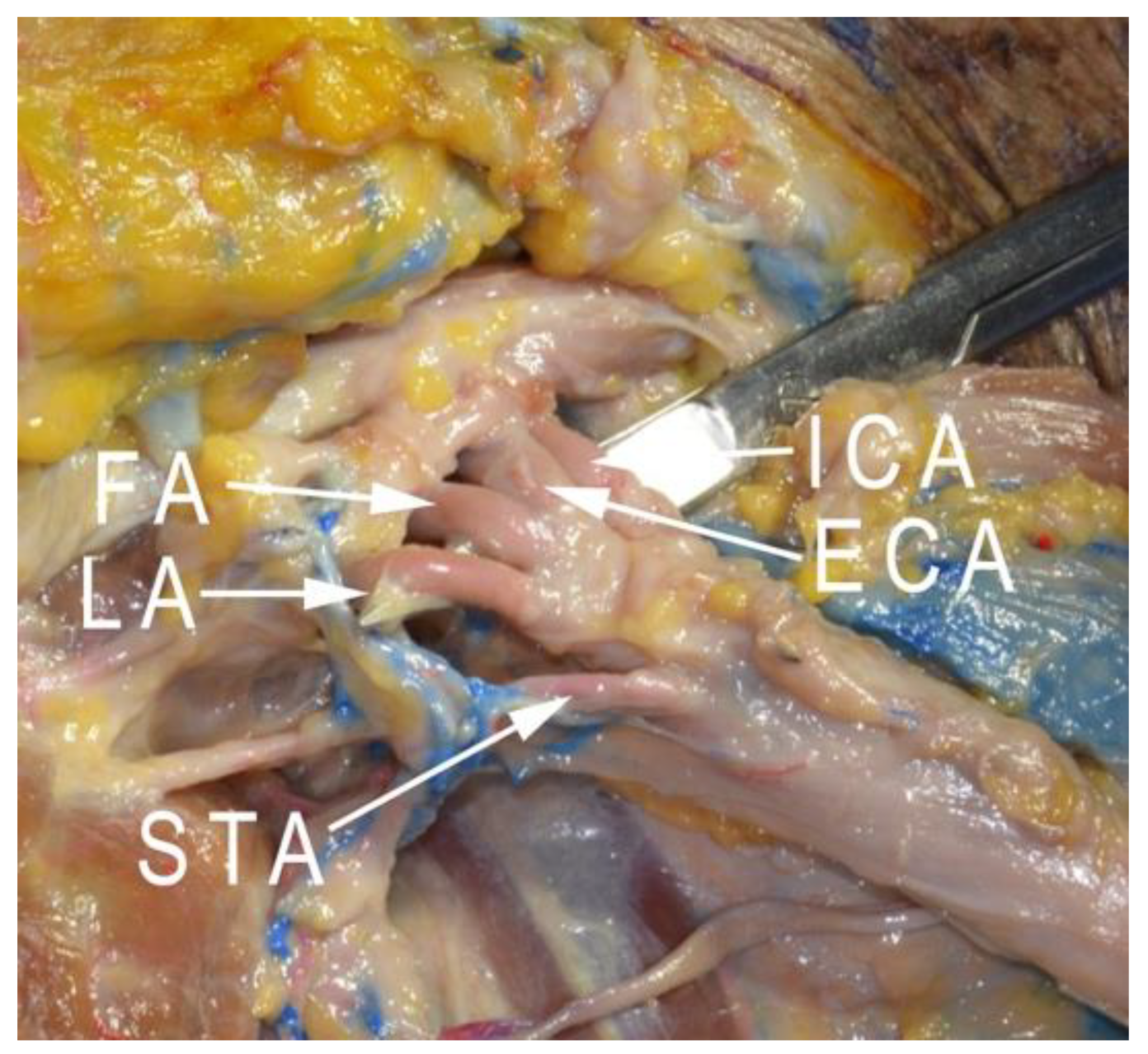
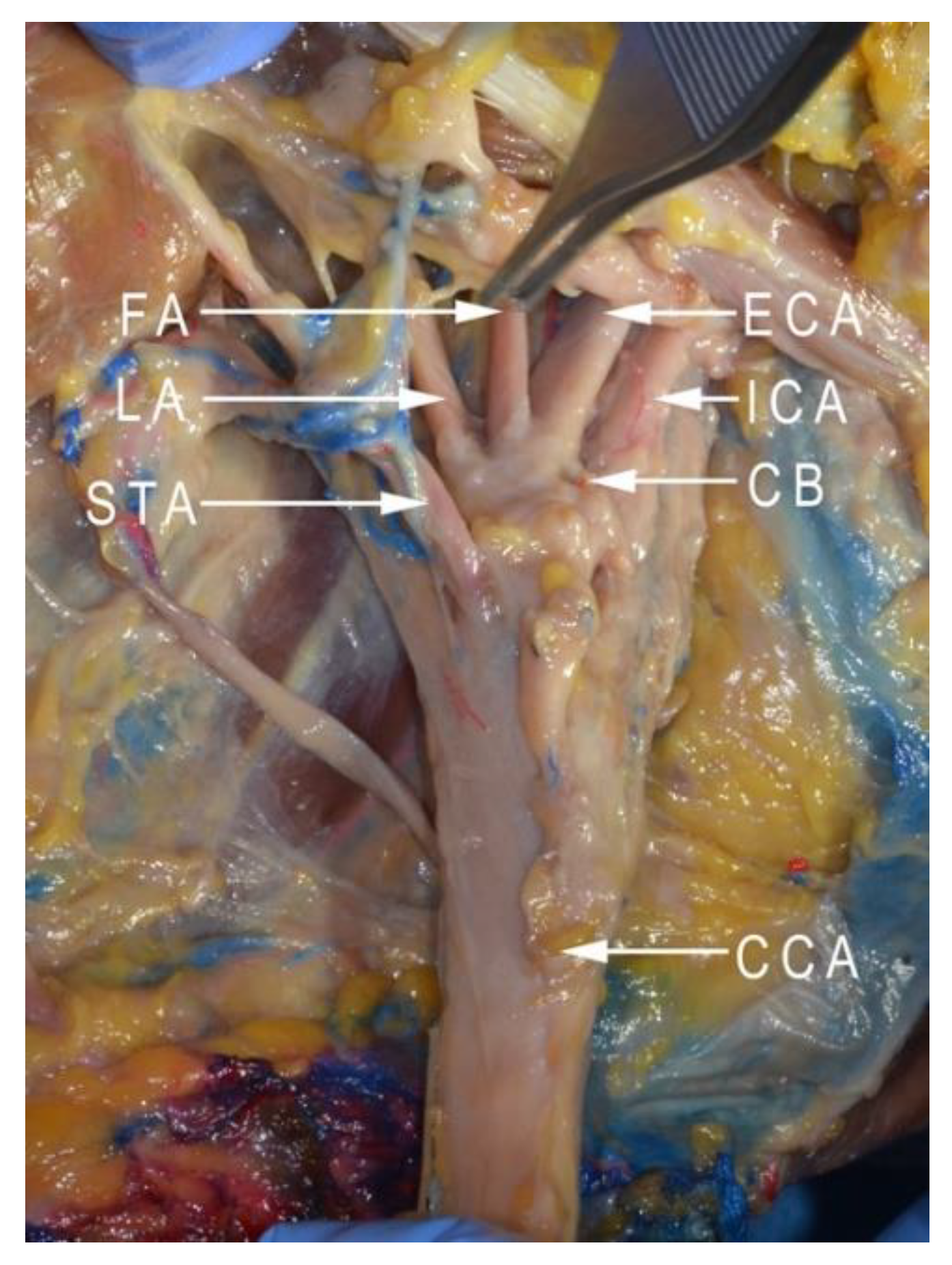
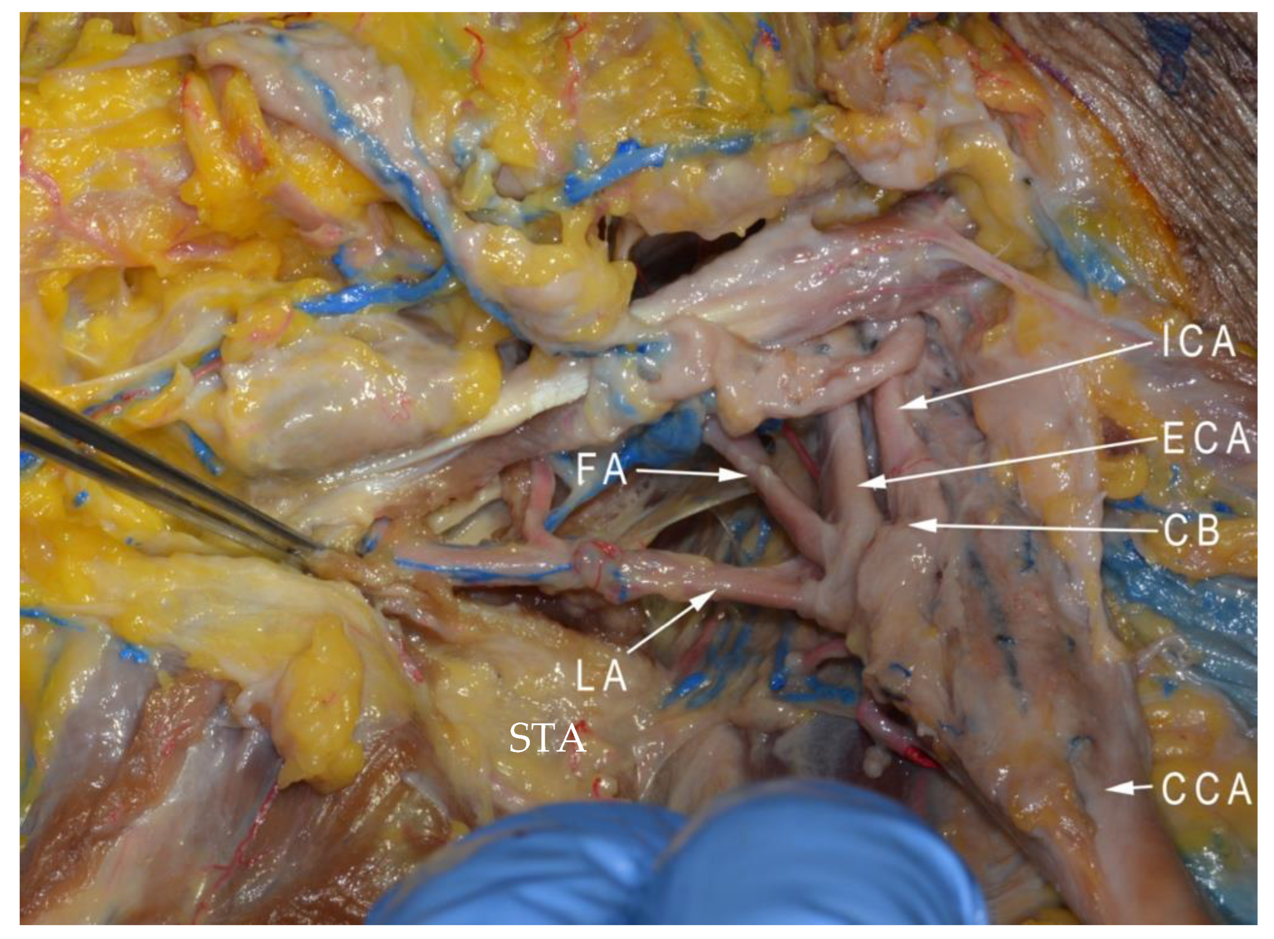
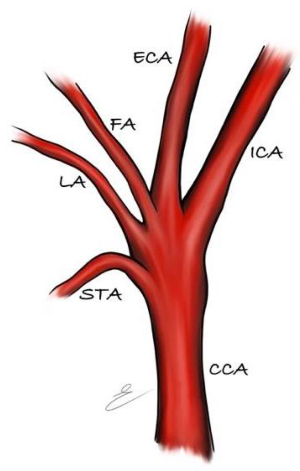
In this rare case, the superior thyroid artery (STA), the lingual artery (LA) and the facial artery (FA) were found to be detached directly from the CCA at the terminal ramus just before the carotid bifurcation (CB). Thus, we obtained a pentafurcation of the common carotid artery with figures resembling a hand (see
Figure 1). This pattern has never been previously reported in the literature as far as we know. The contralateral carotid artery configuration of the analyzed cadaver specimen is a Natsis type I.
Figure 1,
Figure 2,
Figure 3 and
Figure 4 show the abnormal left CCA and the course of its ventral branches. A detailed description of the cadaver’s anatomy follows this paragraph.
The left common carotid artery (CCA) is 9.1 mm in width. It arises from the aortic arch and continues upwards.
The superior thyroid artery (STA) is 1.8 mm in width and arises from the common carotid artery (CCA). The distance between the STA and the origin of the left CCA is 118 mm and 28 mm below the CB. The origin of the STA is located at the height of the C3 vertebral body, 3 mm above the posterior margin of the greater horn of the hyoid bone.
The lingual artery (LA) is 2.1 mm in width and represents the second branch medially arising from the CCA. It is located between the STA and the facial artery (FA). The distance between the STA and LA is 10 mm.
The FA and external carotid artery (ECA) are located above the LA. The lingual artery is anteriorly and superiorly directed, continuing as a frontal course until reaching the posterior margin of the hyoglossus muscle above the hyoid bone. The LA on the right side presents the same level of origin.
The facial artery (FA) is 2.9 mm in width and medially detaches from the CCA at a level of about 2.7 mm upon the lingual artery. Some differences between the right and left side of the facial artery are noted, especially in the distribution. The origin of the left FA is at the same level of the right side, but the distribution of the peripheral branches is different: the left FA crosses the submandibular gland and continues along the internal wall of the mandible, dividing into two main branches. The absence of the left superior labial artery and left angular artery is offset by the contralateral rami of the FA.
The external carotid artery (ECA) can be defined as a common trunk of the posterior auricular, maxillary and superficial temporal arteries. ECA originates at the height of C2 and starts from the glomus caroticum, above the origin of FA. It presents a width of 3.4 mm. From the CB, it runs parallel to the ICA. The posterior auricular artery is 0.9 mm in width and continues superiorly along the back of the head. The maxillary artery and the superficial temporal artery originate 47 mm above the ECA origin from the CCA and present a width of 2.6 mm and 2.4 mm, respectively. We did not find anomalies of the posterior auricular, superficial temporal and internal maxillary arteries.
The internal carotid artery (ICA) shows a width of about 4.1 mm. Its course can be divided into two tracts: cervical and intracranial. The cervical tract does not present collateral branches and continues upward, posteriorly to the ECA. The styloglossus muscle and stylopharyngeus muscle separate the ICA from the ECA. Once the cranial cavity is reached, the intracranial tract starts. No abnormalities were observed on the ICA entrance in the cranial cavity either.
3. Discussion
As a very rare occurrence, the carotid artery can ascend in the neck without dividing into the two usual branches, and either the external or internal carotid artery can be absent [
4]. Julius A Ogeng’o [
5] described some cases where, instead of bifurcating, the common carotid presents with the variant terminations of trifurcation (31.7%), quadrifurcation (5.4%) and pentafurcation (1.4%). The common carotid artery may provide one or more of the branches, usually derived from the external carotid. In this rare case, the pentafurcation of the CCA comprises the STA, LA, FA, ECA and ICA. This pattern has never been described before.
According to the classifications proposed by Natsis et al. and Ozgur et al., the anterior branches of the external carotid artery (ECA) are classified into four types [
6,
7]:
The STA, the LA and the FA originate from separate branches;
The STA and the LA share a common thyrolingual trunk;
The LA and the FA share a common linguofacial trunk;
The STA, the LA and the FA originate from a common thyrolinguofacial trunk.
Moreover. Vazquez et al. divided the classification into three subtypes based on the STA origin [
8]:
From the carotid bifurcation (50.2%);
From the common carotid artery (26.6%);
From the external carotid artery (23.2%).
Once the CCA gives rise to the CB at the level of the thyroid cartilage, the CCA divides into the ECA and the ICA. Following the CB, the ECA gives rise to its anterior branches: the STA, the LA and the FA [
7]. The anterior branches of the ECA have already been described by several authors (STA, LA and FA) [
7,
9,
10,
11,
12,
13,
14,
15]. Natsis 2011 and Zumre 2005 have also described divergences from the ECA in terms of linguofacial trunk (LFT), thyrolingual trunk (TLT) and thyrolinguofacial trunk (TLFT). A variety of origins for these anterior branches have been reported in the literature, including their occurrence either from other carotid arteries or from carotid arterial trunks, but never for the carotid bifurcation before [
16,
17,
18,
19]. Kaneko has previously described a case report where the ECA branches arose directly from the CCA, but the arrangement differed from the one presented here; there was no external carotid artery, and all the external carotid artery branches were derived from the internal carotid artery instead [
4].
4. Conclusions
This case report documents a very rare variant termination of the CCA and an unusual arrangement configuration of the anterior branches of the ECA, which cannot be recognized in the existing classifications.
Due to the wide range of surgical and radiological procedures performed in this area, the distribution of the ECA, the ICA and their branches are clinically relevant.
Possible anatomical variations are documented and classified by various authors and are crucial not only in routine planned surgeries, but in emergencies too. Furthermore, the vascular pattern is important for the evolution and prognosis of oncologic diseases.
Author Contributions
Dissections and writing—original draft preparation, F.Z., F.M., A.G., A.P., G.P. and M.E.C.; supervision, A.C.; original drawing, M.E.C. All authors have read and agreed to the published version of the manuscript.
Funding
This research received no external funding.
Institutional Review Board Statement
This study was conducted in accordance with the Declaration of Helsinki. No additional approval of an ethics board was required for this study.
Informed Consent Statement
All human cadaveric specimens were derived from donors who gave their written consent for the use, postmortem, of their body for educational and scientific purpose.
Acknowledgments
The authors sincerely thank those who donated their bodies to science so that anatomical research could be performed. The results from such research can potentially increase humankind’s overall knowledge, which can then improve patient care. Therefore, these donors and their families deserve our highest gratitude.
Conflicts of Interest
The authors declare no conflict of interest.
Abbreviations
| CB | carotid bifurcation |
| STA | superior thyroid artery |
| CCA | common carotid artery |
| LA | lingual artery |
| FA | facial artery |
| ECA | external carotid artery |
| ICA | internal carotid artery |
| Arteries’ dimensions (Width in mm) |
| 9.1 | left common carotid artery |
| 1.8 | superior thyroid artery |
| 2.1 | lingual artery |
| 2.9 | facial artery |
| 3.4 | external carotid artery |
| 0.9 | posterior auricular artery |
| 2.6 | maxillary artery |
| 2.4 | superficial temporal artery |
| 4.1 | internal carotid artery |
References
- Żytkowski, A.; Tubbs, R.S.; Iwanaga, J.; Clarke, E.; Polguj, M.; Wysiadecki, G. Anatomical normality and variability: Historical perspective and methodological considerations. Transl. Res. Anat. 2020, 23, 100105. [Google Scholar] [CrossRef]
- Klosek, S.K.; Rungruang, T. Topography of carotid bifurcation: Considerations for neck examination. Surg. Radiol. Anat. 2008, 30, 383–387. [Google Scholar] [CrossRef] [PubMed]
- Michalinos, A.; Chatzimarkos, M.; Arkadopoulos, N.; Safioleas, M.; Troupis, T. Anatomical Considerations on Surgical Anatomy of the Carotid Bifurcation. Anat. Res. Int. 2016, 2016, 1–8. [Google Scholar] [CrossRef] [PubMed] [Green Version]
- Kaneko, K.; Akita, M.; Murata, E.; Imai, M.; Sowa, K. Unilateral anomalous left common carotid artery; a case report. Ann. Anat. Anat. Anz. 1996, 178, 477–480. [Google Scholar] [CrossRef] [PubMed]
- Ogeng’o, J.A.; Misiani, M.; Malek, A.; Inyimili, M.; Murunga, A.; Ongeti, K. Variant termination of the common carotid artery: Cases of quadrifurcation and pentafurcation. Anat. J. Afr. 2014, 3, 386–392. [Google Scholar]
- Natsis, K.; Raikos, A.; Foundos, I.; Noussios, G.; Lazaridis, N.; Njau, S.N. Superior thyroid artery origin in Caucasian Greeks: A new classification proposal and review of the literature. Clin. Anat. 2011, 24, 699–705. [Google Scholar] [CrossRef] [PubMed]
- Ozgur, Z.; Govsa, F.; Ozgur, T. Assessment of Origin Characteristics of the Front Branches of the External Carotid Artery. J. Craniofacial Surg. 2008, 19, 1159–1166. [Google Scholar] [CrossRef] [PubMed]
- Vazquez, T.; Cobiella, R.; Maranillo, E.; Valderrama, F.J.; McHanwell, S.; Parkin, I.; Sañudo, J.R. Anatomical variations of the superior thyroid and superior laryngeal arteries. Head Neck 2009, 31, 1078–1085. [Google Scholar] [CrossRef] [PubMed]
- Lo, A.; Oehley, M.; Bartlett, A.; Adams, D.; Blyth, P.; Al-Ali, S. Anatomical variations of the common carotid artery bifurcation. ANZ J. Surg. 2006, 76, 970–972. [Google Scholar] [CrossRef] [PubMed]
- Zümre, Ö.; Salbacak, A.; Çiçekcibaşi, A.E.; Tuncer, I.; Seker, M. Investigation of the bifurcation level of the common carotid artery and variations of the branches of the external carotid artery in human fetuses. Ann. Anat. Anat. Anz. 2005, 187, 361–369. [Google Scholar] [CrossRef]
- Hayashi, N.; Hori, E.; Ohtani, Y.; Ohtani, O.; Kuwayama, N.; Endo, S. Surgical anatomy of the cervical carotid artery for carotid endarterectomy. Neurol. Med.-Chir. 2005, 45, 25–30. [Google Scholar] [CrossRef] [PubMed]
- Fazan, V.P.S.; da Silva, J.H.N.; Borges, C.T.; Ribeiro, R.A.; Caetano, A.G. An anatomical study on the lingual–facial trunk. Surg. Radiol. Anat. 2009, 31, 267–270. [Google Scholar] [CrossRef] [PubMed]
- Thwin, S.S.; Soe, M.M.; Myint, M.; Than, M.; Lwin, S. Variations of the origin and branches of the external carotid artery in a human cadaver. Singap. Med. J. 2010, 51, e40–e42. [Google Scholar]
- Troupis, T.; Michalinos, A.; Dimovelis, I.; Demesticha, T.; Vlasis, K.; Skandalakis, P. Bilateral Abnormal Origin of the Anterior Branches of the External Carotid Artery. Ann. Vasc. Surg. 2013, 28, 494.e5-7. [Google Scholar] [CrossRef] [PubMed]
- Murlimanju, B.V.; Prabhu, L.V.; Pai, M.M.; Jayaprakash, D.; Saralaya, V.V. Variant Origins of Arteries in the Carotid Triangle. Chang Gung Med. J. 2012, 35, 281–284. [Google Scholar] [PubMed]
- Poisel, S.; Golth, D. Variability of the large arteries in the carotid triangle. Wien. Med. Wochenschr. 1974, 15, 229–232. [Google Scholar]
- Czerwiński, F. Variability of the course of external carotid artery and its rami in man in the light of anatomical and radiological studies. Folia Morphol. 1981, 40, 449–453. [Google Scholar]
- Lučev, N.; Bobinac, D.; Maric, I.; Drešćik, I. Variations of the great arteries in the carotid triangle. Otolaryngol. Neck Surg. 2000, 122, 590–591. [Google Scholar] [CrossRef]
- Demirtaş, I.; Ayyıldız, B.; Demirbaş, A.T.; Ayyıldız, S.; Topcu, F.S.; Kuş, K.C.; Kurt, M.A. Geometric morphometric study of anterior branches of external carotid artery and carotid bifurcation by 3D-CT angiography. Surg. Radiol. Anat. 2022, 44, 1029–1036. [Google Scholar] [CrossRef] [PubMed]
| Publisher’s Note: MDPI stays neutral with regard to jurisdictional claims in published maps and institutional affiliations. |
© 2022 by the authors. Licensee MDPI, Basel, Switzerland. This article is an open access article distributed under the terms and conditions of the Creative Commons Attribution (CC BY) license (https://creativecommons.org/licenses/by/4.0/).

 ,
, 


