Article

Construction and Immunogenicity Evaluation of Recombinant Adenovirus-Expressing Capsid Protein of Foot-and-Mouth Disease Virus Types O and A

State Key Laboratory of Veterinary Etiological Biology, OIE/National Foot-and-Mouth Disease Reference Laboratory, Key Laboratory of Animal Virology of Ministry of Agriculture, Lanzhou Veterinary Research Institute, Chinese Academy of Agricultural Sciences, No. 1 Xujiaping, Yanchangpu, Chengguan District, Lanzhou 730046, China
*
Author to whom correspondence should be addressed.
Zoonotic Dis. 2023, 3(2), 104-119; https://doi.org/10.3390/zoonoticdis3020010
Submission received: 9 December 2022 / Revised: 10 February 2023 / Accepted: 8 April 2023 / Published: 19 April 2023
(This article belongs to the Special Issue Feature Papers of Zoonotic Diseases 2021–2022)

Abstract

:
The objective of this study was to construct a recombinant adenovirus expressing the foot-and-mouth disease virus (FMDV) capsid protein of types O and A for future FMDV vaccines to be used in the livestock industry for the reduction in losses caused by FMD outbreaks. Three recombinant adenoviruses, rAdv-P12A3B3C-OZK93, rAdv-P12A3B3C-OA58, and rAdv-P12A3C-AF72, were packaged, characterized, and amplified using the AdMaxTM adenovirus packaging system, and the humoral and cellular immunity levels were further evaluated in guinea pigs with monovalent or bivalent forms. The results showed that the three recombinant adenoviruses could elicit high levels of humoral and cellular immune responses against FMDV types O and A when immunizing monovalent or bivalent forms, and the immune effect changes with the change in the proportion of recombinant adenovirus types O and A, laying an important foundation for the future development of a new FMD live-carrier vaccine. These results implied that the recombinant adenovirus expressing the FMDV capsid protein of types O and A could be used to prevent FMDV in livestock.

1. Introduction

Foot-and-mouth disease (FMD) is a highly exposed infectious disease that, as a major animal disease worldwide, can cause serious economic losses and public health problems. Its etiologic agent is foot-and-mouth disease virus (FMDV), a member of the Picornaviridae family [1]. The virus genome consists of a single-strand, positive-sense RNA molecule of approximately 8500 nucleotides in length that encodes a long open reading frame (ORF) flanked by untranslated regions (UTRs) at the 5′ and 3′ ends.
The ORF encodes a large polyprotein that can be cleaved by viral proteases into four structural proteins, VP4, VP2, VP3, and VP1, and eight nonstructural proteins, L, 2A, 2B, 2C, 3A, 3B, 3C, and 3D [2,3]. Four differently structured peptides of 60 molecules each constitute the capsid of the virion with VP1, VP2, and VP3 proteins exposed to the surface, while VP4 is inside [4]. The VP1 protein is highly immunogenic, and the coding sequence of this protein is commonly used for the molecular diagnosis of FMD and vaccine studies [5,6]. Eight nonstructural proteins control viral replication, protein processing, and protein modification [1]. Proteases include the leading proteins L, 2A, 3C, and unknown enzymes with 3C protease (3Cpro) playing a very important role in the cleavage of FMDV structural proteins. During viral translation and modification, the FMDV capsid protein precursor P12A is cleaved by 3Cpro to produce 1AB (VP0), 1C (VP3), 1D (VP1), and 2A, and VP0 is further lysed by 3Cpro to VP2 and VP4 during genome loading [7]. During assembly of the virions, VP0, VP3, and VP1 first form 5S protomers followed by 14S pentamers, and 12 pentamers finally assemble into 75S FMDV empty capsids [8].
FMDV empty capsids have antigen specificity that is very similar to that of the intact FMDV 146S antigen and are capable of causing similar antibody responses [9]. FMDV empty-capsid vaccines are safe since they do not contain viral genes and do not pose a risk of spreading. FMDV isolates are diverse, are widely distributed, have a high rate of genetic mutations, and have large antigenic differences, mainly seven serotypes (O, A, C, SAT1, SAT2, SAT3, and Asia l) [10]. There is no cross-immunity between the seven serotypes, and only partial cross-immunity occurs between different subtypes of the same serotype [7]. FMDV serotypes are unevenly distributed worldwide with type O being the most widely distributed FMDV serotype, especially in Asia [11]. This is followed by type A FMD transmission. FMD can cause a worldwide pandemic because its susceptible animals are mostly economically important with serious implications for animal production and international trade of the related products [11,12]. The World Organization for Animal Health (WOAH) ranked FMD first in the January 1984 revised classification of infectious diseases in animals [12]. The International Biological Weapons Treaty of some developed countries also include FMD [13], and FMD is also included in the list of a class of animal infectious diseases in China. The wide distribution of FMD worldwide and its consequences have brought great losses to the animal husbandry industry, making the elimination and control of the spread of FMD a common concern of government departments around the world. Currently, vaccination and culling are the primary approaches to controlling the spread of FMD. The main method of preventing the disease is immunization with a FMDV-inactivated vaccine; however, it can only provide short-term protection and is serotype specific for seven serotypes. There is a potential risk of incomplete inactivation of FMDV and virus escape, making the research of new vaccines the focus of FMD research.
Replication-deficient human adenovirus type 5 has the following advantages: it can still be expressed normally after inserting foreign genes into specific areas of the adenovirus genome and does not replicate in inoculated animals; recombinant adenovirus can achieve high titers, and the immunization route is diverse and has the prospect of oral vaccine development; and the adenovirus vector carrying protective antigen genes can stimulate the body to produce strong humoral and cellular immune responses [14,15,16,17]. According to Barrera J et al. [18], a FMDV recombinant adenovirus vaccine prepared with replication-deficient human adenovirus vectors can be efficiently expressed in swine-derived cells and is effective against foot-and-mouth disease virus infection both in vitro and in vivo [19]. Trials in the Plum Island Animal Disease Center laboratory in the United States demonstrated that a recombinant adenovirus vaccine expressing the FMDV-P12A3B3C protein constructed using a highly immunogenic strain can produce up to 100% Immune protective effects in pigs [20]. Adenovirus-specific antibodies are primarily induced by their early protein-coding regions E1 and E3, while E1 and E3 are missing in replication-deficient adenovirus vectors thus reducing the likelihood of immune rejection [21].
In this study, the parent strains of the inactivated vaccines of types O and A FMDV preserved in this laboratory have good immunogenicity. The purpose of this study was to construct the recombinant adenoviruses rAdv-P12A3B3C-OZK93, rAdv-P12A3B3C-OA58, and rAdv-P12A3B3C-AF72, which can stably express the capsid protein precursors of the P12A and 3B3C proteases of the above strains. The immunogenicity of the abovementioned one-component and bivalent three-component recombinant adenovirus was further evaluated through animal immune experiments, which laid a foundation for research on recombinant adenovirus vaccines of O- and A-type FMDV and for the development of new FMDV live-vector vaccines.

2. Materials and Methods

2.1. Plasmids, Cells, and Animals

The adenoviral skeleton plasmid pBHGlox (delta) E1,3Cre and the adenoviral shuttle plasmid pDC316-mCMV-EGFP were purchased from China Beijing Zhuang Meng Biological Company. Guinea pigs were provided by the Lanzhou Veterinary Research Institute (LVRI) of the Chinese Academy of Agricultural Sciences. Human embryonic kidney 293 (HEK-293) cells were provided by this laboratory, the control adenovirus (WtAdv) was obtained in this study, and the control bovine type O and type A FMDV-inactivated virus was provided by the National Innovation Laboratory for Foot-and-Mouth Disease.

2.2. Main Reagents and Consumables

PrimeSTAR® GXL DNA Polymerase and the NotI and AflII restriction enzymes were purchased from TaKaRa (Beijing, China), T4 DNA ligase was purchased from New England Biolabs (Beijing) Ltd. (Beijing, China), and plasmid extraction kits were purchased from Omega (GA, USA). PrimeSTAR® GXL DNA Polymerase and the Adeno-X™ Maxi Purification Kit were purchased from TaKaRa. Lipofectamine 3000 was purchased from Thermo Fisher Scientific (WI, USA). Type O/A FMDV anti-VP1 monoclonal antibody (MAb) was prepared in our laboratory. TRITC-labeled sheep anti-rabbit IgG (TRITC-IgG) and horseradish peroxidase HRP-labeled goat anti-rabbit IgG (HRP-IgG) were purchased from Abcam Corporation (Cambridge, England). BSA was purchased from Sigma (Shanghai, China). PVDF membranes were purchased from Millipore (MA, USA). Skimmed milk powder was purchased from Shanghai Shenggong. Protein Marker (10–250 kDa) was purchased from Shanghai Ya Enzyme Company. The DNA extraction kit (E.Z.N.A® Tissue DNA Kit) was purchased from Omega Bio-Tek Annoren Corporation. Adeno-X™ Maxi Purification Kit was purchased from TaKaRa. MTT Lymphocyte Proliferation Assay Kit, Lymphocyte Isolation Solution Kit, and Knife Bean Protein A were purchased from Beijing Solarbio Biotechnology Co., Ltd. (Beijing, China). Gibco RPMI 1640 medium was purchased from Thermo Fisher. Mouse IL-10 ELISA and IFN-γ ELISA kits were purchased from Xinbosheng Biotechnology Co., Ltd. (Shenzhen, China).

2.3. Construction and Screening of Recombinant Adenoviruses

To construct recombinant replication-deficient human type 5 adenovirus (Ad5) expressing the FMDV capsid protein of types O and A, specific primers for the P12A and 3B3C genes of FMDV-OZK93 were engineered and synthesized. The P12A and 3B3C genes were then amplified and connected with fusion PCR, and the recombinant shuttle plasmid pDC316-mCMV-EGFP-P12A3B3C expressing the FMDV-OZK93 capsid protein precursor P1-2A and 3B3C protease was then obtained by inserting the P12A3B3C gene into the pDC316-mCMV-EGFP plasmids. The P12A3B3C genes of FMDV-OA58 and FMDV-AF72 were artificially synthesized and inserted into the adenoviral shuttle vector pDC316-mCMV-EGFP.
The sequence similarity between the VP1 sequence of the FMDV-OZK93 strain (CATHAY) and the type O isolate WFL (EF175732.1) strain on the NCBI was found to be more than 99%, so the type O strain was referred to as isolate WFL (EF175732.1) strains P12A and 3B3C, which were similar to strains P12A and 3B3C of FMDV-OZK93. Primers were designed using Primer Premier 6.0, and the NotI enzyme resection site and Kozak sequence 5′-GCCGCCACC-3′ were added to the upstream 5′ end. The AflII digestion site was added to the 3′ end. The primer sequence is shown in Table 1 and was synthesized by Shanghai Bioengineering Co., Ltd. (Shanghai, China).
Comparing the VP1 sequence of the FMDV-OA58 strain with the type O strain Akesu/58 (GenBank: AFBank: AF511039) strain VP1, FMDV-AF72 strain, and type A isolate A22/IRQ/24/64 (GenBank: MN447655.1) strain VP1 sequences, the sequence similarity was found to be more than 99%, so the P12A and 3B3C fragments of the type O strain Akesu/58 (CATHAY) were compared with the P12A and 3B3C fragments of the FMDV-OA58 genome, and the P12A and 3B3C fragments of the FMDV-AF72 genome were compared with the type A isolate A22/IRQ/24/64 P12A and 3B3C sequences. Primer Premier 6.0 primers were used. The NotI enzyme cutting site and Kozak sequence 5′-GCCGCCACC-3′ were added to the upstream 5′ end, the AflII enzyme resection site was added at the 3′ end, and the synthetic synthesis of the gene of interest was submitted to Wuhan Jinkairui Company. The constructed plasmid and puncture strain were delivered after being connected to the pDC316-mCMV-EGFP carrier.
Three recombinant shuttle plasmids, pDC316-mCMV-EGFP-P12A3B3C-OZK93, pDC316-mCMV-EGFP-P12A3B3C-OA58, and pDC316-mCMV-EGFP-P12A3B3C-AF72, were successfully constructed.

2.4. Principles of Recombinant Adenovirus Construction

The recombinant adenovirus was packaged using the AdMaxTM adenovirus packaging system. The shuttle plasmid pDC316-mCMV-EGFP-P12A3B3C-OZK93 was cotransfected with the adenoviral skeleton plasmid (pBHGlox (delta) E1,3Cre) into logarithmic HEK-293 cells, and packaging of the recombinant adenovirus was completed by specific site recombination. A schematic diagram of the construction of the FMDV recombinant adenovirus is shown in Figure 1.

2.5. Recombinant Adenovirus Packaging

Cotransfection was carried out according to the instructions for the Lipofectamine 3000 Transfection Kit, and the cells were then placed in a 5% CO2, 37 °C cell culture incubator for 5–7 days until the cells were observed to have a significant cytopathic effect (CPE). They were then considered to be successfully packaged with the recombinant adenoviruses rAdv-P12A3B3C-OZK93, rAdv-P12A3B3C-OA58, and rAdv-P12A3B3C-AF72. The poisoned cells were resuspended and centrifuged at 500× g for 10 min. The supernatant was discarded, the cells were resuspended with 2 mL of PBS, and they were placed in a low-temperature refrigerator at −80 °C and in a water bath at 37 °C for three repeated freeze–thaw cycles. After centrifugation at 12,000× g for 10 min, the virus-containing supernatant was collected, and the pellet was discarded; specifically, the P0 generation and the three successfully packaged recombinant adenoviruses were continuously transmitted to the P10 generation.

2.6. Recombinant Adenovirus Identification

2.6.1. PCR Qualification

Extraction of the P3, P5, and P7 generation rAdv-P12A3B3C-OZK93, rAdv-P12A3B3C-OA58, and rAdv-P12A3B3C-AF72 adenoviruses was performed using DNA extraction kits and PCR using the Primestar® GXL DNA Polymerase kit. PCR was performed as follows: 98 °C for 10 s, 55 °C for 15 s, 68 °C for 1 min, 30 cycles, 68 °C for 5 min, and 4 °C for storage with WtAdv used as a negative control. pDC316 generic primer (upstream primer pDC316-F: 5′-ACGTGGGTATAAGAGGCG-3′ and downstream primer pDC316-R: 5′-CGATGCTAGACGATCCAG-3′) amplification rAdv-P12A3B3C-OZK93, rAdv-P12A3B3C-OA58, and rAdv-P12A3C-AF72. The stability of the target gene P12A3B3C was measured in three recombinant adenoviruses.

2.6.2. Western Blot Analysis

P3-, P5-, and P7-generation rAdv-P12A3B3C-OZK93-, rAdv-P12A3B3C-OA58-, and rAdv-P12A3B3C-AF72-infected HEK-293 cells were collected. Samples were subjected to SDS–PAGE electrophoresis (90 V, 1.5 h), transferred to nitrocellulose (NC) membranes, blocked with 5% skimmed milk powder, and incubated for 2 h with the MAb O-BY-VP1/MAb AF72-VP1 as the primary antibody (1:1000). Cells were washed with PBST every 10 min for a total of 4 times and incubated with goat anti-rabbit HRP-IgG as a secondary antibody (1:10,000) for 1 h. PBST was used to wash the membrane every 10 min, and after 4 washes, ECL luminescent solution was added for Western blot analysis in a Western blot imager. At the same time, bovine-derived FMDV whole-virus protein was established as a positive control, and WtAdv-infected HEK-293 cells were used as a negative control.

2.6.3. Porcine Source Cell (PK Cell) Infection Test

P7 generation rAdv-P12A3B3C-OZK93, rAdv-P12A3B3C-OA58, and rAdv-P12A3B3C-AF72 were inoculated with PK cells at a MOI = 10, and after 24 h, the expression of green fluorescent protein was determined.

2.6.4. VP1 Protein Expression Identification—Indirect Immunofluorescence Test (IFA)

Infective P3-, P5-, and P7-generation rAdv-P12A3B3C-OZK93, rAdv-P12A3B3C-OA58, and rAdv-P12A3B3C-AF72 cells with a MOI = 10 were seeded in a monolayer of PK cells cultured in six-well plates, and the infected cells were fixed overnight after 24 h using paraformaldehyde, 4% at 4 °C. MAb O-BY-VP1 and MAb AF72-VP1 were used as primary antibodies (1:1000), and goat anti-rabbit TRITC-IgG (1:1000) was used as the secondary antibody.

2.7. Recombinant Adenovirus Purification

Recombinant adenovirus was purified using the Adeno-X™ Maxi Purification Kit (purchased from TaKaRa) to improve the titers and purity of the recombinant adenovirus. For specific steps, refer to the method provided by the kit supplier. The method is briefly summarized in the following points. (1) Equilibrate the filter assembly with 5 mL 1× equilibration buffer. (2) Place the inlet tube into the clarified lysate and draw the lysate into the syringe. (3) Load the adenovirus onto the purification filter. (4) Transfer the inlet tube to a sterile tube containing 20 mL 1× wash buffer. Push the wash buffer through the filter at a rate of ~3 mL/min. (5) Remove the filter from the assembly. (6) To elute the adenovirus, attach the filter to a new 5 mL syringe containing 3 mL 1× elution buffer. (7) Determine the adenoviral titer. (8) The adenovirus can be used immediately or aliquoted and stored at −70 °C.

2.8. Recombinant Adenovirus Titer Assay

Purified recombinant adenovirus was seeded with HEK-293 cells cultured in 96-well plates, and the viral titer was determined using the 50% tissue cell infection (TCID50) assay. First, in a 1.5 mL centrifuge tube, DMEM was used to perform a continuous 10-fold dilution of the recombinant adenovirus virus fluid from 10−1–10−12. The diluted virus was then inoculated into a 96-well culture plate, inoculated with a longitudinal row per dilution for a total of 8 wells (100 μL per well), and 100 μL of cell suspension was added to each well, resulting in a cell volume of 2 to 3 × 10⁵/mL. Finally, two longitudinal controls of normal cells were set up, 100 μL DMEM + 100 μL of cell suspension were added, and the results were observed and recorded daily, generally for 5–7 days. The results were calculated according to the Reed–Muench two-clan method.

2.9. Transmission Electron Microscopy

At room temperature, 10 μL of purified 7th generation rAdv-P12A3B3C-OZK93, rAdv-P12A3B3C-AF72, and rAdv-P12A3B3C-OA58 was added on a copper grid coated with an amorphous carbon film. After washing the grid twice for 1 min with PBS, it was stained with 3% phosphotungstic acid and observed under a transmission electron microscope.

2.10. Immunity of Guinea Pigs

Ninety 2-week-old guinea pigs were randomly divided into 9 groups (10 in each group): G1 (blank control), G2 (negative control), G3-G8 (experimental groups), and G9 (commercial O/A bivalent vaccine group). Immunization was performed as described in Table 2. Every guinea pig immunization was administered with an intramuscular injection of 500 μL in the posterior inner thigh.

2.11. Guinea Pig Serum FMDV-Specific IgG Antibodies Were Detected

O-type FMDV-specific IgG antibodies in immune guinea pig serum were detected using the O-type FMDV liquid phase blocking ELISA kit as described by the kit’s instructions. Type A FMDV-specific IgG antibodies were detected in immune guinea pig serum using the A-type FMDV liquid phase blocking ELISA kit, and immunogenicity was evaluated; the A-type and O-type FMDV liquid phase blocking ELISA (LPB-ELISA) kit was provided by the LVRI Diagnostic Center.

2.12. Cytokines IL-4 and γ-IFN in Guinea Pig Serum

Guinea pig IL-4 and γ-IFN levels in serum were determined with ELISA using the manufacturer’s protocol. γ-IFN and IL-4 kits (purchased from China Beijing Dongge Boye Biotechnology Co., Ltd.), i.e., the standard product, were first used to complete the standard curve, and each sample was then assessed for OD450 absorbance. The concentration of IL-4/γ-IFN in each sample was calculated according to the standard curve.

2.13. Statistical Analysis

Using SPSS Statistics 26, an unpaired t-test, a one-way analysis of variance, and the Bonferroni post-test method were used. Significant differences were defined as p ≤ 0.05, where ∗ represents 0.01 < p ≤ 0.05; ∗∗ means 0.001 < p ≤ 0.01; and ∗∗∗ means 0.0001 < p ≤ 0.001.

3. Results

3.1. Identification of the Recombinant Shuttle Vector Digestion

After confirming the correct insertion of the P12A3B3C gene into the constructed FMDV recombinant adenovirus shuttle plasmid, the positive clone was identified using NotI and AflII restriction enzyme bienzyme digestion (Figure 2). The sequence determination results were 100% consistent with the target gene.

3.2. Construction of Recombinant Adenovirus

After cytotoxic passage, significant CPE (cell manifestations as enlarged, rounded, and shedding) was observed in the HEK-293 cells transfected with rAdv-P12A3B3C-OZK93, rAdv-P12A3B3C-AF72, and rAdv-P12A3B3C-OA58 that demonstrated successful packaging of the recombinant adenovirus. After repeated freeze–thawing in a −80 °C refrigerator and 37 °C water bath, the recombinant adenovirus was the P0 generation, and it was successively passaged to the P10 generation with MOI = 10. The fluorescence and CPE of each recombinant adenovirus were consistent with WtAdv; both exhibited significant green fluorescence and cell enlargement and rounded off, while the normal HEK-293 cells in the negative control group had no change (Figure 3).

3.3. Verification of the Stability of the Recombinant Adenovirus Strains

Different generations of rAdv-P12A3B3C-OZK93, rAdv-P12A3B3C-AF72, and rAdv-P12A3B3C-OA58 were harvested, and the corresponding DNA was extracted after PCR amplification in different generations of recombinant adenovirus genomes of approximately 3114-bp PCR product (Figure 4A–C) with the target gene P12A3B3C. The fragment size was consistent and was displayed with sequencing (performed by the Shanghai Sequencing Department). Specifically, the target gene sequence was 100% consistent, which indicates stable inheritance of the target gene P12A3B3C. When the recombinant adenovirus was expanded in the HEK-293 cells, the viral titer increased significantly (Figure 4D). The different recombinant adenoviruses varied in viral titers in viral passages, such as rAdv-P12A3B3C-OZK93 virus titers reaching 1 × 109.1 TCID50/mL at the 10th generation, rAdv-P12A3B3C-OA58 virus titers reaching 1 × 107.8 TCID50/mL at the 10th generation, and rAdV-P12A3B3C-AF72 virus titers reaching 1 × 109.2 TCID50/mL at the 10th generation.

3.4. Identification of Recombinant Adenovirus Protein Expression

To confirm the correct expression of the P12A3B3C gene, the HEK-293 cells were infected using the P3, P5, and P7 generations. rAdv-P12A3B3C-OZK93, rAdv-P12A3B3C-AF72, and rAdv-P12A3B3C-OA58 cells were collected after 36 h, correct expression of their target proteins was identified with Western blot, and O/A-type inactivated FMDV 146S was used as a positive control. The results showed that VP1/VP3 of approximately 23/27-kDa bands and P12A of approximately 82 kDa were detected, consistent with the positive control of inactivated FMDV 146S bands (Figure 5).

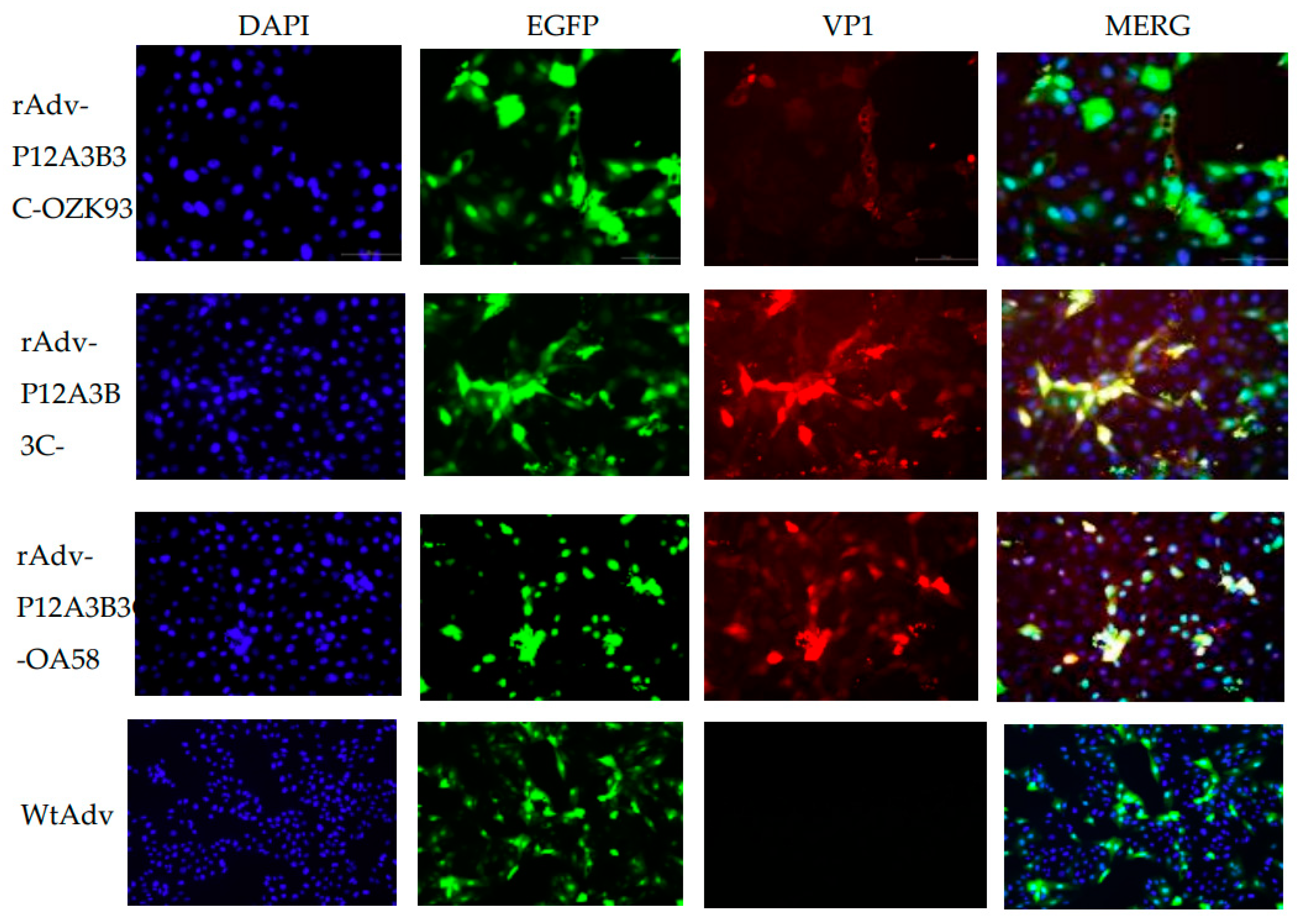
3.5. FMDV Recombinant Adenovirus PK Cell Infection Assay and Indirect Immunofluorescence Identification

P7-rAdv-P12A3B3C-OZK93, P7-rAdv-P12A3B3C-AF72, and P7-rAdv-P12A3B3C-OA58 were expressed 24 h after inoculation into PK cells. The IFA results showed that the VP1 protein was successfully expressed in the rAdv-P12A3B3C-OZK93-, rAdv-P12A3B3C-AF72-, and rAdv-P12A3B3C-OA58-infected PK cells but not expressed in the WtAdv infected control cells (Figure 6). Furthermore, the three constructed recombinant adenoviruses can infect porcine-derived cells.

3.6. Recombinant Adenovirus Particle Detection with Transmission Electron Microscopy

Transmission electron microscopy observations showed that the recombinant adenoviruses rAdv-P12A3B3C-OZK93, rAdv-P12A3B3C-AF72, and rAdv-P12A3B3C-OA58 have a diameter of approximately 75–85 nm, and they are approximately round with slight edges (Figure 7) similar to the size and shape of the adenovirus [22]. Additionally, they can further determine the success of recombinant adenovirus packaging.

3.7. Level of Serum Antibodies in Guinea Pigs after Immunization

Recombinant adenovirus vaccine and inactivated vaccine group guinea pigs’ serum IgG against FMDV were significantly different from the WtAdv group for 21 days (three immunizations once every 14 days for a total of three) after one exemption, and the IgG content increased sequentially between 42 d rAdv-P12A3B3C-O: A 6:4, rAdv-P12A3B3C-O: A 5:5, and rAdv-P12A3B3C-O: A 7:3 seedlings. The rAdv-P12A3B3C-OZK93 monospecies produced a higher antibody content than the rAdv-P12A3B3C-OA58 monospecies (Figure 8A).
The guinea pigs were exempted after 21 d following administration of the inactivated vaccine, and rAdv-P12A3B3C-AF72, rAdv-P12A3B3C-O: A 6:4, rAdv-P12A3B3C-O: A 5:5, and rAdv-P12A3B3C-O: A 7:3 were significantly different from those in the WtAdv group. rAdv-P12A3B3C-AF72 produced antibodies at higher levels than the other four groups, and the other four groups had smaller differences. The guinea pigs were immunized every 14 days after the first three immunizations for 42 days (three times in total). The IgG content of rAdv-P12A3B3C-O: A 6:4, rAdv-P12A3B3C-O: A 5:5, and rAdv-P12A3B3C-O: A 7:3 increased sequentially and was higher than that of the inactivated vaccine to produce antibodies (Figure 8B). The overall results were broadly as expected and varied consistently with the O-type antibody.

3.8. Post-Guinea Pig Immune Cytokine IFN-γ and IL-4 Levels

A kit (ELISA) was used to detect the IFN-γ content in the guinea pig serum. The IFN-γ level induced by rAdv-P12A3B3C-OZK93 and rAdv-P12A3B3C-AF72 between single seedlings was significantly higher than that of rAdv-P12A3B3C-OA58. rAdv-P12A3B3C-O:A6:4, rAdv-P12A3B3C-O:A 5:5, and rAdv-P12A3B3C-O:A 7:3 intergroup IFN-γ levels increased sequentially and varied significantly from those of the PBS group, and recombinant seedlings produced less IFN-γ content than the inactivated vaccine group.
For 42 days after the guinea pigs were exempted (every 14 days after three immunizations for a total of three doses), there was a small difference in IL-4 content between rAdv-P12A3B3C-OZK93, rAdv-P12A3B3C-OA58, and rAdv-P12A3B3C-AF72 seedlings, namely, rAdv-P12A3B3C-O: A 6:4, rAdv-P12A3B3C-O:A: A 5:5, and rAdv-P12A3B3C-O:A 7:3, respectively. The IL-4 content between the seedlings increased sequentially and was significantly different from that in the PBS group, and the IL-4 content of the recombinant seedlings was higher than that in the inactivated vaccine group (Figure 9).

4. Discussion

Studies have shown that O-type FMDV is one of the most widely distributed and important serotypes worldwide followed by type A, which is an important trigger for FMD outbreaks in China, South Korea, Thailand, Japan, and Vietnam [22,23]. Severe economic losses have been inflicted on many countries. Therefore, it is extremely important to prevent and control type O and type A FMDV in these areas. FMDV-inactivated vaccines play a fairly important role in the prevention and control of FMD [24]. However, traditional inactivated vaccines still have some shortcomings. In recent years, the development of new alternative vaccines has been widely explored. Several studies confirmed the protective effects of recombinant adenoviruses expressing P12A and 3C proteins of different serotypes [25,26]. The Plum Island Animal Disease Center in the United States is working on a capsid expressing FMDV with human replication-deficient adenovirus, and the recombinant adenovirus carrying the FMDV P12A3B3C gene constructed by the center can produce up to a 100% immune protective effect on pigs [20]. To enhance the enzymatic activity of 3C protease, 3B (3B1, 3B2, 3B3) was added between 2A and 3C using fusion PCR [20], and different strains of P12A and 3B3C proteins of FMDV were expressed in human replication defective adenovirus type 5 vectors.
FMDV-OZK93, FMDV-OA58, and FMDV-AF72 are the dominant vaccine strains commonly used in the production of FMDV-inactivated vaccines [27], and the packaging of FMDV recombinant adenovirus is the AdMax system, which is different from the AdEasy system used in most previous FMDV recombinant adenovirus studies and does not require plasmid linearization [28]. The system offers advantages, such as simple and convenient operation, high recombination efficiency, and a high level of expression of the gene of interest [29]. When successfully packaged FMDV-OZK93 recombinant adenovirus was passaged to the third generation, significant green fluorescence (Figure 3) was observed in the HEK-293 cells, and the viral titer increased from 103.4 to 109.1 with the passage of the virus; this proved that virus passage can enhance its viral titers (Figure 4D), which is consistent with the study by Xie et al. [30]. Adv-P12A3B3C-AF72 recombinant adenovirus P1 fluorescence is stronger, and significant CPE phenomena can also be observed (Figure 3). As the number of passages increased, the viral titer increased from 105.0 to 109.2 as the recombinant adenovirus expanded in the HEK-293 cells (Figure 4D). The rAdv-P12A3B3C-OA58 recombinant adenovirus had weak fluorescence in the first five generations and exhibited gradually increased fluorescence and viral titer as the number of passages increased (Figure 3,4D). As the recombinant adenovirus expanded in the HEK-293 cells, the viral titer increased from 102.2 to 107.8 (Figure 4D). The packaging of three FMD recombinant adenoviruses as well as the changes in fluorescence and viral titer in the transmission of the virus also proved that the packaging efficiency, infectivity, and protein expression capacity of different FMDV strains of recombinant adenovirus also varied greatly.
This study also successfully amplified the target gene P12A3B3C from different passages (Figure 4A–C), which is consistent with the results of Guo et al. [31]. In summary, these results show that the recombinant adenoviruses rAdv-P12A3B3C-OZK93, Adv-P12A3B3C-AF72, and rAdv-P12A3B3C-OA58 were successfully constructed, and the target gene was stable in the recombinant adenoviruses. IFA test results show that rAdv-P12A3B3C-OZK93, Adv-P12A3B3C-AF72, and rAdv-P12A3B3C-OA58 can infect PK cells and successfully express the protein of interest (Figure 6). In addition, the PK cell infection experiment proved that the recombinant adenoviruses could infect porcine source cells and express FMDV-specific proteins, which are essential for vaccination in pigs. This laid the foundation for the construction of three recombinant adenoviruses for immunity and protection against FMDV in pigs. This result is the same as that reported by José Barrera et al. [18]. The P12A and VP1/VP3 bands detected in the rAdv-P12A3B3C-OZK93-, Adv-P12A3B3C-AF72-, and rAdv-P12A3B3C-OA58-infected HEK-293 cell proteins are the same as the FMDV-inactivated viral antigen 146S bands (Figure 5), indicating that the 3C protein can lyse P1–2A to produce VP3 and VP1 [32]. Pena et al. reported that the addition of a 3B gene to the adenoviral expression FMDV capsid protein helps 3C proteases shear sequences between 3B and 3C [33]. Furthermore, to enable more efficient expression of the FMDV gene, the Kozak sequence was added to this study before the inserted P12A3B3C gene. The adenovirus purification kit is a chromatography-based adenovirus purification system that completes purification and amplification of adenovirus from infected cells within 1.5 h under sterile conditions. During purification, note that when the CPE (cell effector) is near completion, the cell pellet is collected, the cells are lysed, and the virus is purified. The reason for the selection of this kit for purification is that it can purify up to 1 × 10¹² PFU adeno-virion particles, and the viral titers and purity achieved with this purification method are comparable to the effects of the virus obtained with CSCL density-gradient centrifugation.
Humoral immune responses and cellular immune responses play an important role in fighting FMDV infection. Therefore, three recombinant adenoviruses, rAdv-P12A3B3C-OZK93, rAdv-P12A3B3C-OA58, and rAdv-P12A3B3C-AF72, in guinea pig immune experiments that were capable of expressing the O/A FMDV capsid protein were successfully constructed in this study to evaluate immunogenicity (Figure 8). These recombinant adenovirus immunizations were able to induce the body to produce specific antibodies against FMDV as well as high levels of IL-4 and IFN-γ (Figure 9), which yielded strong cellular and humoral immune responses in the guinea pigs. Moreover, the bivalent three-component vaccine can produce both anti-A-FMDV antibodies and anti-O-FMDV antibodies and can produce high levels of IL-4 and IFN-γ, which is similar to the two bivalent vaccines [34] and the chimeric VLP vaccine designed by Yao Lei et al., consisting of multiple epitopes of type A and O FMDV [35]. Research reports that adenovirus expressing alpha interferon rapidly protects swine from foot-and-mouth disease [36], and adenovirus-mediated type I interferon expression delays and reduces disease signs in cattle challenged with foot-and-mouth disease virus [37]. Our study can produce high levels of IL-4 and IFN-γ, which is consistent with the above study. It may be used as a potential divalent vaccine candidate for A and O FMDV. In addition, for rAdv-P12A3B3C-O: A 6:4, rAdv-P12A3B3C-O: A 5:5, and rAdv-P12A3B3C-O: A 7:3 in the combined seedlings, when the ratio of type A to type O recombinant adenovirus was 7:3, the induction of the cellular and humoral immune responses was the best, which may be related to the immunogenicity of type A FMDV.

5. Conclusions

In summary, three recombinant adenoviruses, rAdv-P12A3B3C-OZK93, rAdv-P12A3B3C-OA58, and rAdv-P12A3B3C-AF72, and their immunogenicities were further evaluated, and the three recombinant adenoviruses alone or in combination can induce higher levels of humoral and cellular immunity. Thus, they can be used as vaccine candidates for a novel FMDV live carrier.

Author Contributions

C.W. performed the experiments, and C.W. and X.L. analyzed the data. L.Z.; R.Y.; P.Z.; Z.Z.; X.M.; M.L.; J.L.; L.P.; Y.W. conceived and designed the experiments and and critically reviewed the manuscript. All authors have read and agreed to the published version of the manuscript.

Funding

This work was supported by the National Key Research and Development Program of China (2016YFD0501505).

Institutional Review Board Statement

Guinea pigs were provided by the LVRI and handled in strict accordance with good animal practice according to the Animal Ethics Procedures and Guidelines of the People’s Republic of China, and the study was approved by the Animal Ethics Committee of LVRI, CAAS (No. LVRIAEC2017-003).

Informed Consent Statement

Not applicable, as this research did not involve humans.

Data Availability Statement

The raw data supporting the conclusions of this article will be made available by the authors without undue reservation to any qualified researcher.

Conflicts of Interest

The authors declare no conflict of interest. The funders had no role in the design of the study; in the collection, analyses, or interpretation of data; in the writing of the manuscript; or in the decision to publish the results.

References

  1. Grubman, M.J.; Baxt, B. Foot-and-Mouth Disease. Clin. Microbiol. Rev. 2004, 17, 465–493. [Google Scholar] [CrossRef] [PubMed]
  2. Carrillo, C.; Tulman, E.R.; Delhon, G.; Lu, Z.; Carreno, A.; Vagnozzi, A.; Kutish, G.F.; Rock, D.L. Comparative Genomics of Foot-and-Mouth Disease Virus. J. Virol. 2005, 79, 6487–6504. [Google Scholar] [CrossRef]
  3. Gao, Y.; Sun, S.Q.; Guo, H.C. Biological function of Foot-and-mouth disease virus non-structural proteins and non-coding elements. Virol. J. 2016, 13, 107. [Google Scholar] [CrossRef] [PubMed]
  4. Rueckert, R.R.; Wimmer, E. Systematic nomenclature of picornavirus proteins. J. Virol. 1984, 50, 957–959. [Google Scholar] [CrossRef]
  5. Knowles, N.J.; Samuel, A.R. Molecular epidemiology of foot-and-mouth disease virus. Virus Res. 2003, 91, 65–80. [Google Scholar] [CrossRef] [PubMed]
  6. Mohapatra, J.K.; Subramaniam, S.; Pandey, L.K.; Pawar, S.S.; De, A.; Das, B.; Sanyal, A.; Pattnaik, B. Phylogenetic structure of serotype a foot-and-mouth disease virus: Global diversity and the Indian perspective. J. Gen. Virol. 2011, 92 Pt 4, 873–879. [Google Scholar] [CrossRef]
  7. Jamal, S.M.; Belsham, G.J. Foot-and-mouth disease: Past, present and future. Vet. Res. 2013, 44, 116. [Google Scholar] [CrossRef]
  8. Senthilkumaran, C.; Yang, M.; Bittner, H.; Ambagala, A.; Lung, O.; Zimmerman, J.; Giménez-Lirola, L.G.; Nfon, C. Detection of genome, antigen, and antibodies in oral fluids from pigs infected with foot-and-mouth disease virus. Can. J. Vet. Res. 2017, 81, 82–90. [Google Scholar]
  9. Mignaqui, A.C.; Ferella, A.; Cass, B.; Mukankurayija, L.; L’Abbé, D.; Bisson, L.; Sánchez, C.; Scian, R.; Cardillo, S.B.; Durocher, Y.; et al. Foot-and-Mouth Disease: Optimization, Reproducibility, and Scalability of High-Yield Production of Virus-Like Particles for a Next-Generation Vaccine. Front. Vet. Sci. 2020, 7, 601. [Google Scholar] [CrossRef]
  10. Diab, E.; Bazid, A.-H.I.; Fawzy, M.; El-Ashmawy, W.R.; Fayed, A.A.; El-Sayed, M.M. Foot-and-mouth disease outbreaks in Egypt during 2013–2014: Molecular characterization of serotypes A, O, and SAT2. Vet. World. 2019, 12, 190–197. [Google Scholar] [CrossRef]
  11. Brito, B.P.; Rodriguez, L.L.; Hammond, J.M.; Pinto, J.; Perez, A.M. Review of the global distribution of foot-and-mouth disease virus from 2007 to 2014. Transbound. Emerg. Dis. 2017, 64, 149–161. [Google Scholar] [CrossRef]
  12. World Organisation for Animal Health (OIE). Foot and Mouth Disease; Chapter 8.8; World Organisation for Animal Health: Paris, France, 2017; Available online: http://www.oie.int/fileadmin/Home/eng/Health (accessed on 1 April 2023).
  13. Muleme, M.; Barigye, R.; Khaitsa, M.L.; Berry, E.; Wamono, A.W.; Ayebazibwe, C. Effectiveness of vaccines and vaccination programs for the control of foot-and-mouth disease in Uganda, 2001–2010. Trop. Anim. Health Prod. 2013, 45, 35–43. [Google Scholar] [CrossRef] [PubMed]
  14. Dubey, P. Gene therapy using adenovirus vector. J. Mol. Biomark. Diagn. 2021, 12, 480. [Google Scholar]
  15. Tatsis, N.; Ertl, H.C. Adenoviruses as vaccine vectors. Mol. Ther. 2004, 10, 616–629. [Google Scholar] [CrossRef] [PubMed]
  16. Lasaro, M.O.; Ertl, H.C. New Insights on Adenovirus as Vaccine Vectors. Mol. Ther. 2009, 17, 1333–1339. [Google Scholar] [CrossRef]
  17. Mayr, G.A.; Chinsangaram, J.; Grubman, M.J. Development of replication-defective adenovirus serotype 5 containing the capsid and 3C protease coding regions of foot-and-mouth disease virus as a vaccine candidate. Virology. 1999, 263, 496–506. [Google Scholar] [CrossRef]
  18. Barrera, J.; Brake, D.A.; Schutta, C.; Ettyreddy, D.; Kamicker, B.J.; Rasmussen, M.V.; de Rueda, C.B.; Zurita, M.; Pisano, M.; Hurtle, W.; et al. Versatility of the adenovirus-vectored foot-and-mouth disease vaccine platform across multiple foot-and-mouth disease virus serotypes and topotypes using a vaccine dose representative of the AdtA24 conditionally licensed vaccine. Vaccine. 2018, 36, 7345–7352. [Google Scholar] [CrossRef] [PubMed]
  19. Chen, W.; Liu, M.; Jiao, Y.; Yan, W.; Wei, X.; Chen, J.; Fei, L.; Liu, Y.; Zuo, X.; Yang, F.; et al. Adenovirus-mediated RNA interference against foot-and-mouth disease virus infection both in vitro and in vivo. J. Virol. 2006, 80, 3559–3566. [Google Scholar] [CrossRef] [PubMed]
  20. Fernandez-Sainz, I.; Medina, G.N.; Ramirez-Medina, E.; Koster, M.J.; Grubman, M.J.; de Los Santos, T. Adenovirus-vectored foot-and-mouth disease vaccine confers early and full protection against FMDV O1 Manisa in swine. Virology. 2017, 502, 123–132. [Google Scholar] [CrossRef] [PubMed]
  21. Zhu, F.C.; Li, Y.H.; Guan, X.H.; Hou, L.H.; Wang, W.J.; Li, J.X.; Wu, S.P.; Wang, B.S.; Wang, Z.; Wang, L.; et al. Safety, tolerability, and immunogenicity of a recombinant adenovirus type-5 vectored COVID-19 vaccine: A dose-escalation, open-label, non-randomised, first-in-human trial. Lancet 2020, 395, 1845–1854. [Google Scholar] [CrossRef]
  22. Park, J.H.; Lee, K.N.; Ko, Y.J.; Kim, S.M.; Lee, H.S.; Shin, Y.K.; Sohn, H.J.; Park, J.Y.; Yeh, J.Y.; Lee, Y.H.; et al. Control of Foot-and-Mouth Disease during 2010–2011 Epidemic, South Korea. Emerg. Infect. Dis. 2013, 19, 655–659. [Google Scholar] [CrossRef] [PubMed]
  23. Valdazo-González, B.; Timina, A.; Scherbakov, A.; Abdul-Hamid, N.F.; Knowles, N.J.; King, D.P. Multiple introductions of serotype O foot-and-mouth disease viruses into East Asia in 2010–2011. Vet. Res. 2013, 44, 76. [Google Scholar] [CrossRef]
  24. Caridi, F.; Vázquez-Calvo, Á.; Borrego, B.; McCullough, K.; Summerfield, A.; Sobrino, F.; Martin-Acebes, M.A. Preserved immunogenicity of an inactivated vaccine based on foot-and-mouth disease virus particles with improved stability. Vet. Microbiol. 2017, 203, 275–279. [Google Scholar] [CrossRef] [PubMed]
  25. Kumar, R.; Ba Sagoudanavar, S.H.; Sreenivasa, B.P. Detection of replication competent adenovirus upon serial passaging of recombinant adenovirus expressing FMDV capsid proteins. Biologicals 2015, 43, 209–212. [Google Scholar] [CrossRef] [PubMed]
  26. Xie, Y.; Gao, P.; Li, Z. A Recombinant Adenovirus Expressing P12A and 3C Protein of the Type O Foot-and-Mouth Disease Virus Stimulates Systemic and Mucosal Immune Responses in Mice. BioMed Res. Int. 2016, 2016, 7849203. [Google Scholar] [CrossRef]
  27. Luo, Z.; Fu, X.; Wang, Y. Effect of Attenuated Highly Pathogenic Pig Reproductive and Respiratory Syndrome (HP-PRRS) TJM-F92 Strain Vaccine on Immune Antibody Levels against lassical Swine Fever (CSF) and Foot-and-Mouth Disease (FMD). Anim. Husb. Feed. Sci. 2016, 8, 162–164. [Google Scholar]
  28. Zhou, G.; Wang, H.; Wang, F.; Yu, L. Recombinant adenovirus expressing type Asia1 foot-and-mouth disease virus capsid proteins induces protective immunity against homologous virus challenge in mice. Res. Vet. Sci. 2013, 94, 796–802. [Google Scholar] [CrossRef] [PubMed]
  29. Pan, J.; Shi, H.; Luo, X.; Ma, D.; Liang, W.; Zhang, J.; Zhu, J.; Li, J. Construction of replication-deficient recombinant adenovirus vector with hTFPI-2 gene by AdMax system and expression in U937 monocytes in vitro. J. Biomed. Eng. 2011, 28, 326–331. [Google Scholar]
  30. Xie, Y.; Chang, H.; Li, Z.; Zhang, Y. Adenovirus-Vectored Capsid Proteins of the Serotype A Foot-and-Mouth Disease Virus Protect Guinea Pigs Against Challenge. Front. Microbiol. 2020, 11, 1449. [Google Scholar] [CrossRef]
  31. Guo, C.; Zhang, C.; Zheng, H.; Huang, Y. Recombinant adenovirus expression of FMDV P1-2A and 3C protein and its immune response in mice. Res. Vet. Sci. 2013, 95, 736–741. [Google Scholar] [CrossRef]
  32. Li, H.; Li, Z.; Xie, Y.; Qin, X.; Qi, X.; Sun, P.; Bai, X.; Ma, Y.; Zhang, Z. Novel chimeric foot-and-mouth disease virus-like particles harboring serotype O VP1 protect guinea pigs against challenge. Vet. Microbiol. 2016, 183, 92–96. [Google Scholar] [CrossRef] [PubMed]
  33. Pena, L.; Moraes, M.P.; Koster, M.; Burrage, T.; Pacheco, J.M.; Diaz-San Segundo, F.; Grubman, M.J. Delivery of a foot-and mouth disease virus empty capsid subunit antigen with nonstructural protein 2B improves protection of swine. Vaccine. 2008, 26, 5689–5699. [Google Scholar] [CrossRef]
  34. Yi, J.Z.; Liu, M.Q.; Zhu, C.Z.; Zhang, Q.; Sheng, Z.T.; Du, Q.Y.; Yan, W.Y.; Zheng, Z.X. Recombinant Bivalent Vaccine against Foot-and-Mouth Disease Virus Serotype O/A Infection in Guinea Pig. Acta Biochim. Biophys. Sin. 2004, 36, 589–596. [Google Scholar] [CrossRef] [PubMed]
  35. Lei, Y.; Shao, J.; Zhao, F.; Li, Y.; Lei, C.; Ma, F.; Chang, H.; Zhang, Y. Artificially designed hepatitis B virus core particles composed of multiple epitopes of type A and O foot-and-mouth disease virus as a bivalent vaccine candidate. J. Med. Virol. 2019, 91, 2142–2152. [Google Scholar] [CrossRef]
  36. Chinsangaram, J.; Moraes, M.P.; Koster, M.; Grubman, M.J. Novel viral disease control strategy: Adenovirus expressing alpha interferon rapidly protects swine from foot-and-mouth disease. J. Virol. 2003, 77, 1621–1625. [Google Scholar] [CrossRef]
  37. Wu, Q.; Brum, M.C.S.; Caron, L.; Koster, M.; Grubman, M.J. Adenovirus-mediated type I interferon expression delays and reduces disease signs in cattle challenged with foot-and-mouth disease virus. J. Interf. Cytokine Res. 2003, 23, 359–368. [Google Scholar] [CrossRef] [PubMed]
Figure 1. Schematic diagram of the construction of Ad5 vectors by Cre-mediated, site-specific recombination.
Zoonoticdis 03 00010 g001
Figure 2. Identification of the recombinant adenovirus through double-enzyme digestion. M: 10,000 DNA Marker; 1–3: Identification of the recombinant carrier pDC316-mCMV-EGFP-P12A3B3C-OA58, pDC316-mCMV-EGFP-P12A3B3C-AF72, and pDC316-mCMV-EGFP-P12A3B3C-OZK93 double-enzyme cutting. The enzyme cutting product of pDC316 (5879 bp) and the fusion PCR product P12A3B3C (3114 bp).
Zoonoticdis 03 00010 g002
Figure 3. Recombinant adenovirus fluorescence and CPE phenomenon. WtAdv used as a positive control; Normal HEK-293 cells used as a negative control.
Zoonoticdis 03 00010 g003
Figure 4. Analysis of recombinant adenovirus genome stability (A) PCR identifies the stable presence of the target gene in rAdV-P12A3B3C-OZK93 for different generations. M represents the 5000-bp marker. Lanes 1–3 show the PCR products from the 3rd, 5th, and 7th passages of rAdV-P12A3B3C-OZK93 P12A3B3C (3114 bp). Lane 4 is WtAdv, which was used as a negative control. Lane 5 shows normal HEK-293 cells, which were used as a blank control. (B) PCR identifies the stable presence of the target gene in rAdV-P12A3B3C-AF72 for different generations. M indicates a 5000-bp ladder marker. Lanes 1–2 show the PCR products from the 3rd and 5th passages of rAdV-P12A3B3C-AF72-P12A3B3C (3117 bp). Lane 3 is WtAdv, which was used as a negative control. (C) PCR identifies the stable presence of the target gene in rAdV-P12A3B3C-OA58 for different generations. M indicates a 5000-bp ladder marker. Lanes 1–2 show the PCR products from the 5th and 7th passages of rAdV-P12A3B3C-OA58 P12A3B3C (3114 bp). Lane 3 is WtAdv, which was used as a negative control. (D) The adenoviral titers at different passages (P3, P5, P7, and P10) were monitored with a TCID50 assay.
Zoonoticdis 03 00010 g004
Figure 5. Western blot analysis of FMDV capsid expression by HEK293 cells infected with rAdv-P12A3B3C-OZK93 (A), rAdV-P12A3B3C-AF72 (B), and rAdV-P12A3B3C-OA58 (C). M was a 180-kDa ladder marker. (A) Lanes 1–3 show HEK-293 cells infected with the 3rd, 5th, or 7th passage of rAdv-P12A3B3C-OZK93. Lane 4 is FMDV (146S), which was used as a positive control. Lane 5 is WtAdv, which was used as a negative control. Lane 6 shows normal HEK-293 cells that were used as a blank control. (B) Lanes 1, 3, and 4 show HEK293 cells infected with the 7th, 5th, and 3rd passages of rAdV-P12A3B3C-AF72. Lane 2 is FMDV (146S), which was used as a positive control. Lane 5 is WtAdv, which was used as a negative control. Lane 6 shows normal HEK293 cells that were used as a blank control. (C) Lanes 1–3 show HEK293 cells infected with 3rd, 5th, or 7th passages of rAdV-P12A3B3C-OA58. Lane 4 is FMDV (146S), which was used as a positive control. Lane 5 is WtAdv, which was used as a negative control.
Zoonoticdis 03 00010 g005
Figure 6. Immunofluorescence assay was used to detect PK cells infected with three recombinant adenoviruses. DAPI is used for staining cells and can stain nuclei blue through intact cell membranes. EGFP is green fluorescent protein. VP1 is the main antigen protein of FMDV. MERG is the DAPI, EGFP, and VP1 superimposed. WtAdv was used as a negative control.
Zoonoticdis 03 00010 g006
Figure 7. Recombinant adenovirus assessment with transmission electron microscopy (20k×). These arrows point to the particle as a recombinant adenovirus.
Zoonoticdis 03 00010 g007
Figure 8. Level of serum antibodies in the guinea pigs. WtAdv group, which was used as a negative control, PBS was used as a blank control. rAdv-P12A3B3C-O: A 6:4, rAdv-P12A3B3C-O: A 5:5, and rAdv-P12A3B3C-O: A 7:3 were viral titers of the same condition for volume ratios, rAdv-P12A3B3C-OZK93, rAdv-P12A3B3C-OA58, and rAdv-P12A3B3C-AF72 ratios, where O half of the mixture of the type OZK93 and OA58 each. Significant differences were defined as p ≤ 0.05, where ∗∗∗ means 0.0001 < p ≤ 0.001. (A) Guinea pig-induced O-anti-FMDV IgG antibody levels were detected with LPB-ELISA. (B) Guinea pig-induced A-anti-FMDV IgG antibody levels were detected with LPB-ELISA.
Zoonoticdis 03 00010 g008
Figure 9. Detection of cytokine content in guinea pig serum using the dual-antibody sandwich ELISA kit. Significant differences were defined as p ≤ 0.05, where ∗∗∗ means 0.0001 < p ≤ 0.001. ∗∗∗∗ means 0.00001 < p ≤ 0.0001. WtAdv group, which was used as a negative control, PBS was used as a blank control. (A) Detection of IFN-γ content in guinea pig serum using the dual-antibody sandwich ELISA kit. (B) Detection of IL-4 content in guinea pig serum using the dual-antibody sandwich ELISA kit. WtAdv produces interferon gamma at higher levels when compared to the inactivated vaccine or all the treatments. This can happen because the wild-type adenovirus is a live virus and has a strong ability to infect.
Zoonoticdis 03 00010 g009
Table 1. Primer sequences.
Primer NameSequence (5′–3′)Size (bp)
P12A-FATGCGGCCGCGCCGCCACCATGGGCGCCGGGCAATCCAGCCCGACC46
P12A-RAGTGGCCCAGCGTACCCAGGGTTGGACTCAAC32
3BC-FTGAGTCCAACCCTGGGTACGCTGGGCCACT30
3BC-RAAGCTTAAGTCACTCGTGGTGTGGTTCGGGGTCG34
Table 2. Immunization plan in guinea pigs.
Groups aDesignation Dose bFirst Vaccination DaysSecond Vaccination
Days
Third Vaccination Days
G1PBS500 μL01428
G2WtAdv1 × 107.8 TCID5001428
G3rAdv-P12A3B3C-OZK931 × 107.8 TCID5001428
G4rAdv-P12A3B3C-AF721 × 107.8 TCID5001428
G5rAdv-P12A3B3C-OA581 × 107.8 TCID5001428
G6rAdv-P12A3B3C-O: A 5:5 1 × 107.8 TCID5001428
G7rAdv-P12A3B3C-O: A 6:41 × 107.8 TCID5001428
G8rAdv-P12A3B3C-O: A 7:31 × 107.8 TCID5001428
G9Inactivated vaccine c500 μL01428
a Groups of immunized guinea pigs. b First, second, and third vaccinations were given on days 0, 14, and 28: 0, 21, 42 days serum samples of guinea pigs were collected; TCID50 (Tissue Culture Infectious Dose 50). c Commercial inactivated type O/A FMDV vaccine.
Disclaimer/Publisher’s Note: The statements, opinions and data contained in all publications are solely those of the individual author(s) and contributor(s) and not of MDPI and/or the editor(s). MDPI and/or the editor(s) disclaim responsibility for any injury to people or property resulting from any ideas, methods, instructions or products referred to in the content.

