Article

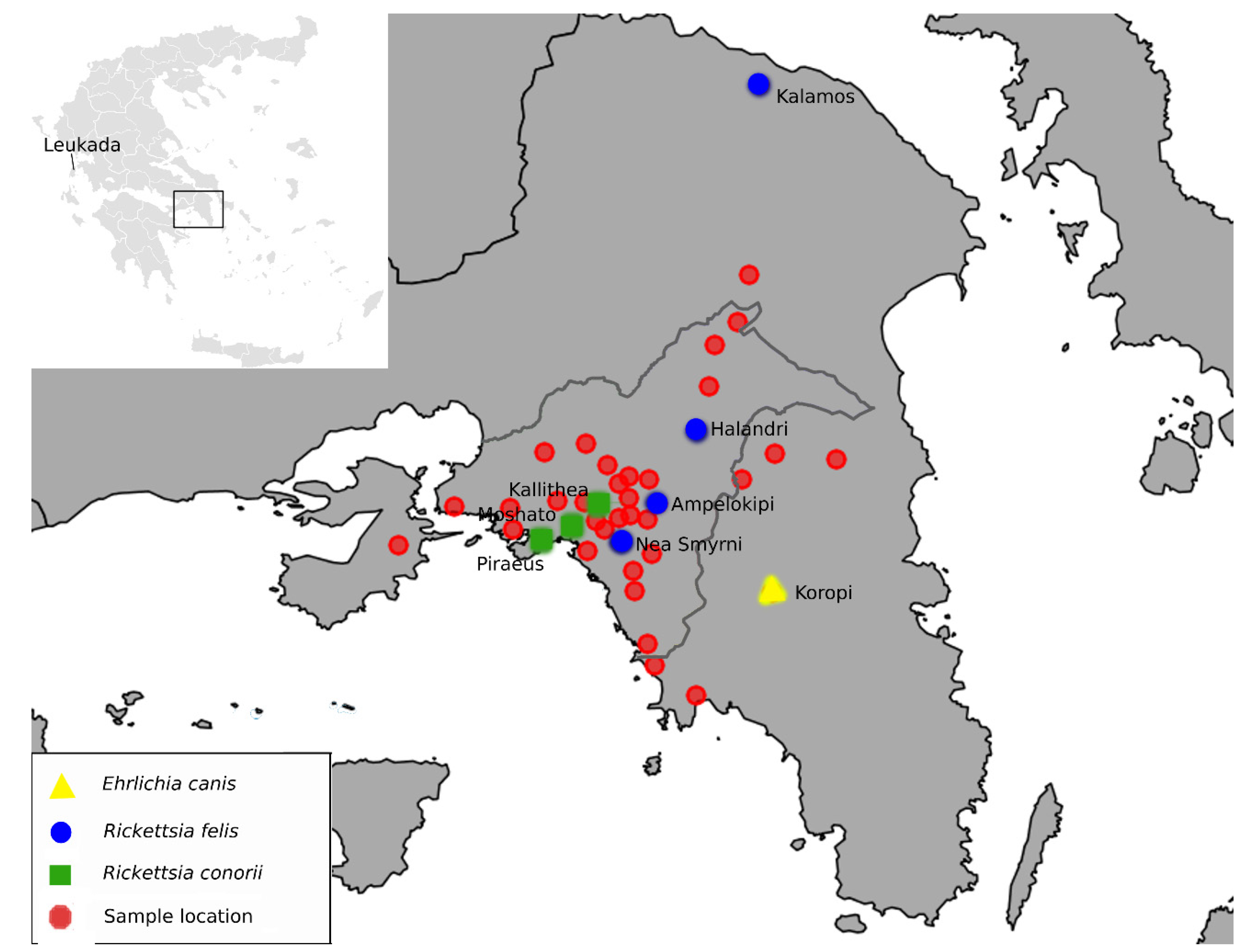
Rickettsial Agents Associated with Ectoparasites in Attica, Greece

by
Maria Liodaki
1,2,
Emmanouil Angelakis
3,
Gregory Spanakos
4,
Ioanna Papadogiannaki
3,
Michael Samarkos
5,
George L. Daikos
6,
Barbara Christopoulou
2 and
Evangelia-Theophano Piperaki
2,*
1
Department of Public and Community Heath, School of Public Health, University of West Attica, 11521 Athens, Greece
2
Department of Microbiology, School of Medicine, National and Kapodistrian University of Athens, 11527 Athens, Greece
3
Diagnostic Department and Public Health Laboratories, Hellenic Pasteur Institute, 11521 Athens, Greece
4
National Public Health Organization, 15123 Marousi, Greece
5
First Department of Medicine, Medical School, National and Kapodistrian University of Athens, 11527 Athens, Greece
6
National and Kapodistrian University of Athens, 11527 Athens, Greece
*
Author to whom correspondence should be addressed.
Zoonotic Dis. 2022, 2(4), 247-257; https://doi.org/10.3390/zoonoticdis2040020
Submission received: 14 October 2022 / Revised: 27 November 2022 / Accepted: 29 November 2022 / Published: 30 November 2022
(This article belongs to the Special Issue Feature Papers of Zoonotic Diseases 2021–2022)

Abstract

:

Simple Summary

Rickettsial pathogens, harbored in ectoparasites such as fleas and ticks found on cats and dogs, may cause vector-borne diseases. The prefecture of Attica in Greece, where the greater Athens metropolitan area is located and a large part of the Greek population resides, is poorly studied regarding these pathogens. To investigate their presence, we screened fleas and ticks collected from cats and dogs in veterinary clinics over 13 months. Using molecular methods, we tested the specimens for rickettsial organisms, such as species of Rickettsia, Wolbachia, and Ehrlichia. Rickettsia felis and Wolbachia were found in the examined fleas. Rickettsia conorii and Ehrlichia canis were found in the examined ticks, as well as Candidatus Midichloria mitochondrii, a little-studied symbiotic organism of ticks, which can be transmitted to humans. Our study shows that in the Attica region, fleas and ticks infesting dogs and cats harbor rickettsial pathogens that may pose a risk to human health. In this context, it contributes to the study of their epidemiology in the area. Additionally, it highlights the need for increased vigilance in the surveillance of vector-borne diseases and continued research to assess their potential implications for public health in this metropolitan area.

Abstract

The bacteria of the families Rickettsiaceae and Anaplasmataceae, harbored by arthropod vectors, may cause disease in animals and humans. The aim of this study was to screen ectoparasites collected from cats and dogs in Attica, Greece for the bacteria of the Rickettsiales group, by molecular methods. The ectoparasites examined were Ctenocephalides felis fleas and Rhipicephalus sanguineus s.l., Rhipicephalus sp., and Ixodes sp. ticks. Rickettsia felis was detected in 4.8% of C. felis fleas, and Rickettsia conorii was detected in 7.3% of R. sanguineus s.l. ticks. Ehrlichia canis was found in one R. sanguineus s.l. tick, and Wolbachia pipientis was detected in the majority of fleas. Another endosymbiont, Cancidatus Midichloria mitochondrii (Cancidatus Midichloriaceae), was detected in one Ixodes sp.. This is the first report of R. conorii and E. canis in R. sanguineus s.l. ticks in this study area. Given the fact that Greece is considered endemic for spotted fever group rickettsioses, further investigation of these rickettsial pathogens’ distribution in their vectors and hosts could enhance our knowledge of their epidemiology, in order to assess their potential implications for public health in this metropolitan area.

1. Introduction

According to recent molecular evidence and phylogenetic data, intracellular bacteria of the order Rickettsiales are classified into three families: Rickettsiaceae, Anaplasmataceae, and “Candidatus Midichloriaceae” [1]. The family Rickettsiaceae includes the genera Rickettsia and Orientia, and the family Anaplasmataceae includes the genera Anaplasma, Ehrlichia, Wolbachia, and Neorickettsia. Bacteria of these genera are associated with hematophagous arthropods and—if implicated in animal and human diseases—are characterized as pathogens, whereas if not, as endosymbionts. The third family comprises nine genera and includes the species “Ca. Midichloria mitochondrii”, which is the first representative of this family, an endosymbiont of the tick Ixodes ricinus and other arthropods [1].
Rickettsial diseases are prevalent and important tick- and flea-borne zoonoses. Rickettsia spp. that have been implicated in animal and human diseases are classified into the spotted fever group (SFG), the typhus group, and the R. belli group [2]. Ixodid ticks transmit the majority of Rickettsiae, but recently many argasid ticks have also been identified as carriers [3]. Rickettsia felis and Rickettsia conorii of the SFG are mainly transmitted by Ctenocephalides felis fleas and Rhipicephalus sanguineus ticks, respectively [4]. In the family Anaplasmataceae, Ehrlichia chaffeensis causes human monocytic ehrlichiosis and Anaplasma phagocytophilum (previously known as Ehrlichia phagocytophila) causes human granulocytic anaplasmosis (previously known as human granulocytic ehrlichiosis) [4]. Ehrlichia canis, the etiological agent of canine monocytic ehrlichiosis, has also been isolated from human blood specimens [5]. In the same family, the genus Wolbachia is found mainly in arthropods and nematodes and includes a single formally recognized species, Wolbachia pipientis [6].
Several rickettsial pathogens have been identified in humans, animal hosts, and vectors in serological or molecular studies in Greece. Regarding ticks, studies have focused mainly on Northern and Central Greece [7,8,9,10,11] and the island of Cephalonia [12]. Although the Attica prefecture was included among others areas in several studies, there are no data on rickettsial pathogens in ticks in this area. Attica is the most densely populated part of the country, with high numbers of stray and free roaming cats and dogs in urban and suburban areas. The aim of this study was to investigate the presence of rickettsial agents in ectoparasites of cats and dogs in the Attica region, in order to assess the possible risk they may pose to human health in the greater Athens metropolitan area.

2. Materials and Methods

Fleas and ticks from cats and dogs were collected from September 2016 to October 2017 from five veterinary clinics. The animals came from 39 areas in the Attica region (Figure 1) and were mostly stray cats and dogs. Only parasitized animals for ticks and fleas were accessed. No multiparasitism was recorded. From each animal, either fleas or ticks were collected. The collection was implemented in the context of a non-experimental clinical veterinary practice, and in the case of owned animals, the owners were informed and oral consent was obtained. Collected ectoparasites were placed in tubes containing 95% ethanol and stored at 4 °C until DNA extraction. The arthropods were photographed and identified using standard morphological identification keys and relevant publications [13,14,15].
Ectoparasites were washed twice for 10 min in sterile distilled water and dried on sterile filter paper. DNA extraction was performed using the NucleoSpin Tissue Kit (Macherey-Nagel GmbH, Düren, Germany) with the following modification: following placement in a microcentrifuge tube containing the T1 buffer and cutting gently into pieces with a sterile scalpel, proteinase K was added and the tube was incubated overnight at 56 °C. The remaining procedure was carried out according to the manufacturer’s instructions. Extracted DNA was stored at −20 °C until testing. All flea and tick samples were tested successfully for the absence of PCR inhibitors by the amplification of a fragment of the ectoparasites’ cytochrome oxidase subunit I (COI) gene using the previously described primers [16].
The ectoparasites were screened for Anaplasmataceae DNA, using the primer set EHR16SR–EHR16SD, which amplifies a 345-bp fragment of the 16S rRNA gene. Conventional PCR was carried out in a total volume of 25 μL containing 3 μL DNA, as previously described [17]. A BioRad S1000 thermocycler was used, and PCR products were visualized using the Bio-Rad GelDoc XR+ Gel Documentation System (Chemidoc XRS+ Gel Imaging System) after electrophoresis on a 2% agarose gel, stained with ethidium bromide. Experiments were performed twice, and negative controls containing sterile water were included.
All positive PCR products from ticks and nine positives from fleas were purified, using NucleoSpin Gel and PCR Clean-up Kit (Macherey-Nagel GmbH, Düren, Germany) according to the manufacturer’s instructions and sequenced in both directions using the PCR primers (CeMIA, Larissa, Greece). 16S rRNA sequences were aligned manually using CLUSTAL X, and phylogenetic analysis was performed using PHYLIP [18] by applying the maximum likelihood analysis.
Additionally, all samples from fleas that tested positive for Anaplasmataceae 16S rRNA were screened for Wolbachia using a Wolbachia spp. (panWolbachia) qPCR targeting the 23S rRNA gene, as previously described [19]. Samples that tested positive were then further examined for Wolbachia pipientis by the qPCR targeting rpoB gene, as previously described [20].
All flea DNA samples were also screened for Rickettsia felis by qPCR targeting the biotin synthase gene as previously described [21]. As cat fleas have been implicated as the most probable vectors of this rickettsial pathogen [22], the fleas in our study were tested directly for R. felis and not for the presence of other SFG rickettsial agents.
All tick samples were initially screened for the presence of SFG rickettsial DNA by qPCR targeting a fragment of the gltA gene as previously described [23]. SFG Rickettsia positive DNA samples were further tested for Rickettsia conorii by qPCR targeting a putative acetyltransferase gene, as previously described [23]. The PCR assays used are presented in Table 1.

3. Results

A total of 145 animals were examined, 92 cats and 53 dogs, from 39 localities in Attica distributed in 7 regional units. The numbers of collected ectoparasites per regional unit were as follows: 44 from South Athens, 37 from Central Athens, 25 from North Athens, 19 from East Attica, 17 from Piraeus, 2 from West Athens, and 1 from Salamina (an island in the Attica area). One ectoparasite from each animal was used: 104 fleas (86 from cats (13 owned/73 strays)/18 from dogs (12 owned/6 strays)) and 41 ticks (6 from cats (1 owned/5 strays)/35 from dogs (16 owned/19 strays)). In the case of many fleas/ticks, we chose to examine only one ectoparasite per animal because of financial issues with the processing of the ectoparasites. All fleas were identified as Ctenocephalides felis (97♀/7♂), and ticks were identified as Rhipicephalus sanguineus s.l. (34♀/4♂), Rhipicephalus sp. (2 nymphs), and Ixodes sp. (1♀).
Conventional PCR was positive for the Anaplasmataceae 16S rRNA gene in 61/104 (58.7%) fleas and 2/41 (4.9%) ticks. The sequencing of PCR products from 9 of the fleas yielded Wolbachia pipientis. A blast analysis of these PCR sequences revealed 100% homology with various W. pipientis strains from Spain (GenBank: LN864488) and USA (GenBank: CP051156, MF944223). The presence of W. pipientis in a total of 64/104 (61.5%) fleas was consequently confirmed using qPCRs for panWolbachia and for W. pipientis targeting rpoB (Table 2).
Sequencing of the two PCR products obtained from the ticks, both from dogs, yielded Ehrlichia canis in one Rhipicephalus sanguineus s.l. and Candidatus Midichloria mitochondrii in the Ixodes sp. (Table 2). The E. canis sequence had 100% homology with strains from Greece (GenBank: MN922610), USA (NCBI Reference Sequence: NR_118741) and China (GenBank: CP025749). The Ca. Midichloria mitochondrii sequence from the Ixodes sp. was 100% homologous with strains from Portugal (GenBank: KX359181), France (GenBank: KU559921), and Italy (GenBank: OM982404, HF568841, CP002130). Sequences derived from this study were deposited in GenBank with Accession Numbers ON678202-ON678207 and OP490629-OP490633.
All fleas and ticks were also tested to assess the prevalence of Rickettsia bacteria using qPCR. R. felis qPCR was positive in 5/104 (4.8%) fleas. SFG Rickettsia qPCR was positive in 3/41 (7.3%) ticks, two from cats and one from a dog. Consequent qPCR identified them all as R. conorii (Table 2).

4. Discussion

Data on rickettsial zoonotic agents in Greece derive mainly from human cases and animal studies, in certain areas of the country. Ectoparasite studies are few and have been mainly conducted in Northern Greece [7,8,9,10,11] and certain islands [12]. There are no studies of arthropod-borne (particularly tick-borne) zoonotic infections in Attica, although a very large proportion of the entire Greek population resides in this prefecture [24]. In the present study, all fleas collected from cats and dogs were identified as C. felis, confirming the wide distribution of cat fleas on dogs, observed in other ectoparasite studies [25]. C. felis, the cat flea, is the commonest flea found on cats and dogs and can inhabit human dwellings [26], where it frequently bites humans. C. felis transmits R. felis and R. typhi and can serve as a vector of R. typhi to humans [27]. Although R. felis is harbored by various hematophagous arthropods, including fleas, ticks, mites, and mosquitoes, its only known biological vector and main reservoir is C. felis [28].

4.1. Rickettsia Felis

In the present study, R. felis was detected in five C. felis fleas from four different localities Figure 1, four from stray cats and dogs and one from an owned dog.
Our results are supported by the findings of a previous study that detected R. felis and R. typhi in cat blood from stray and free-roaming cats in four areas of Greece, including Athens [29]. In another Greek study, R. felis was also found in C. felis fleas [30]. There is serological evidence of the mostly subclinical infection of cats, dogs and other mammals; however, their role as R. felis reservoirs has not been fully elucidated. R. felis causes flea-borne spotted fever (or cat flea fever) in humans, an emerging rickettsial infection that presents with an acute febrile syndrome, similar to murine typhus, with which it may have previously been confused, based on serological diagnosis alone [31]. Many cases have been reported from countries around the Mediterranean [31] and sub-Saharan Africa, where asymptomatic infection in humans has also been described [22]. Our detection of R.felis in C. felis from owned animals indicates a possible route of R. felis transmission to human hosts and merits further investigation.

4.2. Rickettsia Conorii

R. conorii was detected in three Rhipicephalus sanguineus s.l. ticks from three localities Figure 1, two of which were from an owned cat and an owned dog and one from a stray cat. R. conorii has been previously detected in R. sanguineus and R. turanicus ticks from dogs, in different areas of Greece [7,12]. It has also been detected in the blood of dogs from four Greek islands, in the population of Northern Greece, and in a patient from Crete, by serological and molecular methods [32,33,34].
To our knowledge, this is the first detection of R. conorii in R. sanguineus s.l. ticks in the Attica region, notably, from dog ticks found on cats. R. conorii causes Mediterranean Spotted fever (MSF) syndrome. Ιt is an epidemiologically complex disease, affected by numerous factors, including human behavior and vector interaction with the host. Its control, particularly in an epidemic setting, is a challenging feat and requires a better understanding of the reservoirs of the disease and their relationship to human infection [35]. In this context, the detection of R. conorii in cat ectoparasites in our study suggests a potential role for cats, as well as dogs, in the transmission of the pathogen to humans.

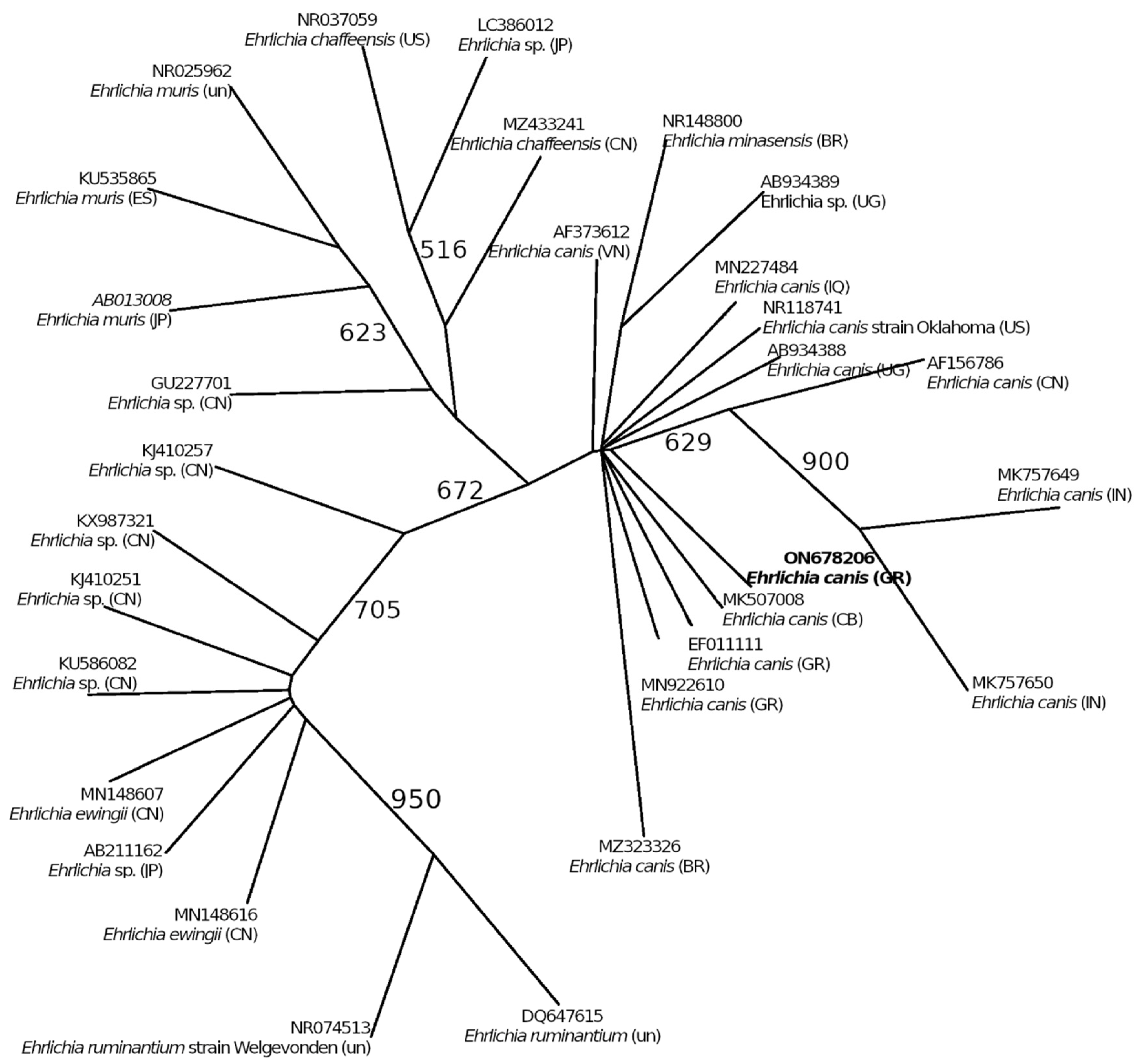
4.3. Ehrlichia canis

In the present study, E. canis was detected in one R. sanguineus s.l. tick from an owned dog, Figure 1. Although many reports from Greece have been recorded, they mainly concern dog epizootology [36,37,38]. Its presence in dog and cat blood in various areas of Greece has been documented by several studies, using molecular [39] and serological [29,32,40] methods. Our 16S rRNA sequence was found to be 100% homologous to one isolated from dog blood in Crete [39], thus supporting a possible role of R. sanguineus s.l. as vector of E. canis in this area. This is also supported by phylogenetic analysis based on 16S rRNA, which clusters our sequence (ON678206) with E. canis from Greece and relates it with other E. canis isolates (Figure 2). To the best of our knowledge, this is the first detection of E. canis in this vector in Greece.
E. canis, the causative agent of canine monocytic ehrlichiosis, is closely related to E. chaffeensis, the agent of human monocytic ehrlichiosis; although chiefly a canine pathogen, it occasionally infects humans and felines [41,42]. It has been isolated from human blood [43,44], and it has been proposed that it can cause both asymptomatic and symptomatic human infection [45].

4.4. Wolbachia pipientis

W. pipientis was detected in 61.5% of our C. felis samples. W. pipientis is found in arthropods as well as filarial nematodes populations [46]. Its presence may affect the host in various ways, including cytoplasmic incompatibility, male killing, induced parthenogenesis, and feminization [46,47]. Wolbachia is frequently found within insects, including potential human disease vectors such as fleas of veterinary importance, e.g., C.felis [48,49]. Our findings are in accordance with surveys from France (17.8%) [50] and Spain (69.4%) [51], more so with the latter, because the majority of fleas in both studies were female.

4.5. Candidatus Midichloria mitochondrii

Candidatus Midichloria mitochondrii was found in an engorged female Ixodes sp. tick from an owned dog. The tick may have been brought to Athens from the island of Leukada Figure 1, where the dog owner was on a trip before visiting the veterinary clinic. This is the first report of the detection of both an Ixodes sp. and Ca. M. mitochondrii in the greater Athens area. In Northern Greece, Ca. M. mitochondrii has been detected in Ixodes ricinus tick pools from goats [10]. Our sequence was 100% homologous to sequences isolated from I. ricinus from Portugal (GenBank: KX359181), from France (GenBank: KU559921), Italy (GenBank: CP002130) and from sequences isolated from human (GenBank: OM982404) and horse blood (GenBank: HF568841) in Italy. Ca. Midichloria mitochondrii is a Gram(-) intracellular bacterium that colonizes the mitochondria of host cells. It is mainly found in I. ricinus, with nearly 100% prevalence reported in females [52]. Although the mechanisms underlying this symbiotic interaction have not been elucidated, in silico metabolic reconstruction suggests it may play a nutrient-provisional role (B complex vitamin biosynthesis) that may enhance host fitness [52]. It has been shown that some patients exposed to I. ricinus bites are seropositive for Ca. M. mitochondrii antigens, suggesting that these antigens may be secreted together with tick saliva, thus triggering an immune response [53]. Alternatively, it is possible that ticks could transmit this endosymbiont to humans and consequently to other ticks, completing a tick-to-human-to tick transmission route, similar to that described for tick-borne pathogens. Indeed, this endosymbiont has been detected in the blood of humans and mammals bitten by ticks [54].
Many endosymbionts have been reported in arthropods. Particularly in ticks, their co-existence with pathogens is the subject of investigation since ticks are major vectors of pathogens affecting humans and animals. According to Bonnet et al. ‘’Ticks represent a compelling yet challenging system in which to study microbiomes and microbial interactions” [55]. The microbiota of important tick species needs to be explored, in view of its potential role in disease ecology and epidemiology and thus of its public health implications. Moreover, given the experimental evidence that the antibiotic cleansing of bacterial endosymbionts had a negative impact on tick fitness, survival, and competence, the development of anti-microbiota vaccines presents an intriguing environmentally friendly alternative for the control of ticks and tick-borne pathogens [52].
In conclusion, the present study provides epidemiological data for the presence of various rickettsial species in the ectoparasites of cats and dogs in a little studied area, the greater Athens urban and peri-urban region, most notably R. conorii and E. canis in R. sanguineus s.l ticks. Certain epidemiological findings are described for the first time in this particular area, an area that is heavily populated, while also housing great numbers of stray cats and dogs, which frequently interact with humans. Moreover, the ectoparasites—cat fleas and brown dog ticks—found on the animals belong to widely distributed species that often enter human settlements in urban and peri-urban settings. All of the above underline a growing need for increased vigilance in the surveillance of vector-borne diseases in the area, as well as for further studies of vector and pathogen distribution to assess their potential implications for public health in this metropolitan area.

Author Contributions

Conceptualization, M.L., E.-T.P. and G.S.; Methodology, E.A., G.S., I.P. and B.C.; Software, G.S.; Investigation, M.L.; Data curation, M.L.; Writing—original draft preparation, M.L.; Writing—review and editing, E.-T.P.; Supervision, M.S. and G.L.D.; Project administration, E.-T.P. All authors have read and agreed to the published version of the manuscript.

Funding

This research received no external funding.

Institutional Review Board Statement

Not applicable.

Informed Consent Statement

Not applicable.

Data Availability Statement

All data used and presented in this study are available in public repositories as described in the results section.

Conflicts of Interest

The authors declare no conflict of interest.

References

  1. Castelli, M.; Sassera, D.; Petroni, G. Biodiversity of “Non-Model” Rickettsiales and Their Association with Aquatic Organisms. In Rickettsiales: Biology, Molecular Biology, Epidemiology, and Vaccine Development; Thomas, S., Ed.; Springer International Publishing: Cham, Swizerland, 2016; pp. 59–91. [Google Scholar] [CrossRef]
  2. Angelakis, E.; Raoult, D. Rickettsia and Rickettsia-Like Organisms. In Infectious Diseases; Elsevier: Amsterdam, The Netherlands, 2017; pp. 1666–1675.e1. [Google Scholar] [CrossRef]
  3. Tomassone, L.; Portillo, A.; Nováková, M.; de Sousa, R.; Oteo, J.A. Neglected Aspects of Tick-Borne Rickettsioses. Parasit. Vectors 2018, 11, 263. [Google Scholar] [CrossRef] [PubMed] [Green Version]
  4. Parola, P.; Davoust, B.; Raoult, D. Tick- and Flea-Borne Rickettsial Emerging Zoonoses. Vet. Res. 2005, 36, 469–492. [Google Scholar] [CrossRef] [PubMed] [Green Version]
  5. Thomas, S.; Alexander, W.; Gilligan, J.; Rikihisa, Y. The Importance of Rickettsiales Infections. In Rickettsiales: Biology, Molecular Biology, Epidemiology, and Vaccine Development; Springer International Publishing: Cham, Switzerland, 2017; pp. 3–21. [Google Scholar] [CrossRef]
  6. Makepeace, B.; Gill, A. Chapter 21: Wolbachia; Springer International Publishing: Cham, Switzerland, 2017; pp. 465–512. [Google Scholar] [CrossRef]
  7. Psaroulaki, A.; Spyridaki, I.; Ioannidis, A.; Babalis, T.; Gikas, A.; Tselentis, Y. First Isolation and Identification of Rickettsia conorii from Ticks Collected in the Region of Fokida in Central Greece. J. Clin. Microbiol. 2003, 41, 3317–3319. [Google Scholar] [CrossRef] [PubMed] [Green Version]
  8. Latrofa, M.S.; Angelou, A.; Giannelli, A.; Annoscia, G.; Ravagnan, S.; Dantas-Torres, F.; Capelli, G.; Halos, L.; Beugnet, F.; Papadopoulos, E.; et al. Ticks and Associated Pathogens in Dogs from Greece. Parasit. Vectors 2017, 10, 301. [Google Scholar] [CrossRef] [Green Version]
  9. Papa, A.; Xanthopoulou, K.; Kotriotsiou, T.; Papaioakim, M.; Sotiraki, S.; Chaligiannis, I.; Maltezos, E. Rickettsia Species in Human-Parasitizing Ticks in Greece. Trans. R. Soc. Trop. Med. Hyg. 2016, 110, 299–304. [Google Scholar] [CrossRef] [Green Version]
  10. Papa, A.; Tsioka, K.; Kontana, A.; Papadopoulos, C.; Giadinis, N. Bacterial Pathogens and Endosymbionts in Ticks. Ticks Tick-Borne Dis. 2017, 8, 31–35. [Google Scholar] [CrossRef]
  11. Moraga-Fernández, A.; Chaligiannis, Ι.; Cabezas-Cruz, A.; Papa, A.; Sotiraki, S.; de la Fuente, J.G.; Fernández de Mera, I. Molecular Identification of Spotted Fever Group Rickettsia in Ticks Collected from Dogs and Small Ruminants in Greece. Exp. Appl. Acarol. 2019, 78, 421–430. [Google Scholar] [CrossRef]
  12. Psaroulaki, A.; Ragiadakou, D.; Kouris, G.; Papadopoulos, B.; Chaniotis, B.; Tselentis, Y. Ticks, Tick-Borne Rickettsiae, and Coxiella burnetii in the Greek Island of Cephalonia. Ann. N. Y. Acad. Sci. 2006, 1078, 389–399. [Google Scholar] [CrossRef]
  13. Lane, R.P.; Crosskey, R.W. (Eds.) Medical Insects and Arachnids; Springer: Dordrecht, The Netherlands, 1993. [Google Scholar] [CrossRef]
  14. Walker, J.B.; Keirans, J.E.; Horak, I. The Genus Rhipicephalus (Acardi, Ixodidae): A Guide to the Brown Ticks of the World; Cambridge University Press: Cambridge, UK; New York, NY, USA, 2000. [Google Scholar]
  15. Estrada-Peña, A.; Bouattour, A.; Camicas, J.-L.; Walker, A. Ticks of Domestic Animals in the Mediterranean Region. A Guide to Identification of Species; University of Zaragoza: Zaragoza, Spain, 2004. [Google Scholar]
  16. Duron, O.; Noël, V.; McCoy, K.D.; Bonazzi, M.; Sidi-Boumedine, K.; Morel, O.; Vavre, F.; Zenner, L.; Jourdain, E.; Durand, P.; et al. The Recent Evolution of a Maternally-Inherited Endosymbiont of Ticks Led to the Emergence of the Q Fever Pathogen, Coxiella burnetii. PLOS Pathog. 2015, 11, e1004892. [Google Scholar] [CrossRef] [Green Version]
  17. Maia, C.; Ferreira, A.; Nunes, M.; Vieira, M.L.; Campino, L.; Cardoso, L. Molecular Detection of Bacterial and Parasitic Pathogens in Hard Ticks from Portugal. Ticks Tick-Borne Dis. 2014, 5, 409–414. [Google Scholar] [CrossRef]
  18. Felsenstein, J. PHYLIP (Phylogeny Inference Package). Version 3.6 Distributed by the Author. Available online: https://evolution.genetics.washington.edu/phylip.html (accessed on 9 October 2022).
  19. Diarra, A.Z.; Kone, A.K.; Doumbo Niare, S.; Laroche, M.; Diatta, G.; Atteynine, S.A.; Coulibaly, M.; Sangare, A.K.; Kouriba, B.; Djimde, A.; et al. Molecular Detection of Microorganisms Associated with Small Mammals and Their Ectoparasites in Mali. Am. J. Trop. Med. Hyg. 2020, 103, 2542–2551. [Google Scholar] [CrossRef] [PubMed]
  20. Taillardat-Bisch, A.-V.; Raoult, D.; Drancourt, M. RNA Polymerase β-Subunit-Based Phylogeny of Ehrlichia Spp., Anaplasma Spp., Neorickettsia Spp. and Wolbachia pipientis. Int. J. Syst. Evol. Microbiol. 2003, 53, 455–458. [Google Scholar] [CrossRef] [PubMed]
  21. Socolovschi, C.; Mediannikov, O.; Sokhna, C.; Tall, A.; Diatta, G.; Bassene, H.; Trape, J.-F.; Raoult, D. Rickettsia felis –Associated Uneruptive Fever, Senegal. Emerg. Infect. Dis. 2010, 16, 1140–1142. [Google Scholar] [CrossRef]
  22. Angelakis, E.; Mediannikov, O.; Parola, P.; Raoult, D. Rickettsia felis: The Complex Journey of an Emergent Human Pathogen. Trends Parasitol. 2016, 32, 554–564. [Google Scholar] [CrossRef] [PubMed]
  23. Bechah, Y.; Socolovschi, C.; Raoult, D. Identification of Rickettsial Infections by Using Cutaneous Swab Specimens and PCR. Emerg. Infect. Dis. 2011, 17, 83–86. [Google Scholar] [CrossRef] [PubMed]
  24. Hellenic Statistical Authority. Census Results of Population and Housing-ELSTAT. 2021. Available online: https://www.statistics.gr/2021-census-pop-hous-results (accessed on 9 November 2022).
  25. Giudice, E.; Pietro, S.D.; Alaimo, A.; Blanda, V.; Lelli, R.; Francaviglia, F.; Caracappa, S.; Torina, A. A Molecular Survey of Rickettsia felis in Fleas from Cats and Dogs in Sicily (Southern Italy). PLoS ONE 2014, 9, e106820. [Google Scholar] [CrossRef]
  26. Bitam, I.; Dittmar, K.; Parola, P.; Whiting, M.F.; Raoult, D. Fleas and Flea-Borne Diseases. Int. J. Infect. Dis. 2010, 14, e667–e676. [Google Scholar] [CrossRef] [Green Version]
  27. Feldman, S.H.; Easton, D.N. Chapter 17—Occupational Health and Safety. In The Laboratory Rat, 2nd ed.; Suckow, M.A., Weisbroth, S.H., Franklin, C.L., Eds.; American College of Laboratory Animal Medicine; Academic Press: Burlington, VT, USA, 2006; pp. 565–586. [Google Scholar] [CrossRef]
  28. Civen, R.; Ngo, V. Murine Typhus: An Unrecognized Suburban Vectorborne Disease. Clin. Infect. Dis. 2008, 46, 913–918. [Google Scholar] [CrossRef] [Green Version]
  29. Diakou, A.; Di Cesare, A.; Accettura, P.M.; Barros, L.; Iorio, R.; Paoletti, B.; Frangipane di Regalbono, A.; Halos, L.; Beugnet, F.; Traversa, D. Intestinal Parasites and Vector-Borne Pathogens in Stray and Free-Roaming Cats Living in Continental and Insular Greece. PLoS Negl. Trop. Dis. 2017, 11, e0005335. [Google Scholar] [CrossRef] [Green Version]
  30. Chochlakis, D.; Germanakis, A.; Chaliotis, G.; Kapetanaki, S.; Kalogeraki, L.; Gkika, E.; Partalis, N.; Polymili, G.; Tselentis, Y.; Psaroulaki, A. Potential Exposure of Humans to Rickettsia felis in Greece. Acta Trop. 2018, 178, 40–45. [Google Scholar] [CrossRef]
  31. Legendre, K.; Macaluso, K. Rickettsia Felis: A Review of Transmission Mechanisms of an Emerging Pathogen. Trop. Med. Infect. Dis. 2017, 2, 64. [Google Scholar] [CrossRef] [PubMed] [Green Version]
  32. Diakou, A.; Di Cesare, A.; Morelli, S.; Colombo, M.; Halos, L.; Simonato, G.; Tamvakis, A.; Beugnet, F.; Paoletti, B.; Traversa, D. Endoparasites and Vector-Borne Pathogens in Dogs from Greek Islands: Pathogen Distribution and Zoonotic Implications. PLoS Negl. Trop. Dis. 2019, 13, e0007003. [Google Scholar] [CrossRef] [PubMed] [Green Version]
  33. Daniel, S.A.; Manika, K.; Arvanmdou, M.; Antoniadis, A. Prevalence of Rickettsia conorii and Rickettsia typhi Infections in the Population of Northern Greece. Am. J. Trop. Med. Hyg. 2002, 66, 76–79. [Google Scholar] [CrossRef] [PubMed] [Green Version]
  34. Psaroulaki, A.; Germanakis, A.; Gikas, A.; Scoulica, E.; Tselentis, Y. First Isolation and Genotypic Identification of Rickettsia conorii Malish 7 from a Patient in Greece. Eur. J. Clin. Microbiol. Infect. Dis. 2005, 24, 297–298. [Google Scholar] [CrossRef] [PubMed]
  35. Spernovasilis, N.; Markaki, I.; Papadakis, M.; Mazonakis, N.; Ierodiakonou, D. Mediterranean Spotted Fever: Current Knowledge and Recent Advances. Trop. Med. Infect. Dis. 2021, 6, 172. [Google Scholar] [CrossRef] [PubMed]
  36. Mylonakis, M.E.; Koutinas, A.F.; Leontides, L.S. Bone Marrow Mastocytosis in Dogs with Myelosuppressive Monocytic Ehrlichiosis (Ehrlichia Canis): A Retrospective Study. Vet. Clin. Pathol. 2006, 35, 311–314. [Google Scholar] [CrossRef]
  37. Komnenou, A.A.; Mylonakis, M.E.; Kouti, V.; Tendoma, L.; Leontides, L.; Skountzou, E.; Dessiris, A.; Koutinas, A.F.; Ofri, R. Ocular Manifestations of Natural Canine Monocytic Ehrlichiosis (Ehrlichia canis): A Retrospective Study of 90 Cases. Vet. Ophthalmol. 2007, 10, 137–142. [Google Scholar] [CrossRef]
  38. Theodorou, K.; Leontides, L.; Siarkou, V.I.; Petanides, T.; Tsafas, K.; Harrus, S.; Mylonakis, M.E. Synovial Fluid Cytology in Experimental Acute Canine Monocytic Ehrlichiosis (Ehrlichia canis). Vet. Microbiol. 2015, 177, 224–227. [Google Scholar] [CrossRef]
  39. Kostopoulou, D.; Gizzarelli, M.; Ligda, P.; Foglia Manzillo, V.; Saratsi, K.; Montagnaro, S.; Schunack, B.; Boegel, A.; Pollmeier, M.; Oliva, G.; et al. Mapping the Canine Vector-Borne Disease Risk in a Mediterranean Area. Parasit. Vectors 2020, 13, 282. [Google Scholar] [CrossRef]
  40. Angelou, A.; Gelasakis, A.I.; Verde, N.; Pantchev, N.; Schaper, R.; Chandrashekar, R.; Papadopoulos, E. Prevalence and Risk Factors for Selected Canine Vector-Borne Diseases in Greece. Parasit. Vectors 2019, 12, 283. [Google Scholar] [CrossRef]
  41. Bowman, D.; Little, S.E.; Lorentzen, L.; Shields, J.; Sullivan, M.P.; Carlin, E.P. Prevalence and Geographic Distribution of Dirofilaria immitis, Borrelia burgdorferi, Ehrlichia canis, and Anaplasma phagocytophilum in Dogs in the United States: Results of a National Clinic-Based Serologic Survey. Vet. Parasitol. 2009, 160, 138–148. [Google Scholar] [CrossRef] [PubMed]
  42. Oliveira, A.C.; Luz, M.F.; Granada, S.; Vilhena, H.; Nachum-Biala, Y.; Lopes, A.P.; Cardoso, L.; Baneth, G. Molecular Detection of Anaplasma bovis, Ehrlichia canis and Hepatozoon felis in Cats from Luanda, Angola. Parasit. Vectors 2018, 11, 167. [Google Scholar] [CrossRef] [PubMed] [Green Version]
  43. Perez, M.; Rikihisa, Y.; Wen, B. Ehrlichia canis-like Agent Isolated from a Man in Venezuela: Antigenic and Genetic Characterization. J. Clin. Microbiol. 1996, 34, 2133–2139. [Google Scholar] [CrossRef] [PubMed] [Green Version]
  44. Unver, A.; Perez, M.; Orellana, N.; Huang, H.; Rikihisa, Y. Molecular and Antigenic Comparison of Ehrlichia canis Isolates from Dogs, Ticks, and a Human in Venezuela. J. Clin. Microbiol. 2001, 39, 2788–2793. [Google Scholar] [CrossRef]
  45. Perez, M.; Bodor, M.; Zhang, C.; Xiong, Q.; Rikihisa, Y. Human Infection with Ehrlichia canis Accompanied by Clinical Signs in Venezuela. Ann. N. Y. Acad. Sci. 2006, 1078, 110–117. [Google Scholar] [CrossRef]
  46. Werren, J.H.; Baldo, L.; Clark, M.E. Wolbachia: Master Manipulators of Invertebrate Biology. Nat. Rev. Microbiol. 2008, 6, 741–751. [Google Scholar] [CrossRef]
  47. Bordenstein, S.R.; Paraskevopoulos, C.; Dunning Hotopp, J.C.; Sapountzis, P.; Lo, N.; Bandi, C.; Tettelin, H.; Werren, J.H.; Bourtzis, K. Parasitism and Mutualism in Wolbachia: What the Phylogenomic Trees Can and Cannot Say. Mol. Biol. Evol. 2009, 26, 231–241. [Google Scholar] [CrossRef] [Green Version]
  48. Madhav, M.; Baker, D.; Morgan, J.A.T.; Asgari, S.; James, P. Wolbachia: A Tool for Livestock Ectoparasite Control. Vet. Parasitol. 2020, 288, 109297. [Google Scholar] [CrossRef]
  49. De Oliveira, C.D.; Gonçalves, D.S.; Baton, L.A.; Shimabukuro, P.H.F.; Carvalho, F.D.; Moreira, L.A. Broader Prevalence of Wolbachia in Insects Including Potential Human Disease Vectors. Bull. Entomol. Res. 2015, 105, 305–315. [Google Scholar] [CrossRef] [Green Version]
  50. Rolain, J.-M.; Franc, M.; Davoust, B.; Raoult, D. Molecular Detection of Bartonella quintana, B. koehlerae, B. henselae, B. clarridgeiae, Rickettsia felis and Wolbachia pipientis in Cat Fleas, France. Emerg. Infect. Dis. 2003, 9, 338–342. [Google Scholar] [CrossRef]
  51. Zurita, A.; Gutiérrez, S.G.; Cutillas, C. Infection Rates of Wolbachia sp. and Bartonella sp. in Different Populations of Fleas. Curr. Microbiol. 2016, 73, 704–713. [Google Scholar] [CrossRef] [PubMed]
  52. Hussain, S.; Perveen, N.; Hussain, A.; Song, B.; Aziz, M.U.; Zeb, J.; Li, J.; George, D.; Cabezas-Cruz, A.; Sparagano, O. The Symbiotic Continuum Within Ticks: Opportunities for Disease Control. Front. Microbiol. 2022, 13, 854803. [Google Scholar] [CrossRef] [PubMed]
  53. Mariconti, M.; Epis, S.; Gaibani, P.; Dalla Valle, C.; Sassera, D.; Tomao, P.; Fabbi, M.; Castelli, F.; Marone, P.; Sambri, V.; et al. Humans Parasitized by the Hard Tick Ixodes ricinus Are Seropositive to Midichloria Mitochondrii: Is Midichloria a Novel Pathogen, or Just a Marker of Tick Bite? Pathog. Glob. Health 2012, 106, 391–396. [Google Scholar] [CrossRef] [PubMed] [Green Version]
  54. Castelli, M.; McCarthy, U.; Petroni, G.; Bazzocchi, C. Transmission of Members of the “Candidatus Midichloriaceae” Family to Vertebrates and Possible Involvement in Disease Pathogenesis. In Rickettsiales: Biology, Molecular Biology, Epidemiology, and Vaccine Development; Thomas, S., Ed.; Springer International Publishing: Cham, Switzerland, 2016; pp. 283–292. [Google Scholar] [CrossRef]
  55. Bonnet, S.I.; Binetruy, F.; Hernández-Jarguín, A.M.; Duron, O. The Tick Microbiome: Why Non-Pathogenic Microorganisms Matter in Tick Biology and Pathogen Transmission. Front. Cell. Infect. Microbiol. 2017, 7. [Google Scholar] [CrossRef]
Figure 1. Sampling sites in Attica region are represented by red circles. Colored shapes indicate locations where ectoparasites positive for R. felis (blue), R. conorii (green), and E.canis (yellow) were collected. (Athens urban area is outlined by a gray line. Leukada island is indicated in the upper left corner of the map).
Zoonoticdis 02 00020 g001
Figure 2. Maximum likelihood phylogenetic tree of Ehrlichia canis based on a 305 bp fragment of 16S rRNA. The numbers at the nodes indicate percentage bootstrap replicates of 1000. The sequence obtained in the present study is indicated in bold type.
Zoonoticdis 02 00020 g002
Table 1. PCR assays used for the screening for rickettsial agents.
Target SpeciesTarget GenePCR Primer and Probe Sequences (5′–3′) Product Size (bp) Reference
SFG Rickettsia genus specificgltAF: GTG AAT GAA AGA TTA CAC TAT TTAT166[23]
R: GTA TCT TAG CAA TCA TTC TAA TAG C
FAM-CTA TTA TGC TTG CGG CTG TCG GTT C-TAMRA
Rickettsia conoriiPutative acetyltransferaseF: TTG GTA GGC AAG TAG CTA AGC AAA116[23]
R: GGA AGT ATA TGG GAA TGC TTT GAA
FAM-GCG GTT ATT CCT GAA AAT AAG CCG GCA-TAMRA
Rickettsia felis specificBiotin synthaseF: ATG-TTC-GGG-CTTCCG-GTA-TG120[21]
R: CCG-ATT-CAG-CAGGTT-CTT-CAA
6-FAM- GCT-GCG-GCGGTA-TTT-TAG-GAA-TGGG-TAMRA
Anaplasmataceae16S rRNAEHR16SD: GGT ACC YAC AGA AGA AGTCC345[17]
EHR16SR: TAG CAC TCA TCG TTT ACAGC
PanWolbachia23S rRNAF: CCA AAA TTA CAG CTA AGT GG100[19]
R: AGT GAG CTG TTA CGC TTT CT
6FAM-TACAGCTAGGAGGTTGGCTT-TAMRA
Wolbachia pipientisrpoBF: TCATGCGCGTTCAGTAGGAC103Unpublished
R: TGCCCAACATTCCATTTCAC
6FAM-AGGAAAGTCTCATTTTGGTGGTAGCG-TAMRA
Table 2. Rickettsial agents detected by PCR in fleas and ticks collected from dogs and cats in Attica, Greece.
Rickettsial AgentPathogen/
Endosymbiont
Prevalence (%) Examined EctoparasiteMethod of Detection Sequence Identity (%) Query Cover (%)GenBank ID
AnaplasmataceaeEhrlichia canis2.4%R. sanguineus s.l.PCR100% (305/305 bp)100%MN922610
Ca. Midichloria mitochondrii2.4%Ixodes sp.PCR100% (305/305 bp)100%KX359181
Wolbachia pipientis58.7% aC. felisPCR100% (305/305 bp)100%CP051156
Wolbachia pipientis61.5%C. felisqPCRna b na b
SFG RickettsiaeRickettsia conorii7.3%R. sanguineus s.l.qPCRna b na b
Rickettsia felis4.8%C. felisqPCRna b na b
a A subgroup of Anaplasmataceae PCR-positives was sequenced. b Not applicable.
Publisher’s Note: MDPI stays neutral with regard to jurisdictional claims in published maps and institutional affiliations.

