Article

Isolation and Identification of Autochthonous Lactic Acid Bacteria from Commonly Consumed African Indigenous Leafy Vegetables in Kenya

1
Department of Physical and Biological Sciences, Bomet University College, Bomet P.O. Box 701-20400, Kenya
2
Department of Food Science and Technology, Jomo Kenyatta University of Agriculture and Technology, Nairobi P.O. Box 62000-00200, Kenya
3
College of Graduate Studies and Research, Mount Kenya University, Thika P.O Box 342-01000, Kenya
4
Institute of Biotechnology Research, Jomo Kenyatta University of Agriculture and Technology, Nairobi P.O. Box 62000-00200, Kenya
*
Author to whom correspondence should be addressed.
Bacteria 2023, 2(1), 1-20; https://doi.org/10.3390/bacteria2010001
Submission received: 5 December 2022 / Revised: 25 December 2022 / Accepted: 29 December 2022 / Published: 5 January 2023

Abstract

:
African indigenous leafy vegetables (AILVs) are plants that have been part of the food systems in Sub-Saharan Africa (SSA) for a long time and their leaves, young shoots, flowers, fruits and seeds, stems, tubers, and roots are consumed. These vegetables are high in vitamins, minerals, protein, and secondary metabolites that promote health. This study aimed at isolating, characterizing, and identifying dominant lactic acid bacteria (LAB) from naturally fermenting commonly consumed AILV in Kenya. A total of 57 LAB strains were isolated and identified based on phenotypic and 16S rRNA gene analyses from three AILVs (23 nightshade leaves, 19 cowpeas leaves, and 15 vegetable amaranth). The highest microbial counts were recorded between 48 h and 96 h of fermentation in all AILVs ranging from approximately log 8 to log 9 CFU/mL with an average pH of 3.7. Fermentation of AILVs was dominated by twenty eight Lactobacillus spp. [Lactiplantibacillus plantarum (22), Limosilactobacillus fermentum (3), Lactiplantibacillus pentosus (2) and Lactiplantibacillus casei (1)], eleven Weissella spp. (Weissella cibaria (8), W. confusa (2), and W. muntiaci) six Leuconostoc spp. [Leuconostoc mesenteroides (3), Leuc. citreum (2) and Leuc. lactis (1)], six Pediococcus pentosaceus, four Enterococcus spp. [Enterococcus mundtii (2), E. faecalis (1) and E. durans (1)] and, finally, two Lactococcus garvieae. These bacteria strains are commonly used in food fermentation as starter cultures and as potential probiotics.

1. Introduction

African indigenous leafy vegetables (AILVs) inhabit Sub-Saharan Africa (SSA) [1,2,3]. Their leaves, young shoots, flowers, fruits and seeds, stems, tubers, and roots are consumable and they have been part of the food systems for generations [1,2,3]. These vegetables contain high levels of vitamins, minerals, protein, and health-promoting secondary metabolites. This could be a valuable source of nutrition in rural areas, where they can help alleviate malnutrition among poor populations [4].
In Kenya, the commonly consumed AILVs include cowpea leaves (Vigna unguiculata), Jude mellow (Corchorus olitorius), moringa leaves (Moringa oleifera), sweet potato leaves (Ipomoea batatas), cassava leaves (Manihot esculenta), slender leaves (Crotalaria ochroleuca), African kale (Brassica carinata), some species of leaf amaranth (Amaranthus spp.), spider plant (Cleome spp.), some species of nightshades (Solanum spp.), and pumpkins (Cucurbita spp.) [5]. The availability of AILVs during the rainy season and their climate-adaptability make them an attractive option for nutritional supplementation for those most in need [6]. Therefore, they must be processed to maintain or improve their nutritional content, organoleptic qualities, and long-term storage properties [2,7].
Fermented foods in Africa contain a broad range of plant-based products derived from maize, sorghum, millet, and cassava, among other sources [8]. Leafy vegetables are rarely fermented in Africa with some recent studies showing that controlled fermentation of some AILVs with well-characterized lactic acid bacteria (LAB) could provide microbiological safety, and enhanced organoleptic and nutritional properties [3,9]. Traditionally, plant-based fermented foods are very common in Asia, Europe, and America where vegetables such as cabbage, radishes, cucumbers, turnips, and beets are commonly used [10].
There is little published data on the isolation and identification of indigenous LAB from AILVs, although some reports indicate that the most common LAB genera that are associated with fermented African indigenous vegetables include Lactobacillus, Lactococcus, and Weissella [11,12]. However, a previous study by Xiong et al. [13] showed that Lactococcus lactis, Leuconostoc mesenteroides, Lactobacillus plantarum, and Lactobacillus casei dominated the natural fermentation of Chinese sauerkraut (fermented cabbage). The microbiota initially present in lactic fermentation processes comes primarily from plant material [14].
Generally, LAB are described as Gram-positive rods or cocci, which are acid tolerant, devoid of cytochromes and porphyrins and therefore are catalase- and oxidase-negative, anaerobic or aero-tolerant, non-spore-forming, and are mostly non-motile bacteria [15,16,17]. Therefore, this study aimed at the isolation, characterization, and identification of dominant LAB from naturally fermenting and commonly consumed AILVs in Kenya.

2. Results

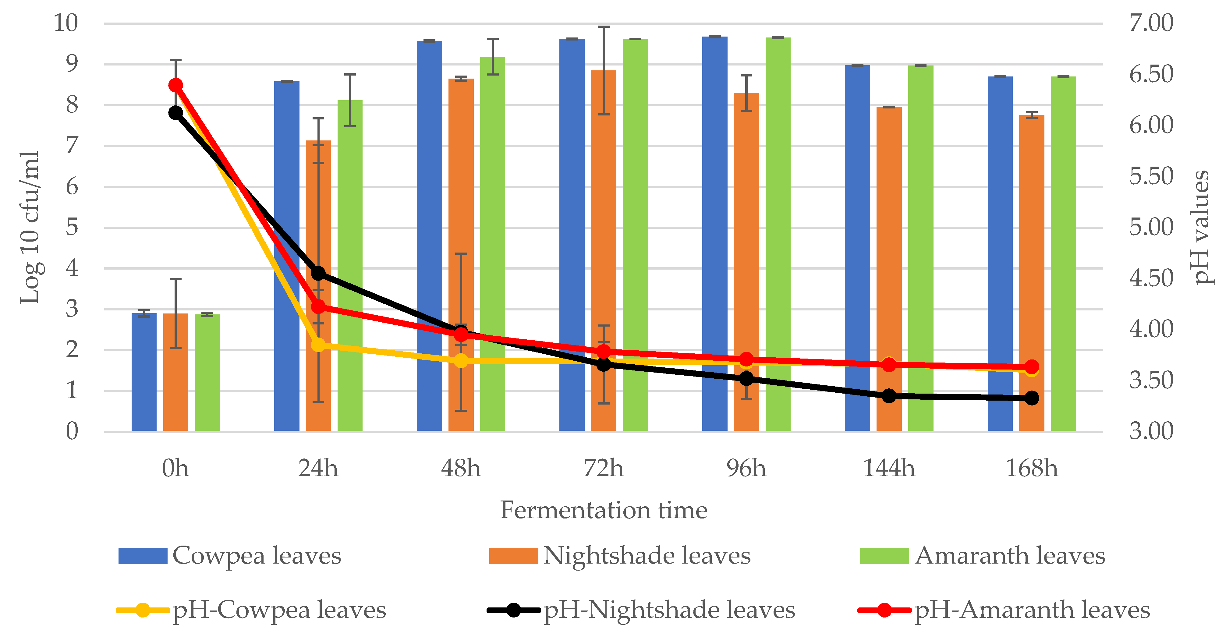
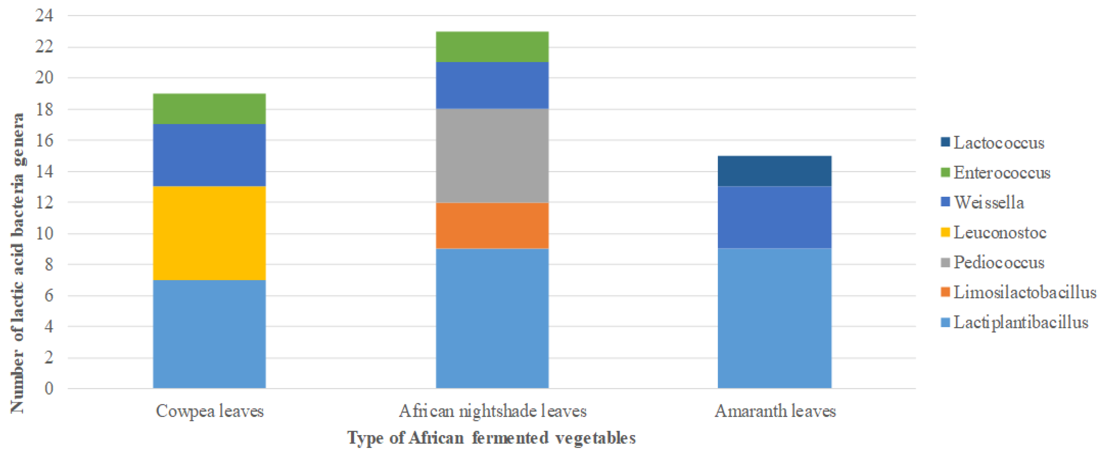
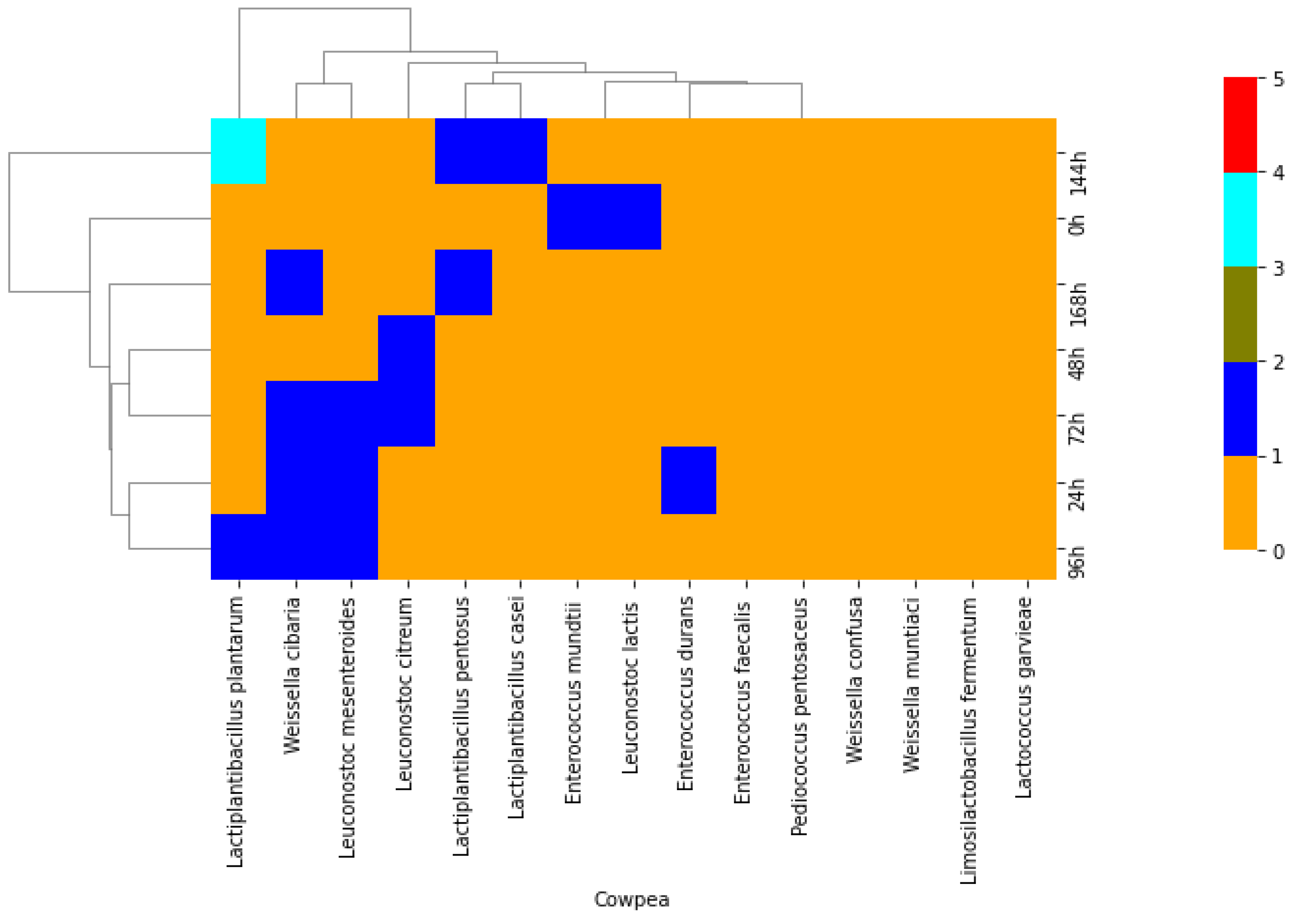
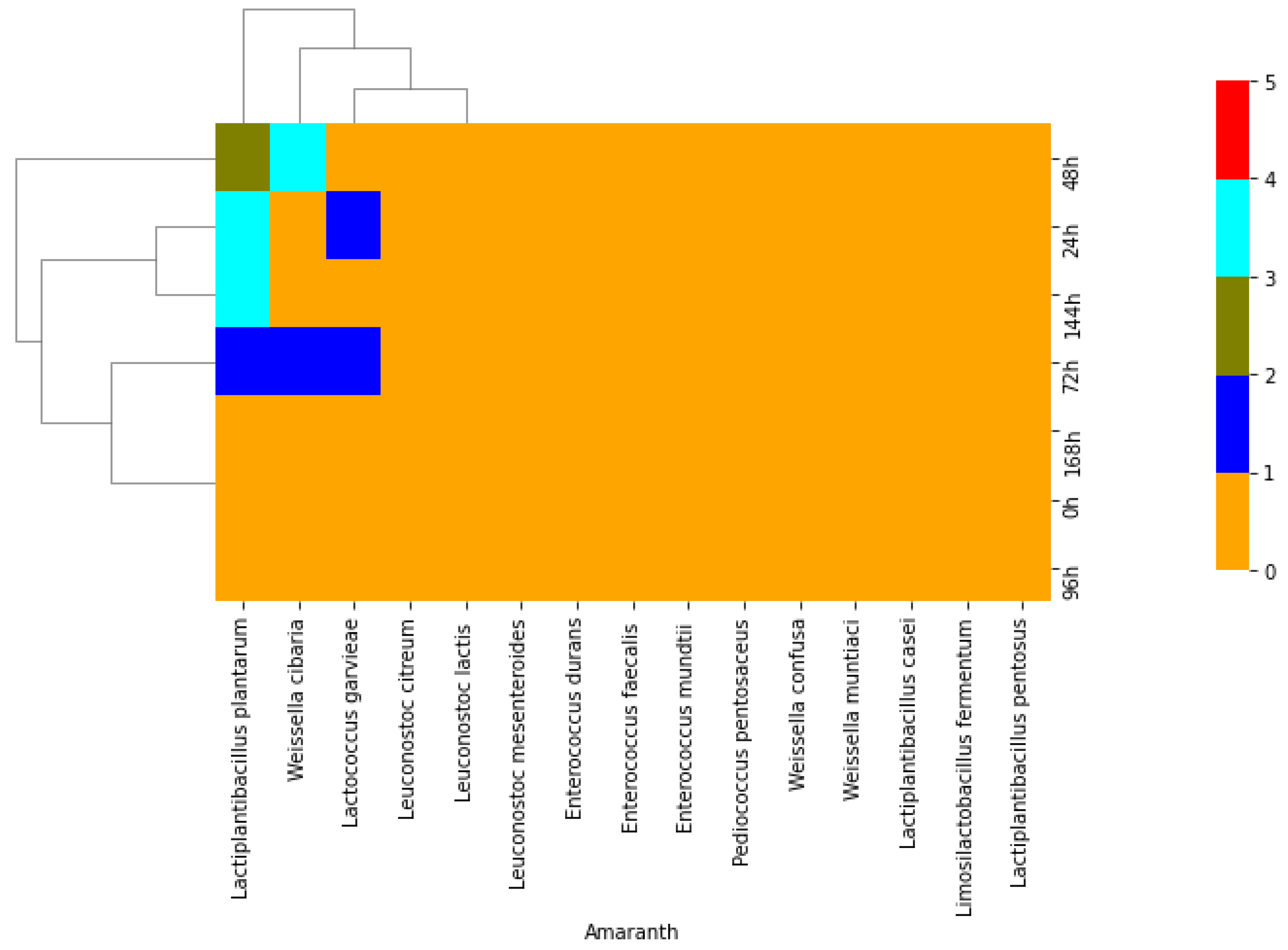
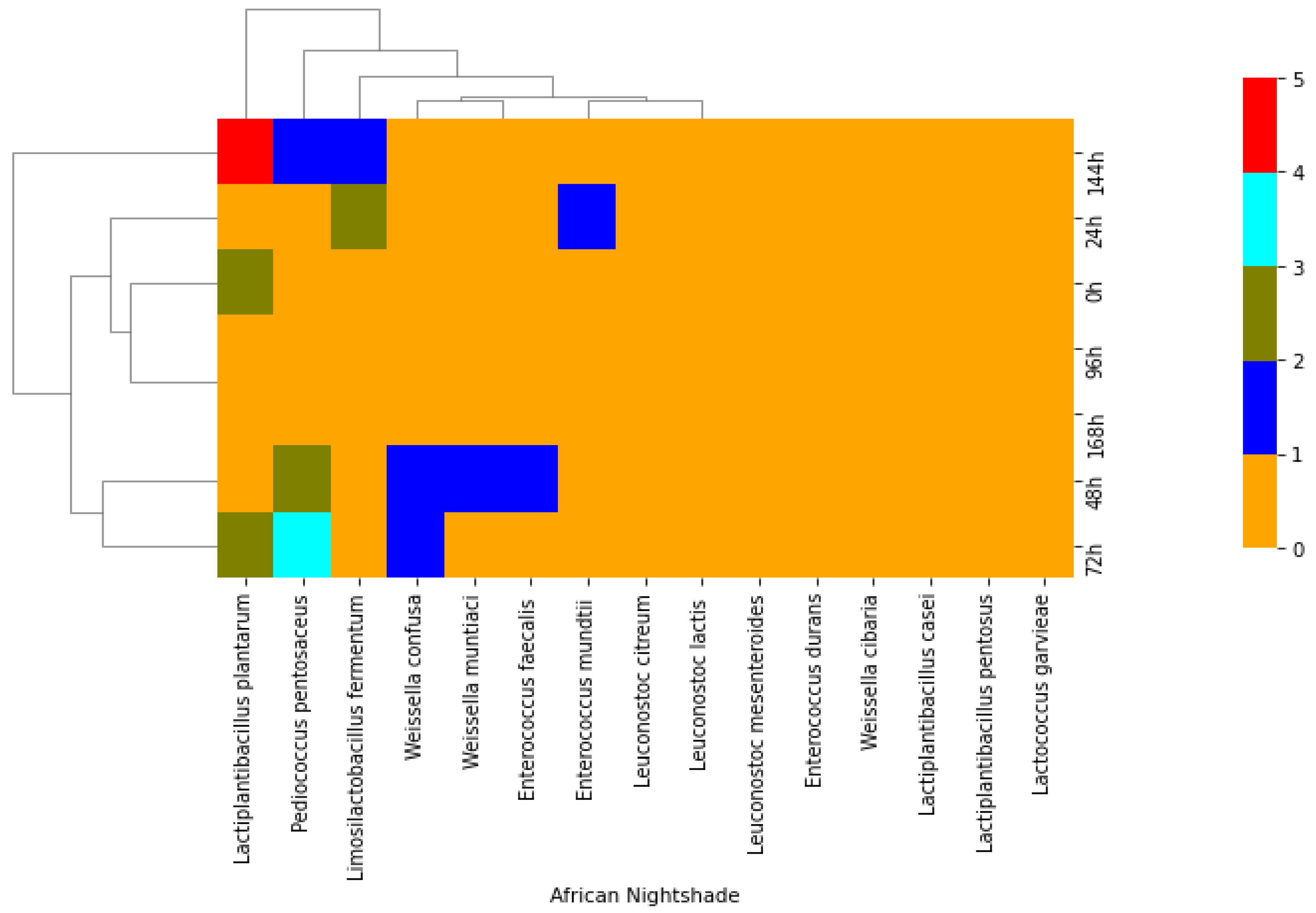
2.1. Isolation of Lactic Acid Bacteria from African Indigenous Vegetables

The microbial counts varied across the three African indigenous leafy vegetables (AILVs). The mean lactic acid bacteria (LAB) count from all AILVs was approximately 3 log cfu/mL at 0 h which slightly increased to approximately log 8 cfu/mL for cowpea and amaranth and log 7 for nightshade leaves after 24 h. The highest microbial counts were recorded between 48–96 h in all AILVs ranging from approximately log 8 to log 9 cfu/mL (Figure 1). The ability of the bacteria to acidify during fermentation is reflected in the pH production. The mean pH during all AILVs fermentation showed a slow decrease over time from pH 6.3 at 0 h to an approximate mean of pH 4.2 after 24 h. The pH decreased slightly further to a mean of 3.8 after 48 h and stayed at this low level up to 168 h, reaching a mean of 3.5 (Figure 1).

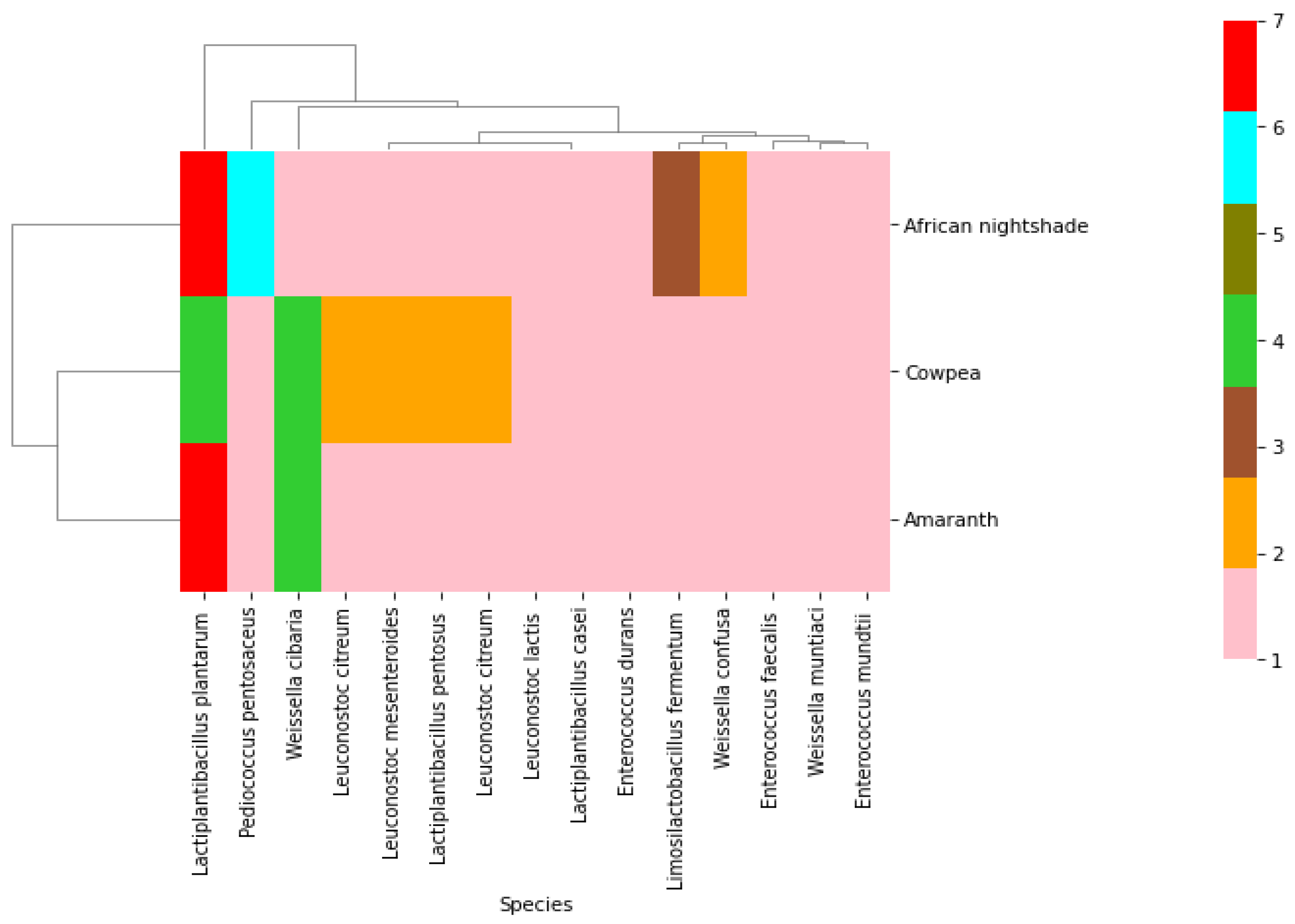
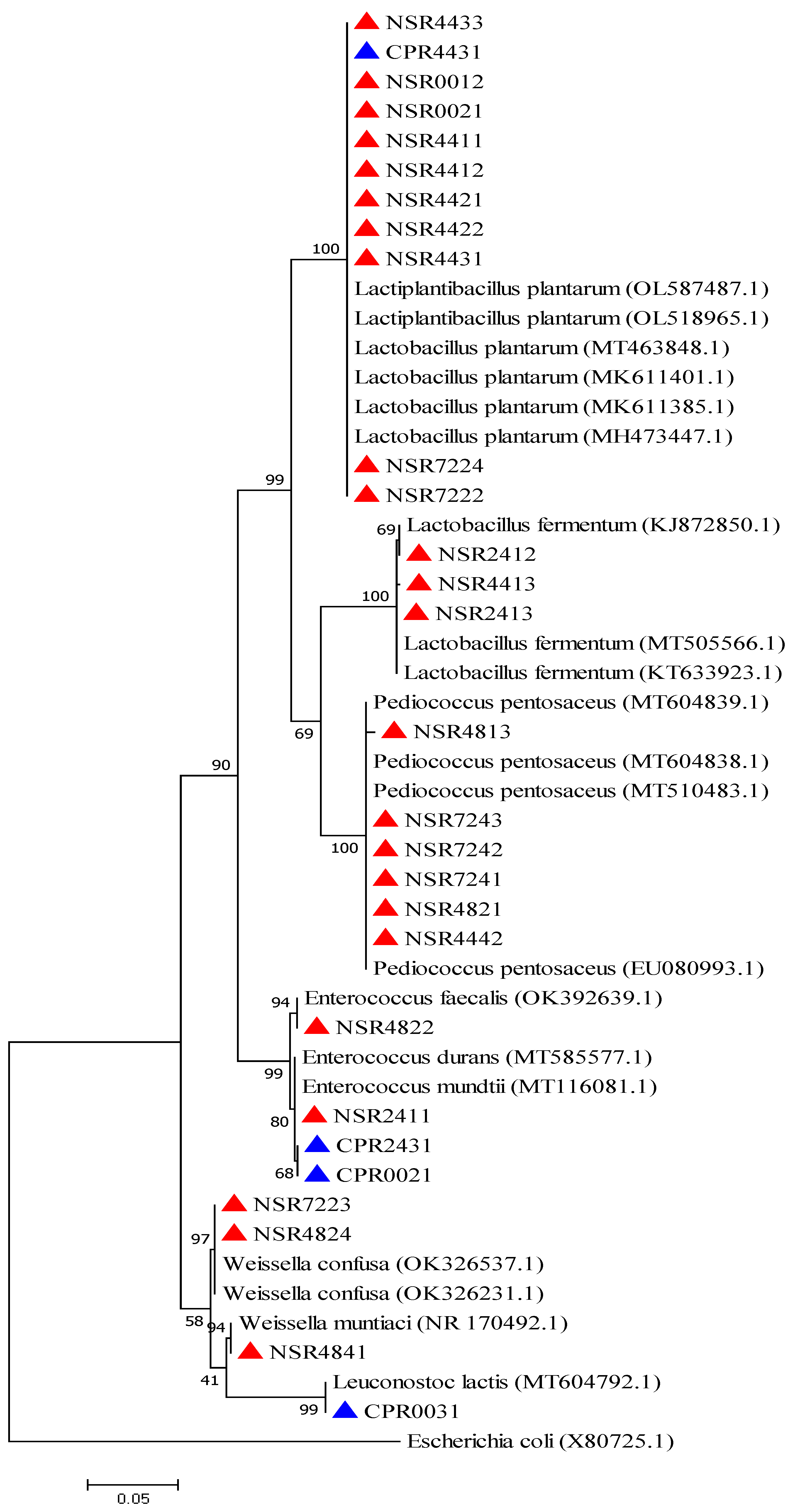
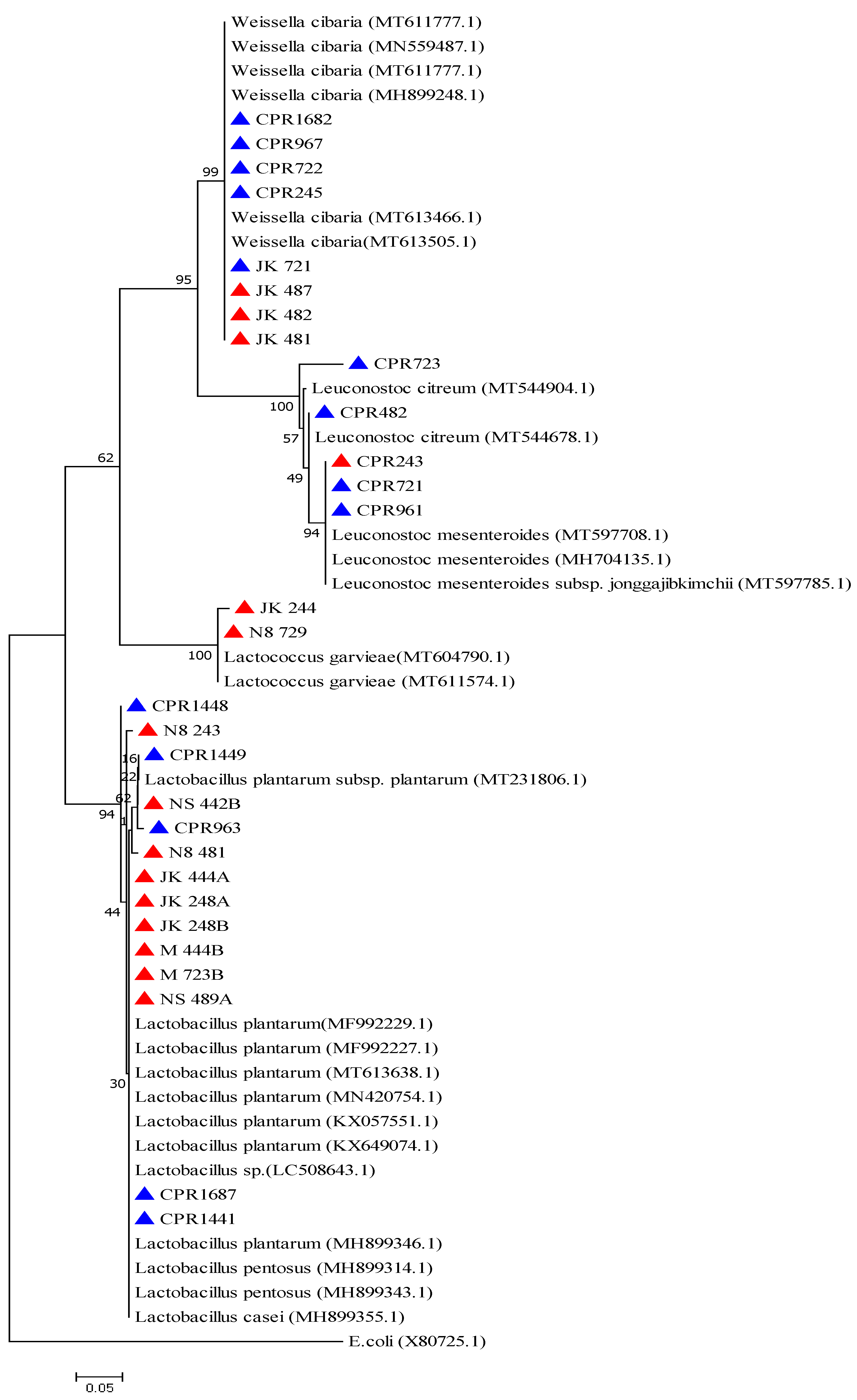
2.2. Phenotypic and Molecular Identification of Lactic Acid Bacteria Strains

A total of 57 strains were isolated and identified according to their phenotypic and molecular characteristics from three African indigenous leafy vegetables (23 nightshade leaves, 19 cowpeas leaves, and 15 amaranth leaves). Phenotypically, the isolates were classified as Gram-positive and catalase-negative, and as either rods or cocci-shaped. Most of the isolates were able to grow in 6.5% NaCl and at 45 °C with a few growing at 10 °C (Table 1 and Table 2. Out of 57 isolates, 20 produced gas from glucose fermentation; thus, they were classified as heterofermentative and 37 isolates were homofermentative or did not produce gas from glucose fermentation [18]. Out of these 20 heterofermentative isolates, 6 were coccus-shaped, produced gas from glucose metabolism, and thus were considered either heterofermentative cocci belonging to the genera Leuconostoc or Weissella, whereas 14 strains were rod-shaped and produced gas from glucose metabolism and thus were characterized as heterofermentative rods, which belonged to either the genus Lactobacillus or Weissella.
Based on the 16S rRNA gene sequencing results, the GenBank databank showed the six heterofermentative strains that were coccus-shaped and produced gas from glucose fermentation that were identified as Leuconostoc mesenteroides (3 strains) with a sequence similarity of 99%, Leuc citreum (2 strains) with a sequence similarity of 99%, and Leuc lactis (1 strain) with a 99% sequence similarity (Table 1). The 14 strains that were rod-shaped produced gas from glucose metabolism and were identified with 16S rRNA gene sequencing such as Lactobacillus fermentum (now Limosilactobacillus fermentum) (3 strains), with a sequence similarity of 99%, and 11 strains were identified as Weissella with sequence similarities of 98 to 100% [Weissella cibaria (8 strains), W. confusa (2 strains) and W. muntiaci (1 strain)] (Table 1 and Table 2).
Out of 37 homofermentative isolates, 22 isolates were Gram-positive, rod-shaped, catalase negative, did not produce gas from glucose metabolism, and thus were considered homofermentative rods belonging to the Lactiplantibacillus (Lactobacillus plantarum) group. These strains were identified as Lactiplantibacillus plantarum with a 16S rRNA gene sequence similarity of 97 to 100%. Two isolates were further identified as Lactiplantibacillus pentosus (Lactobacillus pentosus), with a similarity of 97%, whereas 1 isolate was identified as Lactiplantibacillus casei (Lactobacillus casei) with a sequence similarity of 97% (Table 1). Furthermore, out of 37 homofermentative isolates, 12 were Gram-positive, cocci-shaped, catalase negative, and did not produce gas from glucose fermentation. Based on the 16S rRNA gene sequencing results, 6 isolates were identified as Pediococcus pentosaceus with a sequence similarity of 98 to 100%. Additionally, 4 isolates were identified as Enterococcus, among which 2 were Enterococcus mundtii with a 99% sequence similarity, and E. durans and E. faecalis with percentage sequence similarities of 99% (Table 1 and Table 2). Finally, 2 were Gram-positive, cocci-shaped, catalase negative, did not produce gas from glucose fermentation, grew in 6.5% NaCl, and did not grow either at 10 °C or 45 °C. The 16S rRNA gene sequencing identified these isolates as Lactococcus garvieae with similarities of 98 to 100% (Table 1).
Based on 16S rRNA genes, fermentation of African indigenous leafy vegetables (AILVs) was dominated by the genus Lactobacillus (Lactiplantibacillus and Limosilactobacillus). In cowpea, for example, the genus Lactiplantibacillus (7) dominated followed by Leuconostoc (6), then Weissella (4) and Enterococcus (2), respectively (Figure 2). Africa nightshade leaves were dominated by the genera Lactiplantibacillus (9) and Limosilactobacillus (3), followed by the genera Pediococcus (6), Weissella (3), and Enterococcus (2), respectively. Vegetable amaranth was dominated by Lactiplantibacillus (9), Weissella (4), and Lactococcus (2), respectively (Figure 2).
The hierarchical profile among the bacterial species and AILVs revealed two major clusters and three distinct clusters for the isolated bacteria and AILVs, respectively (Figure 3). Amaranth and cowpeas were clustered separately from the African nightshade. All three vegetables contained Lactiplantibacillus plantarum. Pediococcus pentosaceus was isolated from African nightshades, whereas Weissella cibaria was isolated from cowpea and amaranth (Figure 3).
Analysis of the relationship among the lactic acid bacteria species against different fermentation times in cowpea showed three cluster groups of the bacterial species. Additionally, there was a relationship between 24 h and 96 h and 48 h and 72 h. There was no relationship at 0 h, 144 h, and 168 h. At 144 h, the highest number of bacterial species was recorded compared to the rest of the time. The least number of bacterial species were recorded at 48 h. Lactiplantibacillus plantarum dominated the fermentation at 96 h and 144 h whereas Weissella cibaria dominated at 24 h, 72 h, 96 h, and 168 h in the fermentation. Leuconostoc mesenteroides dominated from 24 h to 96 h whereas Enterococcus was frequently isolated at 0 h and 24 h (Figure 4).
Analysis of the relationship among the bacteria species against the different times of fermentation in vegetable amaranth showed three cluster groups of the bacteria species. Concerning time, there were three cluster groups at 0 h, 72 h, 96 h, and 168 h, 24 h, and 144 h were pressed together, and 48 h was independent. The highest number of bacterial species were recorded at 48 h. Lactiplantibacillus plantarum dominated the fermentation at 24 h, 48 h, 72 h, and 144 h followed by Weissella cibaria, which dominated at 48 h and 72 h and Lactococcus garvieae at 24 h and 72 h of fermentation (Figure 5).
Analysis of the relationship among the bacteria species against the different fermentation times of African nightshade leaves showed five cluster groups of the bacteria species. The highest number of bacterial species was recorded at 144 h. Lactiplantibacillus plantarum dominated nightshade fermentation at 0 h, 72 h, and 144 h. It was followed by Pediococcus pentosaceus, which dominated 48 h, 72 h, and 144 h of fermentation then followed by Weissella muntiaci (48 h) and Weissella confusa (48 h and 72 h) and, finally, Enterococcus mundtii (24 h) and Enterococcus faecalis (48 h) (Figure 6).
Phylogenetically, lactic acid bacteria isolated from nightshade and cowpea leaves had five distinct clusters corresponding to genera Lactiplantibacillus, Limosilactobacillus, Pediococcus, Enterococcus, and Weissella/Leuconostoc. In the phylogenetic group of genus Lactiplantibacillus strains, NSR4433, CPR4431, NSR0012, NSR0021, NSR4411, NSR4421, NSR4421, NSR4431, NSR7224 and NSR7222 were associated with Lactiplantibacillus plantarum (OL587487.1, OL518965.1 and MH473447.1). Strains NSR2412, NSR4413, and NSR2413 were associated with Limosilactobacillus fermentum (Lactobacillus fermentum) (MT505566.1 and KJ872850.1), strains NSR4813, NSR7243, NSR 7241, NSR4821, and NSR4442 were associated with Pediococcus pentosaceus (MT604839.1 and EU080993.1) whereas strains NSR2411, NSR2431, and CPR 0021 formed a cluster associated with Enterococcus sp. (MT585577.1 and MT116081.1). In the cluster, Weissella/Leuconostoc group strains NSR7223 and NSR4824 were clustered with Weissella confusa (OK326537.1) whereas strain NSR4841 was associated with Weissella muntiaci (NR170492.1). One strain, CPR 0031, formed a separate sub-cluster within the Weissella/Leuconostoc group associating with Leuconostoc lactis (MT604792.1) (Figure 7).
Three unique clusters were found in the phylogenetic grouping of lactic acid bacteria that were isolated from fermented cowpea and amaranth leaves and correspond to the genera Weissella/Leuconostoc, Lactococcus, and Lactiplantibacillus. In the cluster of Weissella/Leuconostoc, two distinct sub-clusters were reported; for instance, strains CPR1682, CPR967, CPR722, CPR245, JK721, JK487, JK482, and JK481 clustered with Weissella cibaria (MT611777.1 and MT613505.1), and the other sub-cluster was associated with Leuconostoc spp. For instance, strains CPR723 and CPR 482 were clustered together with Leuc. Citreum (MT544904.1 and MT544678.1); additionally, strains CPR243, CPR721, and CPR961 were associated with Leuc. mesenteroides (MT597708.1). Strains JK244 and N8729 formed a separate cluster associated with Lactococcus garvieae (MT604790.1). The final cluster included strains such as CPR1448, N8243, CPR1449, N8442B, CPR963, N8481, JK444A, JK248A, JK248B, M444B, M723B, NS489A, CPR1687, and CPR1441 that clustered with Lactobacillus plantarum (Lactiplantibacillus plantarum) (Figure 8).

3. Discussion

Fermentation is a biotechnological process with a long history of application. In Africa, there is little information on leafy vegetable fermentation, as opposed to typical fermentations based on animal or plant protein or starchy plant substrates [5]. In natural or spontaneous fermentation, the conditions are set to encourage the growth of the desirable and most adaptable microorganisms, which produce by-products that aid in outgrowing others and dominating the fermentation [19]. Food fermentation offers numerous benefits, several of which are vital for survival and safe nutrition of populations; for example, it enhances food organoleptic properties, aids in food preservation due to the accumulation of organic acids, CO2 and bacteriocins from fermenting microbes and reduction of food spoilage, and it also improves food nutritional value through the production of vitamins, essential amino acids, proteins, and fatty acids. It also improves the toxicological safety of products by degradation of antinutritive factors [16,20]. In this study, three commonly consumed African indigenous leafy vegetables (AILVs) in Kenya, i.e., amaranth, cowpeas, and African nightshade leaves were spontaneously fermented in a brine solution of 3% salt and 3% sugar. AILVs are rich in micronutrients, are widely accessible, and are affordable; hence, they were selected for this investigation [6].
The results showed that the spontaneous fermentation of three AILVs was dominated by six distinct populations of lactic acid bacteria (LAB) genera: Lactobacillus (Lactiplantibacillus and Limosilactobacillus), Pediococcus, Weissella, Leuconostoc, Enterococcus, and Lactococcus (Figure 2). Studies show that plant materials harbor different groups of LAB microbiota, such as Lactobacillus plantarum, Lactobacillus fermentum, Leuconostoc mesenteroides, Lactobacillus acidophilus, and Lactococcus lactis strains [5,21]. The participation of LAB in the spontaneous fermentation of various plant materials has already been demonstrated by Kostinek et al. [22], who found several LAB involved in the natural fermentation of cassava. Spontaneous fermentation of kimchi, a Korean fermented vegetable is commonly carried out by the genera Lactobacillus and Leuconostoc [23,24]. Sauerkraut is also produced by the spontaneous fermentation of cabbage by LAB [25,26].
Among the genera, Weissella, Lactococcus, Enterococcus, and Leuconostoc were abundant at the early stages of all three AILVs fermentation (0–72 h). Previous studies reported that these groups of LAB initiate the fermentation when the pH is low and later a more acid-tolerant group, i.e., Lactobacillus, takes over the fermentation. These results agree with our findings where the pH went below 4.0 after 48 h because of LAB growth (Figure 1). In kimchi fermentation, it was reported that Leuconostoc and other less acid-tolerant bacteria dominate the early stages of fermentation [27], but were later replaced by species of the genera Lactobacillus and Weissella, which are better adapted to grow at high acidity and low pH conditions [28,29].
The genera Lactococcus (Lactococcus garvieae) and Enterococcus (Enterococcus mundtii/Enterococcus faecalis) were among the most abundant LAB bacteria present at 0–48 h. These bacteria belong to a homofermentative group that only produced lactic acid from sugar metabolism [30]. They occur in a wide range of habitats, e.g., on the skin and in the milk and feces of animals, from saliva, breast milk, and the vagina of humans, from plants and vegetables, as well as from a variety of fermented foods [31]. Lactococcus garvieae is commonly used in the manufacture of meat, cheese, and other fermented milk products. It is a good producer of bacteriocins and is hence used in the food industry as a biopreservative agent against various foodborne pathogens [32].
As fermentation progresses, the results showed that members of the genera Lactobacillus, Weissella, and Pediococcus predominated at 24–168 h. Among the LAB isolated, Lactiplantibacillus plantarum (formerly L. plantarum) was the most isolated from 0–144 h (Figure 4, Figure 5 and Figure 6). Lactiplantibacillus plantarum has been previously isolated from different African fermented foods [20,33,34] and it is known to produce plantaricin [35] with acid-tolerant and probiotic properties. Lactiplantibacillus pentosus (formerly L. pentosus) and Lactiplantibacillus casei (formerly L. casei) are phylogenetically clustered with Lactiplantibacillus plantarum, which were isolated from cowpea leaves at 144 h. Lactiplantibacillus pentosus is used as a starter culture in the fermentation of Spanish-style green olives, contributing not only to the organoleptic properties of the final product but also acting as a biopreservative agent of the fermented olives [36]. Lactiplantibacillus casei is frequently isolated from fermented vegetables, milk, dairy products, and the human intestinal tract. It is generally recognized as safe with a long history of use as a probiotic strain and immune system modulator [37].
Limosilactobacillus fermentum (formerly Lactobacillus fermentum) was isolated from nightshade leaves at 24 h and 144 h, and it is obligately heterofermentative rod-shaped, producing CO2 from sugar metabolism. It occurs in diverse habitats, including the human gut, milk products, fermenting plant material, and animals [38]. This species is considered a good probiotic candidate because of its ability to withstand gastrointestinal conditions [39]. It was reported to have the potential for the prevention of community-acquired infections, modulation of the immune systems, and production of antimicrobial compounds [40]. Strains belonging to the genus Weissella were isolated at 48 to 72 h, and they included W. cibaria, W. confuse, and W. muntiaci. Members of this genus are Gram-positive, coccoid, or have a rod-shaped morphology [41]. They are obligately heterofermentative, producing CO2 from sugar metabolism [31]. They occur in a wide range of habitats, e.g., on the skin and in the milk and feces of animals, from saliva, breast milk, feces, and the vagina of humans, from plants and vegetables, as well as from a variety of fermented foods [31]. W. cibaria and W. confusa are linked to the formation of the exopolysaccharide (EPS) dextran, which has prospective applications as a replacement for commercial hydrocolloids in bakery products, as well as health benefits as a prebiotic fiber [42]. Furthermore, members of the genus Leuconostoc were frequently isolated from cowpea leaves at 0–96 h, and they included Leuc. mesenteroides, Leuc. lactis, and Leuc. citreum. Members of this genus are Gram-positive, coccoid, or have a rod-shaped morphology and produced CO2 from sugar fermentation. Leuconostoc species are involved in several food fermentation processes; for example, Leuc. mesenteroides sub sp. mesenteroides are regular aroma-producing starter cultures in the dairy industry and are involved in vegetable and coffee bean fermentations as well as Leuc. mesenteroides and Leuc. citreum are the dominant species in kimchi fermentation [43]. Additionally, Leuc. lactis is a good producer of EPS with potential applications in the food industry [44]. Pediococcus pentosaceus dominated the fermentation of African nightshade leaves between 48 to 144 h. It is an obligate homofermentative, Gram-positive coccus-shaped LAB [45]. It is frequently utilized in the fermentation of fruits, vegetables, dairy products, meat, and silage [46]. It is known to produce pediocins with broad antimicrobial activity against food pathogens [47] and is crucial in the production of various commercial probiotic feeds [48].

4. Materials and Methods

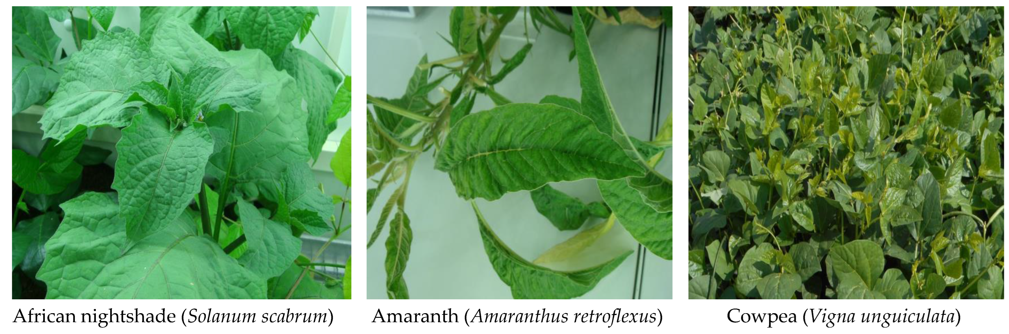
4.1. Growth and Preparation of Plant Materials

The African indigenous leafy vegetables nightshade (Solanum scarbrum), cowpea (Vigna unguiculata), and amaranth (Amaranthus retroflexus) (Figure 9) were cultivated at Jomo Kenyatta University of Agriculture and Technology (JKUAT) in Kenya in open fields for six to eight weeks (17–28 °C, 40–50% relative humidity). The vegetables were cultivated during the dry seasons of December to April. The used soil was supplemented with composite farmyard manure with application of drip-line irrigation 12 h a day. The leafy vegetables were harvested by hand-picking and delivered to the laboratory for processing. The leaves were washed with tap water and dried with paper towels.

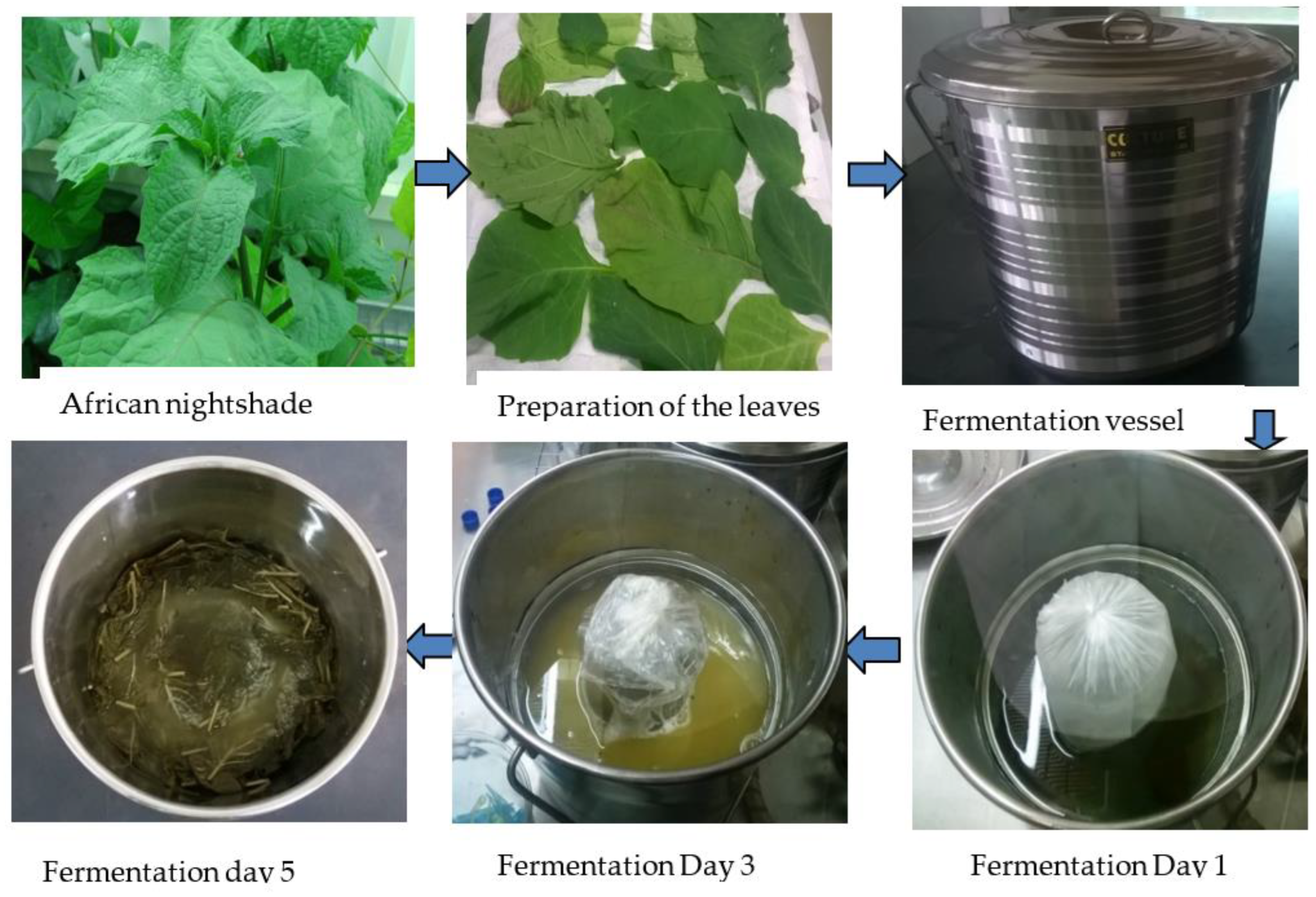
4.2. Fermentation of African Indigenous Leafy Vegetables

Fermentation was performed in 5. L stainless steel buckets. For each vegetable, 1 kg of leaves and 3 L of salt and sugar brine solution were used. The solution consisted of a combination of salt and sugar, 3.0% each. Common table salt (Kensalt ltd, Nairobi, Kenya) and retail sugar (Kabras sugar company ltd, Kakamega, Kenya) were purchased at local stores in Kenya. The brine solution was sterilized by autoclaving for 15 min at 121 °C. Weights were used to hold all plant material below the surface of the liquid. Sampling was done in a sterile setting (Figure 10). The fermentations were done in duplicates at 25 °C.

4.3. Microbiological Testing

The progress of the fermentation was determined by microbial enumeration and pH determination. The buckets were carefully swirled to mix the fermentation brine and materials followed by collecting 7 mL of brine samples for pH determination and 5 mL for microbiological enumeration in sterile test tubes at 0 h, 24 h, 48 h, 72 h, 144 h, and 168 h. The fermentation samples were mixed by vortexing, and then 1 mL of the mixture was transferred to a 9 mL test tube containing sterile quarter-strength ringer’s solution to make serial 10-fold dilutions of 10−1, 10−2, 10−3, 10−4, 10−5, 10−6, 10−7, 10−8, and 10−9. These preparations were further mixed by vortexing. Then, 100 µL of the aliquots from different dilutions were spread-plated onto MRS agar (De Man, Rogosa, and Sharpe), M641, Himedia (Mumbai, India) and M17 agar (M929 Himedia (Mumbai, India). The plates were incubated aerobically at 30 °C for 24–48 h. The incubated plates were counted daily for up to 6 days to determine the bacterial colony forming unit (CFU) as described by Wafula and Murunga [49]. For further characterization, colonies were selected at random from the highest dilution agar plates. The isolates were then grown aerobically in MRS broth at 30 °C and streaked to ensure purity. The isolates stock cultures were cryopreserved in MRS broth with 20% glycerol at −75 °C. The isolates were divided into groups based on phenotypic characteristics, and their identity was confirmed through 16S rRNA gene sequencing.

4.4. Phenotypic Characterization

Presumptive lactic acid bacteria isolated from cowpea, nightshade, and vegetable amaranth leaves were further characterized. Cell morphology was determined using phase-contrast microscopy at 100× magnification (Shimadzu CX41, Tokyo, Japan). Standardized tests including Gram reaction and catalase activity [50,51], gas (CO2) output from glucose in MRS broth, growth at 6.5% NaCl concentration, and growth at 15 °C and 45 °C were also determined [12]. The strains were classified into three groups based on their phenotypic characteristics: obligately heterofermentative rods, facultatively heterofermentative and obligately homofermentative rods, and obligately heterofermentative cocci.

4.5. Genotypic Characterization

Genomic DNA was extracted from overnight cell cultures grown in an MRS broth at 30 °C using a BioWorld Bacterial Miniprep Kit (GeneLink International, Inc., Dublin, OH, USA) according to the manufacturer’s instructions. DNA was quantified using a NanoDrop spectrophotometer (PCRmax, Staffordshire, UK). Amplification was performed in a Primus 96 advanced Peqlab, thermal cycler (PEQLAB, Erlangen, Germany) in relation to positions 8–27 and 1511–1491 of the corresponding 16S rRNA gene of Escherichia coli, respectively [52]. The 16S rRNA gene amplification was performed using the universal primers 27F (5′-AGA GTT TGA TCC TGG CTC AG-3′) and 1492R (5′-GGT TAC CTT GTT ACG ACT T-3′).
PCR was performed in a 50 µL mixture containing 25 µL OneTaq® 2X Master Mix (New England BioLabs, Hertfordshire, UK), 1 µL of each primer, 1 µL of DNA template (10 ng), and 22 µL RNase free water. The reaction mixtures were subjected to the PCR conditions as described by Wafula and Murunga [49]. The amplified PCR products were resolved in 1.2% agarose gel stained with ethidium bromide (1 μg/mL) and visualized using a Uvitec Cambridge gel documentation system (Uvitec, Cambridge, UK). PCR products were purified using the QIAquick PCR purification kit (Qiagen, Hilden, Germany) according to the manufacturer’s instructions. The purified amplicons were Sanger-sequenced at Human Genomics Macrogen Europe (Macrogen Europe B.V, Amsterdam, The Netherlands).

4.6. Phylogenetic Analysis

The 16S rRNA gene sequences of the bacterial isolates were viewed for quality checks and edited using the ChromasPro 2.1.8 software package (http://technelysium.com.au/wp/, accessed on 13 January 2022). They were then compared with available standard sequences of bacteria lineages in the public nucleotide sequence databases in the National Center for Biotechnology Information (NCBI) using nucleotide blast (https://blast.ncbi.nlm.nih.gov/Blast.cgi, accessed on 13 January 2022) to find closely related bacterial 16S rRNA gene sequences. The 16S rRNA gene sequences of the isolates and those of the unknown closely related bacteria strains were aligned using Clustal W software, and phylogenetic trees were constructed using the Kimura 2-parameter model with MEGA (Molecular Evolutionary Genetics analysis) 7.0 software package [53]. The trees’ topologies were evaluated using the bootstrap resampling method [54] based on 1000 replicates. The sequence of Escherichia coli (X80725.1) was used as control.

4.7. Data Analysis

The microbial enumeration and pH results were expressed as the mean and standard deviation of duplicate experiments using Microsoft excel 2021. The hierarchical clustering of LAB vs fermentation and the vegetable type were analyzed using Anaconda-Python version 3.0.

5. Conclusions

Based on 16S rRNA genes, fermentation of African indigenous leafy vegetables was dominated by genera Lactobacillus, Weissella, Leuconostoc, Pediococcus, and Lactococcus. This study revealed that fermented AILVs harbor diverse LAB with Lactiplantibacillus plantarum, Weissella cibaria, and Pediococcus pentosaceus as the dominant species. These bacterial strains could be used in a variety of industrial and commercial settings. Hence, further studies are required to evaluate the isolates’ functional properties as potential probiotics and starter cultures. Molecular and metagenomic techniques might be used to thoroughly investigate how the LAB functions during fermentation and microbial variation throughout vegetable fermentation.

Author Contributions

Conceptualization, E.N.W.; methodology; E.N.W. and T.B.W.; software, E.N.W., T.B.W. and J.O.K.; validation, E.N.W., T.B.W., P.M.W. and J.O.K.; formal analysis, E.N.W. and T.B.W.; investigation, E.N.W., T.B.W., P.M.W. and J.O.K.; resources, E.N.W.; data curation, E.N.W. and T.B.W.; writing—original draft preparation, E.N.W. and T.B.W.; writing—review and editing, E.N.W., T.B.W., P.M.W. and J.O.K.; visualization, E.N.W.; supervision, E.N.W.; project administration, E.N.W.; Funding acquisition, E.N.W. All authors have read and agreed to the published version of the manuscript.

Funding

This project was funded by the International Foundation for Science (IFS) (Grant No. I-3-E-6287-1), and we are grateful for their financial support.

Institutional Review Board Statement

Not applicable.

Informed Consent Statement

Not applicable.

Data Availability Statement

The data used to support the findings of this study are available from the corresponding author upon request.

Acknowledgments

The authors appreciate Jomo Kenyatta University of Agriculture and Technology Department of Food Science and Technology from where this work was done. The authors also appreciate Jennifer Wambugu for the laboratory assistance.

Conflicts of Interest

The authors have declared that no competing interest exists regarding the publication of this paper.

References

  1. Abukutsa-Onyango, M. Strategic repositioning of African indigenous vegetables in the horticulture sector. In Proceedings of the Second RUFORUM Biennial Meeting, Entebbe, Uganda, 20–24 September 2010; pp. 1413–1419. [Google Scholar]
  2. Irakoze, M.L.; Wafula, E.N.; Owaga, E. Potential role of african fermented indigenous vegetables in maternal and child nutrition in sub-saharan Africa. Int. J. Food Sci. 2021, 2021, 3400329. [Google Scholar] [CrossRef] [PubMed]
  3. Oguntoyinbo, F.A.; Cho, G.-S.; Trierweiler, B.; Kabisch, J.; Rösch, N.; Neve, H.; Bockelmann, W.; Frommherz, L.; Nielsen, D.S.; Krych, L.; et al. Fermentation of African Kale ( Brassica Carinata ) Using L. plantarum BFE 5092 and L. fermentum BFE 6620 starter strains. Int. J. Food Microbiol. 2016, 238, 103–112. [Google Scholar] [CrossRef] [PubMed]
  4. Mulaw, G.; Sisay Tessema, T.; Muleta, D.; Tesfaye, A. In vitro evaluation of probiotic properties of lactic acid bacteria isolated from some traditionally fermented ethiopian food products. Int. J. Microbiol. 2019, 2019, 7179514. [Google Scholar] [CrossRef] [Green Version]
  5. Wafula, E.; Franz, C.M.A.P.; Rohn, S.; Huch, M.; Mathara, J.; Trierweiler, B. Fermentation of African indigenous leafy vegetables to lower post-harvest losses, maintain quality and increase product safety. Africa J. Hortic. Sci. 2016, 9, 1–13. [Google Scholar]
  6. Gido, E.O.; Ayuya, O.I.; Owuor, G.; Bokelmann, W. Consumption intensity of leafy african indigenous vegetables: Towards enhancing nutritional security in rural and urban dwellers in Kenya. Agric. Food Econ. 2017, 5, 14. [Google Scholar] [CrossRef]
  7. Uusiku, N.P.; Oelofse, A.; Duodu, K.G.; Bester, M.J.; Faber, M. Nutritional value of leafy vegetables of sub-saharan Africa and their potential contribution to human health: A review. J. Food Compos. Anal. 2010, 23, 499–509. [Google Scholar] [CrossRef]
  8. Franz, C.M.A.P.; Huch, M.; Mathara, J.M.; Abriouel, H.; Benomar, N.; Reid, G.; Galvez, A.; Holzapfel, W.H. African fermented foods and probiotics. Int. J. Food Microbiol. 2014, 190, 84–96. [Google Scholar] [CrossRef]
  9. Stoll, D.A.; Wafula, E.N.; Mathara, J.M.; Trierweiler, B.; Kulling, S.E.; Huch, M. Fermentation of African nightshade leaves with lactic acid bacterial starter cultures. Int. J. Food Microbiol. 2021, 342, 109056. [Google Scholar] [CrossRef]
  10. Liu, S.; Han, Y.; Zhou, Z. Lactic acid bacteria in traditional fermented Chinese foods. Food Res. Int. 2011, 44, 643–651. [Google Scholar] [CrossRef]
  11. Owade, J.O.; Abong’, G.O.; Okoth, M.W.; Mwang’ombe, A.W.; Jobor, J.O. Comparative profiling of lactic acid bacteria isolates in optimized and spontaneous fermentation of cowpea leaves. Food Sci. Nutr. 2021, 9, 1651–1664. [Google Scholar] [CrossRef]
  12. Ibinabo, T.I.; Wafula, E.N.; Josiah, K.; Julius, M.M. Phenotypic and genotypic characterization of lactic acid bacteria isolated from spontaneously fermented vegetable amaranth. African J. Food Sci. 2021, 15, 254–261. [Google Scholar] [CrossRef]
  13. Xiong, T.; Li, X.; Guan, Q.; Peng, F.; Xie, M. Starter culture fermentation of chinese sauerkraut: Growth, acidification and metabolic analyses. Food Control 2014, 41, 122–127. [Google Scholar] [CrossRef]
  14. Bautista-Gallego, J.; Medina, E.; Sánchez, B.; Benítez-Cabello, A.; Arroyo-López, F.N. Role of lactic acid bacteria in fermented vegetables. Grasas Aceites 2020, 71, 358. [Google Scholar] [CrossRef]
  15. Jay, J.M.; Lossener, M.J.; Golden, D.A. Mordern Food Microbiology, 7th ed.; Springer Science: New York, NY, USA, 2005; ISBN 0321109317. [Google Scholar]
  16. Stiles, M.E.; Holzapfel, W.H. Lactic acid bacteria of foods and their current taxonomy. Int. J. Food Microbiol. 1997, 36, 1–29. [Google Scholar] [CrossRef] [PubMed]
  17. Holzapfel, W.H.; Wood, B.J.B. Lactic Acid Bacteria: Biodiversity and Taxonomy, 1st ed.; Holzapfel, W.H., Wood, B.J.B., Eds.; John Wiley & Sons, Ltd: Chichester, UK, 2014; Volume 9781444333, ISBN 9781118655252. [Google Scholar]
  18. Kostinek, M.; Ban-Koffi, L.; Ottah-Atikpo, M.; Teniola, D.; Schillinger, U.; Holzapfel, W.H.; Franz, C.M.A.P. Diversity of predominant lactic acid bacteria associated with cocoa fermentation in Nigeria. Curr. Microbiol. 2008, 56, 306–314. [Google Scholar] [CrossRef] [PubMed]
  19. Holzapfel, W. Use of starter cultures in fermentation on a household scale. Food Control 1997, 8, 241–258. [Google Scholar] [CrossRef]
  20. Kostinek, M.; Specht, I.; Edward, V.A.; Schillinger, U.; Hertel, C.; Holzapfel, W.H.; Franz, C.M.A.P. Diversity and technological properties of predominant lactic acid bacteria from fermented cassava used for the preparation of gari, a traditional African food. Syst. Appl. Microbiol. 2005, 28, 527–540. [Google Scholar] [CrossRef]
  21. McFeeters, R.F.; Pérez-Díaz, I.; Lee, C.-H.; Breidt, F. Fermented vegetables. Food Microbiol. 2013, 27603, 841–855. [Google Scholar] [CrossRef]
  22. Kostinek, M.; Specht, I.; Edward, V.A.; Pinto, C.; Egounlety, M.; Sossa, C.; Mbugua, S.; Dortu, C.; Thonart, P.; Taljaard, L.; et al. Characterisation and biochemical properties of predominant lactic acid bacteria from fermenting cassava for selection as starter cultures. Int. J. Food Microbiol. 2007, 114, 342–351. [Google Scholar] [CrossRef]
  23. Kim, M.; Chun, J. Bacterial community structure in kimchi, a korean fermented vegetable food, as revealed by 16S rRNA gene analysis. Int. J. Food Microbiol. 2005, 103, 91–96. [Google Scholar] [CrossRef]
  24. Lee, J.-Y.; Kim, C.-J.; Kunz, B. Identification of lactic acid bacteria isolated from kimchi and studies on their suitability for application as starter culture in the production of fermented sausages. Meat Sci. 2006, 72, 437–445. [Google Scholar] [CrossRef] [PubMed]
  25. Adams, M.R.; Moss, M.O. Food Microbiology, 3rd ed.; RSC Publishing: Cambridge, UK, 2008. [Google Scholar]
  26. Halász, A.; Baráth, A.; Holzapfel, W. The influence of starter culture selection on sauerkraut fermentation. Z. Fur. Leb. Und-Forsch. 1999, 208, 434–438. [Google Scholar] [CrossRef]
  27. Jung, J.Y.; Lee, S.H.; Kim, J.M.; Park, M.S.; Bae, J.-W.; Hahn, Y.; Madsen, E.L.; Jeon, C.O. Metagenomic analysis of kimchi, a traditional Korean fermented food. Appl. Environ. Microbiol. 2011, 77, 2264–2274. [Google Scholar] [CrossRef] [Green Version]
  28. Cho, J.; Lee, D.; Yang, C.; Jeon, J.; Kim, J.; Han, H. Microbial population dynamics of kimchi, a fermented cabbage product. FEMS Microbiol. Lett. 2006, 257, 262–267. [Google Scholar] [CrossRef] [Green Version]
  29. Lee, J.-S.; Heo, G.-Y.; Lee, J.W.; Oh, Y.-J.; Park, J.A.; Park, Y.-H.; Pyun, Y.-R.; Ahn, J.S. Analysis of kimchi microflora using denaturing gradient gel electrophoresis. Int. J. Food Microbiol. 2005, 102, 143–150. [Google Scholar] [CrossRef]
  30. Sutic, M.; Banina, A. Influence of aflatoxin B1 on gas production by lactic acid bacteria. J Env. Pathol Toxicol Oncol 1990, 10, 149–153. [Google Scholar]
  31. Fusco, V.; Quero, G.M.; Cho, G.-S.; Kabisch, J.; Meske, D.; Neve, H.; Bockelmann, W.; Franz, C.M.A.P. The genus weissella: Taxonomy, ecology and biotechnological potential. Front. Microbiol. 2015, 6, 155. [Google Scholar] [CrossRef] [Green Version]
  32. Moumene, M.; Drissi, F.; Croce, O.; Djebbari, B.; Robert, C.; Angelakis, E.; Benouareth, D.E.; Raoult, D.; Merhej, V. Complete genome sequence and description of Lactococcus Garvieae M14 isolated from algerian fermented milk. New Microbes New Infect. 2016, 10, 122–131. [Google Scholar] [CrossRef] [Green Version]
  33. Mathara, J.M.; Schillinger, U.; Kutima, P.M.; Mbugua, S.K.; Holzapfel, W.H. Isolation, identification and characterisation of the dominant microorganisms of kule naoto: The maasai traditional fermented milk in Kenya. Int. J. Food Microbiol. 2004, 94, 269–278. [Google Scholar] [CrossRef]
  34. Oguntoyinbo, F.A.; Tourlomousis, P.; Gasson, M.J.; Narbad, A. Analysis of bacterial communities of traditional fermented west African cereal foods using culture independent methods. Int. J. Food Microbiol. 2011, 145, 205–210. [Google Scholar] [CrossRef]
  35. Cho, G.-S.; Huch, M.; Hanak, A.; Holzapfel, W.H.; Franz, C.M.A.P. Genetic analysis of the plantaricin EFI locus of Lactobacillus plantarum PCS20 reveals an unusual plantaricin E gene sequence as a result of mutation. Int. J. Food Microbiol. 2010, 141, S117–S124. [Google Scholar] [CrossRef] [PubMed]
  36. Ruiz-Barba, J.L.; Jiménez-Díaz, R. A novel Lactobacillus pentosus-paired starter culture for spanish-style green olive fermentation. Food Microbiol. 2012, 30, 253–259. [Google Scholar] [CrossRef] [PubMed]
  37. Lavermicocca, P.; Dekker, M.; Russo, F.; Valerio, F.; Di Venere, D.; Sisto, A. Lactobacillus paracasei-enriched vegetables containing health promoting molecules. In Probiotics, Prebiotics, and Synbiotics; Elsevier: Amsterdam, The Netherlands, 2016; pp. 361–370. ISBN 9780128023716. [Google Scholar]
  38. Dellaglio, F.; Torriani, S.; Felis, G. Reclassification of Lactobacillus cellobiosus rogosa et al. 1953 as a later synonym of Lactobacillus fermentum beijerinck 1901. Int. J. Syst. Evol. Microbiol. 2004, 54, 809–812. [Google Scholar] [CrossRef] [PubMed] [Green Version]
  39. Jimenez, E.; Langa, S.; Martin, V.; Arroyo, R.; Martin, R.; Fernandez, L.; Rodriguez, J.M. Complete genome sequence of lactobacillus fermentum CECT 5716, a probiotic strain isolated from human Milk. J. Bacteriol. 2010, 192, 4800. [Google Scholar] [CrossRef] [Green Version]
  40. López-Huertas, E. Safety and efficacy of human breast milk Lactobacillus fermentum CECT 5716. A mini-review of studies with infant formulae. Benef. Microbes 2015, 6, 219–224. [Google Scholar] [CrossRef]
  41. Collins, M.D.; Samelis, J.; Metaxopoulos, J.; Wallbanks, S. Taxonomic studies on some Leuconostoc-like organisms from fermented sausages: Description of a new genus Weissella for the Leuconostoc paramesenteroides group of species. J. Appl. Bacteriol. 1993, 75, 595–603. [Google Scholar] [CrossRef]
  42. Arendt, E.K.; Lucid, A.; Lucey, B.; Sleator, R.D.; Coffey, A.; Lynch, K.M. Genomics of weissella cibaria with an examination of its metabolic traits. Microbiology 2015, 161, 914–930. [Google Scholar] [CrossRef]
  43. Kot, W.; Neve, H.; Heller, K.J.; Vogensen, F.K. Bacteriophages of Leuconostoc, Oenococcus, and Weissella. Front. Microbiol. 2014, 5, 186. [Google Scholar] [CrossRef] [Green Version]
  44. Saravanan, C.; Shetty, P.K.H. Isolation and characterization of exopolysaccharide from Leuconostoc lactis KC117496 isolated from idli batter. Int. J. Biol. Macromol. 2016, 90, 100–106. [Google Scholar] [CrossRef]
  45. Franz, C.M.A.P.; Endo, A.; Abriouel, H.; Van Reenen, C.; Galvez, A.; Dicks, L.M. The genus Pediococcus. In Lactic Acid Bacteria: Biodiversity and Taxonomy; Holzapfel, W., Wood, B.J., Eds.; Wiley Blackwell: Chichester, UK, 2014; pp. 361–376. [Google Scholar]
  46. Knorr, D. Technology aspects related to microorganisms in functional foods. Trends Food Sci. Technol. 1998, 9, 295–306. [Google Scholar] [CrossRef]
  47. Todorov, S.D.; Dicks, L.M.T. Bacteriocin production by Pediococcus pentosaceus isolated from marula (Scerocarya Birrea). Int. J. Food Microbiol. 2009, 132, 117–126. [Google Scholar] [CrossRef]
  48. Yirga, H. The use of probiotics in animal nutrition. J. Probiotics Health 2015, 3, 1–10. [Google Scholar] [CrossRef]
  49. Wafula, E.N.; Murunga, S.I. Isolation and Identification of phosphate solubilizing and nitrogen-fixing bacteria from lake ol’ bolossat sediments, Kenya. Mod. Appl. Sci. 2020, 14, 37–51. [Google Scholar] [CrossRef]
  50. Cappuccino, J.G.; Sherman, N. Microbiology: A Laboratory Manual, 10th ed.; Pearson: New York, NY, USA, 2014; ISBN 0321840224. [Google Scholar]
  51. Gregersen, T. Rapid method for distinction of gram-negative from gram-positive bacteria. Eur. J. Appl. Microbiol. Biotechnol. 1978, 5, 123–127. [Google Scholar] [CrossRef]
  52. Brosius, J.; Palmer, M.L.; Kennedy, P.J.; Noller, H.F. Complete nucleotide sequence of a 16S ribosomal RNA gene from Escherichia coli (recombinant plasmids/DNA sequence analysis/RrnB cistron). Biochemistry 1978, 75, 4801–4805. [Google Scholar] [CrossRef] [Green Version]
  53. Kumar, S.; Stecher, G.; Tamura, K. MEGA7: Molecular evolutionary genetics analysis version 7.0 for bigger datasets. Mol. Biol. Evol. 2016, 33, 1870–1874. [Google Scholar] [CrossRef] [PubMed] [Green Version]
  54. Felsenstein, J. Confidence limits on phylogenies: An approach using the bootstrap. Evolution 1985, 39, 783. [Google Scholar] [CrossRef]
Figure 1. Lactic acid bacteria viable counts and pH determination from Kenya’s three commonly consumed African indigenous vegetables. The counts and pH values were determined in duplicates; the standard deviations are indicated.
Bacteria 02 00001 g001
Figure 2. Lactic acid bacteria isolated from three African indigenous leafy vegetables grouped at the genus level.
Bacteria 02 00001 g002
Figure 3. Hierarchical clustering of lactic acid bacteria from three fermented African indigenous leafy vegetables at the species level. Legend: Hierarchical clustergram generated using several bacteria species at different types of African indigenous leaf vegetables. The heatmap (Euclidean matric) shows the relationship between selected bacteria species and varied African vegetables. The colored scale bar indicates the significant quantified strength of the various African vegetables. The red color in the heatmap indicates the highest, and pink indicates the lowest significance at p ≤ 0.05 for the assayed treatments.
Bacteria 02 00001 g003
Figure 4. Hierarchical clustering of lactic acid bacteria at the species from fermented cowpea leaves at different fermentation times. Legend: Hierarchical clustergram generated using several bacterial species from cowpeas at different times. The heatmap (Euclidean matric) shows the relationship between selected bacteria species and varied times. The colored scale bar indicates the significant quantified strength of the various times. The red color in the heatmap indicates the highest and orange indicates the lowest significance at p ≤ 0.05 for the assayed treatments.
Bacteria 02 00001 g004
Figure 5. Hierarchical clustering of lactic acid bacteria species from fermented vegetable amaranth at different fermentation times. Legend: Hierarchical clustergram generated using several bacterial species from Amaranth at different Times. The heatmap (Euclidean matric) shows the relationship between selected bacteria species (Amaranth) and varied times. The colored scale bar indicates the significant quantified strength of the various times. The red color in the heatmap indicates the highest and orange indicates the lowest significance at p ≤ 0.05 for the assayed treatments.
Bacteria 02 00001 g005
Figure 6. Hierarchical clustering of LAB species from fermented African nightshade leaves at different fermentation times. Legend: Hierarchical clustergram generated using the number of bacterial species from African nightshades at different times. The heatmap (Euclidean matric) shows the relationship between selected bacteria species and varied times. The colored scale bar indicates the significant quantified strength of the various times. The red color in the heatmap indicates the highest, and orange indicates the lowest significance at p ≤ 0.05 for the assayed treatments.
Bacteria 02 00001 g006
Figure 7. A phylogenetic tree based on 16S rRNA gene sequences shows the relationship between lactic acid bacteria isolated from nightshade/cowpea leaves and representatives of other related taxa. The scale bar indicates 0.05 substitutions per nucleotide position. The red arrows indicate isolates from nightshade and blue arrows indicate isolates from cowpea. The number beside the node is the statistical bootstrap value. In brackets are the GenBank accession numbers. The gene sequence of E. coli (X80725.1) was used as an out-group.
Bacteria 02 00001 g007
Figure 8. A phylogenetic tree based on 16S rRNA gene sequences shows the relationship between lactic acid bacteria isolated from cowpea/amaranth and representatives of other related taxa. The scale bar indicates 0.05 substitutions per nucleotide position. The red arrows indicate isolates from amaranth and blue arrows indicate isolates from cowpea. The number beside the node is the statistical bootstrap value. In brackets are the GenBank accession numbers. The gene sequence of E. coli (X80725.1) was used as an out-group.
Bacteria 02 00001 g008
Figure 9. The African indigenous leafy vegetables nightshade (Solanum scarbrum), cowpea (Vigna unguiculata), and amaranth (Amaranthus retroflexus).
Bacteria 02 00001 g009
Figure 10. Fermentation of African indigenous leafy vegetables.
Bacteria 02 00001 g010
Table 1. Phenotypic and molecular identification of lactic acid bacteria from cowpea and amaranth leaves.
Phenotypic Characterization Molecular Identification
Sample IDCell ShapeGram StatusCatalaseCO26.5% NaClGrowth at 10 °CGrowth at 45 °CClosest Relatives% IdentityAccession No.
CPR243Cocci+-++--Leuconostoc mesenteroides99.90MT597785.1
CPR245Rods+-++++Weissella cibaria100.00MN559487.1
CPR482Cocci+-++-+Leuconostoc citreum99.54MT544678.1
CPR721Cocci+-++--Leuconostoc mesenteroides99.21MH704135.1
CPR722Rods+-++-+Weissella cibaria100.00MT611777.1
CPR723Cocci+-++++Leuconostoc citreum91.35MT544904.1
CPR961Cocci+-+---Leuconostoc mesenteroides99.61MT597708.1
CPR963Rods+--+-+Lactobacillus plantarum98.86MT231806.1
CPR967Rods+-++++Weissella cibaria99.72MT611777.1
CPR1448Rods+--+--Lactobacillus casei97.15MH899355.1
CPR1449Rods+--+-+Lactobacillus pentosus97.44MH899343.1
CPR1682Rods+-++-+Weissella cibaria98.47MH899248.1
CPR1687Rods+--+++Lactobacillus pentosus97.01MH899314.1
CPR14410Rods+--+++Lactobacillus plantarum97.57MH899346.1
CPR0021Cocci+--+-+Enterococcus mundtii99.71MT116081.1
CPR0031Cocci+-+---Leuconostoc lactis99.26MT604792.1
CPR2431Cocci+----+Enterococcus durans99.37MT585577.1
CPR4431Rods+--+-+Lactobacillus plantarum100.00OL587487.1
CPR4433Rods+--+-+Lactobacillus plantarum99.52MK611385.1
JK244Cocci+--+--Lactococcus garvieae98.22MT611574.1
JK248ARods+--+++Lactobacillus plantarum98.94MN640561.1
JK248BRods+--+++Lactobacillus plantarum98.13MF992227.1
JK444ARods+--+-+Lactobacillus plantarum100.00KX649074.1
JK481Rods+-++--Weissella cibaria99.63MT613505.1
JK482Rods+-++++Weissella cibaria100MT613466.1
JK487Rods+-++-+Weissella cibaria100MT613505.1
JK721Rods+-++-+Weissella cibaria99.82MT613505.1
M444BRods+--+++Lactobacillus plantarum99.27KX057551.1
M723BRods+--+-+Lactobacillus plantarum99.06MF992227.1
N8243Rods+--+++Lactobacillus plantarum99.72MT613638.1
N8481Rods+--+-+Lactobacillus plantarum96.75MN420754.1
N8729Cocci+--+-+Lactococcus garvieae100MT604790.1
NS442BRods+--+++Lactobacillus plantarum98.67MF992227.1
NS489ARods+--+++Lactobacillus plantarum99.76MF992229.1
Table 2. Phenotypic and molecular identification of lactic acid bacteria from African nightshade leaves.
Phenotypic Characterization Molecular Identification
Sample IDCell ShapeGram StatusCatalaseCO26.5% NaClGrowth at 10 °CGrowth at 45 °CClosest Relatives% IdentityAccession No.
NSR0012Rods+--+++Lactiplantibacillus plantarum99.50OL587487.1
NSR0021Rods+--+++Lactobacillus plantarum99.14MT463848.1
NSR2411Cocci+--+-+Enterococcus mundtii99.49AP019810.1
NSR2412Rods+-++-+Lactobacillus fermentum99.90KJ872850.1
NSR2413Rods+-+--+Lactobacillus fermentum99.07MT505566.1
NSR4411Rods+--+++Lactobacillus plantarum99.61MK611401.1
NSR4412Rods+--+++Lactobacillus plantarum100.00OL587487.1
NSR4413Rods+-++-+Lactobacillus fermentum99.65KT633923.1
NSR4421Rods+--+++Lactobacillus plantarum99.50MH473447.1
NSR4422Rods+--+++Lactiplantibacillus plantarum100.00OL518965.1
NSR4431Rods+--+-+Lactobacillus plantarum99.25MK611401.1
NSR4442Cocci+--+++Pediococcus pentosaceus99.90MT510483.1
NSR4813Cocci+--+++Pediococcus pentosaceus98.67EU080993.1
NSR4821Cocci+--+++Pediococcus pentosaceus99.90MT604839.1
NSR4822Cocci+--+-+Enterococcus faecalis99.88OK392639.1
NSR4824Rods+-++++Weissella confusa100.00OK326537.1
NSR4841Rods+-+++-Weissella muntiaci99.71NR_170492.1
NSR7222Rods+--+++Lactobacillus plantarum99.70OL587487.1
NSR7223Rods+-++--Weissella confusa100.00OK326231.1
NSR7224Rods+--+++Lactiplantibacillus plantarum100.00OL587487.1
NSR7241Cocci+--+++Pediococcus pentosaceus100.00MT604838.1
NSR7242Cocci+--+++Pediococcus pentosaceus99.90MT604839.1
NSR7243Cocci+--+++Pediococcus pentosaceus100.00MT604839.1
Disclaimer/Publisher’s Note: The statements, opinions and data contained in all publications are solely those of the individual author(s) and contributor(s) and not of MDPI and/or the editor(s). MDPI and/or the editor(s) disclaim responsibility for any injury to people or property resulting from any ideas, methods, instructions or products referred to in the content.

