Article

Isolation and Characterization of Bacillus velezensis from Lake Bogoria as a Potential Biocontrol of Fusarium solani in Phaseolus vulgaris L.

1
Institute of Biotechnology Research, Jomo Kenyatta University of Agriculture and Technology, Nairobi P.O. Box 62000-00200, Kenya
2
Dudutech IPM Limited, Naivasha P.O. Box 1927-20117, Kenya
3
Department of Public Health, Bomet University College, Bomet P.O. Box 701-20400, Kenya
*
Author to whom correspondence should be addressed.
Bacteria 2022, 1(4), 279-293; https://doi.org/10.3390/bacteria1040021
Submission received: 16 October 2022 / Revised: 7 November 2022 / Accepted: 10 November 2022 / Published: 17 November 2022

Abstract

:
The common bean (Phaseolus vulgaris L.) is a significant vegetable crop, grown because it is a rich source of protein, carbohydrates, and vitamin B complex. Fusarium solani and Rhizoctonia solani are the most widely known pathogens contributing to large yield losses for this crop. The use of cultural and chemical control practices has been ineffective. Therefore, a sustainable, affordable, and effective control method is urgently required. In this study, we aimed to isolate and characterize Bacillus velezensis from Lake Bogoria as a potential biocontrol agent for Fusarium solani. Bacteria were isolated from soil and sediments using the serial dilution technique. Molecular characterization was performed using the 16S rRNA gene. A total of 13 bacteria were isolated from soil and sediments. Based on the partial sequences, BLAST analysis showed two isolates, B20 (Bacillus velezensis strain QH03-23) and B30 (Bacillus velezensis strain JS39D), belonging to Bacillus velezensis. Other isolates were identified as Bacillus tequilensis, Brevibacillus brevis, Bacillus subtilis, Bacillus amyloliquefaciens, and Bacillus licheniformis. The effectiveness of their antifungal properties was determined via co-culturing, and we found mycelial inhibition rates of 28.17% (for B20) and 33.33% (for B30) for the Fusarium solani isolates. The characterization of the Bacillus velezensis strain revealed that they were Gram-positive and grew well at pH 7.0 and 8.5, although growth was recorded at pH 5.0 and 10.0. In terms of temperature, the optimal temperature conditions were 30−35 °C, with an optimum salinity of 0–0.5 M NaCl. When these isolates were tested for their ability to produce secondary metabolites, they were found to produce phosphate, pectinase, chitinase, protease, indole -3- acetic acid (IAA), and hydrogen cyanide (HCD), making them potential biocontrol agents.

1. Introduction

Phaseolus vulgaris L. is a yearly herbaceous plant, grown for its edible dry seeds [1]. Depending on their uses, common beans can be classified as dry beans, snap beans, and shell beans. The leaves are used as vegetables and animal feed. According to their botanical classification as members of the Fabaceae legume family, they obtain nitrogen through a symbiotic relationship with Rhizobia, a nitrogen-fixing bacterium of beans [1]. Common beans’ dry seeds and leaves contain antioxidants, phenolic compounds, flavonoids, vitamins B complex, and essential metal ions, such as copper, zinc, and calcium. They have also exhibited anticancer and anti-inflammatory effects and can increase capillary resistance [2]. About half a million tons (Mt) of beans are harvested each year in Kenya.
A major difficulty in growing beans is the presence of pests and diseases. Cutworms, bean flies, red spider mites, aphids, pod borers, whiteflies, and thrips are some problematic crop pests. Fusarium solani fungus is an important soil-borne pathogen of common beans that causes fusarium wilt [2,3]. This fungus can remain viable for several years in the soil due to its ability to produce chlamydospores [1]. Fusarium wilt is one of the most difficult diseases to control due to its ability to inhabit the soil and penetrate the plant through the root systems. It can infect more than 600 species of legumes [4]. Therefore, this has increased its survival rate and growers’ inability to manage it. Water stress, soil acidification, and calcium deficiency also favor fungal development. Given the lack of a fully effective control mechanism for Fusarium solani, several studies have been conducted to develop biological approaches for managing this pathogen [2,3]. Several Bacillus species are known to act as antagonists of phytopathogenic fungi and have been used in biological control programs [5,6,7]. As described in [8], Bacillus velezensis has been used as a potential biocontrol agent for fusarium head blight. Furthermore, the research presented in [3] indicates that Bacillus velezensis is responsible for controlling bitter apple rot caused by Colletotrichum gloeosporioides.
There is a need for the biocontrol of this pathogen due to the prolonged use and reduced efficacy of synthetic fungicides. These chemicals also pollute the environment, leaving toxic residues that broaden the range of resistant pathogens [9]. Therefore, biological control has been reported as an alternative method to control Fusarium wilt [7,10,11,12].
In our study, we aimed to isolate and characterize Bacillus velezensis from Lake Bogoria as a potential biocontrol agent of Fusarium solani in Phaseolus vulgaris L. Lake Bogoria is an example of a Kenyan soda lake, known for its hot springs and thus providing a habitant for extremophilic bacteria and fungi. Soda lake microorganisms have been used for decades, but very little is known about their effectiveness in combating and controlling Fusarium solani. The use of Bacillus velezensis as a biological control agent may have several advantages, such as being suitable for the environment and providing efficient management [13,14] The bioactivity of Bacillus velezensis isolated from Lake Bogoria as a potential biocontrol agent against Fusarium solani represents a novel discovery and will open up more opportunities for research in agriculture.

2. Results

2.1. Isolation and Morphological Characteristics

A total of 13 bacterial isolates were obtained from Lake Bogoria.
Morphological characterization was performed using microscopic techniques to determine colony and cell morphology. The colony morphology characteristics included forms ranging from circular to irregular, as well as various elevations (Table 1). The isolates also exhibited differences in their margins, ranging from circular to irregular, wavy, lobate, smooth, and filamentous. The color of the isolates ranged from white to cream, cream-white, teal, blue, and cream-yellow. Their sizes ranged from small to medium and large.
The cell morphology of the pure isolates was assessed in terms of their shape, ranging from rods to cocci. In addition, the surface of the isolates was determined and characterized as smooth, dull, or rough. The Gram staining technique showed that 10 bacterial isolates were Gram-positive and 3 bacterial isolates were Gram-negative (Table 1).

2.2. Molecular Characterization

From the partial sequences, the BLAST analysis showed two isolates B20 (Bacillus velezensis strain QH03-23) and B30 (Bacillus velezensis strain JS39D) as Bacillus velezensis strains. Among the other isolates were Bacillus tequilensis, Brevibacillus brevis, Bacillus subtilis, Bacillus amyloliquefaciens, and Bacillus licheniformis (Figure 1). Three isolates belonged to Gammaproteobacteria in the proteobacteria phylum with a 99.59% to 100% similarity index. Among the proteobacteria groups were the Alcaligenaceae bacterium, Pseudomonas sp., and Pseudomonas aeruginosa (Figure 1).

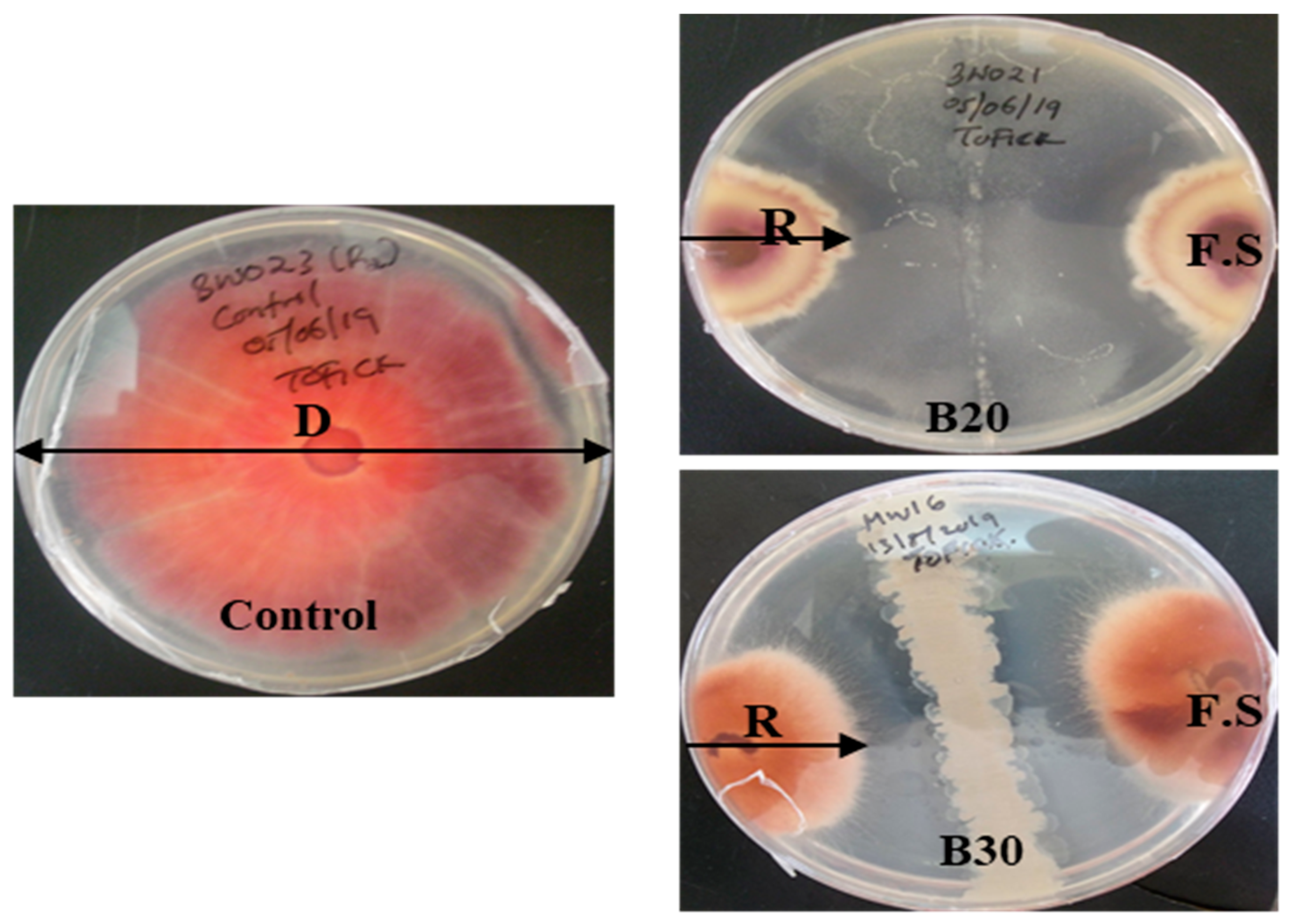
2.3. Evaluation of Bacillus velezensis against Fusarium solani

In Table 2, the ANOVA analysis showed a significant (p < 0.05) variation in the diameter of the F. solani mycelium after the tested bacterial treatment. Isolate B30 (33.33%) had the highest inhibition rate compared to B20 (28.17%) (Figure 2).

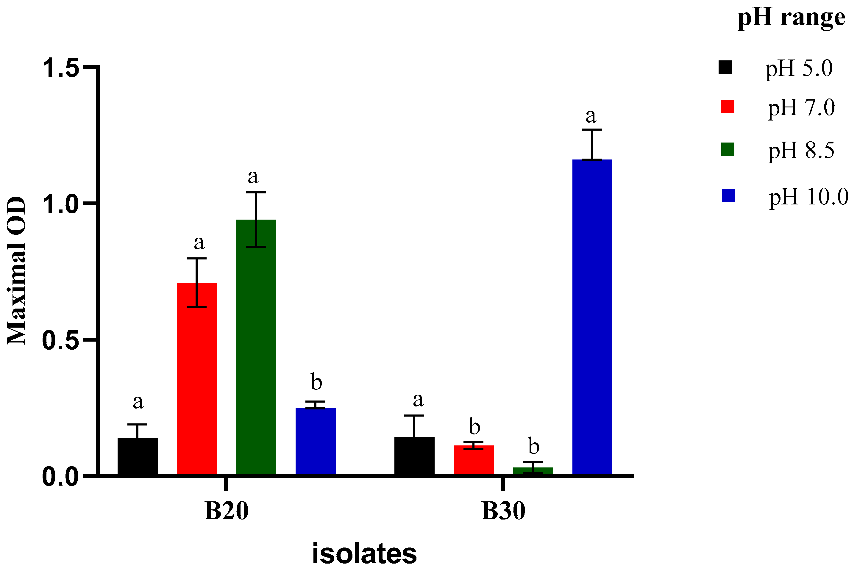
2.3.1. Growth at Different pH

Figure 3 shows a varied growth of Isolates B20 and B30 at different pH. Although the isolates were obtained from an alkaline environment, both isolates could grow at pH 5.0 and 7.0. Isolate B20 recorded the highest O.D. at pH 8.5, while isolate B30 was at pH 10.0. The growth trend at pH 5.0, pH 7.0, and pH 8.5 increased significantly for isolate B20 compared to isolate B30.

2.3.2. Growth at Different Salinity

The isolates B20 and B30 showed varied growth at different concentrations of sodium chloride concentration. The salt concentration of 0.0 M recorded the highest O.D. as an indicator of the highest growth, followed by 0.5 M NaCl, and gradually decreased toward 2.0 M with minimal growth (Figure 4). Additionally, the growth trend decreased with an increase in sodium chloride concentration.

2.3.3. Growth at Different Temperatures

The isolates B20 and B30 grew at a wide range of temperatures (20–60 °C). Optimal growth was observed at 30–40 °C (Figure 5). However, optical growth was observed at 35 °C.

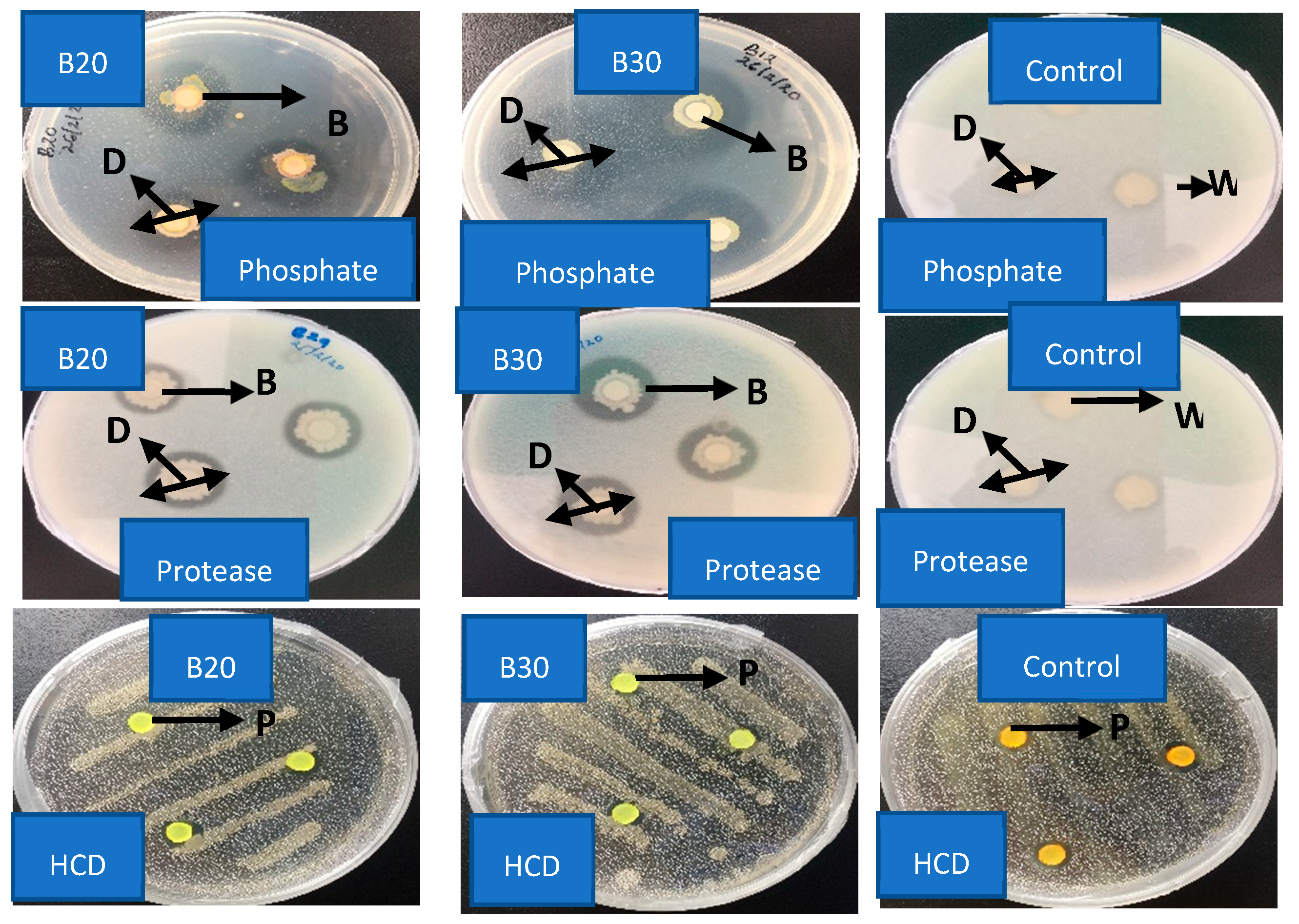
2.4. Enzymatic Bioassay of Isolate B20 and B30

The isolates B20 and B30 on skim milk agar showed a clear zone of inhibition formed around the isolates, indicating a protease-producing strain (Figure 6). B20 and B30 did not show a clear zone of inhibition for pectinase (Table 3).

3. Discussion

Soil-borne fungal diseases cause worldwide economic losses in the agriculture sector. Farmers over the past years have relied on the use of chemical fungicides, which can potentially be harmful to the environment and human health. Additionally, the use of fungicides is linked to soil infertility due to pH change and low yield. In this study, we highlight the alternative use of Bacillus velezensis as an eco-friendly solution for the control of soilborne diseases. In recent years, the use of Bacillus as biological control has been reported. A member of this genus is B. velezensis, a species considered valuable due to its potent bioactive molecule. In this context, two B. velezensis were recovered from the soil and sediment of Lake Bogoria, which is thermophilic because of its high-temperature range. The lake is also classified as a highland lake by [15], with a surface more than 990 m above sea level.
The isolation of bacteria through serial dilution technique clearly showed that lake Bogoria harbors diverse microbes of different morphological characteristics. The study indicates that microbes occur in various ecological environments, such as soil and sediment of the alkaline ecosystem; hence, had the potential to survive a harsh environment. Several studies have indicated that Lake Bogoria is rich in the microbial community. For example, Kambura Et al., [16] reported lake’s microbial community, ranging from Bacillus to Gammaproteobacteria species. Furthermore, the study by [17] indicates that Lake Bogoria has a wide range of Bacillus species with antibiotic-producing properties. The lake, which is saline and thermophilic, has a wide range of enzyme-producing bacteria that have been exploited, identified, and used in various biotechnological fields [18,19,20]. Soil microbial communities are responsible for the cycling of nutrients, stabilizing the ecosystem, and maintaining the structure of the soil. Approximately 90% of the bacteria are found in the soil, contributing to the soil’s nutrient content compared to sediment. Grasslands have also been shown to have more diverse bacterial communities due to biomass stability than water and sediments [21]. Studies have shown a relationship between microbes and environmental factors, geographical location [22,23], pH [22,23], nutrients [21,23], and temperature.
In our study, B. velezensis was identified based on partial 16S rDNA genes where two different strains were obtained. They include Isolate B20, closely related to Bacillus velezensis strain QH03-23 (MT072102.1), and B30, related to Bacillus velezensis strain JS39D (KX129842.1), with a similarity index of 98.71–100%. Additionally, other Bacillus spp. such as B. subtilis, B. amyloliquefaciens, B. licheniformis and Brevibacillus were identified. Others belong to the class of Gammaproteobacteria in the phylum proteobacteria with a range of 98.71–100% similarity index, such as Alicagenes sp and Pseudomonas spp. These results are consistent with previous studies on L. Bogoria, where phylogenetic analysis of the bacterial 16S rRNA gene sequence of the bacteria sequence showed Firmicutes and Gammaproteobacteria as the dominant bacteria domains [17,18,24,25].
In vitro tests, through co-culturing experiments, clearly showed that B20 and B30 could inhibit the fungal radial growth of Fusarium solani. Interestingly, B30 (B. velezensis Strain JS39) produced the highest inhibition rate compared to B20 (B. velezensis Strain QH03-23). Numerous strains of this species have been reported to suppress the growth of microbial pathogens, including bacteria, fungi, and nematodes. Genomic analysis has revealed that B. velezensis possesses strain-specific clusters of genes related to the biosynthesis of secondary metabolites, which play significant roles in pathogen suppression and plant growth promotion. Specifically, B. velezensis exhibits a high genetic capacity to synthesize cyclic lipopeptides such as surfactin, bacillomycin D, fengycin, and bacillibactin, and polyketides such as macrolactin, bacillaene, and difficidin [26]. Secondary metabolites produced by B. velezensis can also trigger trigger-induced systemic resistance in plants, a process by which plants defend themselves against recurrent attacks by virulent microorganisms. According to [14], putative butanediol dehydrogenase, a critical enzyme in butanediol production, is known to induce systemic resistance (ISR) in plants. Therefore, the presence of this gene suggests that B. velezensis may contribute to plant defenses through ISR, allowing plants to prepare themselves for faster and stronger defenses against pathogens [13]. Surfactins are known to have antimicrobial properties that act against both bacteria and fungi. They can be inserted into bacterial cell membranes, solubilizing the fluid phospholipid bilayer and creating pores and ion channels [26,27]. Surfactins have also been shown to interfere with protein processing and secretion. For antifungal activity, surfactins can inhibit glucan synthase, which is involved in cell wall synthesis, and induce apoptotic markers [26]. Furthermore, surfactins are believed to play a key role in ISR triggering [28]. They may also contribute to swarming motility [29], which can help achieve effective rhizosphere colonization and facilitate plant growth-promoting traits while simultaneously inhibiting competing microorganisms through antimicrobial activity. The use of B. velezensis as a potential control against F. solani has been reported by [3,8,13,30,31,32]. Therefore, more exploitation must be evaluated to quantify its mode of action. Additionally, B. velezensis is attracting attention as a valuable biocontrol agent. Consequently, to develop and formulate biobased products, it is increasingly important to understand the antifungal potential of biosynthesis of B. velezensis. Furthermore, the elucidation of genes responsible for bioactive secondary metabolites and the ability to control such genes are important and are additional steps to increase the production of metabolites by beneficial microbes and facilitate metabolic engineering. B. velezensis may be represented as a practical and powerful biocontrol agent that can be used as an effective alternative to synthetic agrochemicals.
The morphological results of the isolated bacteria indicate that they are Gram-positive and rod-shaped; they are also cream-white, though there is variation in their texture, elevation, form, and size due to different strains. The results are consistent with the earlier morphological description of B. velezensis strains of bacteria as aerobic, Gram-positive, rod-shaped, and spore-forming bacteria [3].
In our study, physiochemical characterization of the isolates B20 and B30 showed that they could grow in a wide pH range. The highest growth was observed at pH 7.0, 8.5, and 10.0, respectively. However, the results also indicated that isolates B20 and B30 could grow at an acidic pH of 5.0. The results were consistent with a previous study by [33] which indicated that a pH range of 5.7 to 9.0 favors the growth of alkaliphiles. However, the pH range of 7.0 to 11.0 serves as a selective optimum pH. Soda lakes have a high pH range. Therefore, microbes can only survive by maintaining their cytoplasm at the same pH as their mesophilic relatives. The mechanism for achieving this is secondary proton uptake mediated by membrane-associated antiporters. Alkaliphiles can also maintain an internal pH > 7, 5 regardless of environmental pH. Most bacteria activate the sodium-ion pump at this high pH, which lowers the internal pH by transporting hydrogen ions into the cells. According to [34], alkaliphiles have cell membranes with a special composition of tissues that protect them from highly alkaline conditions. The study was consistent with previous findings [35], which reported a wide pH range (4.5–10.0) where bacillus can grow. It also agrees with the findings of [35], who reported varied bacterial growth at different pH.
For Sodium chloride concentration, our study showed that isolates B20 and B30 could tolerate different concentrations of salinity conditions. They had a maximum growth at 0.0 M NaCl. This is because they were isolated from soil and sediment samples. Isolate B30 was able to grow at 2.0 M NaCl. These isolates can be classified as halophiles. The isolate B20 grew at all salt concentrations, hence known as halotolerant. The ability of the isolates to grow at different salt concentrations indicates that they are tolerant to salinity and as an adaptive strategy to survive in adverse growth conditions. This is in agreement with earlier studies on Bacillus spp. [36]. Soda lake bacteria can survive millions of years in the fluid inclusions of salt deposits, including evaporates, by adapting to these potentially deadly ecosystems. To prevent water loss from their cell membrane, the halophile compensates for offsets the high salt in the environment by accumulating potassium and glycine-betaine compounds. This mechanism balances the salt concentration inside the cell and the environment, preventing the cell from bursting due to the diffusion of water. The ability of isolates to grow at different salt concentrations has also been reported by [15,37,38,39] for Lake Bogoria isolates.
The isolate B20 and B30 grew at a wide range of temperatures. The optimal temperature was recorded at 35 °C. Compared to the temperature recorded at sampling points, most bacterial isolates were obtained in the soil sample with a lower temperature than in the sediments. Although 35 °C was recorded as the optimal temperature, some bacteria survived up to 60 °C. The growth of bioactive bacteria at varied temperatures indicates that Bogoria harbors diverse microbes that can adapt to a wide temperature range. This property is ideal for biocontrol since different environments have different temperatures and can adapt to them, making them good biocontrol agents. The study agrees with previous studies that show the ability of Lake Bogoria bacterial isolates to adapt at different temperatures [16,19,20,38].
In our study, enzymatic assay for isolates B20 and B30 showed varied presence and absence of protease, phosphatase, chitinase, pectinase, indole -3-Acetic Acid (IAA), and hydrogen cyanide (HCD). This feature is highly suitable helping suppress the disease and promote plant growth and productivity [40]. Additionally, our results emphasized that selected antagonist bacteria with high antifungal activity shared excellent attributes. Most of these bacterial isolates exerted different biocontrol mechanisms such as the production of cell wall degrading enzymes, cyanide (HCD) production, and plant growth promotion traits. Producing lytic enzymes is among the major mechanisms’ biocontrol agents employed to control fungal pathogens. Our results showed that B20 and B30 displayed protease and phosphate production, while chitinase was observed in B30 only, and IAA in B20. There was no production of pectinase and HCD for both isolates. Chitinase isis important because the microbes attack the fungal cell wall and cause lysis by degrading chitin [3,41]. Similar results were found for pseudomonas fluoresces, which produced chitinase against the growth of Sclerotina sclerotium [42]. The production of IAA is characterized by a strong affinity for iron which is one of the important mechanisms used by biocontrol [32,43,44].
Additionally, IAA contributes to the increasing root surface area and length and, therefore, better uptake of nutrients. Phosphate solubilization is considered potential bioinoculant to increase crop production by utilizing phosphate in the soil to soluble form for plant use. In our study, both B20 and B30 were able to solubilize the phosphate. The results concur with previous findings [45,46] that Bacillus spp. could potentially enhance plant growth by producing IAA, NH3, and ACC deaminase.

4. Materials and Methods

4.1. Sampling Site and Sample Collection

Lake Bogoria is located in the northern part of the Kenyan Rift Valley (0°20′ N and 36°15′ E). It is approximately 35 km long and 3.4 km wide. It is a drainage sink for water over 700 km2, and its surface has increased by 990 m. The lake is alkaline and has a higher concentration of Na+, HCO3−, and CO32− ions [15]. It is sometimes home to one of the world’s largest populations of lesser flamingos. It is famous for geysers and hot springs, which erupt up to 5 m high. Seven soil and sediments were collected from the shores of the lake using a purposive sampling technique. Temperature, pH, total dissolved salts, and conductivity were measured at the sampling location. The collected samples were put in a sterile bottle, labeled, and preserved in a cool box (4 °C). They were transported to the Institute of Biotechnology Research Laboratory in Jomo Kenyatta University of Agriculture and Technology (JKUAT, Nairobi, Kenya), for further analysis.

4.2. Isolation and Morphological Characterization of Lake Bogoria

The isolation of Bacillus velezensis strains from soil and sediment was carried out using methods described by [47,48]. In brief, 1 g of soil and sediment were weighed and then homogenized in a sterile test tube containing 9 mL of sterile physiological saline (0.85% NaCl). The resulting soil and sediment suspensions were then vigorously vortexed at 150 rpm for 1 min. A five-fold serial dilution of soil and sediment suspension with physiological saline (0.85% NaCl) was done in 1 mL to 9 mL. The dilutions were: 100, 10−1, 10−2, 10−3, and 10−4. An aliquot of 30 µL from dilutions 10−3 and 10−4 was cultured according to [49] on modified nutrient agar-Himedia containing cycloheximide (antifungal) and 35 g/L (w/v) of NaCl. The plates were incubated at 39.5 °C for 24 h, followed by a subculture of the bacterial colonies using an isolation medium (modified nutrient agar) until pure colonies were obtained. The isolates were cryopreserved at −86 °C in an isolation medium (Nutrient Agar) supplemented with 20% glycerol for further analysis. Cultural morphological features were observed as follows: form, elevation, margin, size, pigmentation, surface, texture, and opacity, following standard microbiological techniques described by [50]. Cell morphology was done by Gram staining technique as described by [51,52] and light microscope used to determine the shape and Gram reaction (Gram-positive or negative) of the bacterial cells.

4.3. Molecular Characterization

4.3.1. Genomic DNA Extraction

Genomic DNA extraction was done using a bacterial DNA isolation kit (Norgen Biotek Corp., Thorold, ON, Canada) described by the manufacturer. A spectrophotometer was used to measure the DNA concentration and purity. The fragments were separated by electrophoresis on 0.8% agarose gel stained with ethidium bromide (Sigma-Aldrich, St. Louis, Missouri, United States) gel for 1 h. The DNA template was then stored at −20 °C for further analysis.

4.3.2. PCR Amplification of 16S rRNA Genes

To amplify the 16S rRNA genes, genomic DNA from each bacterial isolate was used as a template. A pair of 27F Forward (5′-AGAGTTTGATCCTGGCTCAG-3′) and 1492R Reverse (5′-CGGCTACCTTGTTACGACTT-3′) bacterial primers were used to amplify the 16S rRNA gene in response to the Escherichia coli gene sequence. The amplification was performed using Peqlab Primus 96 PCR equipment. It was amplified in a 40 µL mixture comprising 20 µL of Master mix, 18.2 µL of PCR water, 0.4 µL of 27F forward primer, 0.4 µL of 1492R reverse primer, and 1 µL of template DNA (750 ng/L) DNA.
The following temperature cycling profiles were applied for the reaction mixtures: A 10 min enzyme activation at 96 °C for a single cycle, which was followed by 35 cycles of 45 s of denaturation at 95 °C, 45 s of primer annealing at 53 °C, 1 min of the chain of elongation at 72 °C, and 10 min of the chain of final extension at 72 °C [17]. The presence and size of PCR amplicon were verified on 1.2% agarose gel and visualized under U.V. light [17]. PCR amplicons were purified using the QIAquick PCR amplification kit protocol (Qiagen) according to the manufacturer’s instructions. The PCR amplicon was sent to Macrogen for sequencing.

4.3.3. Phylogenetic Analysis of Bioactive Isolates

The Basic Local Alignment Search Tool (BLAST) from the National Center for Biotechnology Information (NCBI) site was used to compare the 16S rRNA of the bacterial isolates. The BLAST findings selected 16S rRNA gene sequences with the highest similarity index. The sequences were aligned using CLUSTAL W 2.0. and incorporated in MEGA 7 for pairwise and multiple sequence alignment [53]. Phylogenetic analysis was conducted using MEGA 7, where the Kimura-2 parameter model was used to calculate the evolutionary distances and construct the phylogeny tree. The evolutionary distance was calculated using the maximum composite likelihood techniques [53]. A bootstrap analysis of 1000 replicates denote the evolutionary history of the already analyzed taxa.

4.4. Evaluation of Bacillus velezensis against Fusarium solani

The antifungal efficacy of selected bacteria isolates B20 and B30 was examined in vitro by inhibiting the growth of the phytopathogenic fungus, Fusarium solani using co-culturing as described by [10] on the PDA medium. A 5-day active growth fungal agar plug (8 mm diameter) was cut from a plate of potato dextrose agar (PDA) with Fusarium solani. The plugs were placed at the end of the plate, and bacteria (108 CFU/mL) were streaked perpendicularly across the plate in a PDA medium. In all antagonistic studies, a plate with only a plug placed at the center was used as a control. The experiment and the control were arranged in a randomized design with three replicates per experiment. The Petri dishes were incubated at 30 °C for 7 days. The mycelium percentage inhibition rate (I.R.) of Fusarium solani was calculated using the formula described by [10].
Formula; IR% = [(C2 − C1)/C2] × 100 N/B I.R.: Inhibition rate, C2: colony diameter of the pathogen in control, and C1: colony diameter of the pathogen co-cultured with the bacterial isolate. The mean was obtained and recorded as the final data.

4.5. Physiochemical Characterization

4.5.1. Growth at Different Sodium Chloride Concentrations

Isolates B20 and B30 were cultured in Luria Bertani Broth (L.B.) Himedia (10.0 g of tryptone, 5.0 g of yeast extract, 10.0 g of NaCl) at various NaCl concentrations per liter (0.0 M, 0.5 M, 1.0 M, 1.5 M, and 2.0 M NaOH), according to the manufacturer. The optical density (O.D. 600 nm) of the Isolate B20 and B30 were recorded after 48 h at 30 °C in a shaking incubator at a speed of 120 rpm/min. An uninoculated L.B was used as a blank tube.

4.5.2. Growth at Various Temperatures

Isolates B20 and B30 were cultured on L.B. broth at varying temperatures of 20 °C, 25 °C, 30 °C, 35 °C, 40 °C, 50 °C, and 60 °C. They were incubated in a shaking incubator of 120 rpm at 30 °C. The growth of the isolates was determined by measuring optical density at 600 nm after 48 h.

4.5.3. Effect of pH on the Growth of the Isolates

Isolates B20 and B30 were cultured in L.B. broth at a varying pH of 5.0, 7.0, 8.5, and 10.0. A tube with uninoculated L.B. was used as blank. The tubes were incubated in the shaking incubator at a speed of 120 rpm/min at 30 °C for 48 h. The optical density (O.D.) at 600 nm was measured and recorded.

4.6. Enzymatic Characterization

4.6.1. Protease Activity

The proteolytic activity of the bacterial strains B20 and B30 was assessed following the protocol described by [54] on skim milk agar-Himedia (28.0 g S.M. powder, 5.0 g Tryptone, 2.5 g yeast extract, 1.0 g Dextrose or glucose, and 15.0 g Agar) per liter (3%) (v/v) medium. The bacteria strains B20 and B30 were cultured overnight, and 5 µL of the bacterial suspension was soaked on a filter-paper disc, allowed to dry, and placed on a Petri-dish containing skim milk agar. Sterilized water was used as a control. The experiment was performed in triplicate and incubated at 30 °C for 24 h. Proteolytic activity was detected by forming clear zones or a halo around the bacterial spots, and the inhibition zone diameter was measured and recorded.

4.6.2. Chitinase Activity

The isolates B20 and B30 were screened for chitin hydrolysis by spotting on the center of 1% CCA (1% Colloidal Chitin, 0.2 g NaNO3, 0.1 g K2HPO4, 0.1 g MgSO4, 0.1 g CaCO3, 0.001 g FeSO4·7H2O, 0.05 g KCL) media at pH 7.0. Sterilized water was used as a control. The plates were conducted in triplicates and incubated at 30 °C for 72 h. Chitinase activity was detected by forming a clear zone around the bacterial spots.

4.6.3. Pectinase Activity

Pectinase enzyme assay was based on the determination of reducing sugars produced as a result of enzymatic hydrolysis of pectin by the dinitrosalicylic acid reagent (DNS) method [55]. A total of 1.5 mL of freshly grown culture was taken for enzyme assay and centrifuged at 10,000 rpm for 5 min. The enzyme’s source was the supernatant (100 μL) from the culture broth. In addition, the substrate was prepared by mixing 0.5% (w/v) citrus pectin in 0.1 M of pH 7.5 phosphate buffer.
From the prepared substrate, 900 µL was added to three clean labeled test tubes; one for the enzyme, one for the enzyme blank, and one for the reagent blank. Then, 100 µL of the crude enzyme was added to the test tube labeled as an enzyme, and 100 µL of distilled water was added to the test tube labeled as regent blank while the test tube labeled as enzyme blank remained as it was. Then, the test tubes were incubated at 50 °C for 10 min in the water bath. After incubation, 2000 µL of dinitrosalicylic acid reagent (DNS) was added to all test tubes to stop the reaction. Meanwhile, in a test tube labeled enzyme blank, 100 μL of the crude enzyme was added after the DNS. Then, all the test tubes were placed in a boiling water bath (92 °C) for 10 min. Finally, the tubes were cooled, and optical density (O.D.) was measured using a spectrophotometer at 540 nm. Enzyme activity was measured against enzyme blank and reagent blank. The enzyme unit was defined as the amount of enzyme that catalyzes μmol of galacturonic acid per minute (μmol min−1) under the assay conditions.
Relative activity was calculated as the percentage enzyme activity of the sample concerning the sample for which maximum activity is obtained:
Relative   Activity = Activity   of   sample   ( U ) × 100 Maximun   enzyme   activity   ( U )

4.6.4. Hydrogen Cyanide (HCD) Production Ability

The application of the Lorck method allowed the qualitative detection of hydrogen cyanide (HCD) [56]). Fresh B20 and B30 isolates were cultured on a Petri plate with nutrient agar supplemented with glycine (4.4 g/L: w/v). Filter paper discs sterilized (9 cm in diameter) were soaked in a picric alkaline solution (2% sodium carbonate and 0.5% picric acid) and then placed on the top of each Petri dish. Positive infected control plates were used for comparison. They were incubated at 25 °C for four days. If HCD generation is produced, the color will shift from yellow to a pale reddish brown.

4.6.5. Phosphate Solubilization Ability

The phosphate solubilization assay was performed qualitatively as described by [57]. The bacteria strains B20 and B30 were cultured on Pikovskaya’s medium. For comparison, uninoculated plates were used. The plates were incubated at 30 °C for 7 days. The presence or absence of a clear zone around the colonies was checked.

4.6.6. Indole-3-Acetic Acid (IAA) Production Ability

The ability of isolates B20 and B30 to produce indole-3-acetic acid was evaluated using the colorimetric approach [10,44]. Isolates of bacteria were plated in 20 mL of Luria-Bertani Broth supplemented with L-tryptophan (2.0 g/L; w/v). In addition, 1 mL of the culture supernatant was treated with 2 mL of Salkowski’s reagent and 3 drops of orthophosphoric acid. The medium without inoculation was used as a negative control. Positive IAA production is represented by red [44].

4.7. Data Analysis

Molecular data were analyzed using MEGA 7, where the maximum composite likelihood method was used to analyze phylogenetic data. Antifungal activity and enzymatic assay were subjected to one-way analysis of variance (ANOVA) using SAS software. The data was recorded in triplicates, and means were separated using Fisher’s Least Significant Difference (LSD) test at p ≥ 0.05. GraphPad-Prism version 6.0 was used for physiochemical characterization to present the data in graph format.

5. Conclusions

The study was able to identify Bacillus velezensis strains from Lake Bogoria and demonstrated that they inhibited the growth of the mycelia of Fusarium solani under in vitro conditions. The isolates also varied in physiochemical characteristics regarding temperature, salinity, and pH. The fungal mycelia reduction could be because of several actions such as chitinase, HCD, protease, and phosphate IAA among others. However, the detailed mode of action should be investigated. The strains of Bacillus velezensis may represent a prospective candidate for biocontrol in managing diseases caused by Fusarium solani. However, in vivo and field trials should be conducted to assess the potential of Bacillus velezensis strains in managing diseases caused by Fusarium solani.

Author Contributions

Conceptualization, T.B.W. and V.W.W.; methodology, T.B.W.; software, T.B.W.; validation, E.N.W., V.W.W. and J.M.O.; formal analysis, T.B.W.; investigation, T.B.W., V.W.W., J.M.O. and E.N.W.; resources, J.M.O. and V.W.W.; data curation, T.B.W. and E.N.W.; writing—original draft preparation, T.B.W.; writing—review and editing, E.N.W., N.K., J.M.O. and V.W.W.; visualization, T.B.W.; supervision, V.W.W. and J.M.O.; project administration, T.B.W., V.W.W. and J.M.O. All authors have read and agreed to the published version of the manuscript.

Funding

This project was funded by the Global Environment Facility (GEF-UNEP) fund, and we are grateful for their financial support.

Institutional Review Board Statement

Not applicable.

Informed Consent Statement

Not applicable.

Data Availability Statement

The data used to support the findings of this study are available from the corresponding author upon request.

Acknowledgments

The authors appreciate Jomo Kenyatta University of Agriculture and Technology from where this work was done. The authors also appreciate Dudutech IPM Limited Company for providing pathogens and space for further project studies. Kenya Wildlife Service (KWS) for providing technical support during sample collection. Other institutions such as KIRDI, UON, and Moi University are the project partners.

Conflicts of Interest

The authors have declared that no competing interest exists regarding the publication of this paper.

References

  1. Rathna Priya, T.S.; Manickavasagan, A. Common Bean. In Pulses: Processing and Product Development; Springer International Publishing: Berlin/Heidelberg, Germany, 2020; pp. 77–97. ISBN 9783030413767. [Google Scholar]
  2. Naseri, B. Bean Production and Fusarium Root Rot in Diverse Soil Environments in Iran. J. Soil Sci. Plant Nutr. 2014, 14, 177–188. [Google Scholar] [CrossRef] [Green Version]
  3. Kim, Y.S.; Lee, Y.; Cheon, W.; Park, J.; Kwon, H.-T.; Balaraju, K.; Kim, J.; Yoon, Y.J.; Jeon, Y. Characterization of Bacillus Velezensis AK-0 as a Biocontrol Agent against Apple Bitter Rot Caused by Colletotrichum Gloeosporioides. Sci. Rep. 2021, 11, 1–14. [Google Scholar] [CrossRef] [PubMed]
  4. Teixeira, H.; Júnior, T.J.P.; Vieira, R.F.; Lehner, M.S.; Lima, R.C.; Ponte, E.M. Del Seasonal Dynamics of Soil-Borne Inoculum and Severity of Fusarium Root Rot of Common Beans Affected by Sequential Planting of Legume or Cereal Crops. Trop. Plant Pathol. 2015, 40, 335–338. [Google Scholar] [CrossRef]
  5. Jiao, X.; Takishita, Y.; Zhou, G.; Smith, D.L. Plant Associated Rhizobacteria for Biocontrol and Plant Growth Enhancement. Front. Plant Sci. 2021, 12, 634796. [Google Scholar] [CrossRef]
  6. Baazeem, A.; Almanea, A.; Manikandan, P.; Alorabi, M.; Vijayaraghavan, P.; Abdel-Hadi, A. In Vitro Antibacterial, Antifungal, Nematocidal and Growth Promoting Activities of Trichoderma Hamatum FB10 and Its Secondary Metabolites. J. Fungi 2021, 7, 331. [Google Scholar] [CrossRef]
  7. Tamiru, G.; Muleta, D. The Effect of Rhizobia Isolates Against Black Root Rot Disease of Faba Bean (Vicia Faba L) Caused by Fusarium Solani. Open Agric. J. 2018, 12, 131–147. [Google Scholar] [CrossRef]
  8. JM, P.; CA, D.; MJ, B.; SN, C. Bacillus Velezensis RC 218 as a Biocontrol Agent to Reduce Fusarium Head Blight and Deoxynivalenol Accumulation: Genome Sequencing and Secondary Metabolite Cluster Profiles. Microbiol. Res. 2016, 192, 30–36. [Google Scholar] [CrossRef]
  9. Habtegebriel, B.; Boydom, A. Biocontrol of Faba Bean Black Root Rot Caused by Fusarium solani Using Seed Dressing and Soil Application of Trichoderma harzianum. J. Biol. Control 2017, 30, 169. [Google Scholar] [CrossRef] [Green Version]
  10. Aydi, R.; Abdallah, B.; Jabnoun-Khiareddine, H.; Nefzi, A.; Mokni-Tlili, S.; Daami-Remadi, M. Endophytic Bacteria from Datura Stramonium for Fusarium Wilt Suppression and Tomato Growth Promotion. J. Microb. Biochem. Technol. 2016, 8, 30–041. [Google Scholar] [CrossRef]
  11. Dukare, A.; Paul, S.; Arambam, A. Isolation and Efficacy of Native Chitinolytic Rhizobacteria for Biocontrol Activities against Fusarium Wilt and Plant Growth Promotion in Pigeon Pea (Cajanus cajan L.). Egypt. J. Biol. Pest Control 2020, 30, 56. [Google Scholar] [CrossRef]
  12. Mahmoudi, E.; Naderi, D. Anti-Fungal and Bio-Control Properties of Chitinolytic Bacteria against Safflower Fusarium Root Rot. J. Crop Prot. 2017, 2017, 225–234. [Google Scholar]
  13. Rabbee, M.F.; Ali, M.S.; Choi, J.; Hwang, B.S.; Jeong, S.C.; Baek, K. Bacillus Velezensis: A Valuable Member of Bioactive Molecules within Plant Microbiomes. Molecules 2019, 24, 1046. [Google Scholar] [CrossRef] [PubMed] [Green Version]
  14. Grady, E.N.; MacDonald, J.; Ho, M.T.; Weselowski, B.; McDowell, T.; Solomon, O.; Renaud, J.; Yuan, Z.-C. Characterization and Complete Genome Analysis of the Surfactin-Producing, Plant-Protecting Bacterium Bacillus Velezensis 9D-6. BMC Microbiol. 2019, 19, 5. [Google Scholar] [CrossRef] [PubMed] [Green Version]
  15. Harper, D.M.; Childress, R.B.; Harper, M.M.; Boar, R.R.; Hickley, P.; Mills, S.C.; Otieno, N.; Drane, T.; Vareschi, E.; Nasirwa, O.; et al. Aquatic Biodiversity and Saline Lakes: Lake Bogoria National Reserve, Kenya. Aquat. Biodivers. 2003, 259–276. [Google Scholar] [CrossRef] [Green Version]
  16. Kambura, A.; Mwirichia, R.; Ngaira, J.; Boga, H. Isolation and Characterization of Bacterial Isolates from Lake Magadi. J. Trop. Microbiol. Biotechnol. 2013, 8, 17–25. [Google Scholar] [CrossRef]
  17. Torome*, T.K.; Matasyoh, L.G.; Orinda, G.; Gakuya, F. Isolation and Characterization of Antibiotic Producing Bacillus Species in Lake Bogoria, Kenya. Afr. J. Microbiol. Res. 2015, 9, 1037–1043. [Google Scholar] [CrossRef] [Green Version]
  18. Simasi, L. Isolation, Identification and Characterization of Alkalithermophiles from the Hot Springs of Lake Bogoria of the Kenyan Rift Valley. 2013. Available online: http://ir.jkuat.ac.ke/handle/123456789/954 (accessed on 15 October 2022).
  19. Duckworth, A.; Grant, W.; Jones, B.; Steenbergen, R. Van Phylogenetic Diversity of Soda Lake Alkaliphiles. FEMS Microbiol. Ecol. 1996, 19, 181–191. [Google Scholar] [CrossRef]
  20. Duckworth, A.; Grant, W.; Jones, B.; Meijer, D.; Márquez, M.; Ventosa, A. Halomonas Magadii Sp. Nov., a New Member of the Genus Halomonas, Isolated from a Soda Lake of the East African Rift Valley. Extremophiles 2000, 4, 53–60. [Google Scholar] [CrossRef]
  21. Lawlor, K.; Knight, B.P.; Barbosa-Jefferson, V.L.; Lane, P.W.; Lilley, A.K.; Paton, G.I.; McGrath, S.P.; O’Flaherty, S.M.; Hirsch, P.R. Comparison of Methods to Investigate Microbial Populations in Soils under Different Agricultural Management. FEMS Microbiol. Ecol. 2000, 33, 129–137. [Google Scholar] [CrossRef]
  22. Lauber, C.L.; Hamady, M.; Knight, R.; Fierer, N. Pyrosequencing-Based Assessment of Soil PH as a Predictor of Soil Bacterial Community Structure at the Continental Scale. Appl. Environ. Microbiol. 2009, 75, 5111–5120. [Google Scholar] [CrossRef] [Green Version]
  23. Fierer, N.; Jackson, R.B. The Diversity and Biogeography of Soil Bacterial Communities. Proc. Natl. Acad. Sci. USA 2006, 103, 626–631. [Google Scholar] [CrossRef] [PubMed]
  24. Szilagyi-Zecchin, V.J.; Mógor, F.; Figueiredo, G.G.O. Strategies for Characterization of Agriculturally Important Bacteria. In Microbial Inoculants in Sustainable Agricultural Productivity: Vol. 1: Research Perspectives; Springer: Delhi, India, 2016; pp. 1–21. ISBN 9788132226475. [Google Scholar]
  25. Mwirichia, R.; Muigai, A.; Tindall, B.; Boga, H.; Stackebrandt, E. Isolation and Characterisation of Bacteria from the Haloalkaline Lake Elmenteita, Kenya. Extremophiles 2010, 14, 339–348. [Google Scholar] [CrossRef] [PubMed]
  26. Zhao, H.; Shao, D.; Jiang, C.; Shi, J.; Li, Q.; Huang, Q.; Rajoka, M.; Yang, H.; Jin, M. Biological Activity of Lipopeptides from Bacillus. Appl. Microbiol. Biotechnol. 2017, 101, 5951–5960. [Google Scholar] [CrossRef]
  27. Hamley, I.W. Lipopeptides: From Self-Assembly to Bioactivity. Chem. Commun. 2015, 51, 8574–8583. [Google Scholar] [CrossRef] [PubMed] [Green Version]
  28. Goto, K.; Fujita, R.; Kato, Y.; Asahara, M.; Yokota, A. Reclassification of Brevibacillus brevis Strains NCIMB 13288 and DSM 6472 (=NRRL NRS-887) as Aneurinibacillus danicus sp. Nov. and Brevibacillus limnophilus sp. Nov. Int. J. Syst. Evol. Microbiol. 2004, 54, 419–427. [Google Scholar] [CrossRef] [Green Version]
  29. Paraszkiewicz, K.; Bernat, P.; Siewiera, P.; Moryl, M.; Paszt, L.S.; Trzciński, P.; Jałowiecki, Ł.; Płaza, G. Agricultural Potential of Rhizospheric Bacillus Subtilis Strains Exhibiting Varied Efficiency of Surfactin Production. Sci. Hortic. 2017, 225, 802–809. [Google Scholar] [CrossRef]
  30. Jiang, C.H.; Liao, M.J.; Wang, H.K.; Zheng, M.Z.; Xu, J.J.; Guo, J.H. Bacillus Velezensis, a Potential and Efficient Biocontrol Agent in Control of Pepper Gray Mold Caused by Botrytis Cinerea. Biol. Control 2018, 126, 147–157. [Google Scholar] [CrossRef]
  31. Elmahdi, S.; Kadir, J.; Mohamed, M.T.M.; Vadamalai, G.; Akter, S. Isolation, Screening and Characterization of Effective Microbes with Potential for Biological Control of Fusarium Wilt of Rock Melon. World J. Agric. Res. 2015, 3, 11–16. [Google Scholar] [CrossRef]
  32. Myo, E.M.; Liu, B.; Ma, J.; Shi, L.; Jiang, M.; Zhang, K.; Ge, B. Evaluation of Bacillus Velezensis NKG-2 for Bio-Control Activities against Fungal Diseases and Potential Plant Growth Promotion. Biol. Control 2019, 134, 23–31. [Google Scholar] [CrossRef]
  33. Morita, R.Y.; Horikoshi, K.; Grant, W.D. Extremophiles: Microbial Life in Extreme Environments. Bioscience 1999, 49, 245. [Google Scholar] [CrossRef] [Green Version]
  34. DeLong, E.; Pace, N. Environmental Diversity of Bacteria and Archaea. Syst. Biol. 2001, 50, 470–478. [Google Scholar] [CrossRef] [PubMed]
  35. Krulwich, T.A.; Agus, R.; Schneier, M.; Guffanti, A.A. Buffering Capacity of Bacilli That Grow at Different PH Ranges. J. Bacteriol. 1985, 162, 768. [Google Scholar] [CrossRef] [PubMed] [Green Version]
  36. Li, Z.; Kawamura, Y.; Shida, O.; Yamagata, S.; Deguchi, T.; Ezaki, T. Bacillus Okuhidensis Sp. Nov., Isolated from the Okuhida Spa Area of Japan. Int. J. Syst. Evol. Microbiol. 2002, 52, 1205–1209. [Google Scholar] [CrossRef] [PubMed] [Green Version]
  37. Antony, C.P.; Kumaresan, D.; Hunger, S.; Drake, H.L.; Murrell, J.C.; Shouche, Y.S. Microbiology of Lonar Lake and Other Soda Lakes. ISME J. 2012, 7, 468–476. [Google Scholar] [CrossRef]
  38. Scoon, R.N. Lakes of the Gregory Rift Valley: Baringo, Bogoria, Nakuru, Elmenteita, Magadi, Manyara and Eyasi. In Geology of National Parks of Central/Southern Kenya and Northern Tanzania; Springer: Cham, Switzerland, 2018; pp. 167–180. [Google Scholar] [CrossRef]
  39. How Kenya’s Lake Bogoria Is Feeding the Global Biotech Industry. Available online: https://www.unep.org/news-and-stories/story/how-kenyas-lake-bogoria-feeding-global-biotech-industry (accessed on 23 June 2021).
  40. Bano, A.; Muqarab, R. Plant Defence Induced by PGPR against Spodoptera Litura in Tomato (Solanum lycopersicum L.). Plant Biol. 2017, 19, 406–412. [Google Scholar] [CrossRef]
  41. Cernava, T.; Aschenbrenner, I.A.; Grube, M.; Liebminger, S.; Berg, G. A Novel Assay for the Detection of Bioactive Volatiles Evaluated by Screening of Lichen-Associated Bacteria. Front. Microbiol. 2015, 6, 398. [Google Scholar] [CrossRef]
  42. Panpatte, D.G.; Jhala, Y.K.; Shelat, H.N.; Vyas, R.V. Pseudomonas Fluorescens: A Promising Biocontrol Agent and PGPR for Sustainable Agriculture. In Microbial Inoculants in Sustainable Agricultural Productivity: Vol. 1: Research Perspectives; Springer: Delhi, India, 2016; pp. 257–270. ISBN 9788132226475. [Google Scholar]
  43. Gang, S.; Sharma, S.; Saraf, M.; Buck, M.; Schumacher, J. Analysis of Indole-3-Acetic Acid (IAA) Production in Klebsiella by LC-MS/MS and the Salkowski Method. Bio-Protocol 2019, 9, e3230. [Google Scholar] [CrossRef]
  44. Etesami, H.; Alikhani, H.A.; Hosseini, H.M. Indole-3-Acetic Acid (IAA) Production Trait, a Useful Screening to Select Endophytic and Rhizosphere Competent Bacteria for Rice Growth Promoting Agents. MethodsX 2015, 2, 72. [Google Scholar] [CrossRef]
  45. Korir, H.; Mungai, N.W.; Thuita, M.; Hamba, Y.; Masso, C. Co-Inoculation Effect of Rhizobia and Plant Growth Promoting Rhizobacteria on Common Bean Growth in a Low Phosphorus Soil. Front. Plant Sci. 2017, 8, 141. [Google Scholar] [CrossRef] [Green Version]
  46. Sundaramoorthy, S.; Raguchander, T.; Ragupathi, N.; Samiyappan, R. Combinatorial Effect of Endophytic and Plant Growth Promoting Rhizobacteria against Wilt Disease of Capsicum Annum L. Caused by Fusarium Solani. Biol. Control 2012, 60, 59–67. [Google Scholar] [CrossRef]
  47. Thomas, P.; Sekhar, A.C.; Upreti, R.; Mujawar, M.M.; Pasha, S.S. Optimization of Single Plate-Serial Dilution Spotting (SP-SDS) with Sample Anchoring as an Assured Method for Bacterial and Yeast Cfu Enumeration and Single Colony Isolation from Diverse Samples. Biotechnol. Rep. 2015, 8, 45–55. [Google Scholar] [CrossRef] [PubMed] [Green Version]
  48. Waksman, S.A. A Method for Counting the Number of Fungi in the Soil. J. Bacteriol. 1922, 7, 339. [Google Scholar] [CrossRef] [PubMed] [Green Version]
  49. Hartman, D. Perfecting Your Spread Plate Technique. J. Microbiol. Biol. Educ. 2011, 12, 204–205. [Google Scholar] [CrossRef] [PubMed] [Green Version]
  50. Mullane, P. Research Guides: BIO 2410: Microbiology: Bacterial Colonial Morphology. Available online: https://guides.baker.edu/c.php?g=303096&p=2022446 (accessed on 15 October 2022).
  51. Tripathi, N.; Sapra, A. Gram Staining; StatPearls: Tampa, FL, USA, 2020. [Google Scholar]
  52. Beveridge, T.J. Use of the Gram Stain in Microbiology. Biotech. Histochem. 2001, 76, 111–118. [Google Scholar] [CrossRef]
  53. Tamura, K.; Dudley, J.; Nei, M.; Kumar, S. MEGA7: Molecular Evolutionary Genetics Analysis (MEGA) Software Version 7.0. Mol. Biol. Evol. 2007, 24, 1596–1599. [Google Scholar] [CrossRef] [PubMed]
  54. Saran, S.; Isar, J.; Saxena, R.K. A Modified Method for the Detection of Microbial Proteases on Agar Plates Using Tannic Acid. J. Biochem. Biophys. Methods 2007, 70, 697–699. [Google Scholar] [CrossRef]
  55. Miller, G.L. Use of Dinitrosalicylic Acid Reagent for Determination of Reducing Sugar. Anal. Chem. 1959, 31, 426–428. [Google Scholar] [CrossRef]
  56. Rijavec, T.; Lapanje, A. Hydrogen Cyanide in the Rhizosphere: Not Suppressing Plant Pathogens, but Rather Regulating Availability of Phosphate. Front. Microbiol. 2016, 0, 1785. [Google Scholar] [CrossRef] [Green Version]
  57. Katznelson, H.; Bose, H. Metabolic Activity and Phosphate-Dissolving Capability of Bacterial Isolates from Wheat Roots, Rhizosphere, and Non-Rhizosphere Soil. Can. J. Microbiol. 1959, 5, 79–85. [Google Scholar] [CrossRef]
Figure 1. Phylogenetic tree of bacterial isolates from Lake Bogoria based on 16S rRNA sequences. (B) Bogoria isolates. The evolutionary history was inferred using the maximum likelihood method based on the Kimura 2-parameter model. A scale bar of 0.02 was used.
Bacteria 01 00021 g001
Figure 2. Antibiosis assay of Bacillus velezensis strains (B20 & B30) against Fusarium solani after 14 days of incubation at 30 °C ± 2.0 using the co-culturing technique: (D) diameter of pathogen mycelium in control, (R) radius of pathogen mycelium co-cultured with bacterial isolate, (F.S) Fusarium solani, (B20) Bacillus velezensis strain QH03-23, and (B30) Bacillus velezensis strain JS39D.
Bacteria 01 00021 g002
Figure 3. The growth of isolates B20 and B30 at different pH. The results are the mean values of three replications. The vertical bars represent standard errors (S.E.), and the different letter above the errors bar indicate the separation of means according to different letter means significant differences according to Fisher’s LSD test at p < 0.05. The same letters per pH indicates no significant differences and different letters per pH indicates significant differences according to Fisher’s LSD test.
Bacteria 01 00021 g003
Figure 4. The growth of Isolates B20 and B30 at different salt concentrations. The results are the mean values of three replications. The vertical bars represent standard errors (S.E.), and the letter above the errors bar indicate the separation of means according to Fisher’s LSD test at p < 0.05. The same letters per salt concentration indicates no significant differences and different letters per salt concentration indicates significant differences according to Fisher’s LSD test.
Bacteria 01 00021 g004
Figure 5. The growth of isolate B20 and B30 at different temperatures. The results are the mean values of three replicates. The vertical bars represent standard errors (S.E.), and the letter above the errors bar indicate the separation of means according to different letter means significant differences according to Fisher’s LSD test at p < 0.05. The same letters per temperature indicates no significant differences and different letters per temperature indicates significant differences according to Fisher’s LSD test.
Bacteria 01 00021 g005
Figure 6. Enzymatic activity of isolates B20 and B30: (B) disc with bacteria, (D) diameter of zone of inhibition, (P) disc with picric alkaline, and (W) disc with sterile water.
Bacteria 01 00021 g006
Table 1. Morphological characterization of bacteria isolated from Lake Bogoria.
Colony MorphologyCell Morphology
IsolateFormElevationMarginSizeColorSurfaceShapeGram Staining
B7CircularRaisedCircularMediumCreamSmoothStreptococcusPositive
B11CircularRaisedIrregularSmallWhiteDullDiplococcusPositive
B12CircularFlatCircularSmallCreamSmoothCoccusNegative
B17CircularRaisedCircularSmallTealSmoothCoccusNegative
B19IrregularRaisedCircularSmallBlueSmoothCoccusNegative
B20PenctiformFlatWavyMediumCream-whiteSmoothCoccusPositive
B21IrregularFlatLobateMediumCreamRoughBacillusPositive
B26CircularRaisedCircularSmallCreamSmoothStreptococcusPositive
B29IrregularRaisedIrregularMediumCream-whiteSmoothCoccusPositive
B30IrregularRaisedCircularMediumCream-whiteSmoothStreptobacillusPositive
B32IrregularFlatFilamentousLargeCreamDullStreptococcusPositive
B38CircularRaisedSmoothMediumCream-yellowSmoothCoccusPositive
B39CircularRaisedSmoothMediumCream-whiteSmoothBacillusPositive
Table 2. Antifungal activity of Bacillus velezensis strains (B20 and B30) on Fusarium solani mycelium growth after 14 days.
LakesIsolate CodeMycelium Length (cm)Inhibition Rate %
BogoriaControl8.4 ± 0.00 a0.00
B206.03 ± 0.25 b28.17
B305.60 ± 0.20 c33.33
The mean values with the same superscript letter(s) within the same column are not significantly different by the Fisher test. According to the Fisher’s LSD test (p < 0.05), different superscript letters (a, b, c) indicate significantly different means within a column 2.4.
Table 3. Functional characteristics of Isolates B20 and B30 from Lake Bogoria against Fusarium solani.
Isolate CodeProteasePectinasePhosphateChitinaseHCDIndole-3-Acetic Acid
Control0.00 ± 0.00 c0.00 ± 0.00 a0.00 ± 0.00 c0.00 ± 0.00 b−ve−ve
B201.23 ± 0.03 b0.00 ± 0.00 a1.93 ± 0.03 a0.00 ± 0.00 b−ve+ve
B301.70 ± 0.06 a0.00 ± 0.00 a1.40 ± 0.00 b1.37 ± 0.03 a−ve+ve
CV6.82 3.007.32
LSD0.130.000.070.07
p>0.0001 0.00010.0001
The mean values with the same superscript letter(s) within the same column are not significantly different by the Fisher test. According to the Fisher’s LSD test (p < 0.05), different superscript letters (a, b, c) indicate significantly different means within a column. Hydrogen cyanide (HCD).
Publisher’s Note: MDPI stays neutral with regard to jurisdictional claims in published maps and institutional affiliations.

