Article

Comparative Genomics and In Silico Evaluation of Genes Related to the Probiotic Potential of Bifidobacterium breve 1101A

by
Juan Valdez-Baez
1,
Francielly Morais Rodrigues da Costa
1,
Anne Cybelle Pinto Gomide
1,
Rodrigo Profeta
1,
Alessandra Lima da Silva
1,
Thiago de Jesus Sousa
1,
Marcus Vinícius Canário Viana
1,
Rodrigo Bentes Kato
1,
Monique Ferrary Americo
1,
Andria dos Santos Freitas
1,
Rodrigo Dias de Oliveira Carvalho
1,2,
Bertram Brenig
3,
Flaviano Santos Martins
4,
Flavia Aburjaile
5,*,† and
Vasco Azevedo
1,*,†
1
Laboratory of Cellular and Molecular Genetics, Institute of Biological Sciences, Federal University of Minas Gerais, Belo Horizonte 31270-901, MG, Brazil
2
Department of Biochemistry and Biophysics, Institute of Health Sciences, Federal University of Bahia, Salvador 40231-300, BA, Brazil
3
Institute of Veterinary Medicine, University of Göttingen, Burckhardtweg 2, 37077 Göttingen, Germany
4
Department of Microbiology, Institute of Biological Sciences, Federal University of Minas Gerais, Belo Horizonte 31270-901, MG, Brazil
5
Department of Preventive Veterinary Medicine, Veterinary School, Federal University of Minas Gerais, Belo Horizonte 31270-901, MG, Brazil
*
Authors to whom correspondence should be addressed.
These authors contributed equally to this work.
Bacteria 2022, 1(3), 161-182; https://doi.org/10.3390/bacteria1030013
Submission received: 5 April 2022 / Revised: 15 June 2022 / Accepted: 4 July 2022 / Published: 13 July 2022

Abstract

:
Bifidobacterium breve is among the first microorganisms colonizing the intestinal tract in humans and is a predominant species in the gut microbiota of newborns and children. This bacterium is widely used in the probiotic industry due to its capacity to improve host health. The search for new targets with probiotic properties is an increasing trend with the help of next-generation sequencing as they facilitate the characterization of the bacterial features. B. breve 1101A was isolated from the faeces of healthy children in Brazil and therefore could play a protective role in the gut. To investigate the beneficial properties of this strain, the present study performed a comprehensive characterization of the genetic features involved in the bacterium resistance and adaptation to gastrointestinal conditions, production of nutrients, and immunomodulatory compounds. Furthermore, this study carried out the prediction of genomic elements (plasmids, prophages, CRISPR-Cas systems, insertion sequences, genomic islands, antibiotic resistance genes) to evaluate the safety of B. breve 1101A. A comparative genomics approach using 45 B. breve complete genomes based on pangenome and phylogenomic analysis was also performed to identify specific genes in B. breve 1101A. The prediction of genetic elements, possibly safety-related, did not detect plasmids, but only one incomplete prophage, two non-functional CRISPR systems, and seven genomic islands. Additionally, three antibiotic resistance genes were identified: ileS (resistance to mupirocin), rpoB, and erm(X). In the comparative genomic analysis, the pangenome was revealed to be open, and B. breve 1101A presented 63 unique genes associated with several processes, such as transmembrane transport, membrane components, DNA processes, and carbohydrate metabolism. In conclusion, B. breve 1101A is potentially safe and well-adapted for intestinal disorder therapeutics, although the role of its unique genetic repertoire needs further investigation.

1. Introduction

Bifidobacterium species are Gram-positive bacteria, non-spore-forming, non-motile, with irregular rod-shaped cells, but sometimes with a Y or V shape [1,2]. Species from this genus are among the first colonizers of the intestinal tract in newborns [3,4]. Specifically, B. breve is the dominant species in the gut of breastfed babies [5].
Probiotics are microorganisms that confer health benefits on the host [6]. A considerable part of probiotics is from Lactobacillus and Bifidobacterium [7]. The principal mechanisms of action of probiotics are resistance to acid and bile salts [8], the capacity of adhesion to the host epithelium cells [9], improvement of the intestinal epithelial barrier [10], competition with pathogens and production of antimicrobial compounds [11], immunomodulatory effects [12], among others which are considered of relevance in the evaluation process of probiotics.
Several studies have explored genes in a bacterial genome using in silico analysis when looking for beneficial properties in new strains for probiotics [13]. It is noteworthy that some genes are involved in the aspects discussed above. However, other factors are related to the safety aspect (antibiotic resistance, virulence factors) and the genome stability (phages, plasmids, insertion sequences).
Therefore, genomic islands and CRISPR-Cas systems must be evaluated because they could confer some additional features [14,15,16]. For instance, the genome evaluation of Lactobacillus reuteri PNW1 evidenced the presence of genes related to antibiotic resistance, and it has allowed us to test virulence factors and genes of specific interest, among other elements [17]. Moreover, the functional analysis of Bacillus velezensis FTCo1 permitted the identification of genes related to adhesion and acid resistance [18]. Additionally, the in silico evaluation of B. coagulans HS243 showed the adaptation and probiosis potential of that strain [19].
Comparative genomics is another strategy used to evaluate several genomes of probiotic bacterial strains, being defined as pan-probiosis [20]. This Pan-genomic derivative approach allows the identification of genes related with probiotic properties, which are shared among strains or are either unique within a bacterium genus or species. Moreover, integration with phylogenomic analyses provides studies with the ability to connect genotypes and phenotypes to select strains for specific clinical or biotechnological applications [21]. Some studies have followed this perspective, such as the comparative evaluation of the Lactobacillus johnsonii ZLJ010 genome with others from the same group, to characterize the probiotic profile of this species [22]. Similarly, other recent studies have been successful in understanding the probiotic potential of other species like Lactobacillus helveticus [23].
In a previous study, Bifidobacterium breve 1101A was evaluated in vitro with other strains of Bifidobacterium, isolated from the faeces of healthy children in Brazil [24]. Some of the parameters were growth rate, oxygen tolerance, antagonism, cell wall hydrophobicity, and antimicrobial susceptibility. In that study, B. breve 1101A showed high aerotolerance, a feature considered as a positive property for industrial processes, a high inhibition rate (66.7%) of pathogens (Clostridium difficile, Listeria monocytogenes, Shigella sonnei, Vibrio cholera, among others), and an antibiotic resistance related to cefoxitin, erythromycin, and metronidazole classes [24]. In this respect, the genomic analysis could help elucidate some of these features and mechanisms and help to facilitate the exploration of others of interest. Therefore, the aim of the present study was the in silico characterization of the B. breve 1101A genome using a comparative genome approach with 45 available complete genomes of the species, along with the searching of genes related to beneficial features to explore the probiotic potential of this strain.

2. Materials and Methods

2.1. Sample Preparation and DNA Extraction

B. breve 1101A was isolated from infant faecal samples in Belo Horizonte, Brazil [24]. This strain was maintained in De Man, Rogosa, and Sharpe (MRS; Difco, Sparks, NV, USA) broth supplemented with 0.5% L-cysteine for 48h in an anaerobic chamber to 37 °C. The genomic DNA extraction was performed following an adapted protocol [25].

2.2. Whole-Genome Sequencing of Bifidobacterium breve 1101A Comparative Genome Analysis

The genomic DNA was sequenced using the HiSeq 2500 platform (Illumina, San Diego, CA, USA) with paired-end (2 × 150 bp), library inserts size of ~450. Read sequences were assembled using SPAdes v3.9.1 [26], the scaffolding with CONTIGuator v2 was obtained from the reference genome B. breve S27 (CP006716.1) [27], and gaps were closed using FGAP v1.7 [28] and CLC Genomics Workbench v7 (https://digitalinsights.qiagen.com, accessed on 22 February 2022). The complete genome of the B. breve 1101A was deposited at GenBank under the accession number CP053655.
The 45 complete genome sequences of B. breve available in the NCBI GenBank database were downloaded in nucleotide FASTA format. All genomes were annotated using Prokka v1.14.5 [29]. Synteny was evaluated on the B. breve 1101A genome with the other complete genomes of the species. Multiple whole-genome sequence alignments were conducted using the implemented Mauve v2.4 [30].

2.3. Taxonomy, Phylogenomics, and Evolutionary Analysis

Average Nucleotide Identity (ANI) values were calculated for the 46 genomes of B. breve and the outgroup species (Bifidobacterium longum NCTC 11818, B. bifidum JCM 1255, and B. animalis subsp. animalis ATCC 25527). The results were visualized with the heatmap R package (https://cran.r-/pheatmap/, accessed on 15 January 2022). The phylogenomic tree was performed using the Codon Tree Test method of the Pathosystems Resource Integration Center (PATRIC) (http://www.patricbrc.org, accessed on 15 January 2022) with 376 genes of a single copy. Support values were generated using replicates of 100 by the RaxML tool.

2.4. Prediction of Mobile Elements, Insertion Sequences, Bacteriocins, and CRISPR-Cas Systems in Bifidobacterium breve 1101A

The in silico identification of plasmids on the genome of B. breve 1101A was done with PlasmidFinder 2.0 [31]. The presence of phages was evaluated with PHASTER [32], and the prediction of insertion sequences (IS) was performed with Insertion Sequence Semi-Automatic Genome Annotation (ISsaga v2.0) from the ISfinder tool [33]. Therefore, the prediction of putative bacteriocins was performed with BAGEL4 [34], and the presence of Clustered Regularly Interspaced Short Palindromic Repeats (CRISPRs) and Cas proteins were analyzed with the CRISPRCasfinder tool [35].

2.5. Prediction of Antibiotic Resistance Genes

The prediction of antibiotic resistance genes was evaluated with ABRIcate v1.0.1 using NCBI-AMRFinderPlus [36], CARD [37], ARG-ANNOT [38], Resfinder [39], MEGARES 2.0 [40] databases for antibiotic resistance genes (last update of databases: April 2020).

2.6. Genomic Plasticity Analysis

The prediction of putative genomic islands in the B. breve 1101A genome, such as the product of possible horizontal gene transfer (HGT) events, was performed using GIPSy v1.1.2 [41] using B. breve Bifido_07 genome (ENA: FTRK01000000) as a reference genome, both genomes in Genbank format. This last bacterium was present in a report in a bacteremia case [42]. The circular genomic maps were visualized with BRIG v0.95 [43]. To explore the genes in the islands, functional annotation was performed with EggNOG 5.0 [44].

2.7. Pangenome Analysis

The pangenome size calculation was performed with Roary v3.11.2 [45]. The pangenome’s openness was estimated with Bacterial Pan Genome Analysis, BPGA v1.3 [46], and it was considered the exponential parameter b of the empirical power law equation of the pangenome curve obtained with protein sequences. They were pre-processed, the posterior clustering step was done with USEARCH v11 with default parameters and an identity cut-off of 95%, considering an atypical average GC content (2*standard deviation). The functional analysis was done with Cluster of Orthologous Genes (COG) assignments based on representative sequences for core, accessory, and unique gene families. The number of unique genes of B. breve strains was represented in a flower chart, and the unique genes for B. breve 1101A were processed with the GOfeat tool by functional annotation [47]. For the core genome SNP phylogenetic analysis, a rapid multiple-alignment of B. breve genomes was performed with ParSNP [48] and used for building the tree.

2.8. Identification of Genes Related to Probiotic Features

Genes involved in mechanisms of adhesion; resistance to stress conditions (acid, bile salts, heat, osmotic); repair and protection of DNA and proteins, and production of vitamins were retrieved from the literature regarding the genera Bifidobacterium and Lactobacillus [8,17,49,50] Protein sequences of these genes were aligned with our genome of study with Basic Local Alignment Search Tool (BLAST) with a cut-off 1E5 and a minimal identity percentage of 70%.

3. Results

3.1. Whole-Genome Characterization of Bifidobacterium breve 1101A

The complete genome of B. breve 1101A was a circular chromosome of 2,371,121 bp with a GC content of 58.8%. Initially, the genome assembly reached 17 contigs with a coverage of 1234; N50 value of 643.298 bp, and after the gap-closure process, it was possible to obtain the complete genome in a single contig. The annotation process identified 1986 CDS (being 907 hypothetical proteins), 55 tRNA, 9 rRNA, and one tmRNA. Regarding its source information, most samples were isolated from children’s faeces, whereas a few were obtained from adult faeces, vagina, environment, and human milk (Table 1).
In evaluating the conservation on the genome structure, B. breve 1101A showed collinearity of the gene blocks with most of the evaluated genomes (Supplementary Figure S1A). In contrast, other few strains presented rearrangements over large parts of the genome (Supplementary Figure S1B). In this respect, B. breve ACS-071-V-Sch8b showed a large inversion and B. breve JCM7017 presented a minor inversion in the central region of its genome (around the 1.2Mbp locus). In addition, B. breve BR3 seems to have two inversion events, one covering most part of the genome and the other in the terminal region (200 kb size, approximately).

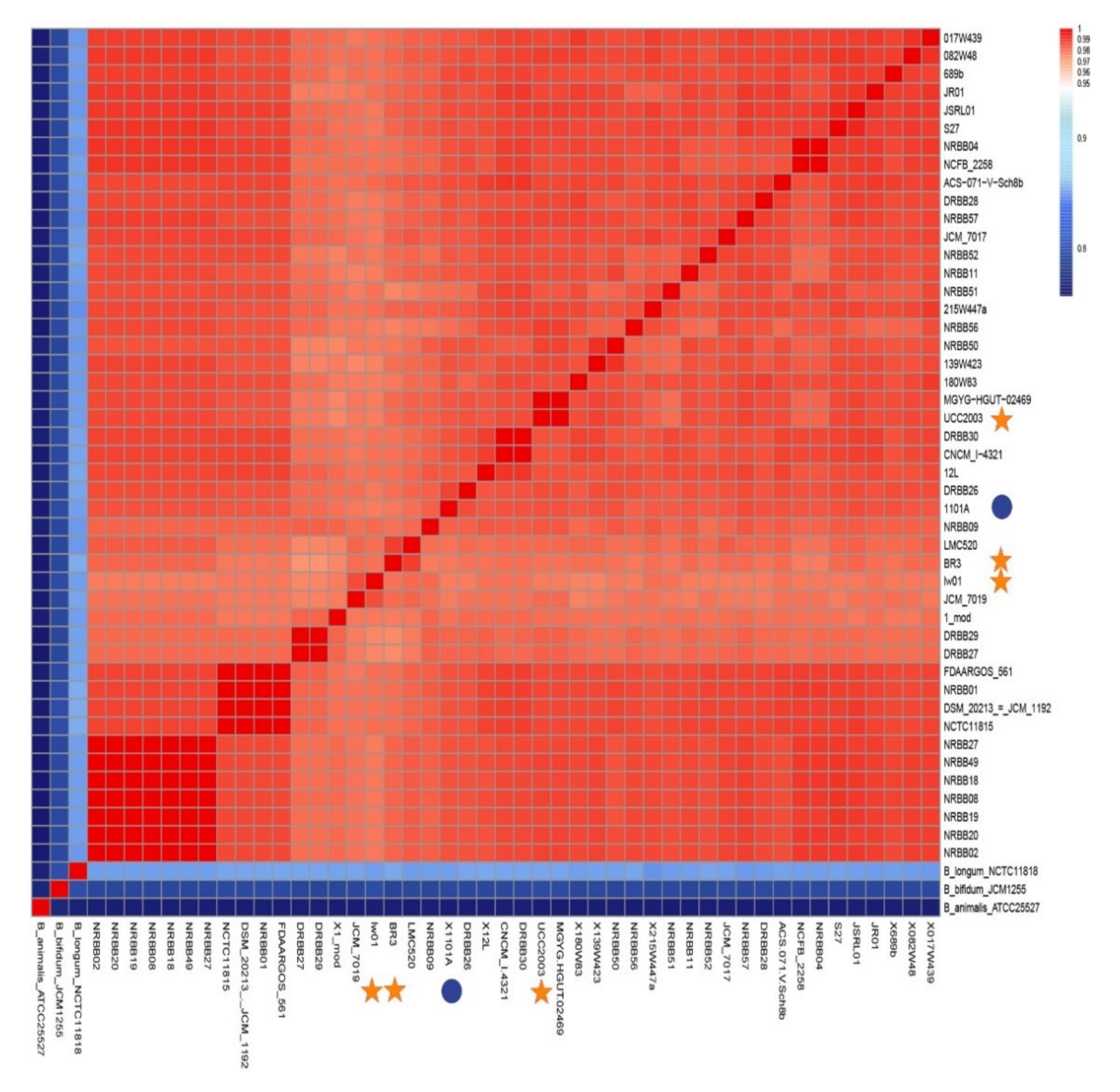
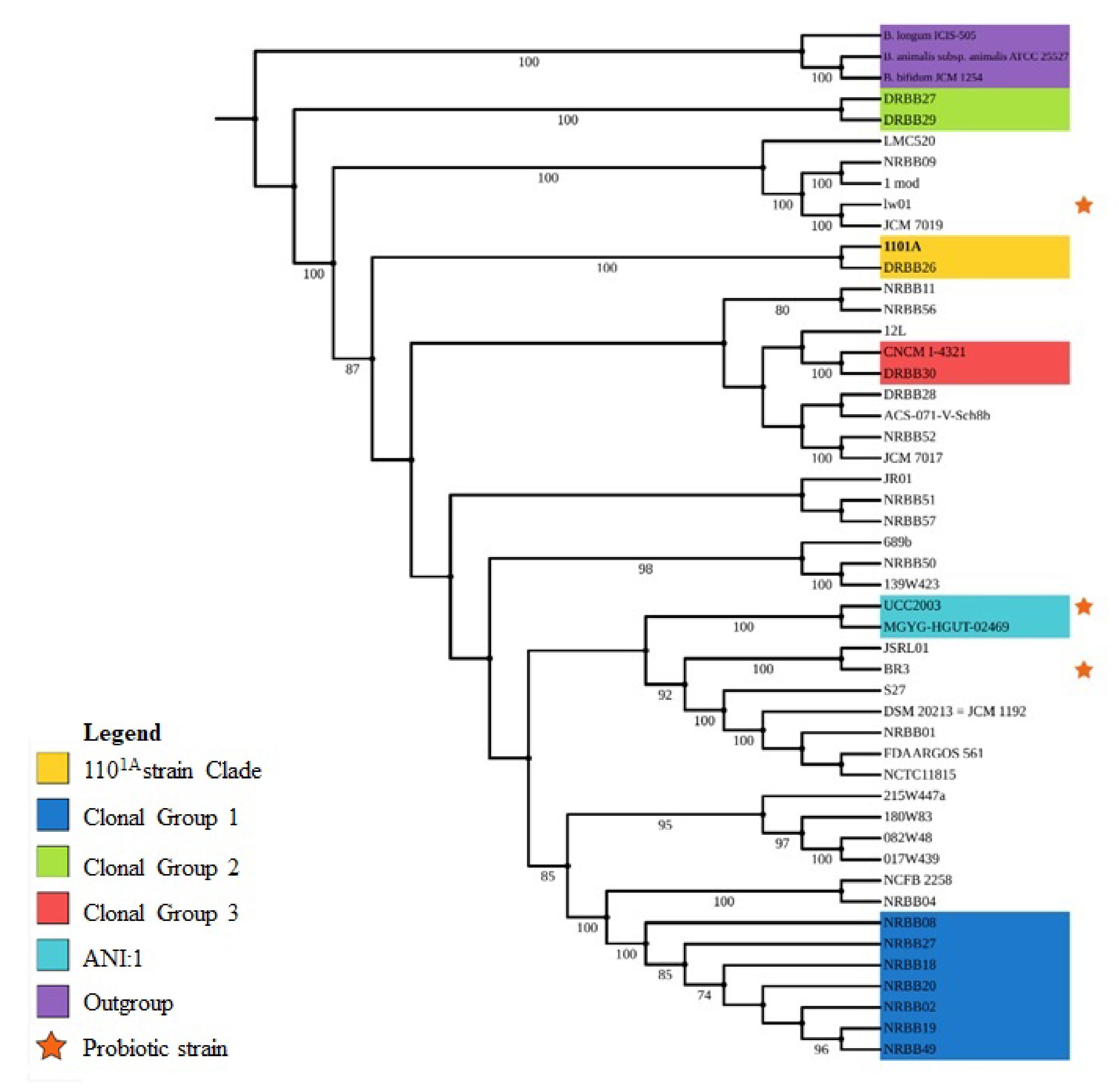
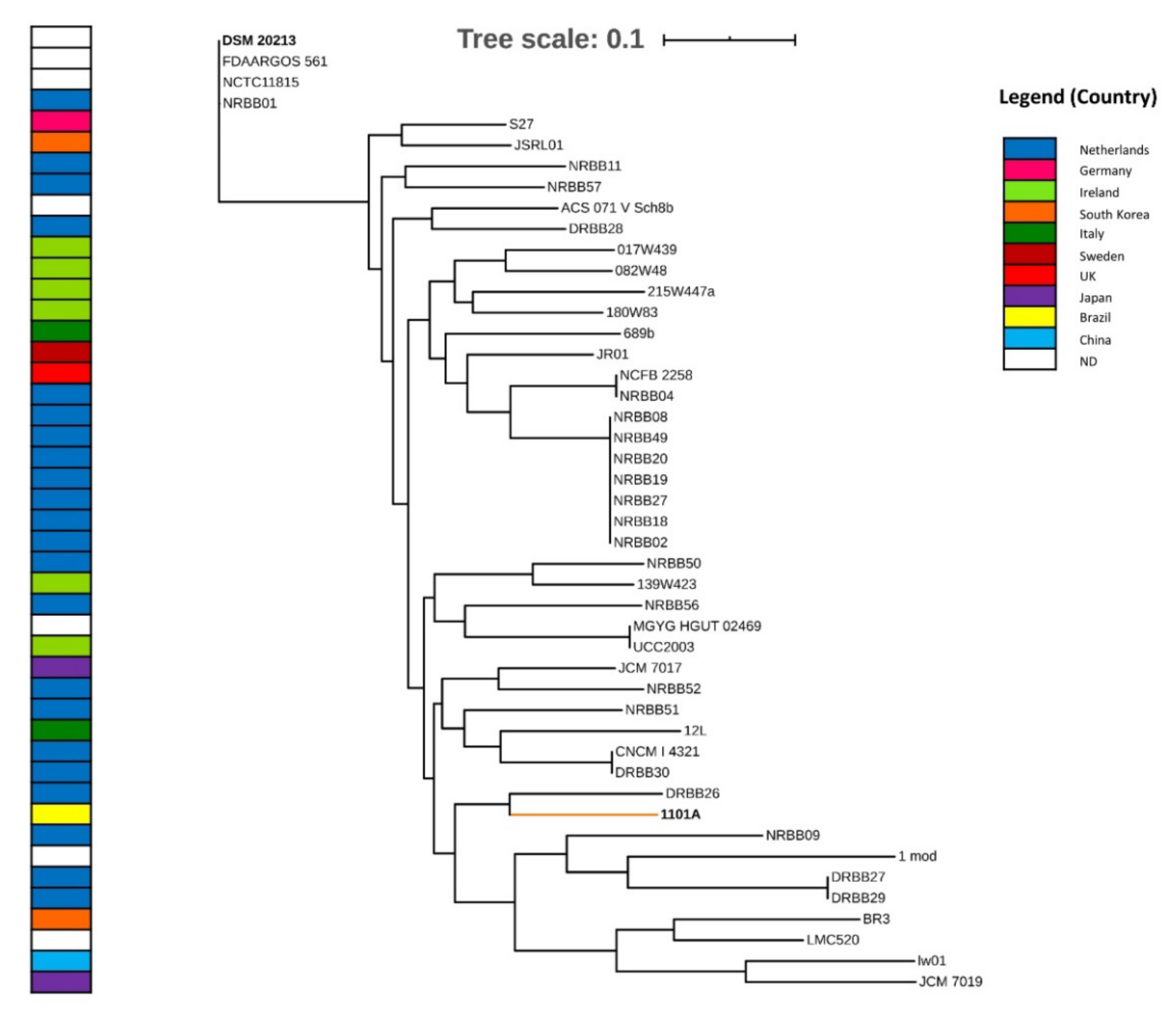
3.2. Taxonomy and Phylogenomics Analysis

Taxonomic characterization through similarity comparison using ANI values estimated for the 46 strains of B. breve is shown in Figure 1. All comparisons of B. breve 1101A with other B. breve strains showed ANI values between 0.97–0.98 grouped with these strains, confirming its high nucleotide identity with this species.
The phylogenomic tree based on 376 single-copy genes showed B. breve 1101A formed a strongly supported clade with B. breve DRBB26 (100), suggesting this strain closeness according to the ANI analysis (Figure 2). Experimentally, B. breve strains demonstrated as probiotics, such as UCC2003, lw01, and BR03, represented by stars, showed a relationship with other clades (Figure 2).

3.3. Prediction of Mobile Elements, Bacteriocins, Insertion Sequences, CRISPR-Cas Systems, and Antibiotic Resistance Genes in Bifidobacterium breve 1101A

The analysis performed with PlasmidFinder2.0 did not identify plasmids and bacteriocin-coding genes in the genome of B. breve 1101A. The PHASTER tool predictions has identified an incomplete prophage region of 8518 bp coding six phage-like and three hypothetical proteins (Supplementary Figure S2, Table 2). The screening of insertion sequences (IS) carried out in ISfinder revealed 29 elements, being seven of them complete ORFs. The IS were classified into six family groups (IS3, IS256, IS21, ISL3, IS30, and IS5) (Figure 3). Considering other genomic elements identified, three regions were related to Clustered Regularly Interspaced Short Palindromic Repeats (CRISPR) in the genome of B. breve 1101A (Table 3); however, there was a lack of arrangements codifying for Cas proteins. The detection of resistance genes to antibiotics revealed, in total, three genes: ileS, rpoB, and erm(X). The importance of coverage percentage for every hit was above 99.2% (Table 4).

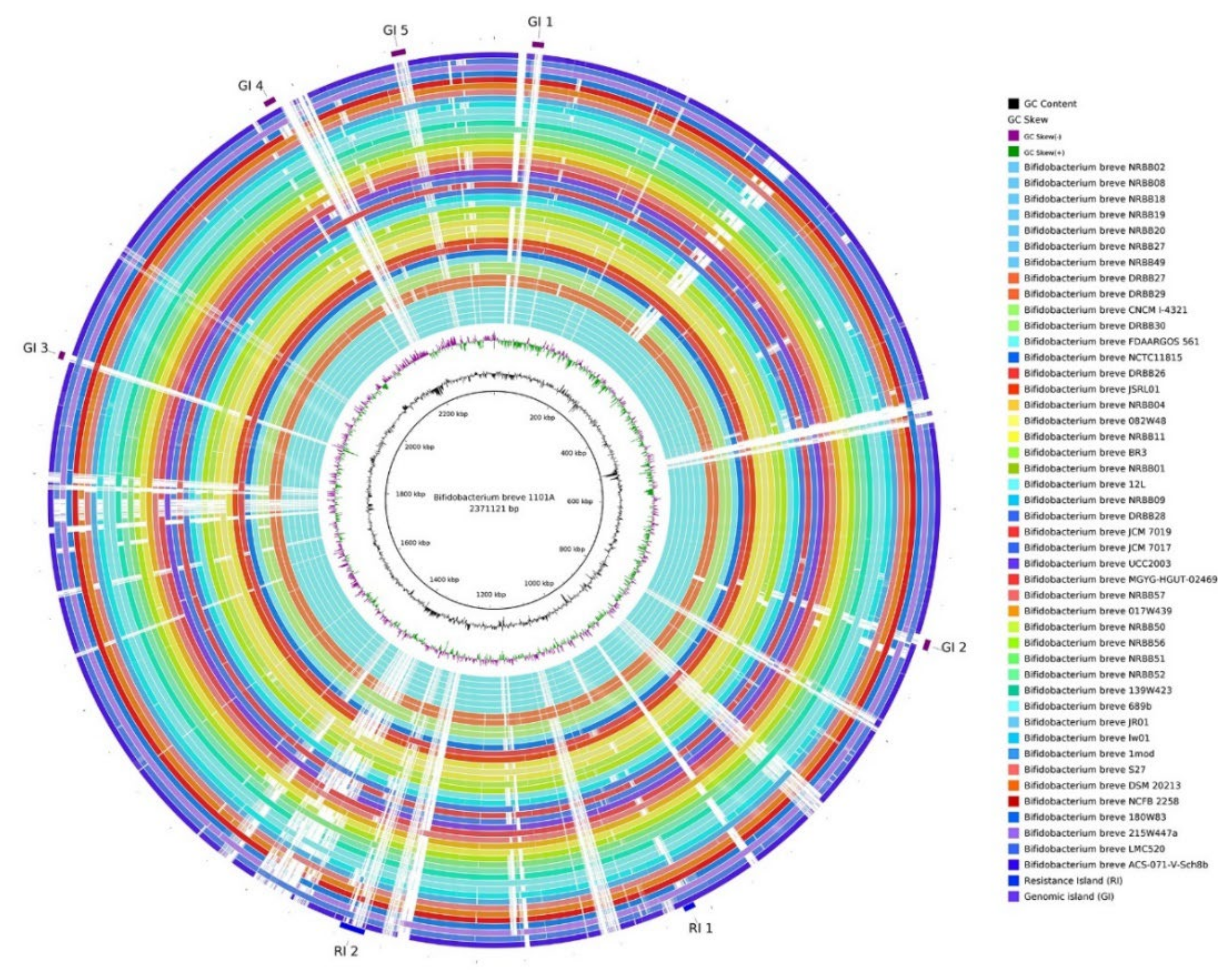
3.4. Genomic Plasticity Analysis

Seven genomic islands (GEI) were predicted in B. breve 1101A: two Resistance Islands (RI) and five Genomic Islands (GI) (Figure 4). Some genes in resistance islands (RI1, RI2) are related to antibiotic resistance (methyltransferase domain, bacterial regulatory proteins-tetR family, VanZ-like family) and metal ion binding (Cupin two conserved barrel domain protein). Moreover, genes involved in DNA binding and transcription regulation (two genes for RelB antitoxin), DNA binding (addiction module antidote protein HigA, PemK-like, MazF-like toxin of type II toxin-antitoxin system, and two gene helix-turn-helix domain), transferases (HipA-like C-terminal domain, FR47-like protein) and Major facilitator Superfamily (MFS) were identified (Supplementary Tables S1 and S2).
Gene content in Genomic Islands (GI1–GI5) were involved in processes such as transport proteins (ytfL, transporter associated domain, ABC transporter) and DNA binding to transcription regulation (transcriptional regulatory protein C terminal). Other genes involved in the active transport of solutes (four genes codifying for bacterial extracellular solute-binding protein, transmembrane transport), an integral membrane component (six codifying for binding-protein-dependent transport system inner membrane component, branched-chain amino acid transport system/permease component, and branched-chain amino acid ABC transporter, permease protein). Also, genes related to metabolism and transport of carbohydrates (glycosyl hydrolase family 36 N-terminal domain) with α-galactosidase activity, and others (FGGY kinase family, raffinose synthase or seed imbibition protein Sip1, ABC-type sugar transport system periplasmic component), uptake and translocation of the essential macronutrient phosphorus (phosphate transporter) (Supplementary Tables S3–S7).

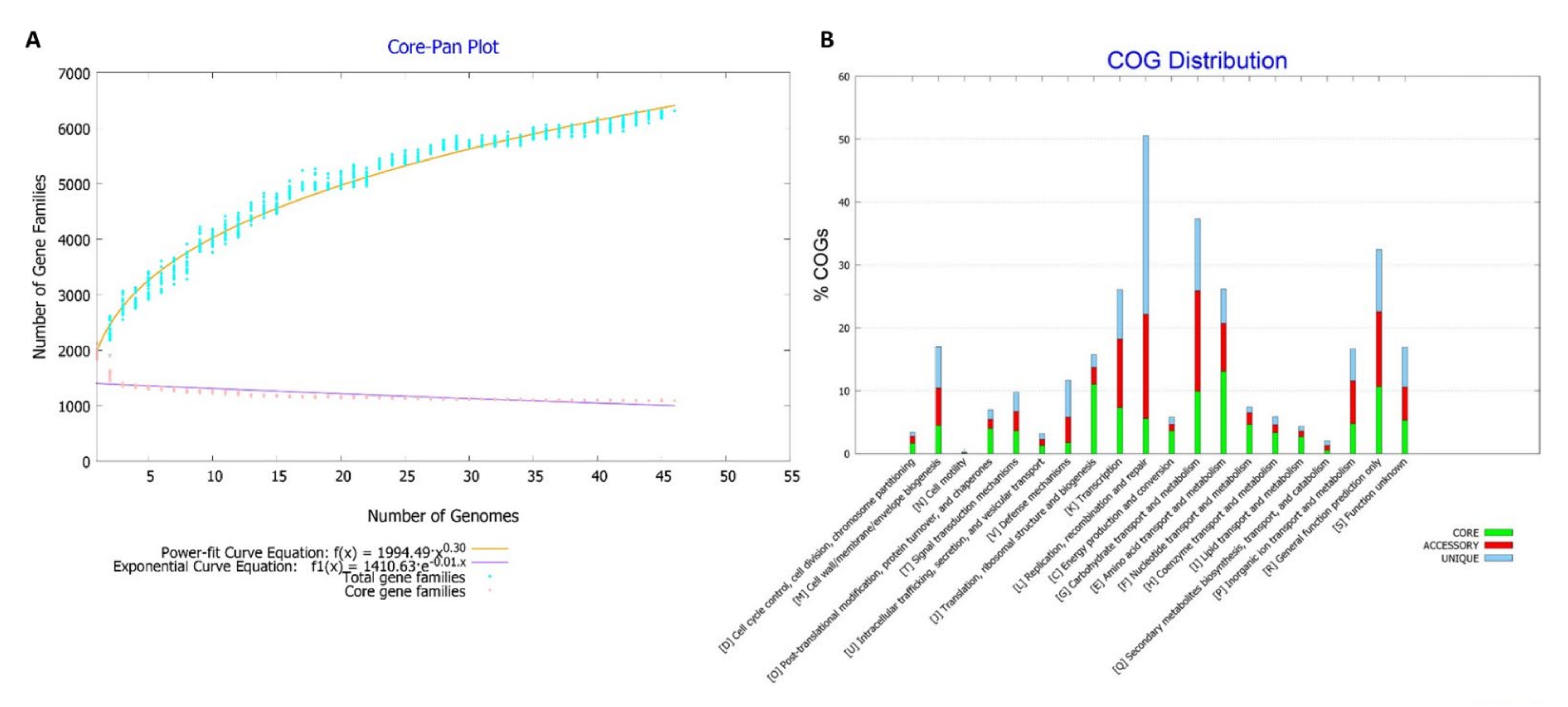
3.5. Pangenome Analysis

Pangenome size estimation identified 5943 genes in total based on their distribution in the 46 genomes. Core genes were 1174, soft-core genes were 163, shell genes were 1197, and cloud genes were 3409 (Supplementary Figure S3).
The pangenome was visualized in a matrix that shows a core and accessory genome (Supplementary Figure S4). Therefore, the estimation of the pangenome showed the exponential parameter (b = 0.3047) that suggested to be considered as an open pangenome, which has been increased in size with the addition of new genomes in the analysis (Figure 5A).
The distribution of functional classifications in the COG for the pangenome showed the prevalence of several categories, according to the organization of genes present in the core, accessory, and unique (Figure 5B).
The unique genes that were strain-specific in the pangenome analysis were in the range of 0–181 with a mean value of 34 genes per genome (Supplementary Figure S5). B. breve 1101A presented 63 unique genes associated with processes, such as transmembrane transport, membrane components, DNA processes, carbohydrate metabolism, among others (Supplementary Table S8). Thirty genes were uncharacterized protein (47%), of which ten genes had GO terms related to intermembrane integral components and related to DNA binding. There were five genes involved in carbohydrate metabolism (α-galactosidase, β-galactosidase, dTDP-4-dehydrorhamnose reductase, glycosidase) and one gene for transport of maltose (maltose ABC transporter permease). Regarding genes related to DNA process events, genes received GO terms of DNA integration, transposition, and DNA binding (IS3 family transposase, integrase, proteins containing domains for IS or integrase). In addition, other genes related only to DNA binding and regulation of transcription were predicted (putative transcriptional regulator XRE family, transcriptional regulator LacI family). Moreover, six genes associated with the cellular membrane were identified (ABC transporter substrate-binding protein, ABC transporter permease protein probably xylobiose porter, MFS transporter, putative membrane protein, histidine kinase, sortase family protein, LPXTG-motif cell wall anchor domain protein) with functions, according to GO terms, such as an integral component of membrane, membrane transport, plasma membrane.
About the phylogenomic tree based on SNP variants from the core genome, the B. breve 1101A formed a clade with DRBB26 from the Netherlands (Figure 6). Only in part, B. breve strains from Ireland were grouped based in SNP variants; therefore, there were no evident differences concerning the source of isolation (faecal and gut samples). However, there were few samples from other sources, such as 12L isolate from human milk which formed a clade with gut samples (CNCMI4321 and DRBB30); ACS071VSch8b isolate from the vagina, which formed a clade with a gut sample (DRBB28). The LMC520 isolate formed a clade with faecal samples (BR3, lw01, and JCM 7019). In contrast, the unique sample with clinical origin was FDAARGOS561, rooting the tree, and representatives from the intestine (NCTC11815) and gut (NRBB01) (Figure 6).

3.6. Identification of Genes Related to Probiotic Features

The analysis found 18 genes related to adhesion, some of them were sequences codifying for sortases, related as Tad-like protein (A, B, C, Z), TadE, and TadF. Two sequences were codifying for secretion proteins, such as LPXTG-type cell surface and only one sequence for luxS or autoinducer-2 (AI-2) (Supplementary Table S9).
Furthermore, it was identified that 39 CDS were involved in stress resistance, among them, multiple subunits of F0F1-type ATPase (a, b, alpha, beta, delta, epsilon, and gamma). In addition to this, some genes codifying for chaperons and SOS response genes (Supplementary Table S10).
Concerning genes associated with repairing and protection, genes were identified with ten sequences codifying for nucleoside triphosphate pyrophosphohydrolase (mutT) and DNA-binding ferritin-like protein (dps) methionine sulfoxide reductase (msr). Moreover, other sequences were found codifying for copper-transporting ATPase (copA), such as subunit A of the exonuclease ATP-binding cassette ABC complex (uvrA) (Supplementary Table S11). Additionally, there were 11 genes involved in the production of vitamins, such as B2, B9, and B12 (Supplementary Table S12).

4. Discussion

4.1. Evolutionary Relationships from Phylogenomic Analysis

The B. breve strain 1101A taxonomy was confirmed based on the orthologous multiple loci phylogenomic analysis showing a close relationship to other B. breve strains. Moreover, our results revealed DRBB26, isolated in the Netherlands from infant faeces as well (Table 1), to be the most closely related strain from 1101A. Interestingly, the core genome SNP analysis also supported this finding, although no correlation was evident between the strain clusters and the countries of origin. It is important to notice that some of the B. breve clades observed in the multilocus approach were in contrast to previous phylogenomic analysis [51]. Although some clades remain as in the previous study, such as the clonal groups 1, 2, and 3 (Figure 2), the DRBB26 strain formed a different clade with B. breve NRBB01 in contrast to our study. In this context, B. breve 1101A phylogenetic group could be replaced. As was shown in phylogenomic results, the closer relationship of B. breve strains, B. breve fell into the B. longum clade based on orthologous genes and a multilocus approach [52,53].
In addition, only three strains with complete genomes were confirmed to present probiotic properties in previous experimental studies, UCC2003 [54], lw01 [55,56], and BR3 [57,58]. Therefore, the phylogenetic analysis may not provide further insights regarding the probiotic profile of B. breve 1101A as these strains do not show a close relationship with its clade. However, our study suggests that JCM 7019, MGYG-HGUT-02469, and JSRL01 strains possibly present beneficial properties due to their proximity with the probiotic strains, although experimental studies should be performed as well.
Genomic rearrangements are a very important feature regarding the safety of probiotic strains as it might be useful to detect genetic plasticity and instability. Genetic alterations associated with the growth of bifidobacteria, such as small deletions throughout the genome, make them able to adapt to extreme conditions, such as in the gastrointestinal tract or during a fermentation process [59]. The Bifidobacterium genus has been reported to present a highly conservative level of synteny [60], despite there being a degree of diversity related to each species. As expected, our study has shown the synteny of gene blocks was kept in most of the evaluated genomes, including the B. breve 1101A, indicated by the locally collinear blocks (LCBs) on multiple alignment analysis. Nevertheless, three genomes presented a different genome organization, such as inversions on regions of different sizes. Two of these genomes (ACS-071-V-Sch8b and JCM7017) were reported previously in a comparative study of this species [52], suggesting that Bifidobacterium breve exhibits genomic regions which are affected by events of inversions, insertions, and deletions [60]. For example, urea metabolism genes and the way they utilize carbon from different sources (such as milk and plants) contribute to the classification of these subspecies of B. longum [61]. Prediction of mobile elements, bacteriocins, and CRISPR-Cas systems in Bifidobacterium breve 1101A.
Regarding mobile elements, it is known that plasmids provide new characteristics to probiotic bacteria that increases the possibility to survive in other environmental conditions, acquire additional properties in bacterial metabolism, adherence, and antibiotic resistance, being the last one, is an undesired attribute for a probiotic bacterium [14]. Although plasmids were not detected in B. breve 1101A, they were previously reported in the genus Bifidobacterium [62]. Regarding B. breve, plasmids were detected in 40% of 42 genomes analyzed [63]; in contrast, they were absent in 106 evaluated isolates of this species in another study [64]. A reduced number of plasmids has been reported for B. breve strains with complete genomes, particularly only the strains, NCFB 2258 [65] and BR3 [57].
Concerning the predicted prophage in the genome of B. breve 1101A, the region was classified as incomplete and did not contain typical genes that codify for structural proteins (such as the tail, capsid), DNA regulation, integrases, lysis, and others for a functional bacteriophage. Therefore, this prophage region could be considered as a defective prophage. It presented some genes involved in response to oxidative stress (Fe-S cluster assembly ATPase SufC, cysteine desulfurase, SUF system NifU family Fe-S cluster assembly protein), biosynthesis of glycogen (glucose-1-phosphate adenylyltransferase), among other processes (RNA methyltransferase, histidine triad domain protein, PhoH family protein). Prophages could represent a future lysis event for the bacteria, or they could provide some additional properties as a double-edged sword [66]. The frequency of integrated prophages is standard in bacterial genomes [67]. A considerable part of them is defective, possibly due to the phage domestication event [68]. Previous studies have reported the mentioned IS families in the Bifidobacterium genus [52,69]. For example, IS3 was referred to as the IS family with the most widespread and significant number being identified in Bifidobacterium [69]. Contrastingly, the study of the mobilome of B. breve showed the IS30 as the most frequent IS family [52].
Genes codifying for bacteriocins were not identified in the analyzed B. breve 1101A. These molecules allow the competence against other bacteria, being specific for an intra-genus or less clear for inter-genus strains, in the gut environment; it is a desirable feature for probiotic bacteria. Some studies have reported some produced bacteriocins in the genus Bifidobacterium [9,70]. However, in other studies, the production of these compounds in Bifidobacterium was relatively rare [71] or not reported in gut samples [72]. Although any gene for bacteriocin was not predicted, the antagonism showed in in vitro [24] could be due to other mechanisms of the strain that were evident in probiotic bacteria, such as adhesive capability, which allows for the inhibition of pathogen colonization [73] or the reduction of pH in the environment with the glucose fermentation and production of short-chain fat chain (SCFA) seen in bifidobacteria [74,75].
At the same time, CRISPR regions were identified. These components are essential for the bacteria to deal with phage sequences from the environment, especially in the gut tract where there is a viral community. Phages can lyse bacteria, and it can drastically affect the survival of the bacterial population in the production of probiotics, fermentation time, taste, among other parameters [76]. Considering that phages are resistant to the pasteurization process and its elimination is difficult, the search of probiotics with the ability to be protected from phages and other DNA invaders, such as plasmids [16]. Our results identified three CRISPR loci in the B. breve 1101A genome. These CRISPR regions were considered not functional because there are no Cas proteins; it was impossible to determine the type and subtype of the systems due to the necessary presence of Cas proteins for its classification. In a previous study, the occurrence of CRISPR-Cas systems in the genus Bifidobacterium was 77% of the 48 analyzed species, representing a high presence in the genus [77].

4.2. Assessing the Risk of Antibiotic Resistance Genes in Probiotic Bacteria

The antibiotic resistance genes in the B. breve 1101A were ileS, rpoB, and erm(X). The ileS confers resistance to mupirocin [78] and rpoB gene confers resistance to rifampicin [79]. Both genes were detected in the other 45 genomes of B. breve to extend this predictive analysis. These results were supported by other studies, where resistance to mupirocin is considered as intrinsic resistance in bifidobacteria [78,80]. Using different resistance databases, the erm(X) gene was identified in B. breve 1101A. This gene can enzymatically modify the DNA sequence of the 23S rRNA gene by methylation to avoid the binding of macrolides, lincosamides, streptogramin B, and conferring resistance [81].
The presence of homologous erm gene was reported in B. longum and some strains of B. breve (BR-14 and DPC6330), and it was suggested the acquisition by HGT [82]. More specifically, erm(X) was reported in B. thermophilum and B. animalis subsp. lactis strains from pigs, founded in the Tn5432-like transposon, and similar transposon found in the pathogenic bacteria, such as Corynebacterium [83]. This antibiotic resistance gene was found in two strains of B. breve in another study [84]: NRBB51 (three copies) and DRBB26 (two copies) interleaved by genes codifying transposases. In the present study, only a transposase IS1249 (IS256 family) was detected in the vicinity of erm(X), and both within the RI2 could suggest that the island was acquired by HGT. Contrasting to these in silico results with the experimental essays of antibiotic susceptibility in B. breve 1101A [24], this suggests that the identified erm(X), in this study, could be the responsible gene for the showed resistance to erythromycin. The species B. breve is currently considered with QPS status, Quality Presumption of Safety [85], due to not being related to any infective process. Although the presence of genes of resistance is not considered a safety issue per se, it is necessary to determine the possible transference of this resistance [79].

4.3. Genome Plasticity Analysis

The analysis of genomic islands (GEIs) prediction in B. breve 1101A revealed seven islands (Figure 4, Tables S1 and S2). Gene content GEI1-GEI5 islands were concerned with transport proteins (ytfL, transporter associated domain, ABC transporter), membrane components, DNA binding (two genes helix-turn-helix lactose operon repressor), C-terminal transcriptional regulatory protein (two genes), and the metabolism of carbohydrates, such as genes related to galactose and raffinose that could contribute to these other functions of the bacteria.
Other genes involved in the active transport of solutes (four codifying for bacterial extracellular solute-binding protein, transmembrane transport), are an integral component of the membrane (six codifying for binding-protein-dependent transport system inner membrane component, branched-chain amino acid transport system/permease component and branched-chain amino acid ABC transporter, permease protein). In addition, genes related to metabolism and the transport of carbohydrates (glycosyl hydrolase family 36 N-terminal domain) with α-galactosidase activity, and others (FGGY kinase family, raffinose synthase or seed imbibition protein Sip1, ABC-type sugar transport system periplasmic component), uptake and translocation of the essential macronutrient phosphorus (phosphate transporter). Among other processes (lysR substrate binding domain, DNA-binding transcription factor 2, sugar-phosphate isomerase involved in capsule formation) (Tables S3–S7).

4.4. Pangenome Analysis

In a previous study, an analysis with MCL to perform a pangenome analysis with B. breve, the estimation of pangenome size was 3667 gene families, and the core genome was composed of 1307 gene families using 13 genomes (including 8 complete and 5 draft genomes) [52]. In another study, the pangenome size estimation was 6138 gene families, and the core genome size was 1282 gene families, using 73 genomes with 37 complete and 36 drafts [51]. In a recent study, the species’ pangenome was evaluated with 55 genomes, with 46 drafts originally from China that were composed of 6707 gene families and the core genome composed of 1111 gene families [86]. Considering our results with 46 complete genomes of B. breve, with a pangenome size of 5943 genes and a core genome size of 1174, we are closer to the second study [84], even with the considerable difference in the number of used genomes. In this respect, genome assembly and annotation are two main factors determining the performance of a pangenome analysis and an adequate number of complete genomes [87]. Furthermore, it is essential to mention that draft genomes are helpful in some studies; however, due to the underrepresentation of some genes, it could affect comparative studies because of low-read coverage or assembly errors [88].
Concerning the result of the matrix representation of the pangenome, the similarity of a group composed of: (i) NRBB02, NRBB8, NRBB18, NRBB19, NRBB20, NRBB27, and NRBB49; (ii) DSM 20213, NCTC11815, NRBB01, and FDAARGOS561; and (iii) CNCM I4321 and DRBB30 by the ANI values = 0.9999 in each group. Moreover, the first two groups were reported as clonal strains [84]. Considering the estimation of the curve parameters of the pangenome suggested, it as an open pangenome because it tends to continue to increase in size with the addition of new genomes in the simulations (Figure 5). Our results agree with a previous study with thirteen genomes that considered the pangenome as opened [52] and the seventy-three genomes as opened [51] of the B. breve pangenome. However, the last reference [86] considered the pangenome as not entirely closed but gradually saturated. The distribution of COG showed a high percentage of genes related to replication, recombination, and repair processes concentrated in the core and accessory genome. In the second place, many genes are involved in carbohydrate transport, and the metabolism in the core and accessory genome. The COG items were distributed according to basic (core genome) and adaptive processes (accessory genome).
According to the phylogenomic tree based on SNPs of the core genome, there was no marked distribution of the strains according to geographical distribution and source of isolation. Referring to B. breve 1101A, the closest strain was DRBB26 isolated from the gut in the Netherlands and without additional information. A comparative perspective and the pangenome analysis allow for its segmentation and the identification of core, accessory, and unique genes [89]. This last part is relevant to know the strain-specific genetic repertoire that could represent adaptive features to different niches, metabolic advantages, or even genes related to pathogenicity. The comparative approach has been previously used in industrial starter cultures [21]. The results about mean value of unique genes per genome, in this study, was of 34 genes considering 46 complete genomes; contrasting with a previous study for the species of 53 unique genes per genome using eight complete genomes [52]. From the pool of unique genes, 47% were considered uncharacterized proteins in this study that coincide with the predominant presence of genes with this label in a previous B. breve genomic comparison [52]. More generally, genome projects identified hypothetical proteins that have a range of 30–40% of total identified proteins in prokaryotes [90] that partially interfere in the characterization of the strain. Moreover, mobile elements were present in minor frequency [52], represented by transposases and integrases. This could be related to the unique genes and the acquisition by HGT. Therefore, unique genes were involved in the metabolism and transport of carbohydrates that could represent additional features that allow the utilization of a wide variety of sources of energy. Especially, galactosidase and β -galactosidase are enzymes of interest because they are used commercially to improve symptoms of lactose intolerance and use this carbon source [91]. These enzymes were identified in bifidobacterial, and probiotics were used to alleviate lactose intolerance [92,93]. Other genes codified for the sortase family protein and LPXTG-motif cell wall anchor domain protein, which were involved in cellular adhesion, represent a desirable characteristic in potential candidates for probiotic bacteria in the gut colonization stage [17]. Moreover, an MFS transporter was also predicted in the unique gene group, and its overexpression was reported under acid bile exposition [94].

4.5. Identification of Genes Related to Probiotic Features in B. breve 1101A

Some of these genes were related to adhesion mechanisms, among them: sortases (srtA1, srtA2, and srtA3) LPXTG-type proteins that probably have a role in the attachment to cells or mucus in the gut [49]. Another identified protein was Tad (ABCEFZ), essential for the pili structure necessary for bacterial colonization [95]. Therefore, luxS has been involved with acid tolerance and adherence to intestinal epidermal cells in Lactobacillus plantarum [96]. Acid and bile resistance is common in Lactobacillus strains, as in the work of Chou and Weimer, causing the bacteria to survive and adapt to stressful conditions, being potentially probiotic targets [97].
Some genes related to stress resistance, such as F0F1-type ATPase, work as proton pumps in Gram-positive bacteria. These transcription activities were evidenced in acid conditions in Bifidobacterium species [98,99,100]. Genes also identified (dnaK, groEL, groES) in codifying for chaperones are involved in a general response to stress by protection, removal of damaged proteins, and other related functions. Moreover, dnaJ and grpE, identified in the analysis, have shown response (upregulation) to acid environments [101]. Some other genes (lexA, ruvA, recA, and mutY) are involved in SOS response. The lexA has shown to be induced under temperature stress in B. breve, and recA showed an upregulated response in heat stress [102]. Moreover, reduced survival in L. reuteri was demonstrated in mutation in clp, in exposure to bile [103], indicating its importance for survival in conditions of acidity and general stress. In addition, it was identified that 10 CDS with functions were related to the repair and protection of DNA or proteins, such as DNA-binding ferritin-like protein and nucleoside triphosphate pyrophosphohydrolase (mutT) that act in the reparation due to the oxidative damage removing hydroxyl radicals [49]. Other identified genes, such as mutants of methionine sulfoxide reductase, have shown a reduced capacity in stress conditions in L. reuteri 100-23 [104], and mutants of copper-transporting ATPase (copA) in L. plantarum WCFS1 have shown a reduced competitive ability in mouse [105]. Therefore, copA is involved in the nucleotide reparation in acid conditions in L. helveticus CNBL 1156 [106]. Furthermore, there were 11 genes involved in the biosynthesis of vitamins. Some of them form parts of operons for specific vitamins. The integrity of the operons for vitamins is of interest for considering the functionality of its production by the B. breve 1101A.

5. Conclusions

Probiotics are known for their beneficial health properties and there is an increasing tendency to look for and select new candidates for human usage. The present study applied a comparative genomic approach to explore the features of the first complete genome of B. breve from Brazil, strain 1101A, isolated from healthy children’s faeces. The analysis of this strain showed positive characteristics to be considered as a candidate based on the identified genes related to probiotic properties (adhesion, resistance to stress for acidity and heat, and production of vitamins) that suggest good survival ability in the gastrointestinal tract. Furthermore, some unique genes for B. breve 1101A are involved in the adhesion and metabolism of some carbohydrates representing the desired condition for bacteria to consume different sources of energy from the gut niche. Moreover, the antagonism against pathogens observed in previous in vitro studies seems be promoted by other factors (i.e., competence by adherence, reduction of pH) due to the absence of bacteriocin genes. On the other hand, considering the safety criteria, a crucial point for further elucidation is whether the transference of the antibiotic resistance erm(x) gene in exploratory essays is possible. Moreover, the evaluation of unique genes with unknown functions and possible pathways should be explored. In this context, in vitro and in vivo studies with B. breve 1101A should provide supportive evidence.

Supplementary Materials

The following supporting information can be downloaded at: https://www.mdpi.com/article/10.3390/bacteria1030013/s1, Figure S1: Representation of multiple sequence alignments of whole genomes of 46 Bifidobacterium breve strains shows conserved synteny and collinearity among gene blocks; Figure S2: Representation of the incomplete prophage region in Bifidobacterium breve 1101A. The prophage showed 9 CDS: 6 CDS in dark turquoise identified as phage-like protein and 3 CDS in light green as a hypothetical protein; Figure S3: Pie chart representing the number of genes shared in the core, softcore, shell and cloud genome for the 46 Bifidobacterium breve; Figure S4: Matrix representation of the pangenome based on the presence-absence of genes of the pan-genome; Figure S5: Plot diagram showing the core-genome size of all 46 genomes of Bifidobacterium breve (central area) and unique genes for each strain; Table S1: Functional annotation of genes in RI1 of B. breve 1101A; Table S2: Functional annotation of genes in RI2 of B. breve 1101A; Table S3: Functional annotation of genes in GI1 of B. breve 1101A; Table S4: Functional annotation of genes in GI2 of B. breve 1101A; Table S5: Functional annotation of genes in GI3 of B. breve 1101A; Table S6: Functional annotation of genes in GI4 of B. breve 1101A; Table S7: Functional annotation of genes in GI5 of B. breve 1101A; Table S8: Functional annotation information about the 63 unique genes of B. breve 1101A; Table S9: Identified genes potentially involved in adhesion mechanisms in B. breve 1101A; Table S10: Identified genes potentially involved in resistance mechanisms to stress in B. breve 1101A; Table S11: Identified genes potentially involved in repair and protection of DNA and protein mechanisms in B. breve 1101A; Table S12: Identified genes potentially involved in the biosynthesis of vitamins in B. breve 1101A.

Author Contributions

J.V.-B., R.P., A.L.d.S. and T.d.J.S.: conducted in silico analyses, review, and editing. A.C.P.G., M.V.C.V., F.M.R.d.C., R.B.K., M.F.A., A.d.S.F., R.D.d.O.C., B.B., F.S.M. and F.A.: data curation, review and editing. F.A. and V.A.: conceptualization, supervision, review and editing. All authors have read and agreed to the published version of the manuscript.

Funding

This research received no external funding.

Institutional Review Board Statement

Not applicable.

Informed Consent Statement

Not applicable.

Data Availability Statement

Genomes sequences are available in NCBI database.

Acknowledgments

We thank the agencies Coordination for the Improvement of Higher Education Personnel (CAPES) and Minas Gerais Research Funding Foundation (FAPEMIG). Our thanks to the Post Graduate Program in Bioinformatics at the Federal University of Minas Gerais and the Omics Science Network (RECOM).

Conflicts of Interest

The authors declare no conflict of interest.

References

  1. Klijn, A.; Mercenier, A.; Arigoni, F. Lessons from the genomes of bifidobacteria. FEMS Microbiol. Rev. 2005, 29, 491–509. [Google Scholar] [CrossRef] [PubMed]
  2. Lee, J.-H.; Li, X.; O’Sullivan, D.J. Transcription Analysis of a Lantibiotic Gene Cluster from Bifidobacterium longum DJO10A. Appl. Environ. Microbiol. 2011, 77, 5879–5887. [Google Scholar] [CrossRef] [PubMed] [Green Version]
  3. O’Callaghan, A.; Van Sinderen, D. Bifidobacteria and Their Role as Members of the Human Gut Microbiota. Front. Microbiol. 2016, 7, 925. [Google Scholar] [CrossRef] [PubMed] [Green Version]
  4. Turroni, F.; Peano, C.; Pass, D.A.; Foroni, E.; Severgnini, M.; Claesson, M.J.; Kerr, C.; Hourihane, J.; Murray, D.; Fuligni, F.; et al. Diversity of Bifidobacteria within the Infant Gut Microbiota. PLoS ONE 2012, 7, e36957. [Google Scholar] [CrossRef] [PubMed] [Green Version]
  5. Cionci, N.B.; Baffoni, L.; Gaggìa, F.; Di Gioia, D. Therapeutic Microbiology: The Role of Bifidobacterium breve as Food Supplement for the Prevention/Treatment of Paediatric Diseases. Nutrients 2018, 10, 1723. [Google Scholar] [CrossRef] [Green Version]
  6. FAO. Health and Nutritional Properties of Probiotics in Food Including Powder Milk with Live Lactic Acid Bacteria; Food and Agricultural Organization of the United Nations; World Health Organization: Rome, Italy, 2001. [Google Scholar]
  7. Foligne, B.; Daniel, C.; Pot, B. Probiotics from research to market: The possibilities, risks and challenges. Curr. Opin. Microbiol. 2013, 16, 284–292. [Google Scholar] [CrossRef]
  8. Ruiz, L.; Margolles, A.; Sánchez, B. Bile resistance mechanisms in Lactobacillus and Bifidobacterium. Front. Microbiol. 2013, 4, 396. [Google Scholar] [CrossRef] [Green Version]
  9. Sarkar, A.; Mandal, S. Bifidobacteria—Insight into clinical outcomes and mechanisms of its probiotic action. Microbiol. Res. 2016, 192, 159–171. [Google Scholar] [CrossRef]
  10. Ohland, C.L.; Macnaughton, W.K. Probiotic bacteria and intestinal epithelial barrier function. Am. J. Physiol. Liver Physiol. 2010, 298, G807–G819. [Google Scholar] [CrossRef] [Green Version]
  11. Bermudez-Brito, M.; Plaza-Díaz, J.; Muñoz-Quezada, S.; Gómez-Llorente, C.; Gil, A. Probiotic Mechanisms of Action. Ann. Nutr. Metab. 2012, 61, 160–174. [Google Scholar] [CrossRef]
  12. Azad, M.A.K.; Sarker, M.; Li, T.; Yin, J. Probiotic Species in the Modulation of Gut Microbiota: An Overview. BioMed Res. Int. 2018, 2018, 9478630. [Google Scholar] [CrossRef] [PubMed] [Green Version]
  13. De Jesus, L.C.L.; Drumond, M.M.; Aburjaile, F.F.; de Sousa, T.J.; Coelho-Rocha, N.D.; Profeta, R.; Brenig, B.; Mancha-Agresti, P.; Azevedo, V. Probiogenomics of Lactobacillus delbrueckii subsp. lactis CIDCA 133: In Silico, In Vitro, and In Vivo Approaches. Microorganisms 2021, 9, 829. [Google Scholar] [CrossRef] [PubMed]
  14. Abriouel, H.; Montoro, B.P.; de la Fuente Ordoñez, J.J.; Lerma, M.L.; Knapp, C.W.; Benomar, N. New insights into the role of plasmids from probiotic Lactobacillus pentosus MP-10 in Aloreña table olive brine fermentation. Sci. Rep. 2019, 9, 10938. [Google Scholar] [CrossRef] [PubMed] [Green Version]
  15. Bennedsen, M.; Stuer-Lauridsen, B.; Danielsen, M.; Johansen, E. Screening for Antimicrobial Resistance Genes and Virulence Factors via Genome Sequencing. Appl. Environ. Microbiol. 2011, 77, 2785–2787. [Google Scholar] [CrossRef] [PubMed] [Green Version]
  16. Hidalgo-Cantabrana, C.; Crawley, A.; Sanchez, B.; Barrangou, R. Characterization and Exploitation of CRISPR Loci in Bifidobacterium longum. Front. Microbiol. 2017, 8, 1851. [Google Scholar] [CrossRef] [PubMed] [Green Version]
  17. Alayande, K.A.; Aiyegoro, O.A.; Nengwekhulu, T.M.; Katata-Seru, L.; Ateba, C.N. Integrated genome-based probiotic relevance and safety evaluation of Lactobacillus reuteri PNW1. PLoS ONE 2020, 15, e0235873. [Google Scholar] [CrossRef]
  18. Pereira, J.Q.; Ritter, A.C.; Cibulski, S.; Brandelli, A. Functional genome annotation depicts probiotic properties of Bacillus velezensis FTC01. Gene 2019, 713, 143971. [Google Scholar] [CrossRef]
  19. Kapse, N.; Engineer, A.; Gowdaman, V.; Wagh, S.; Dhakephalkar, P. Functional annotation of the genome unravels probiotic potential of Bacillus coagulans HS243. Genomics 2019, 111, 921–929. [Google Scholar] [CrossRef]
  20. Debmalya Barh, D.; Soares, S.; Tiwari, S.; Azevedo, V. Pan-Genomics: Applications, Challenges, and Future Prospects; Elsevier: Amsterdam, The Netherlands, 2020; ISBN 978-0-12-817076-2. [Google Scholar]
  21. Garrigues, C.; Johansen, E.; Crittenden, R. Pangenomics—An avenue to improved industrial starter cultures and probiotics. Curr. Opin. Biotechnol. 2013, 24, 187–191. [Google Scholar] [CrossRef]
  22. Zhang, W.; Wang, J.; Zhang, D.; Liu, H.; Wang, S.; Wang, Y.; Ji, H. Complete Genome Sequencing and Comparative Genome Characterization of Lactobacillus johnsonii ZLJ010, a Potential Probiotic With Health-Promoting Properties. Front. Genet. 2019, 10, 812. [Google Scholar] [CrossRef] [Green Version]
  23. Fontana, A.; Falasconi, I.; Molinari, P.; Treu, L.; Basile, A.; Vezzi, A.; Campanaro, S.; Morelli, L. Genomic Comparison of Lactobacillus helveticusstrains Highlights Probiotic Potential. Front. Microbiol. 2019, 10, 1380. [Google Scholar] [CrossRef] [Green Version]
  24. Souza, T.; Silva, A.; Drews, J.; Gomes, D.; Vinderola, C.; Nicoli, J. In vitro evaluation of Bifidobacterium strains of human origin for potential use in probiotic functional foods. Benef. Microbes 2013, 4, 179–186. [Google Scholar] [CrossRef]
  25. De, S.; Kaur, G.; Roy, A.; Dogra, G.; Kaushik, R.; Yadav, P.; Singh, R.; Datta, T.K.; Goswami, S.L. A Simple Method for the Efficient Isolation of Genomic DNA from Lactobacilli Isolated from Traditional Indian Fermented Milk (dahi). Indian J. Microbiol. 2010, 50, 412–418. [Google Scholar] [CrossRef] [Green Version]
  26. Bankevich, A.; Nurk, S.; Antipov, D.; Gurevich, A.A.; Dvorkin, M.; Kulikov, A.S.; Lesin, V.M.; Nikolenko, S.I.; Pham, S.; Prjibelski, A.D.; et al. SPAdes: A new genome assembly algorithm and its applications to single-cell sequencing. J. Comput. Biol. 2012, 19, 455–477. [Google Scholar] [CrossRef] [Green Version]
  27. Galardini, M.; Biondi, E.G.; Bazzicalupo, M.; Mengoni, A. CONTIGuator: A bacterial genomes finishing tool for structural insights on draft genomes. Source Code Biol. Med. 2011, 6, 11. [Google Scholar] [CrossRef] [Green Version]
  28. Piro, V.C.; Faoro, H.; Weiss, V.A.; Steffens, M.B.; Pedrosa, F.O.; Souza, E.M.; Raittz, R.T. FGAP: An automated gap closing tool. BMC Res. Notes 2014, 7, 371. [Google Scholar] [CrossRef] [Green Version]
  29. Seemann, T. Prokka: Rapid Prokaryotic Genome Annotation. Bioinformatics 2014, 30, 2068–2069. [Google Scholar] [CrossRef]
  30. Darling, A.E.; Mau, B.; Perna, N.T. progressiveMauve: Multiple Genome Alignment with Gene Gain, Loss and Rearrangement. PLoS ONE 2010, 5, e11147. [Google Scholar] [CrossRef] [Green Version]
  31. Carattoli, A.; Zankari, E.; Garcìa-Fernandez, A.; Larsen, M.; Lund, O.; Voldby Villa, L.; Møller Aarestrup, F.; Hasman, H. In Silico Detection and Typing of Plasmids. Antimicrob using PlasmidFinder and plasmid multilocus sequence typing. Antimicrob. Agents Chemother. 2014, 58, 3895–3903. [Google Scholar] [CrossRef] [Green Version]
  32. Arndt, D.; Grant, J.R.; Marcu, A.; Sajed, T.; Pon, A.; Liang, Y.; Wishart, D.S. PHASTER: A better, faster version of the PHAST phage search tool. Nucleic Acids Res. 2016, 44, W16–W21. [Google Scholar] [CrossRef] [Green Version]
  33. Siguier, P.; Perochon, J.; Lestrade, L.; Mahillon, J.; Chandler, M. ISfinder: The reference centre for bacterial insertion sequences. Nucleic Acids Res. 2006, 34, D32–D36. [Google Scholar] [CrossRef] [PubMed] [Green Version]
  34. Van Heel, A.J.; De Jong, A.; Song, C.; Viel, J.H.; Kok, J.; Kuipers, O.P. BAGEL4: A user-friendly web server to thoroughly mine RiPPs and bacteriocins. Nucleic Acids Res. 2018, 46, W278–W281. [Google Scholar] [CrossRef] [PubMed]
  35. Couvin, D.; Bernheim, A.; Toffano-Nioche, C.; Touchon, M.; Michalik, J.; Néron, B.; Rocha, E.P.C.; Vergnaud, G.; Gautheret, D.; Pourcel, C. CRISPRCasFinder, an update of CRISRFinder, includes a portable version, enhanced performance and integrates search for Cas proteins. Nucleic Acids Res. 2018, 46, W246–W251. [Google Scholar] [CrossRef] [Green Version]
  36. Feldgarden, M.; Brover, V.; Haft, D.H.; Prasad, A.B.; Slotta, D.J.; Tolstoy, I.; Tyson, G.H.; Zhao, S.; Hsu, C.-H.; McDermott, P.F.; et al. Validating the AMRFinder Tool and Resistance Gene Database by Using Antimicrobial Resistance Genotype-Phenotype Correlations in a Collection of Isolates. Antimicrob. Agents Chemother. 2019, 63. [Google Scholar] [CrossRef] [PubMed] [Green Version]
  37. Alcock, B.P.; Raphenya, A.R.; Lau, T.T.Y.; Tsang, K.K.; Bouchard, M.; Edalatmand, A.; Huynh, W.; Nguyen, A.-L.V.; Cheng, A.A.; Liu, S.; et al. CARD 2020: Antibiotic resistome surveillance with the comprehensive antibiotic resistance database. Nucleic Acids Res. 2020, 48, D517–D525. [Google Scholar] [CrossRef] [PubMed]
  38. Gupta, S.K.; Padmanabhan, B.R.; Diene, S.M.; Lopez-Rojas, R.; Kempf, M.; Landraud, L.; Rolain, J.-M. ARG-ANNOT, a New Bioinformatic Tool to Discover Antibiotic Resistance Genes in Bacterial Genomes. Antimicrob Agents Chemother. 2014, 58, 212–220. [Google Scholar] [CrossRef] [PubMed] [Green Version]
  39. Zankari, E.; Hasman, H.; Cosentino, S.; Vestergaard, M.; Rasmussen, S.; Lund, O.; Aarestrup, F.M.; Larsen, M.V. Identification of acquired antimicrobial resistance genes. J. Antimicrob. Chemother. 2012, 67, 2640–2644. [Google Scholar] [CrossRef]
  40. Doster, E.; Lakin, S.M.; Dean, C.J.; Wolfe, C.; Young, J.G.; Boucher, C.; Belk, K.E.; Noyes, N.R.; Morley, P.S. MEGARes 2.0: A database for classification of antimicrobial drug, biocide and metal resistance determinants in metagenomic sequence data. Nucleic Acids Res. 2019, 48, D561–D569. [Google Scholar] [CrossRef]
  41. Soares, S.C.; Geyik, H.; Ramos, R.T.; de Sá, P.H.; Barbosa, E.G.; Baumbach, J.; Figueiredo, H.C.; Miyoshi, A.; Tauch, A.; Silva, A.; et al. GIPSy: Genomic island prediction software. J. Biotechnol. 2015, 232, 2–11. [Google Scholar] [CrossRef]
  42. Esaiassen, E.; Hjerde, E.; Cavanagh, J.P.; Simonsen, G.S.; Klingenberg, C. Bifidobacterium bacteremia: Clinical Characteristics and a Genomic Approach To Assess Pathogenicity. J. Clin. Microbiol. 2017, 55, 2234–2248. [Google Scholar] [CrossRef] [Green Version]
  43. Alikhan, N.-F.; Petty, N.K.; Ben Zakour, N.L.; Beatson, S.A. BLAST Ring Image Generator (BRIG): Simple prokaryote genome comparisons. BMC Genom. 2011, 12, 402. [Google Scholar] [CrossRef] [Green Version]
  44. Huerta-Cepas, J.; Szklarczyk, D.; Heller, D.; Hernández-Plaza, A.; Forslund, S.K.; Cook, H.V.; Mende, D.R.; Letunic, I.; Rattei, T.; Jensen, L.J.; et al. eggNOG 5.0: A hierarchical, functionally and phylogenetically annotated orthology resource based on 5090 organisms and 2502 viruses. Nucleic Acids Res. 2018, 47, D309–D314. [Google Scholar] [CrossRef] [Green Version]
  45. Page, A.J.; Cummins, C.A.; Hunt, M.; Wong, V.K.; Reuter, S.; Holden, M.T.G.; Fookes, M.; Falush, D.; Keane, J.A.; Parkhill, J. Roary: Rapid large-scale prokaryote pan genome analysis. Bioinformatics 2015, 31, 3691–3693. [Google Scholar] [CrossRef]
  46. Chaudhari, N.M.; Gupta, V.; Dutta, C. BPGA-an ultra-fast pan-genome analysis pipeline. Sci. Rep. 2016, 6, 24373. [Google Scholar] [CrossRef] [Green Version]
  47. Araujo, F.A.; Barh, D.; Silva, A.; Guimarães, L.; Ramos, R.T.J. GO FEAT: A rapid web-based functional annotation tool for genomic and transcriptomic data. Sci. Rep. 2018, 8, 1794. [Google Scholar] [CrossRef] [Green Version]
  48. Treangen, T.; Ondov, B.; Koren, S.; Phillippy, A. The Harvest suite for rapid core-genome alignment and visualization of thousands of intraspecific microbial genomes. Genome Biol. 2014, 15, 524. [Google Scholar] [CrossRef] [Green Version]
  49. Lee, J.-H.; O’Sullivan, D.J. Genomic Insights into Bifidobacteria. Microbiol. Mol. Biol. Rev. 2010, 74, 378–416. [Google Scholar] [CrossRef] [Green Version]
  50. O’Flaherty, S.; Goh, Y.J.; Klaenhammer, T.R. Genomics of Probiotic Bacteria. In Prebiotics and Probiotics Science and Technology; Charalampopoulos, D., Rastall, R.A., Eds.; Springer: New York, NY, USA, 2009; pp. 681–723. ISBN 978-0-387-79057-2. [Google Scholar]
  51. Bottacini, F.; Morrissey, R.; Esteban-Torres, M.; James, K.; Van Breen, J.; Dikareva, E.; Egan, M.; Lambert, J.; Van Limpt, K.; Knol, J.; et al. Comparative genomics and genotype-phenotype associations in Bifidobacterium breve. Sci. Rep. 2018, 8, 10633. [Google Scholar] [CrossRef] [Green Version]
  52. Bottacini, F.; O’Connell Motherway, M.; Kuczynski, J.; O’Connell, K.J.; Serafini, F.; Duranti, S.; Milani, C.; Turroni, F.; Lugli, G.A.; Zomer, A.; et al. Comparative Genomics of the Bifidobacterium Brevetaxon. BMC Genomics 2014, 15, 170. [Google Scholar] [CrossRef] [Green Version]
  53. Ventura, M.; Canchaya, C.; Del Casale, A.; Dellaglio, F.; Neviani, E.; Fitzgerald, G.F.; Van Sinderen, D. Analysis of bifidobacterial evolution using a multilocus approach. Int. J. Syst. Evol. Microbiol. 2006, 56, 2783–2792. [Google Scholar] [CrossRef]
  54. Fanning, S.; Hall, L.J.; Van Sinderen, D. Bifidobacterium breve UCC2003 surface exopolysaccharide production is a beneficial trait mediating commensal-host interaction through immune modulation and pathogen protection. Gut Microbes 2012, 3, 420–425. [Google Scholar] [CrossRef] [PubMed] [Green Version]
  55. Li, Q.; Li, Y.; Wang, Y.; Xu, L.; Guo, Y.; Wang, Y.; Wang, L.; Guo, C. Oral Administration of Bifidobacterium breve Promotes Antitumor Efficacy via Dendritic Cells-Derived Interleukin 12. OncoImmunology 2021, 10, 1868122. [Google Scholar] [CrossRef]
  56. Wang, L.; Wang, Y.; Li, Q.; Tian, K.; Xu, L.; Liu, G.; Guo, C. Exopolysaccharide, Isolated From a Novel Strain Bifidobacterium breve Lw01 Possess an Anticancer Effect on Head and Neck Cancer – Genetic and Biochemical Evidences. Front. Microbiol. 2019, 10, 1044. [Google Scholar] [CrossRef] [PubMed] [Green Version]
  57. Kwak, M.-J.; Yoon, J.-K.; Kwon, S.-K.; Chung, M.-J.; Seo, J.-G.; Kim, J.F. Complete genome sequence of the probiotic bacterium Bifidobacterium breve KCTC 12201BP isolated from a healthy infant. J. Biotechnol. 2015, 214, 156–157. [Google Scholar] [CrossRef] [PubMed]
  58. Lee, J.-S.; Chung, M.-J.; Seo, J.-G. In Vitro Evaluation of Antimicrobial Activity of Lactic Acid Bacteria against Clostridium difficile. Toxicol. Res. 2013, 29, 99–106. [Google Scholar] [CrossRef]
  59. Lee, J.-H.; Karamychev, V.; Kozyavkin, S.; Mills, D.; Pavlov, A.; Pavlova, N.; Polouchine, N.; Richardson, P.; Shakhova, V.; Slesarev, A.; et al. Comparative genomic analysis of the gut bacterium Bifidobacterium longum reveals loci susceptible to deletion during pure culture growth. BMC Genom. 2008, 9, 247. [Google Scholar] [CrossRef] [Green Version]
  60. Ventura, M.; Canchaya, C.; Tauch, A.; Chandra, G.; Fitzgerald, G.F.; Chater, K.F.; van Sinderen, D. Genomics of Actinobacteria: Tracing the Evolutionary History of an Ancient Phylum. Microbiol. Mol. Biol. Rev. 2007, 71, 495–548. [Google Scholar] [CrossRef] [Green Version]
  61. LoCascio, R.G.; Desai, P.; Sela, D.A.; Weimer, B.; Mills, D.A. Broad Conservation of Milk Utilization Genes in Bifidobacterium longum subsp. Infantis as Revealed by Comparative Genomic Hybridization. Appl. Environ. Microbiol. 2010, 76, 7373–7381. [Google Scholar] [CrossRef] [Green Version]
  62. Cui, Y.; Hu, T.; Qu, X.; Zhang, L.; Ding, Z.; Dong, A. Plasmids from Food Lactic Acid Bacteria: Diversity, Similarity, and New Developments. Int. J. Mol. Sci. 2015, 16, 13172–13202. [Google Scholar] [CrossRef] [Green Version]
  63. Iwata, M.; Morishita, T. The presence of plasmids in Bifidobacterium breve. Lett. Appl. Microbiol. 1989, 9, 165–168. [Google Scholar] [CrossRef]
  64. Sgorbati, B.; Scardovi, V.; Leblanc, D.J. Plasmids in the Genus Bifidobacterium. Microbiology 1982, 128, 2121–2131. [Google Scholar] [CrossRef] [PubMed] [Green Version]
  65. O’Riordan, K.; Fitzgerald, G.F. Molecular characterisation of a 5.75-kb cryptic plasmid from Bifidobacterium breve NCFB 2258 and determination of mode of replication. FEMS Microbiol. Lett. 1999, 174, 285–294. [Google Scholar] [CrossRef] [PubMed]
  66. Mahony, J.; Lugli, G.A.; van Sinderen, D.; Ventura, M. Impact of gut-associated bifidobacteria and their phages on health: Two sides of the same coin? Appl. Microbiol. Biotechnol. 2018, 102, 2091–2099. [Google Scholar] [CrossRef] [PubMed]
  67. Casjens, S. Prophages and bacterial genomics: What have we learned so far? Mol. Microbiol. 2003, 49, 277–300. [Google Scholar] [CrossRef] [PubMed]
  68. Bobay, L.-M.; Touchon, M.; Rocha, E.P.C. Pervasive domestication of defective prophages by bacteria. Proc. Natl. Acad. Sci. USA 2014, 111, 12127–12132. [Google Scholar] [CrossRef] [Green Version]
  69. Mancino, W.; Lugli, G.A.; van Sinderen, D.; Ventura, M.; Turroni, F. Mobilome and Resistome Reconstruction from Genomes Belonging to Members of the Bifidobacterium Genus. Microorganisms 2019, 7, 638. [Google Scholar] [CrossRef] [Green Version]
  70. Liu, G.; Ren, L.; Song, Z.; Wang, C.; Sun, B. Purification and characteristics of bifidocin A, a novel bacteriocin produced by Bifidobacterium animals BB04 from centenarians’ intestine. Food Control 2015, 50, 889–895. [Google Scholar] [CrossRef]
  71. Walsh, C.J.; Guinane, C.; Hill, C.; Ross, R.P.; O’Toole, P.W.; Cotter, P.D. In silico identification of bacteriocin gene clusters in the gastrointestinal tract, based on the Human Microbiome Project’s reference genome database. BMC Microbiol. 2015, 15, 183. [Google Scholar] [CrossRef] [Green Version]
  72. Zheng, J.; Gänzle, M.G.; Lin, X.B.; Ruan, L.; Sun, M. Diversity and dynamics of bacteriocins from human microbiome. Environ. Microbiol. 2015, 17, 2133–2143. [Google Scholar] [CrossRef]
  73. Plaza-Diaz, J.; Ruiz-Ojeda, F.J.; Gil-Campos, M.; Gil, A. Mechanisms of Action of Probiotics. Adv. Nutr. Int. Rev. J. 2019, 10, S49–S66. [Google Scholar] [CrossRef] [Green Version]
  74. Den Besten, G.; van Eunen, K.; Groen, A.K.; Venema, K.; Reijngoud, D.-J.; Bakker, B.M. The role of short-chain fatty acids in the interplay between diet, gut microbiota, and host energy metabolism. J. Lipid Res. 2013, 54, 2325–2340. [Google Scholar] [CrossRef] [PubMed] [Green Version]
  75. Duncan, S.; Louis, P.; Thomson, J.M.; Flint, H.J. The role of pH in determining the species composition of the human colonic microbiota. Environ. Microbiol. 2009, 11, 2112–2122. [Google Scholar] [CrossRef] [PubMed]
  76. Yang, L.; Li, W.; Ujiroghene, O.J.; Yang, Y.; Lu, J.; Zhang, S.; Pang, X.; Lv, J. Occurrence and Diversity of CRISPR Loci in Lactobacillus casei Group. Front. Microbiol. 2020, 11, 624. [Google Scholar] [CrossRef]
  77. Briner, A.E.; Lugli, G.A.; Milani, C.; Duranti, S.; Turroni, F.; Gueimonde, M.; Margolles, A.; Van Sinderen, U.; Ventura, M.; Barrangou, R. Occurrence and Diversity of CRISPR-Cas Systems in the Genus Bifidobacterium. PLoS ONE 2015, 10, e0133661. [Google Scholar] [CrossRef] [Green Version]
  78. Gueimonde, M.; Sánchez, B.; de los Reyes-Gavilán, C.G.; Margolles, A. Antibiotic Resistance in Probiotic Bacteria. Front. Microbiol. 2013, 4. [Google Scholar] [CrossRef] [Green Version]
  79. Lokesh, D.; Parkesh, R.; Kammara, R. Bifidobacterium adolescentis is intrinsically resistant to antitubercular drugs. Sci. Rep. 2018, 8, 11897. [Google Scholar] [CrossRef] [PubMed]
  80. Serafini, F.; Bottacini, F.; Viappiani, A.; Baruffini, E.; Turroni, F.; Foroni, E.; Lodi, T.; van Sinderen, D.; Ventura, M. Insights into Physiological and Genetic Mupirocin Susceptibility in Bifidobacteria. Appl. Environ. Microbiol. 2011, 77, 3141–3146. [Google Scholar] [CrossRef] [Green Version]
  81. Chen, J.; Yu, Z.; Michel, F.C.; Wittum, T.; Morrison, M. Development and Application of Real-Time PCR Assays for Quantification of erm Genes Conferring Resistance to Macrolides-Lincosamides-Streptogramin B in Livestock Manure and Manure Management Systems. Appl. Environ. Microbiol. 2007, 73, 4407–4416. [Google Scholar] [CrossRef] [Green Version]
  82. Martínez, N.; Luque, R.; Milani, C.; Ventura, M.; Bañuelos, O.; Margolles, A. A Gene Homologous to rRNA Methylase Genes Confers Erythromycin and Clindamycin Resistance in Bifidobacterium breve. Appl. Environ. Microbiol. 2018, 84, e02888-17. [Google Scholar] [CrossRef] [Green Version]
  83. van Hoek, A.H.; Mayrhofer, S.; Domig, K.J.; Aarts, H.J. Resistance determinant erm(X) is borne by transposon Tn5432 in Bifidobacterium thermophilum and Bifidobacterium animalis subsp. lactis. Int. J. Antimicrob. Agents 2008, 31, 544–548. [Google Scholar] [CrossRef]
  84. Bottacini, F.; Morrissey, R.; Roberts, R.J.; James, K.; van Breen, J.; Egan, M.; Lambert, J.; van Limpt, K.; Knol, J.; Motherway, M.O.; et al. Comparative Genome and Methylome Analysis Reveals Restriction/Modification System Diversity in the Gut Commensal Bifidobacterium breve. Nucleic Acids Res. 2018, 46, 1860–1877. [Google Scholar] [CrossRef] [PubMed]
  85. Andreoletti, O.; Lau Baggesen, D.; Bolton, D.; Butaye, P.; Cook, P.; Davies, R.; Sofos, J. Scientific Opinion on the maintenance of the list of QPS biological agents intentionally added to food and feed. EFSA J. 2012, 10, 3020. [Google Scholar] [CrossRef]
  86. Liu, R.; Yang, B.; Stanton, C.; Paul Ross, R.; Zhao, J.; Zhang, H.; Chen, W. Comparative genomics and gene-trait matching analysis of Bifidobacterium breve from Chinese children. Food Biosci. 2020, 36, 100631. [Google Scholar] [CrossRef]
  87. Xiao, J.; Zhang, Z.; Wu, J.; Yu, J. A Brief Review of Software Tools for Pangenomics. Genom. Proteom. Bioinform. 2015, 13, 73–76. [Google Scholar] [CrossRef] [Green Version]
  88. Klassen, J.L.; Currie, C.R. Gene fragmentation in bacterial draft genomes: Extent, consequences and mitigation. BMC Genom. 2012, 13, 14. [Google Scholar] [CrossRef] [Green Version]
  89. Rouli, L.; Merhej, V.; Fournier, P.-E.; Raoult, D. The bacterial pangenome as a new tool for analysing pathogenic bacteria. New Microbes New Infect. 2015, 7, 72–85. [Google Scholar] [CrossRef] [Green Version]
  90. Naveed, M.; Chaudhry, Z.; Ali, Z.; Amjad, M.; Zulfiqar, F.; Numan, A. Annotation and curation of hypothetical proteins: Prioritizing targets for experimental study. Adv. Life Sci. 2018, 5, 73–87. [Google Scholar]
  91. Vonk, R.; Reckman, G.; Harmsen, H.; Priebe, M. Probiotics and Lactose Intolerance. In Probiotics; Rigobelo, E., Ed.; InTech: London, UK, 2012; ISBN 978-953-51-0776-7. [Google Scholar]
  92. De Vrese, M.; Stegelmann, A.; Richter, B.; Fenselau, S.; Laue, C.; Schrezenmeir, J. Probiotics-compensation for lactase insufficiency. Am. J. Clin. Nutr. 2001, 73 (Suppl. S2), 421s–429s. [Google Scholar] [CrossRef] [Green Version]
  93. Han, Y.R.; Youn, S.Y.; Ji, G.E.; Park, M.S. Production of α- and β-galactosidases from Bifidobacterium longum subsp. longum RD47. J. Microbiol. Biotechnol. 2014, 24, 675–682. [Google Scholar] [CrossRef]
  94. Pfeiler, E.A.; Klaenhammer, T.R. Role of Transporter Proteins in Bile Tolerance of Lactobacillus acidophilus. Appl. Environ. Microbiol. 2009, 75, 6013–6016. [Google Scholar] [CrossRef] [Green Version]
  95. Motherway, M.O.; Zomer, A.; Leahy, S.C.; Reunanen, J.; Bottacini, F.; Claesson, M.J.; O’Brien, F.; Flynn, K.; Casey, P.G.; Munoz, J.A.M.; et al. Functional genome analysis of Bifidobacterium breve UCC2003 reveals type IVb tight adherence (Tad) pili as an essential and conserved host-colonization factor. Proc. Natl. Acad. Sci. USA 2011, 108, 11217–11222. [Google Scholar] [CrossRef] [Green Version]
  96. Jia, F.-F.; Zheng, H.-Q.; Sun, S.-R.; Pang, X.-H.; Liang, Y.; Shang, J.-C.; Zhu, Z.-T.; Meng, X.-C. Role of luxS in Stress Tolerance and Adhesion Ability in Lactobacillus plantarum KLDS1.0391. BioMed Res. Int. 2018, 2018, 4506829. [Google Scholar] [CrossRef] [Green Version]
  97. Chou, L.-S.; Weimer, B. Isolation and Characterization of Acid- and Bile-Tolerant Isolates from Strains of Lactobacillus acidophilus. J. Dairy Sci. 1999, 82, 23–31. [Google Scholar] [CrossRef] [Green Version]
  98. Hamon, E.; Horvatovich, P.; Izquierdo, E.; Bringel, F.; Marchioni, E.; Aoudé-Werner, D.; Ennahar, S. Comparative proteomic analysis of Lactobacillus plantarum for the identification of key proteins in bile tolerance. BMC Microbiol. 2011, 11, 63. [Google Scholar] [CrossRef] [Green Version]
  99. Kullen, M.J.; Klaenhammer, T.R. Identification of the pH-inducible, proton-translocating F1F0-ATPase (atpBEFHAGDC) operon of Lactobacillus acidophilus by differential display: Gene structure, cloning and characterization. Mol. Microbiol. 2002, 33, 1152–1161. [Google Scholar] [CrossRef]
  100. Matsumoto, M.; Ohishi, H.; Benno, Y. H+-ATPase activity in Bifidobacterium with special reference to acid tolerance. Int. J. Food Microbiol. 2004, 93, 109–113. [Google Scholar] [CrossRef]
  101. Pfeiler, E.A.; Azcarate-Peril, M.A.; Klaenhammer, T.R. Characterization of a Novel Bile-Inducible Operon Encoding a Two-Component Regulatory System in Lactobacillus acidophilus. J. Bacteriol. 2007, 189, 4624–4634. [Google Scholar] [CrossRef] [Green Version]
  102. Zomer, A.; Fernandez, M.; Kearney, B.; Fitzgerald, G.F.; Ventura, M.; Van Sinderen, D. An interactive regulatory network controls stress response in Bifidobacterium breve UCC2003. J. Bacteriol. 2009, 191, 7039–7049. [Google Scholar] [CrossRef] [Green Version]
  103. Whitehead, K.; Versalovic, J.; Roos, S.; Britton, R.A. Genomic and Genetic Characterization of the Bile Stress Response of Probiotic Lactobacillus reuteri ATCC 55730. Appl. Environ. Microbiol. 2008, 74, 1812–1819. [Google Scholar] [CrossRef] [Green Version]
  104. Walter, J.; Chagnaud, P.; Tannock, G.W.; Loach, D.M.; Bello, F.D.; Jenkinson, H.F.; Hammes, W.P.; Hertel, C. A High-Molecular-Mass Surface Protein (Lsp) and Methionine Sulfoxide Reductase B (MsrB) Contribute to the Ecological Performance of Lactobacillus reuteri in the Murine Gut. Appl. Environ. Microbiol. 2005, 71, 979–986. [Google Scholar] [CrossRef] [Green Version]
  105. Bron, P.A.; Grangette, C.; Mercenier, A.; de Vos, W.M.; Kleerebezem, M. Identification of Lactobacillus plantarum Genes That Are Induced in the Gastrointestinal Tract of Mice. J. Bacteriol. 2004, 186, 5721–5729. [Google Scholar] [CrossRef] [Green Version]
  106. Cappa, F.; Cattivelli, D.; Cocconcelli, P.S. The uvrA gene is involved in oxidative and acid stress responses in Lactobacillus helveticus CNBL1156. Res. Microbiol. 2005, 156, 1039–1047. [Google Scholar] [CrossRef]
Figure 1. Heatmap of ANI values between Bifidobacterium breve strains with the complete genome. Outgroup genomes: B. longum NCTC11818, B. bifidum JCM1255, and B. animalis ATCC25527. Blue circles represent Bifidobacterium breve 1101A and stars represent demonstrated probiotic strains.
Bacteria 01 00013 g001
Figure 2. Phylogenomic tree of Bifidobacterium breve strains. A phylogenomic tree was built based on 376 genes of a single copy. The outgroup genomes were B. longum ICIS-505, B. bifidum JCM1254, and B. animalis ATCC25527. Stars represent demonstrated probiotic strains.
Bacteria 01 00013 g002
Figure 3. Percentage distribution of predicted IS family in the genome of Bifidobacterium breve 1101A.
Bacteria 01 00013 g003
Figure 4. Circular comparative map of 46 genomes of Bifidobacterium breve by BRIG. The B. breve 1101A was the reference in the central position with the first three inner rings showing its size, GC content, and GC skew. The outer rings are representations of genomic islands founded with GIPSy (RI and GI).
Bacteria 01 00013 g004
Figure 5. (A) Pangenome-core plot, and (B) COG distribution of core, accessory, and unique genes of Bifidobacterium breve strains.
Bacteria 01 00013 g005
Figure 6. Phylogenomic of Bifidobacterium breve based on variants (SNP) of core-genome.
Bacteria 01 00013 g006
Table 1. Genome features of 46 complete genomes of Bifidobacterium breve were used in the present study.
StrainGC%Size (Mb)Prokka AnnotationSourceHostCountryAccession Number
CDStRNArRNAtmRNACRISPR
1101A58.762.3719865591-FecalChildrenBrazilPresent study
JCM 119258.92.2719305461-FecalChildrenJapanNZ_AP012324
NCTC1181558.92.2819275491-IntestineChildrenUKNZ_LR134348
215W447a59.32.5922587291-Gut *ChildrenIrelandNZ_CP021558
NRBB5759.42.5121627291-Gut *ChildrenNetherlandsNZ_CP021389
DRBB3058.92.4721395691-Gut *ChildrenNetherlandsNZ_CP023199
CNCM I-4321592.4621425661-Gut *ChildrenNetherlandsNZ_CP021559
DRBB28592.4621405491-Gut *ChildrenNetherlandsNZ_CP021553
DRBB2958.92.4421325661-Gut *ChildrenNetherlandsNZ_CP023198
DRBB2758.92.4421355661-Gut *ChildrenNetherlandsNZ_CP021552
NRBB5658.92.43203055612Gut *ChildrenNetherlandsNZ_CP021394
UCC200358.72.42202655612FecalChildren (breastfed) **IrelandNC_020517
MGYG-HGUT-0246958.72.42202655612GutHuman-NZ_LR699003
BR359.12.43209855911FecalHuman **KoreaNZ_CP010413
139W42358.62.41205654911Gut *ChildrenIrelandNZ_CP021556
NRBB5058.82.41205755911Gut *ChildrenNetherlandsNZ_CP021391
LMC520592.420505691-Environment--NZ_CP019596
NRBB51592.4200154912Gut *ChildrenNetherlandsNZ_CP021392
DRBB2658.52.420215491-Gut *ChildrenNetherlandsNZ_CP021390
NRBB5258.92.38201253912Gut *ChildrenNetherlandsNZ_CP021393
NRBB1158.72.38195154911Gut *ChildrenNetherlandsNZ_CP021388
1 mod58.82.3619755361----NZ_LR655209
JCM 701958.62.36201757611FecalAdultJapanNZ_CP006713
689b58.72.3319295361-FecalChildrenItalyNZ_CP006715
ACS-071-V-Sch8b58.72.33192954913VaginaHumanUSANC_017218
NRBB0458.72.32193253912Gut *ChildrenNetherlandsNZ_CP021386
NCFB 225858.72.32192053612FecalChildrenUKNZ_CP006714
lw0158.82.31195354611FecalChildren **ChinaNZ_CP034192
JR0158.92.319595491-StoolHumanSwedenNZ_CP040931
017W43958.72.319555461-Gut *ChildrenIrelandNZ_CP021554
S2758.72.29188755912FecalChildren (breastfed)GermanyNZ_CP006716
NRBB2058.62.29191755612Gut *ChildrenNetherlandsNZ_CP023195
NRBB0258.62.29191455612Gut *ChildrenNetherlandsNZ_CP021385
NRBB2758.62.29191655612Gut *ChildrenNetherlandsNZ_CP023196
NRBB4958.62.29191755612Gut *ChildrenNetherlandsNZ_CP023197
NRBB0858.62.29191955612Gut *ChildrenNetherlandsNZ_CP023192
NRBB1958.62.29192155612Gut *ChildrenNetherlandsNZ_CP023194
NRBB1858.62.29191955612Gut *ChildrenNetherlandsNZ_CP023193
JCM 701758.72.29188354612FecalChildrenJapanNZ_CP006712
082W4858.82.2919195391-Gut *ChildrenIrelandNZ_CP021555
FDAARGOS 56158.92.2819325491-Clinical IsolateHuman-NZ_CP033841
JSRL0158.62.27186054912FecalBabySouth KoreaNZ_CP045646
180W8358.82.27192254911Gut *ChildrenIrelandNZ_CP021557
NRBB0158.92.2719375461-Gut *ChildrenNetherlandsNZ_CP021384
NRBB0958.72.27191654912Gut *ChildrenNetherlandsNZ_CP021387
12L58.92.2418455361-Human MilkHumanItalyNZ_CP006711
* Specific information of sample origin was absent in the GenBank description. These samples were reported within the project “Comparative genomics and methylome analysis of the gut commensal Bifidobacterium breve [51]. ** Strains with probiotic properties confirmed experimentally by in vivo studies.
Table 2. Genes identified in the prophage region in Bifidobacterium breve 1101A.
StartEndOrientationDescriptionCompletenessIdentity
11126021113381ForwardFe-S cluster assembly ATPase SufC 100100
11135501114824ForwardCysteine desulfurase *100100
11148361115390ForwardSUF system NifU family Fe-S cluster assembly protein100100
11153981115982ForwardMetal-sulfur cluster assembly factor *100100
11161021117346ReverseGlucose-1-phosphate adenylyltransferase 10099.76
11175501118428ReverseRNA methyltransferase100100
11186711119468ForwardRNA methyltransferase *100100
11195791119917ForwardHistidine triad domain protein100100
11199361121120ForwardPhoH family protein10099.75
* Previouly identified as hypothetical protein.
Table 3. CRISPR regions and related information about the Repeat Sequences and several spacers.
CRISPR IDStartEndSpacerRepeat SequencesEvidence
Level
1101A_11404201407566TACTGGTGGTTTTGCCCCGCTGAGG2
1101A_2110481311048981GCTTAGTGCAATAAATTCTCGAAAT1
1101A_3209517920953252AATCTCCTAAAATCCTGTCACTAAG1
Table 4. Antibiotic resistance genes identified in Bifidobacterium breve 1101A.
DatabaseileSrpoBerm(X)
ARG-ANNOT--99.18 (99.88)
CARD88.16 (99.22)88.56 (99.86)99.18 (99.88)
MEGARES88.16 (99.22)-99.88 (100)
NCBI-AMRFinderPlus--99.88 (100)
ResFinder--99.18 (99.88)
Publisher’s Note: MDPI stays neutral with regard to jurisdictional claims in published maps and institutional affiliations.

