Article

Vitamin E and A Availability in Goose Embryos and Goslings and Improvement of Reproduction Traits Depending on the Starting Temperature Regime of Egg Incubation

by
Igor A. Ionov
1,*,
Oleg O. Katerinich
2,
Viktor O. Kuchmistov
3,
Olga V. Anisimova
4,
Darren K. Griffin
5,
Michael N. Romanov
5,* and
Irina O. Zhukova
6
1
Y. R. Synelnikov Department of Human Anatomy and Physiology, H. S. Skovoroda Kharkiv National Pedagogical University, 61002 Kharkiv, Ukraine
2
State Poultry Research Station, National Academy of Agrarian Sciences of Ukraine, Birky, Chuguyiv District, 63421 Kharkiv, Ukraine
3
Ukrainian Military Medical Academy, 01015 Kyiv, Ukraine
4
National Scientific Center “Institute for Soil Science and Agrochemistry Research named after O. N. Sokolovsky”, 61024 Kharkiv, Ukraine
5
School of Biosciences, University of Kent, Canterbury CT2 7NZ, UK
6
Institute of Veterinary Medicine and Animal Husbandry, Kharkiv State Biotechnological University, Mala Danylivka, Kharkiv District, 62341 Kharkiv, Ukraine
*
Authors to whom correspondence should be addressed.
Poultry 2023, 2(2), 305-319; https://doi.org/10.3390/poultry2020023
Submission received: 30 March 2023 / Revised: 18 April 2023 / Accepted: 27 April 2023 / Published: 17 May 2023

Abstract

:
One of the major problems impeding the sustainable development of goose production is low egg hatchability. Thus, it is imperative to develop more efficient ways to improve the hatching qualities of goose eggs. The present study aimed to investigate the effects of changes in the temperature regime of incubation on the availability of vitamins A (retinol) and E (α-tocopherol), as well as important elements of antioxidant protection in embryos and goslings. The initial heating of eggs at temperatures of 39 and 39.5 °C during the first 24 or 36 h was used as the main factor influencing goose embryo development. The dynamics in the content of antioxidant retinol and α-tocopherol were analyzed in the liver of embryos and goslings (up to 9 days of age) using the standard regime of incubation (at 38 °C) and the prior heating of eggs (at 39 °C and 39.5 °C) in the first 24 and 36 h of incubation. The obtained results provided new information about the effect of the initial heating of eggs on the function of antioxidants in the gosling’s body. It was confirmed that the age-related changes of retinol and α-tocopherol levels in goslings are similar to those found in chicks, ducklings, and turkey-poults. In addition, the effect of an egg’s weight on hatchability was established, with the hatchability of eggs in the medium weight class being higher than that of smaller and larger eggs. The efficiency of the redistribution of retinol from the yolk to the liver in day-old goslings was higher by 6% when exposing eggs to a temperature of 39.5 °C for 36 h. The initial heating regime at 39.5 °C led to an increased hatch of goslings, and to the evenness of their hatch from eggs of different weights, approaching the hatch values of the medium class. Overall, the α-tocopherol concentration in the liver of embryos and goslings, using the starting heating of eggs, was higher than that with the standard temperature mode of incubation. At the same time, the hatch rate of goslings increased by 9–13%, and the hatchability of eggs by 10–16%. For use in the practice of hatcheries and breeding poultry farms, it is recommended that goose eggs are heated for 36 h at 39.5 °C.

1. Introduction

Domestic geese originated from the graylag (Anser anser (Linnaeus, 1758)) and swan (A. cygnoides (Linnaeus, 1758)) goose, with some presumable influence from the greater (A. albifrons (Scopoli, 1769)) and lesser (A. erythropus (Linnaeus, 1758)) white-fronted goose, bar-headed goose (A. indicus (Latham, 1790)), and a few other wild species. They persist as an important poultry species [1,2]. They are reared in both large-scale industrial and small courtyard conditions for such valuable products as meat, foie gras, fat, feathers and down, eggs, edible offals, and paws [2,3]. Goose production is a recognized branch of animal agriculture in various countries, including Ukraine, and has been improved by genetics, breeding, reproduction biology, and technology advances (e.g., [4,5,6,7,8,9,10,11,12,13]). However, it is hampered by a low egg hatchability [2,14].
The development of an embryo in ovo is affected by many abiotic and biotic factors of the environment (temperature, humidity, composition of the atmosphere, toxic substances, microorganisms, etc.) e.g., [14,15,16,17]. As with all birds, the goose egg acts not only as a supply of nutrients (proteins, lipids, vitamins, etc.) needed for the embryo, but also as an integral system of physical protection aimed at preserving the embryo and securing its development stages [15,16,17,18].
New strategies under development in the contemporary poultry industry involve the implementation of approaches aimed at improving embryo development by enriching feed with polyunsaturated fatty acids, antioxidants, vitamins, and minerals (e.g., [19,20,21]). In particular, high concentrations of such natural antioxidants as vitamins E and A play a significant physiological role in the body of avian embryos by inhibiting the intense peroxidation of lipids in embryonic tissues during the incubation period [22,23]. Previously, we investigated important quality indicators of hatching poultry eggs, including the content of vitamins E and A, as well as carotenoids in the yolk [19,24]. This, in turn, depends on the content of these vitamins in the embryonic tissues, primarily in the liver and yolk sac. It was also found that vitamin E is present in the form of α-tocopherol in the liver of embryos and day-old young birds in various poultry species [23,24,25]. According to our data [24], the α-tocopherol concentration in the egg yolk of Large Gray breed geese averaged 45.62 ± 2.29 μg/g, while it was 54.61 ± 3.11 μg/g in ducks of the Ukrainian Gray breed and 127.62 ± 2.48 μg/g in the Rhode Island Red chickens, with the highest vitamin E concentration being noted in the yolk of quail eggs (252.46 ± 7.80 μg/g). When studying the dynamics of the vitamin E level in the liver during egg incubation and after hatch [24], the α-tocopherol concentration in this organ was maximized in day-old goslings and grew from 42.3 ± 3.6 μg/g in 15-day (E15) embryos to 286.1 ± 5.9 μg/g at the time of hatching (Figure 1).
Due to the adjustment of the feeding program for parent stocks and the relevant changes in the mode and technology of incubation, it is possible to improve the hatching egg indicators and the quality of young birds [26,27,28]. The microbiological contamination of eggs, and the time, temperature, and humidity of egg storage also have a significant impact on hatchability results; however, the latter are more dependent on egg fertilization [14,17,21]. Goose eggs have one of the longest incubation periods of any poultry species (generally for 28–35 days; [3]), and an embryo mortality during incubation is relatively high compared to other poultry species. In addition, egg characteristics are influenced by the age of geese of the parent stock and the laying period [28]. One of the technological methods of waterfowl egg incubation is to turn the eggs during incubation for an even distribution of temperature on the embryo [26,29]. Changes in the mRNA expression of regulatory genes of muscle development (e.g., [30,31]), such as myogenic factor 5 (MYF5), myogenic differentiation 1 (MYOD1), myogenin (MYOG), and myogenic factor 6 (MYF6, or MRF4), along with the activation of the genes for paired box 3 (PAX3) and paired box 7 (PAX7), explain the enhanced growth and development of limb muscles using a wider angle of rotation of the eggs during incubation. Herewith, the development of goslings and the secretion of hormones of the somatotropic and thyrotropic axes are also intensified [29,32]. In addition, when calibrating eggs by weight in various poultry species, it was shown that the hatching qualities of the medium weight (modal) class were higher than those of small and large eggs (e.g., [33,34]).
Investigating the effects of temperature stimuli on egg incubation outcomes was particularly intriguing (e.g., [35]). An analysis of the impact of different temperature regimes of goose egg incubation on the egg hatchability and the quality of day-old goslings suggested their impairment when using hatching eggs of large sizes [36]. At the same time, the use of a differentiated temperature regimes makes it possible to increase the hatchability of goose eggs (by 7–8%), as well as the quality of the produced day-old young geese [36].
One of differentiated temperature stimuli can be a short-term heating of eggs at the beginning of incubation, as it was established for turkeys in order to facilitate their embryogenesis [37]. There is an issue of low hatchability due to the imperfect artificial incubation of goose eggs, although almost 100% of goslings can be hatched when natural incubation occurs. The latter conducive effect of the natural incubation can be explained by a slightly higher temperature during the initial period of goose embryo organogenesis [38]. The egg, while being naturally incubated under the broody mother, cannot be overheated since the embryo easily tolerates higher temperatures. The embryo, therefore, seems to be able to tolerate high temperatures in the first hours (h) of artificial incubation [38]. Armed with the knowledge of the dynamics of α-tocopherol and retinol content in the embryos and goslings in the conventional incubation conditions (Figure 1, [24]), it would be worthwhile to ascertain whether this can be applied as an effective strategy for the initial short-term heating of goose eggs.
In this regard, the goal of our research was to investigate ways to improve the hatching qualities of goose eggs by exploring (1) The availability of vitamins E and A in embryos and young geese; and (2) The effects of egg weight on hatchability when changing the incubation temperature regime. By elevating the temperature to 39 and 39.5 °C for the first 24 and 36 h of incubation, the positive effects on reproduction traits, including egg hatchability, hatch of goslings, and vitamin availability, were established. The ultimate purpose was to establish whether the implementation of this procedure should become commonplace in the practice of hatcheries and goose production farms.

2. Materials and Methods
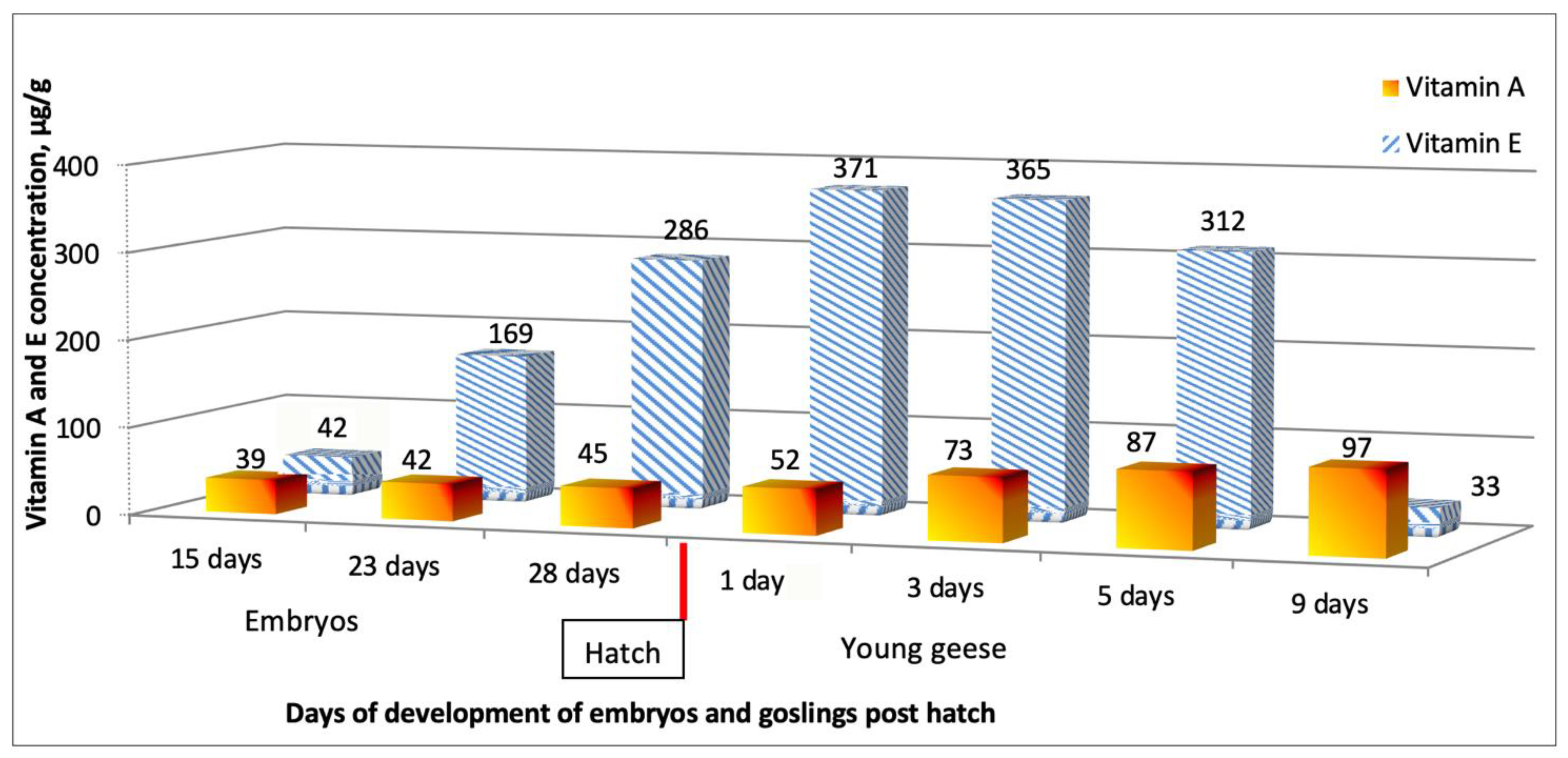
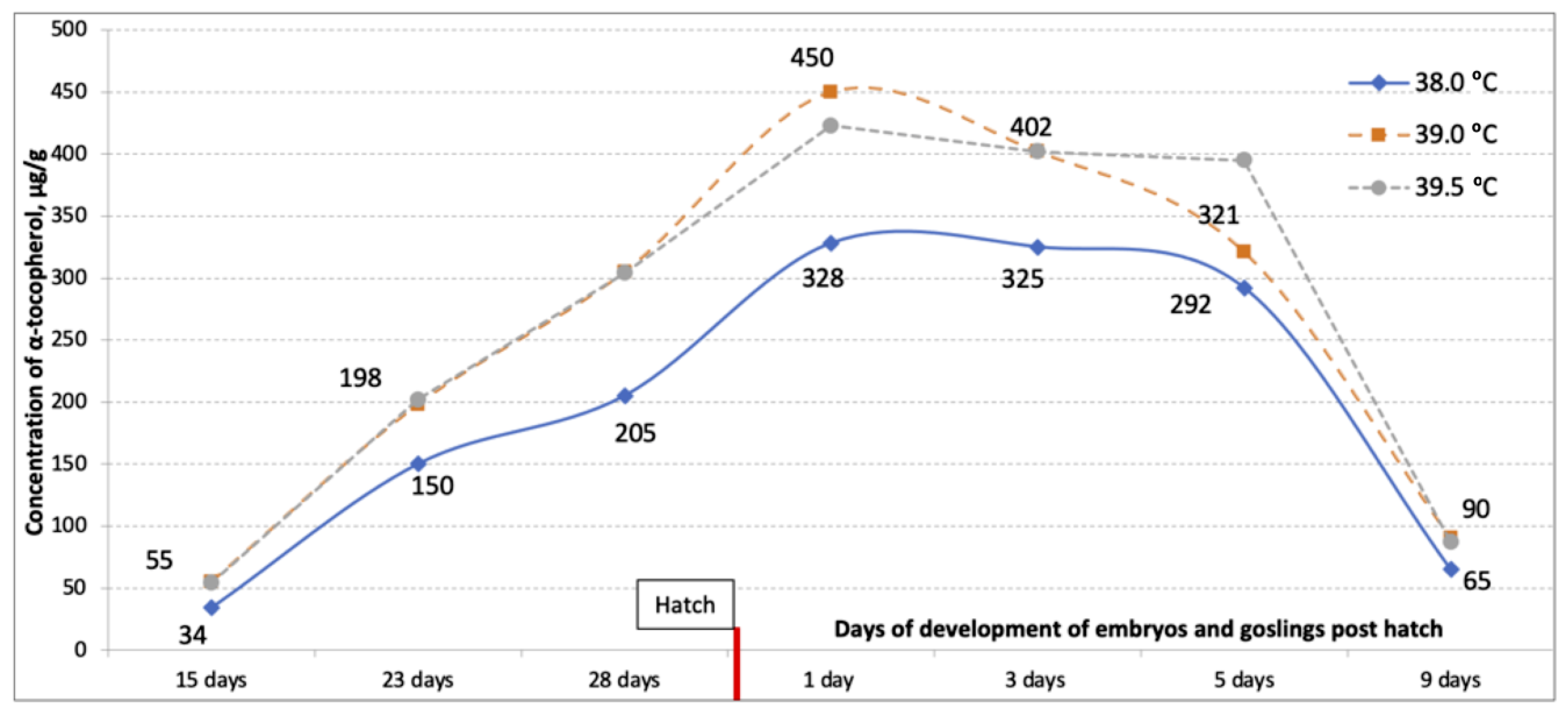
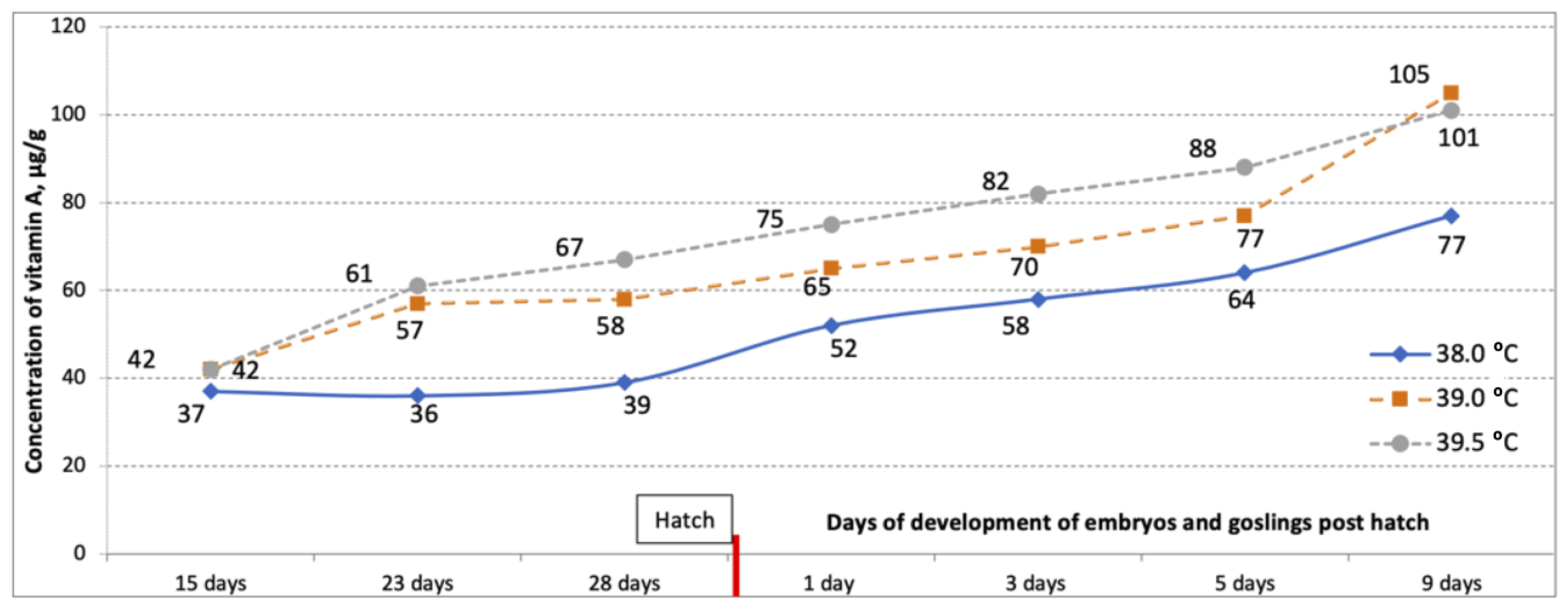
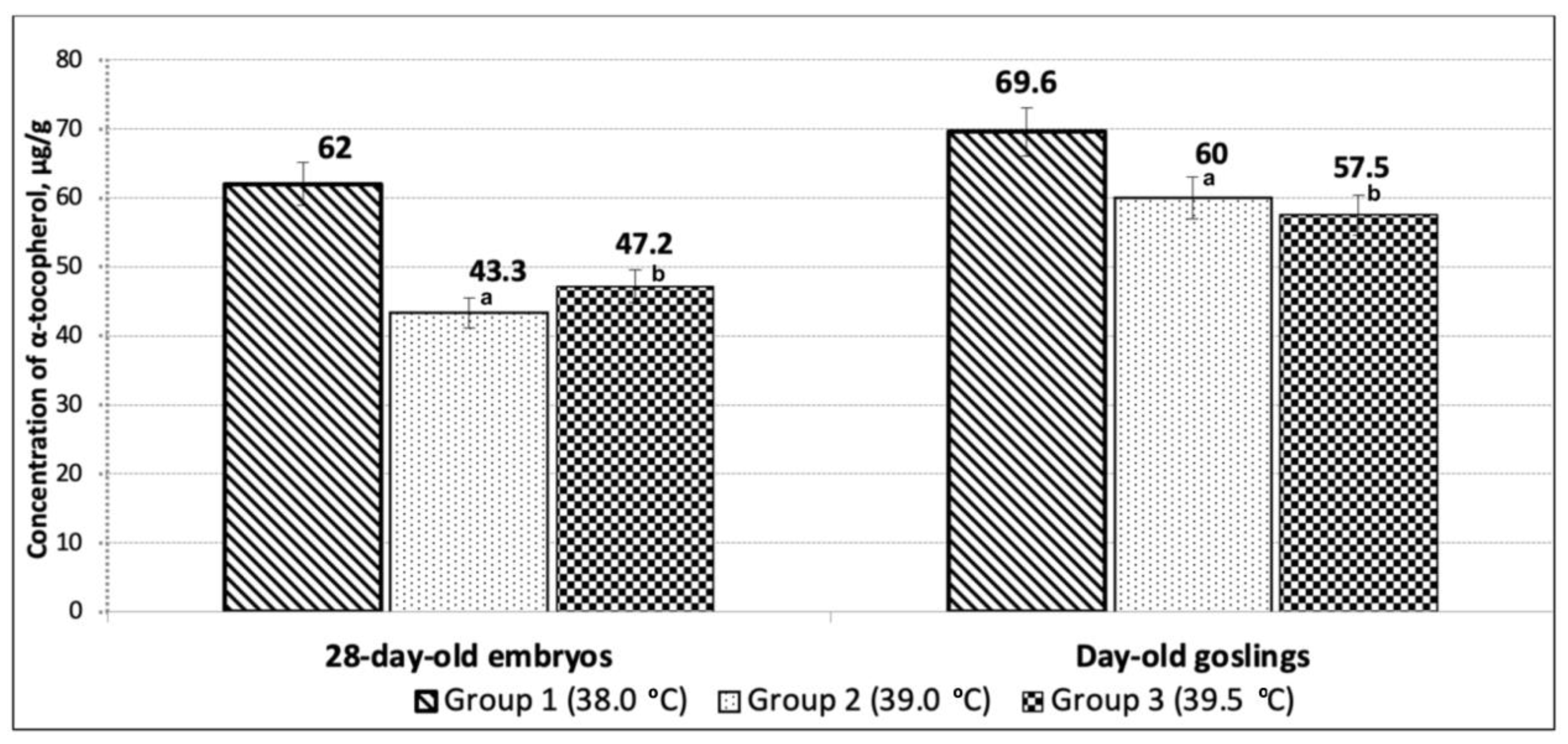
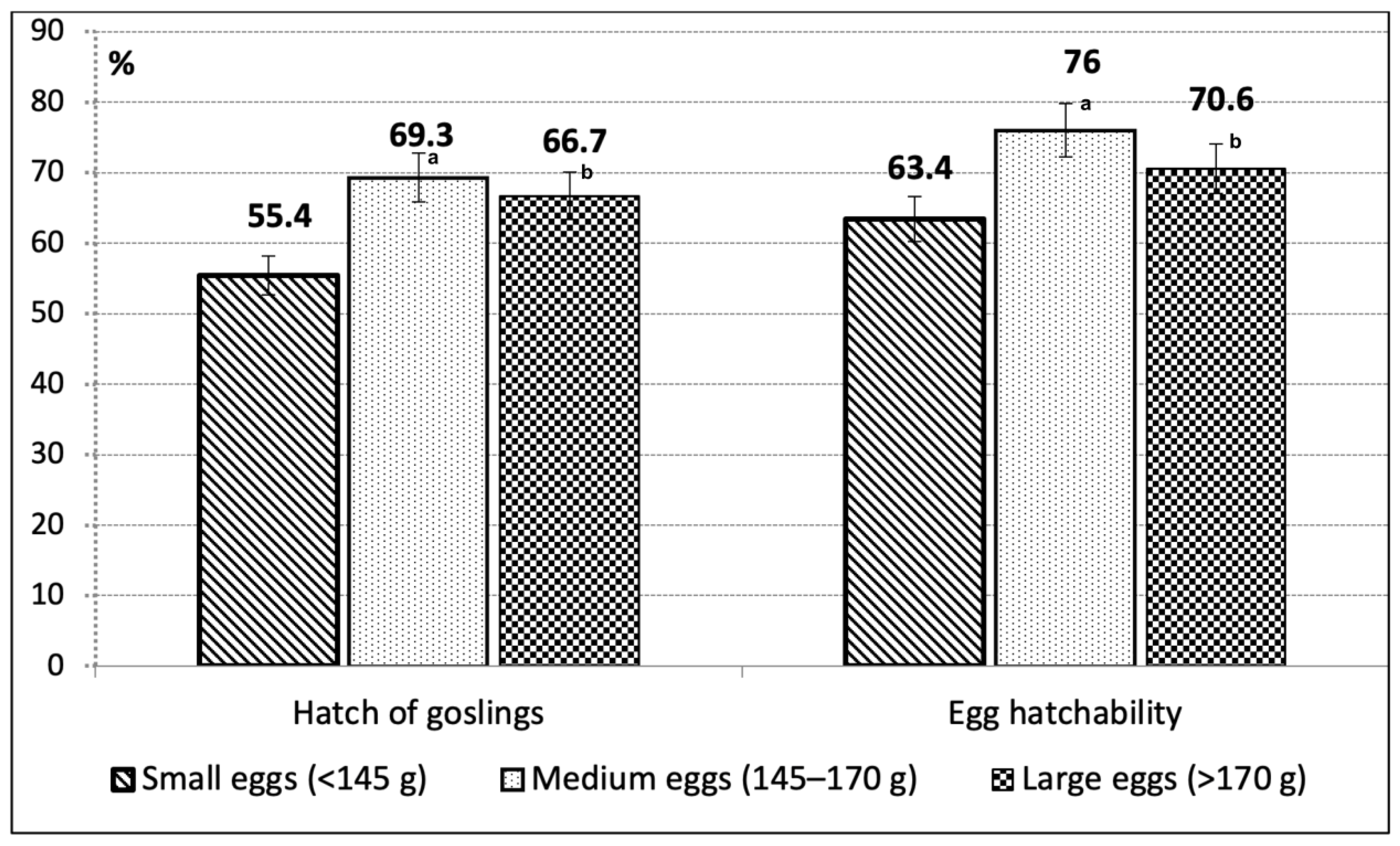
The present study included two experiments carried out at the State Poultry Research Station (SPRS, formerly Poultry Research Institute), National Academy of Agrarian Sciences of Ukraine, Birky, Kharkiv Region. It incorporated fertile eggs, embryos, and goslings from a population of the native Large Gray breed (Figure 2; [39]) maintained at the Rozdolne Experimental Farm, an SPRS branch. Eggs were incubated in a hatchery of the Birky Experimental Farm, an SPRS research base, while groups of hatched goslings were then housed in an SPRS vivarium.
In Experiment 1, the effects of starting heating of goose eggs on their reproduction traits, including hatchability and vitamin availability in embryos during incubation (up to 28 days of incubation) and in goslings post-hatch (up to 9 days of age), were examined. For this purpose, eggs were subjected to short-term (within 24 and 36 h) initial heating at 39.0 and 39.5 °C, respectively, and the following five groups were formed: Group 1 (control, 770 eggs), with incubation according to generally accepted technology [3] at 38 °C for the entire time; Group 2 (766 eggs), where heating begins at 39 °C in the first 24 h; Group 3 (772 eggs), where heating begins at 39.5 °C for the first 24 h; Group 4 (705 eggs), where heating begins at 39 °C during the first 36 h; and Group 5 (706 eggs), where the eggs are heated at 39.5 °C for the first 36 h. The temperatures were selected as close as possible to the temperature under the mother during natural incubation. After applying the initial heating, the eggs of Groups 2 to 5 were placed in one incubator (setter) and further incubated together at 38 °C.
Setters IUP-F-45 and hatchers IUV-F-15 (CJSC Pyatigorskselmash, Pyatigorsk, Stavropol Krai, Russia) were exploited, with each apparatus chamber capacity being ~6000 goose eggs. The length of egg storage before incubation did not exceed five days, and the storage conditions were set at 10–12 °C with 70% relative humidity and darkness. Disinfection of hatching eggs was performed using formaldehyde fumigation. Eggs were candled before incubation to identify cracks and on E15 for possible embryo deaths. Air cooling of the eggs during incubation was applied according to the following scheme: E15 to E19, two times a day; E19 to E22, three times a day; and E22 to E26, four times a day. Starting on E22, the cooling process was accelerated by spraying the eggs with water at room temperature (with the addition of potassium permanganate to a slightly pink color). Egg hatchability was measured as percentage of eggs that provide hatched goslings relative to the number of fertile eggs. Hatch rate of goslings, also designated as hatchability of set eggs, was determined as percentage of hatched goslings relative to the total number of eggs set for incubation.
To examine the dynamics of the vitamin E and A levels on embryogenesis, the liver of goose embryos was sampled for subsequent biochemical analysis on the following key days of their development: E15 (when observing the presence of a formed liver), E23 (transition from the protein type of nutrition to yolk), and E28 (transfer of eggs to a hatcher). On each key day of embryogenesis, samples of the liver were collected from five developing embryos in each group. Additionally, five samples per group were collected from the yolk sac and residual yolk contents in E28 embryos and day-old goslings, respectively. In order to explore the vitamin E and A level dynamics in the first days of life, 30 day-old goslings per group were placed in a vivarium. At the age of 1, 3, 5, and 9 days, five goslings per group were slaughtered, liver samples were obtained, and the concentrations of vitamins E and A were determined. Vitamin E and A content was identified, as described elsewhere [40,41].
The widely used tables of Hamburger and Hamilton [42] were employed to determine the stage of embryo development in this investigation. This information is instrumental in providing an objective step-by-step description of embryo morphogenesis. Previously, such tables were described for chicken, turkey, quail, and duck embryos, while the works of Łukaszewicz et al. [38] and Li et al. [43] made it possible to characterize accurately the development of goose embryos. Applicability of the Hamburger and Hamilton tables [42] was shown here for determining the goose embryo stage, too. In particular, the tables of development of goose embryos were useful for assessing their specific morphogenetic status during incubation, including number of somite pairs (Figure 3).
In Experiment 2, to establish the effect of starting heating of goose eggs and different weights on their hatchability and vitamin availability of embryos during incubation, all eggs from Experiment 1 were weighed on the day of setting the eggs for incubation and additionally calibrated into three weight classes: small, <145 g; medium weight (modal), 145–170 g; and large, >170 g, with at least 300 eggs from each class being placed for incubation.
Statistical data analysis was performed using Excel 2013 (Microsoft Corporation, Redmond, WA, USA). The software package STATISTICA 8 (Statsoft, Inc./TIBCO, Palo Alto, CA, USA) was also employed for repeated measures analysis of variance (rANOVA). Parameter values were presented as group means and standard errors (M ± SE) and further subjected to statistical processing using the non-parametric Mann–Whitney U-test.

3. Results

As can be seen in Figure 3, the results obtained in Experiment 1 with the starting heating at 39.0 and 39.5 °C during the first 24 and 36 h of incubation demonstrated a positive effect of the applied methodological approach on the early development of embryos. Specifically, embryos in the eggs treated with short-term heating (Groups 2 to 5) had a greater number of somite pairs (14.3–16.5) as compared to those in Group 1 (11.8; p < 0.05).
As shown in Figure 4, Figure 5 and Figure 6, differences in the dynamics of the vitamin E and A content in the liver of embryos and goslings were observed in response to the modified temperature regime, when the eggs were subjected to the 24- and 36-h heating at 39 and 39.5 °C, respectively, as compared to the standard incubation conditions (Group 1). Particularly, the concentration of α-tocopherol in the liver of embryos at all the incubation time points was higher in the experimental Groups 2 to 5, as compared to the control Group 1 (p < 0.05). The same regularity was found in the early postnatal ontogenesis (Figure 4 and Figure 5). The concentration of vitamin A in the liver of the embryos and young animals of an early age was also higher in the experimental groups as compared to the control, both when using the starting heating within the first 24 and 36 h (Figure 6; p < 0.05).
We also identified that in response to the 24-h initial heating, the total reserves of α-tocopherol in the residual yolk sac of E28 embryos amounted to 1002 ± 196.7 μg in Group 2 (T = 39.0 °C) and 1119 ± 72.8 μg in Group 3 (T = 39.5 °C). This was 742 and 625 µg less as compared to the control group (1744 ± 242.1 µg; p < 0.01). The content of α-tocopherol in the residual sac of E28 embryos and day-old goslings due to 24- and 36-h egg exposure to starting temperatures is shown in Figure 7 and Table 1.
During the process of embryonic development, the redistribution of vitamin E between the residual yolk and the liver was established. During the incubation period, 25% of the total α-tocopherol reserve of the yolk passed into the liver of day-old goslings in the control group, and 29–34% in the experimental Groups 2–5 (p < 0.01). In the residual yolk sac, the α-tocopherol concentration was lower in the experimental groups, suggesting its more efficient transfer to the liver and other tissues of the goslings during the embryogenesis when using the initial heating of eggs. The efficiency of retinol redistribution from the yolk to the liver of day-old goslings in the control was 14.5%, while this indicator in the experimental groups was 15.3% at 24-h exposure and 20.6% at 36-h exposure (p < 0.05). Therefore, a higher concentration of vitamins E and A in the liver of embryos and their lower concentration in the residual yolk were indicative of their better assimilation from the yolk when applying elevated temperatures at the beginning of incubation.
In terms of the improvement of incubation and hatch indicators, the egg-heating effectiveness in the first 24 and 36 h of embryo development was also determined, as shown in Table 2. Overall, when eggs were initially heated, egg hatchability increased by 10–16%, and gosling hatch rates increased by 9–13 points (p < 0.05).
In addition to the increased hatch of young birds, the use of elevated temperature at the beginning of egg incubation also led to an evenness of the hatch percentage from eggs of different weights, approaching the values of the medium class of eggs. Overall, in Experiment 2, using goose eggs of different weight categories, it was established that the hatching quality of eggs of the medium class was higher than that of small and large eggs (Figure 8; p < 0.05). One of the factors that can explain this pattern is the presence of increased availability of carotenoids and vitamin A in eggs of the medium class as compared to small and large egg classes. Since the goose egg heating for 36 h at 39.5 °C (i.e., the temperature that was closest to the actual temperature under the mother) led to higher hatching rates (by 13.5%; p < 0.05), this incubation mode seems to be preferable for use in the work of hatcheries and poultry farms.

4. Discussion

Knowledge of the dynamics of changes in the content of vitamins (e.g., Figure 1) is important in the reproduction research and pivotal for understanding the processes of embryonic and early postnatal development of young animals, as well as the initial formation phases of antioxidant defense systems to withstand oxidative stress [22,44]. Here, we examined the vitamin E and A dynamics during the standard and modified incubation regimes of goose eggs and assessed the effects of short-term heating of eggs in the early stages of embryogenesis on the vitamin availability in embryos and goslings post-hatch. The vitamin E concentration in the liver drops sharply after the hatching of the goslings and decreases up to almost 10 times by the age of 9 days (Figure 1). We suggest that a high level of α-tocopherol, known as a very effective native antioxidant, is needed for its use in the first days after hatching from the egg and should act as a starting stock to ensure an adequate antioxidant status at the time of the hatching of goslings. This complies with the important physiological role of high concentrations of this natural antioxidant in the body of embryos to prevent the intense peroxidation of lipids in their tissues during this period [22,23].
As for the accumulation of vitamin A in the liver of embryos and young geese, its concentration tended to grow constantly throughout incubation and in the first days of life (Figure 1). We also demonstrated these vitamin A dynamics in our study. Previously, we noticed this pattern for adult birds as well, reaching a maximum at the end of the productive period [24]. It should be emphasized that the amount of retinol in other embryonic tissues during the development of goslings changed insignificantly and is fewer than 10% of its concentration in the liver. Thus, already at the early stages of embryo development, the liver functions as a depot for vitamin A. Subsequently, this is even more evident in adult geese [24].
Our findings were concordant with the previous studies on the young birds of other poultry species, showing that the age dynamics of the vitamin A level in goslings is similar to that in chicks, ducklings, and turkey-poults [45,46]. In addition, the presence of a high amount of vitamin A in the diet of the parent flock of poultry has a significant effect on the concentration of vitamins E and C and carotenoids in the egg yolk and liver of embryos [47].
It is known that 90% of vitamin A in the liver is localized in the stellate cells [48,49]. Being a lipid, vitamin A exhibits a membranotropic effect on all types of membranes, which is manifested in its antioxidant effect [50]. The accumulation of vitamin A in the target organ depends on the functional state of the liver, which can be explained by the peculiarities, level, and direction of metabolism of this organ. With hypervitaminosis A, the membrane is destroyed due to the lysis process. This leads to the release of hydrolytic enzymes, the destruction of cells, and the development of inflammatory reactions. The disruption of mitochondrial membranes can result in apoptosis and those of erythrocytes in hemolysis. On the other hand, vitamin A in normative (natural) quantities plays a very important role in the cell, ensuring the processes of regeneration and antioxidant protection. Therefore, we consider its increased level in the liver of embryos and goslings due to the initial heating of eggs at 39.5 °C during the first 36 h of incubation as a positive factor.
Our research output is also consistent with the fact that vitamin E in the egg yolk and liver of embryos and day-old goslings is present exclusively in the form of α-tocopherol [51], and its total reserves in the liver of goslings can be used as a physiological marker to evaluate their health condition. An active accumulation of vitamin E in the liver of young poultry begins in the last week of development, and the maximum level of α-tocopherol was noted immediately after hatching. The deposition of α-tocopherol in the liver of embryos before hatching is of a general biological significance since a similar pattern is observed in all poultry species [45].
In previous studies (e.g., [17,52,53,54,55,56,57]), the egg weight effects in various poultry species were established on the significantly higher mortality of embryos that develop in smaller and larger eggs than that in eggs of the medium class. In the current study, we mainly confirmed this observation. Differentiation of incubation regimes taking into account the egg weight reduced the difference in hatchability between the medium and extreme classes, although it did not completely eliminate it. The biological mechanisms of this phenomenon require further investigation. We also established that the short-term heating of goose eggs in the initial stages of incubation enabled the increase of vitamin E and A statuses of embryos and young geese, while the hatchability differences between the egg weight classes essentially disappeared, which contributed to a higher egg hatchability and hatch of young birds in general. The most effective in terms of increasing the hatching of goslings and the hatchability of eggs was the initial heating at 39.5 °C during the first 36 h of incubation. Therefore, we recommend this technological approach for implementation in incubation stations.
The results of our experiments suggested the redistribution of vitamin E between the residual yolk, the yolk sac membrane, and the liver of the embryo during the incubation process. Importantly, vitamin E is accumulated in the yolk sac membrane much earlier than in the liver and reaches its maximum (almost 220 μg/g) on E19 or E20, i.e., close to the hatching of the goslings. Considering the fact that the yolk sac membrane serves as a continuation of the gastrointestinal tract of bird embryos and takes an active part in the adsorption of nutrients [58], including vitamins from the residual yolk, we suggest the following mechanism of vitamin E accumulation in the liver of embryos by the hatch. First, vitamin E is adsorbed by the yolk sac membrane from the yolk contents. This happens in the earlier stages of incubation. After that, it passes from the yolk sac membrane through the circulatory system of the embryo to the liver, where it accumulates. It should be borne in mind that the process of vitamin E redistribution between the yolk sac membrane and the liver continues even after the hatching of goslings.
The presence of vitamin E in such high concentrations in the liver of day-old birds can be explained by a number of factors. First, almost the entire amount of vitamin E in the egg yolk is transferred through the yolk membrane to the liver of the goslings. The total amount of tocopherol in an average egg yolk (about 16 g) reaches 2000–2500 μg. This vitamin E amount is thought to be required for its use in the first days after hatching and should act as an initial stock of α-tocopherol, ensuring an adequate antioxidant status at the time of hatching. Secondly, another reason for the high concentration of α-tocopherol in the liver of day-old young animals is the absence of an active form of NADH-dependent quinone reductase in their liver, which ensures further utilization of α-tocopherylquinone, a product of vitamin E metabolism. As a consequence, the latter accumulates in the liver in free form, thus inhibiting the α-tocopherol conversion.
Surai et al. [22] established that the production of reactive oxygen molecules in the poultry body and oxidative stress are the major negative outcomes of the most common stress factors in the poultry industry as a whole. However, the antioxidant defense system includes a complex network of internally synthesized (antioxidant enzymes, glutathione, etc.) and external (vitamins E, A, C, carotenoids, etc.) antioxidants and protects cells from the negative effects of various peroxides. Maintaining an optimal oxidation-reduction balance in the cell/body is a key element in providing the necessary conditions for adapting to stress and maintaining homeostasis in the body. Surai et al. [22] suggested that the development of a system of optimal antioxidant supplements in the diet of the parent stock is one of the alternative ways of maintaining this balance. This new direction in improving the antioxidant protection of poultry under stress conditions is associated with the possibility of activating a number of vitagenes [59,60].
High superoxide dismutase activity in organs and tissues is considered to be a compensatory protection mechanism during the transition from hypoxia at the end of embryonic development to relative hyperoxia in the first days of life [22]. Inadequate protection of the body of young birds from reactive oxygen species in the second and third decades of life leads to a shift of oxidizing processes towards free radicals.
It is known that vitamin E is able to perform its antioxidant functions only in the form of α-tocopherol-alcohol [45]. At the same time, the activity of acylation–deacylation enzymes in the body at the time of hatching is very low, and this enzyme system, practically, does not function. This is the biological meaning of the high concentration of vitamin E in the form of free tocopherol. Ensuring adequate antioxidant capacity, in particular, a high concentration of vitamin E in the liver at various stages of embryo development, is necessary to protect the increased level of unsaturation in embryo lipids. Research in this area represents the first attempts to explain the protective mechanisms of vitamin E action in tissues during embryogenesis [23]. The results we report here provide a new insight into the function of vitamin E in the early postnatal period of development. Considering the fact that the moment of hatching is a very strong stress factor, the high concentration of vitamin E in the liver (i.e., an organ that actively participates in its metabolism and regulation of its distribution among other tissues) is fully understandable [25]. In addition, the high level of such lipids as polyunsaturated fatty acids (PUFAs) in tissues plays an important role at the time of hatching. Collectively, the results of our own research and other studies suggest that, along with the PUFAs accumulation in embryonic tissues, there is an increase in the concentration of the natural antioxidant vitamin E. Herewith, the maximum concentration of this vitamin is found in embryonic tissues at the time of hatching, which is accompanied by a significant oxygen stress. The adaptation of young animals to the stressful conditions of the external environment in the first day of life leads to an increase in the consumption of α-tocopherol reserves in the liver, which contributes to the reliable protection of tissues from lipid peroxidation [61]. The endogenous level of free radical inhibitors in the body, such as vitamin E, is crucial for maintaining the physiological level of oxygen radicals and the integrity of cell membranes [62] so the level of free α-tocopherol in the liver can reflect the health status of day-old young birds as an indicator related to the main life processes.

5. Conclusions

We established here that initially heating eggs at 39 and 39.5 °C within the first 24 or 36 h (as compared to the generally accepted technology at 38 °C for the entire time) was effective. A higher level of α-tocopherol and retinol in the liver of embryos and young goslings was established when initially heating goose eggs during 24 and 36 h of incubation at 39–39.5 °C in all studied age periods in comparison with the standard mode of incubation of eggs at 38 °C. Our results confirmed that the age dynamics of vitamin E and A levels in goslings are similar to that found in chicks, ducklings, and turkey-poults. We argue that the formation of the antioxidant system of poultry, the components of which are α-tocopherol and retinol, occurs at the earliest stages of ontogenesis. The embryogenesis period is characterized by high levels of α-tocopherol in the liver, which may be due to sufficiently good protection of the embryos from external impacts and a low level of synthesis of antioxidant enzymes in the embryonic tissues. The existence of a high endogenous level of vitamin E in the liver and the sensitivity of this organ to lipid peroxidation may evidence the specifics of the antioxidant system functioning in embryogenesis and early postnatal ontogenesis of birds. We also identified that the modified incubation schemes, when eggs were initially treated at 39 and 39.5 °C, facilitated an improvement in the reproduction traits, including the hatching qualities of eggs and hatch rate of goslings. For use in the work of hatcheries and breeding poultry farms, it can be recommended to implement the initial heating of goose eggs for 36 h at 39.5 °C.

Author Contributions

Conceptualization, I.A.I. and V.O.K.; methodology, I.A.I., V.O.K. and O.O.K.; software, I.A.I. and V.O.K.; validation, O.V.A., I.O.Z. and M.N.R.; formal analysis, I.A.I., V.O.K. and O.O.K.; investigation, I.A.I. and V.O.K.; resources, I.A.I. and O.V.A.; data curation, I.A.I. and V.O.K.; writing—original draft preparation, I.A.I., O.O.K. and O.V.A.; writing—review and editing, I.A.I., O.V.A., D.K.G. and M.N.R.; visualization, I.A.I., O.O.K., I.O.Z. and M.N.R.; supervision, I.A.I. and D.K.G.; project administration, I.A.I. and D.K.G.; funding acquisition, I.A.I. and M.N.R. All authors have read and agreed to the published version of the manuscript.

Funding

This research was supported by the Ukrainian Academy of Agrarian Sciences.

Institutional Review Board Statement

The present research involving embryos and young geese was carried out in accordance with the requirements of the provisions as specified by the European Convention for the Protection of Vertebrate Animals used for Experimental and other Scientific Purposes (Strasbourg, 1986) and in compliance with the relevant laws of Ukraine, and was approved by the Commission on Ethics and Bioethics, of the Kharkiv National Pedagogical University named after H. S. Skovoroda (Protocol No. 1, dated 10 January 2023).

Informed Consent Statement

Not applicable.

Data Availability Statement

Main data is contained within the article, with the raw data being available on request.

Acknowledgments

The authors are most grateful to Olga N. Verko, of the National Scientific Center “Institute for Soil Science and Agrochemistry Research named after O. N. Sokolovsky”, Kharkiv, Ukraine, for technical assistance in translation from Ukrainian into English. The skilled technical assistance of Olga M. Romanova in preparing Figure 2 is kindly appreciated.

Conflicts of Interest

The authors declare no conflict of interest.

References

  1. Romanov, M.N. Evolution of domestic geese. Centres of domestication and dispersion. In New Investigations on Palearctic Geese; Gudina, A.N., Ed.; Zaporizhya Branch of the Ukrainian Ornithological Society, Zaporizhya State University: Zaporizhya, Ukraine, 1995; pp. 120–126. [Google Scholar]
  2. Romanov, M.N. Geese. In Encyclopedia of Animal Science, Two-Volume Set, 2nd ed.; Baer, C.K., Ullrey, D.E., Pond, W.G., Eds.; Taylor & Francis Inc.: London, UK; CRC Press Inc.: Boca Raton, FL, USA, 2018; Volume 1, pp. 487–489. [Google Scholar] [CrossRef]
  3. Tereshchenko, A.V.; Mikityuk, D.N.; Ryabokon, Y.A.; Ivko, I.I.; Melnik, V.A.; Ryabinina, E.V.; Khvostik, V.P.; Gorbanev, A.P. Geese on the Farm and in the Courtyard, 2nd ed.; Tereshchenko, A.V., Ed.; Poultry Research Institute, Ukrainian Academy of Agrarian Sciences: Borky, Ukraine, 2008. (In Russian) [Google Scholar]
  4. Bondarenko, Yu.V.; Zharkova, I.P.; Romanov, M.N. Study of down colour genotype in the collection flock geese at the All-Union Poultry Research and Technological Institute. Naučno-Teh. Bûll.—Ukr. Naučno-Issled. Inst. Pticevod. (Ukr. Poult. Res. Inst. Kharkiv Ukr.) 1986, 21, 3–7. (In Russian) [Google Scholar]
  5. Bondarenko, Y.V.; Tereshchenko, O.V.; Tkachyk, T.E.; Zakharchenko, O.P.; Hadiuchko, O.T. Recommendations on the Use of Modern Methods for Determining the Sex of Young Birds of Poultry (Theory and Practice); Kharkiv, Ukraine, 2007. (In Ukrainian) [Google Scholar]
  6. Sakhatsky, N.I.; Tereshchenko, A.V.; Artemenko, A.B. An express-method of estimation of fertilizing ability of freezing-thawing of poultry spermatozoa. Sel’skokhozyaistvennaya Biol. (Agric. Biol.) 1987, 22, 77–80. (In Russian) [Google Scholar]
  7. Romanov, M.N. Population-morphological Studies on Domestic Geese. In Proceedings of the Ukrainian Conference of Young Scientists and Postgraduates on Poultry Problems, Borky, Kharkiv, Ukraine; Poultry Research Institute, Ukrainian Academy of Agrarian Sciences: Kharkiv, Ukraine, 1992; pp. 8–9. (In Ukrainian) [Google Scholar]
  8. Romanov, M.N. Genetic Studies on Geese of Ukraine and Russia. 1. Historical Preface. In Proceedings of the 9th International Symposium on Waterfowl, Pisa, Italy, 16–18 September 1992; World’s Poultry Science Association: Pisa, Italy, 1992; pp. 102–104. [Google Scholar]
  9. Romanov, M.N.; Veremeyenko, R.P.; Bondarenko, Y.V. Conservation of Waterfowl Germplasm in Ukraine. In Proceedings of the 10th International Symposium on Waterfowl, Halle (Saale), Germany, 26–31 March 1995; World’s Poultry Science Association: Halle (Saale), Germany, 1995; pp. 401–414. [Google Scholar]
  10. Romanov, M.N.; Weigend, S.; Bondarenko, Y.V.; Podstreshny, A.P.; Kutnyuk, P.I.; Sakhatsky, N.I. Studies on Poultry Germplasm Diversity and Conservation in Ukraine. In Proceedings of the Poultry Genetics Symposium, Mariensee, Germany, 6–8 October 1999; Working Group 3 of WPSA: Mariensee, Germany, 1999; p. 140. [Google Scholar]
  11. Romanov, M.N.; Betuel, A.M.; Chemnick, L.G.; Ryder, O.A.; Kulibaba, R.O.; Tereshchenko, O.V.; Payne, W.S.; Delekta, P.C.; Dodgson, J.B.; Tuttle, E.M.; et al. Widely Applicable PCR Markers for Sex Identification in Birds. Russ. J. Genet. 2019, 55, 220–231. [Google Scholar] [CrossRef]
  12. Gadyuchko, O.T.; Sakhatsky, N.I.; Tereshchenko, A.V.; Anisimov, V.I.; Kislaya, E.D.; Svinarev, V.F. Genetic potential of breeds and populations of geese in Ukraine. Ptakhivnytstvo (Poult. Farming) 2003, 53, 54–62. (In Russian) [Google Scholar]
  13. Tagirov, M.T.; Tereshchenko, L.V.; Tereshchenko, A.V. Substantiation of the possibility of using primary germ cells as material for the preservation of poultry genetic resources. Ptakhivnytstvo (Poult. Farming) 2006, 58, 464–473. (In Russian) [Google Scholar]
  14. Tagirov, M.T.; Ogurtsova, N.S.; Tereshchenko, A.V. Analysis of hatchability problems for incubated eggs. Ptakhivnytstvo (Poult. Farming) 2009, 63, 199–215. (In Russian) [Google Scholar]
  15. Narushin, V.G.; Chausov, M.G.; Shevchenko, L.V.; Pylypenko, A.P.; Davydovych, V.A.; Romanov, M.N.; Griffin, D.K. Shell, a naturally engineered egg packaging: Estimated for strength by non-destructive testing for elastic deformation. Biosyst. Eng. 2021, 210, 235–246. [Google Scholar] [CrossRef]
  16. Narushin, V.G.; Romanov, M.N.; Griffin, D.K. Non-destructive evaluation of the volumes of egg shell and interior: Theoretical approach. J. Food Eng. 2021, 300, 110536. [Google Scholar] [CrossRef]
  17. Tagirov, M.T.; Shomina, N.V.; Artemenko, A.B.; Tkachenko, S.N.; Baydevlyatova, O.N.; Tereshchenko, A.B.; Sakhatsky, N.I. Incubation of Poultry Eggs: Guidelines; Poultry Research Institute, Ukrainian Academy of Agrarian Sciences: Kharkiv, Ukraine, 2009. (In Russian) [Google Scholar]
  18. Khvostyk, V.P.; Bondarenko, Y.V. Genetic polymorphism of egg white proteins of geese of different breeds of domestic selection. Ef. Ptakhivnytstvo (Eff. Poult. Farming) 2008, 12, 48. (In Ukrainian) [Google Scholar]
  19. Surai, P.F.; Ionov, I.A.; Bondarenko, Y.V. The content of carotenoids and vitamin a in the yolk of poultry eggs. Naučno-Teh. Bûll.—Inst. Pticevod. (Poult. Res. Inst. Ukr. Acad. Agrar. Sci. Kharkiv Ukr.) 1992, 33, 28–33. (In Russian) [Google Scholar]
  20. Bratyshko, N.I.; Haviley, O.V.; Tereshchenko, O.V.; Prytulenko, O.V. The effect of triticale on the vitamin content of eggs of chickens of different breeds. Ptakhivnytstvo (Poult. Farming) 2010, 65, 93–100. (In Ukrainian) [Google Scholar]
  21. Kokoszyński, D. Chapter 4—Guinea fowl, goose, turkey, ostrich, and emu eggs. In Egg Innovations and Strategies for Improvements; Hester, P.Y., Ed.; Academic Press: Cambridge, MA, USA; Elsevier Inc.: London, UK, 2017; pp. 33–43. [Google Scholar] [CrossRef]
  22. Surai, P.F.; Kochish, I.I.; Fisinin, V.I.; Kidd, M.T. Antioxidant defence systems and oxidative stress in poultry biology: An update. Antioxidants 2019, 8, 235. [Google Scholar] [CrossRef]
  23. Surai, P.F.; Kochish, I.I.; Romanov, M.N.; Griffin, D.K. Nutritional modulation of the antioxidant capacities in poultry: The case of vitamin E. Poult. Sci. 2019, 98, 4030–4041. [Google Scholar] [CrossRef] [PubMed]
  24. Ionov, I.A. Physiological Status of the Birds in Embryogenesis and Postnatal Ontogenesis Depending on Its A-, E- and K-Vitamin Availability. Ph.D. Thesis, Poultry Research Institute, Ukrainian Academy of Agrarian Sciences, Kharkiv, Ukraine, 1997. (In Ukrainian). [Google Scholar]
  25. Surai, P.F.; Fisinin, V.I.; Karadas, F. Antioxidant systems in chick embryo development. Part 1. Vitamin E, carotenoids and selenium. Anim. Nutr. 2016, 2, 1–11. [Google Scholar] [CrossRef] [PubMed]
  26. Guo, B.; Yan, L.; Lei, M.; Dai, Z.; Shi, Z. Wider angle egg turning during incubation enhances yolk utilization and promotes goose embryo development. Animals 2021, 11, 2485. [Google Scholar] [CrossRef] [PubMed]
  27. Kucharska-Gaca, J.; Adamski, M.; Biesek, J. Effect of parent flock age on hatching, growth rate, and features of both sexes goose carcasses. Poult. Sci. 2022, 101, 101920. [Google Scholar] [CrossRef] [PubMed]
  28. Kucharska-Gaca, J.; Adamski, M.; Biesek, J. The age of the geese from the parent flock and the laying period affect the features of the eggs. Poult. Sci. 2022, 101, 102094. [Google Scholar] [CrossRef]
  29. Guo, B.B.; Dai, Z.C.; Ren, Y.H.; Zhu, H.X.; Shao, X.B.; Sun, A.D.; Shi, Z.D. Improvement of goose embryonic and muscular developments by wider angle egg turning during incubation and the regulatory mechanisms. Poult. Sci. 2021, 100, 101477. [Google Scholar] [CrossRef]
  30. Kochish, I.I.; Titov, V.Y.; Nikonov, I.N.; Brazhnik, E.A.; Vorobyov, N.I.; Korenyuga, M.V.; Myasnikova, O.V.; Dolgorukova, A.M.; Griffin, D.K.; Romanov, M.N. Unraveling signatures of chicken genetic diversity and divergent selection in breed-specific patterns of early myogenesis, nitric oxide metabolism and post-hatch growth. Front. Genet. 2023, 13, 1092242. [Google Scholar] [CrossRef]
  31. Kochish, I.I.; Brazhnik, E.A.; Vorobyov, N.I.; Nikonov, I.N.; Korenyuga, M.V.; Myasnikova, O.V.; Griffin, D.K.; Surai, P.F.; Romanov, M.N. Features of fractal conformity and bioconsolidation in the early myogenesis gene expression and their relationship to the genetic diversity of chicken breeds. Animals 2023, 13, 521. [Google Scholar] [CrossRef]
  32. Guo, B.; Dai, Z.; Chen, R.; Liu, J.; Shi, Z. Enhancing gosling growth and secretion of somatotrophic and thyrotrophic axis hormones through egg turning during incubation. Br. Poult. Sci. 2022, 64, 122–128. [Google Scholar] [CrossRef]
  33. Bondarenko, Yu.V.; Kovalenko, A.T.; Kutnyuk, P.I. Efficiency of modal selection in poultry populations. Naučno-Teh. Bûll.—Ukr. Naučno-Issled. Inst. Pticevod. (Ukr. Poult. Res. Inst. Kharkiv Ukr.) 1979, 7, 3–7. (In Russian) [Google Scholar]
  34. Bondarenko, Yu.V. The study of the comparative variability of the weight of eggs and day-old chicks of nine poultry species. Naučno-Teh. Bûll.—Ukr. Naučno-Issled. Inst. Pticevod. (Ukr. Poult. Res. Inst. Kharkiv Ukr.) 1989, 26, 6–10. (In Russian) [Google Scholar]
  35. Artemenko, O.B.; Tereshchenko, O.V.; Shomina, N.V.; Baidevlyatova, O.M.; Gavilei, O.V. Study of the Effect of Circadian Temperature Stimuli on the Results of Egg Incubation. In Proceedings of the Materials of the XIII International Conference “Poultry Industry 2017”, Truskavets, Ukraine, 19–21 September 2017; pp. 83–88. (In Ukrainian). [Google Scholar]
  36. Lomako, D.V. Technology of incubation of goose eggs in the conditions of Myrhorod private rental agricultural hatchery and poultry enterprise. Visnyk Poltavsʹkoyi Derzhavnoyi Ahrarnoyi Akad. (Bull. Poltava State Agrar. Acad.) 2015, 4, 44–46. (In Ukrainian) [Google Scholar] [CrossRef]
  37. Christensen, V.L.; Bagley, L.G. Improved hatchability of turkey eggs at high altitudes due to added oxygen and increased incubation temperature. Poult. Sci. 1988, 67, 956–960. [Google Scholar] [CrossRef] [PubMed]
  38. Łukaszewicz, E.; Lasoń, M.; Rosenberger, J.; Kowalczyk, A.; Bakst, M. Goose embryonic development from oviposition through 16 h of incubation. Poult. Sci. 2017, 96, 1934–1938. [Google Scholar] [CrossRef] [PubMed]
  39. Podstreshny, O.P.; Nalyvaiko, L.I.; Bondarenko, Y.V. Genetic polymorphism in populations of the Large Gray geese. Ptakhivnytstvo (Poult. Farming) 2001, 50, 19–26. (In Ukrainian) [Google Scholar]
  40. Mcmurray, C.H.; Blanchflower, W.J.; Rice, D.A. Influence of Extraction Techniques on Determination of α-Tocopherol in Animal Feedstuffs. J. AOAC Int. 1980, 63, 1258–1261. [Google Scholar] [CrossRef]
  41. Vahl, H.A.; Klooster, A.T. Effects of excessive vitamin A levels in broiler rations. J. Anim. Physiol. Anim. Nutr. 1987, 57, 204–218. [Google Scholar] [CrossRef]
  42. Bellairs, R.; Osmond, M. Appendix II—Normal Table of Hamburger and Hamilton (1951; 1992). In The Atlas of Chick Development, 3rd ed.; Academic Press: Oxford, UK; Waltham, MA, USA, 2014; pp. 603–621. [Google Scholar] [CrossRef]
  43. Li, S.; Bai, S.; Qin, X.; Zhang, J.; Irwin, D.M.; Zhang, S.; Wang, Z. Comparison of whole embryonic development in the duck (Anas platyrhynchos) and goose (Anser cygnoides) with the chicken (Gallus gallus). Poult. Sci. 2019, 98, 3278–3291. [Google Scholar] [CrossRef]
  44. Liang, J.; Dai, H.; Yang, H.; Yang, Z.; Wang, Z. The effect of dietary vitamin A supplementation in maternal and its offspring on the early growth performance, liver vitamin A content, and antioxidant index of goslings. Poult. Sci. 2019, 98, 6849–6856. [Google Scholar] [CrossRef]
  45. Surai, P.F.; Ionov, I.A.; Kuchmistova, E.F.; Noble, R.C.; Speake, B.K. The relationship between the levels of α-tocopherol and carotenoids in the maternal feed, yolk and neonatal tissues: Comparison between the chicken, turkey, duck and goose. J. Sci. Food Agric. 1998, 76, 593–598. [Google Scholar] [CrossRef]
  46. Surai, P.; Kuklenko, T.; Ionov, I.; Noble, R.; Sparks, N. Effect of vitamin A on the antioxidant system of the chick during early postnatal development. Br. Poult. Sci. 2000, 41, 454–458. [Google Scholar] [CrossRef]
  47. Surai, P.F.; Ionov, I.A.; Kuklenko, T.V.; Kostjuk, I.A.; Acpherson, A.M.; Speake, B.K.; Noble, R.C.; Sparks, N.H. Effect of supplementing the hen's diet with vitamin A on the accumulation of vitamins A and E, ascorbic acid and carotenoids in the egg yolk and in the embryonic liver. Br. Poult. Sci. 1998, 39, 257–263. [Google Scholar] [CrossRef]
  48. Shirakami, Y.; Lee, S.-A.; Clugston, R.D.; Blaner, W.S. Hepatic metabolism of retinoids and disease associations. Biochim. Biophys. Acta 2012, 1821, 124–136. [Google Scholar] [CrossRef] [PubMed]
  49. Senoo, H.; Mezaki, Y.; Fujiwara, M. The stellate cell system (vitamin A-storing cell system). Anat. Sci. Int. 2017, 92, 387–455. [Google Scholar] [CrossRef]
  50. Bozhkov, A.; Ionov, I.; Kurhuzova, N.; Novikova, A.; Katerynych, O.O.; Akzhyhitov, R. Vitamin A intake forms resistance to hypervitaminosis A and affects the functional activity of the liver. Clin. Nutr. Open Sci. 2021, 41, 82–97. [Google Scholar] [CrossRef]
  51. Ivchenko, V.V.; Bondarenko, Y.V.; Rozhen, T.M. Method of Selecting Goose Eggs for Incubation. Patent SU 1535505 A1, 1990. Available online: https://patents.google.com/patent/SU1535505A1/en (accessed on 26 April 2023). (In Russian).
  52. Zhang, J.; Huang, B.; Cai, Z.; Xu, J.; Zheng, Y.; Wang, M. Fat-soluble vitamins in different eggs and egg products in Hangzhou City. Wei Sheng Yan Jiu 2020, 49, 984–989. (In Chinese) [Google Scholar] [CrossRef] [PubMed]
  53. Narushin, V.G.; Romanov, M.N. Influence of egg parameters on hatchability (a research review). Ptakhivnytstvo (Poult. Farming) 2001, 51, 422–429. (In Russian) [Google Scholar]
  54. Narushin, V.G.; Romanov, M.N. Physical Characteristics of Chicken Eggs in Relation to Their Hatchability and Chick Weight. In Proceedings of the ASAE Annual International Meeting/CIGR World Congress, Chicago, IL, USA, 28–31 July 2002; Paper #026066. American Society of Agricultural and Biological Engineers: St. Joseph, MI, USA, 2002. [Google Scholar] [CrossRef]
  55. Narushin, V.G.; Romanov, M.N. Relationship between physical characteristics and results of hatching in chicken eggs. Arch. Geflugelkd. 2002, 66 (Suppl. S2), 128. [Google Scholar]
  56. Narushin, V.G.; Bogatyr, V.P.; Romanov, M.N. Relationship between hatchability and non-destructive physical measurements of chicken eggs. J. Agric. Sci. 2016, 154, 359–365. [Google Scholar] [CrossRef]
  57. Prokudina, N.A.; Duyunova, A.A.; Ogurtsova, N.S.; Ryabokon, Y.A.; Artyomenko, A.B.; Ivko, I.I.; Melnik, V.A.; Bondarenko, Y.V. Turkey Egg Incubation Guide; Poultry Research Institute, Ukrainian Academy of Agrarian Sciences: Borki, Ukraine, 2007. (In Russian) [Google Scholar]
  58. Yang, H.; Liang, J.; Dai, H.; Wan, X.; Wang, Z. Effects of vitamin A supplementation in the diet of breeding geese on offspring intestinal tissue morphology and immune performance. Asian-Australas. J. Anim. Sci. 2020, 33, 1463–1469. [Google Scholar] [CrossRef]
  59. Surai, P.; Fisinin, V. Vitagenes in poultry production: Part 1. Technological and environmental stresses. World's Poult. Sci. J. 2016, 72, 721–734. [Google Scholar] [CrossRef]
  60. Surai, P.; Fisinin, V. Vitagenes in poultry production: Part 2. Nutritional and internal stresses. World's Poult. Sci. J. 2016, 72, 761–772. [Google Scholar] [CrossRef]
  61. Surai, P.F. Antioxidants in poultry nutrition and reproduction: An update. Antioxidants 2020, 9, 105. [Google Scholar] [CrossRef] [PubMed]
  62. Surai, P.F.; Earle-Payne, K. Antioxidant defences and redox homeostasis in animals. Antioxidants 2022, 11, 1012. [Google Scholar] [CrossRef] [PubMed]
Figure 1. Dynamics of α-tocopherol and retinol content in the liver of embryos and young geese (according to [24]).
Poultry 02 00023 g001
Figure 2. Large Gray breed of geese. (a) Goslings post hatch; (b) a parent flock of adult geese.
Poultry 02 00023 g002
Figure 3. Stages of development of goose embryos after two days of incubation at different starting temperatures during the first 24 (b,c) and 36 (d,e) h of incubation. (a) Group 1 (control, with the basic mode of incubation at 38.0 °C during the entire incubation period), 11.80 ± 0.52 pairs of somites; (b,d) Groups 2 and 4 (with the starting warm-up at 39.0 °C), 14.27 ± 0.54 and 15.04 ± 0.44 pairs of somites, respectively; (c,e) Groups 3 and 5 (with the starting warm-up at 39.5 °C), 14.98 ± 0.49; and 16.51 ± 0.62 pairs of somites, respectively.
Poultry 02 00023 g003
Figure 4. Concentration of α-tocopherol in the liver of embryos and young geese in the control Group 1 (38.0 °C) and Groups 2 and 3 (with the initial egg heating during the first 24 h of incubation at 39 and 39.5 °C, respectively).
Poultry 02 00023 g004
Figure 5. Concentration of α-tocopherol in the liver of embryos and young geese in the control Group 1 (38 °C) and Groups 4 and 5 (with the initial egg heating during the first 36 h of incubation at 39 and 39.5 °C, respectively).
Poultry 02 00023 g005
Figure 6. Concentration of vitamin A in the liver of embryos and young geese in the control Group 1 (38.0 °C) and Groups 4 and 5 (with the initial egg heating during the first 36 h of incubation at 39.0 and 39.5 °C, respectively).
Poultry 02 00023 g006
Figure 7. Concentration of α-tocopherol in the yolk sac of embryos and goslings in response to the initial heating of eggs for 24 h. Significant differences in 28-day-old embryos between the experimental groups and Control Group 1: a, Group 2, p < 0.01; b, Group 3, p < 0.01. Those in day–old goslings: a, Group 2, p < 0.05; b, Group 3, p < 0.01.
Poultry 02 00023 g007
Figure 8. Influence of the three different egg weight classes on their hatching quality results. Significant differences relative to the small eggs: a, medium eggs, p < 0.01; b, large eggs, p < 0.05.
Poultry 02 00023 g008
Table 1. Vitamin E content (M ± SE) in the residual yolk of embryos and goslings in response to the initial heating for 36 h.
Mode of Starting Heating28-Day EmbryosDay-old Goslings
Content, µg/gTotal, µgContent, µg/gTotal, µg
38.0 °C (Group 1)53.8 ± 1.891688.0 ± 171.457.6 ± 4.45590.8 ± 116.0
39.0 °C (Group 4)48.7 ± 1.521290.5 ± 61.7 *52.8 ± 2.90288.7 ± 65.6 **
39.5 °C (Group 5)41.2 ± 3.00 *903.1 ± 111.7 **45.3 ± 3.20 *309.6 ± 73.4 **
Differences between Group 1 and Group 4 or 5 are significant at: * p < 0.05; ** p < 0.01.
Table 2. Results of goose egg incubation and hatch of goslings in response to the starting heating at 39 and 39.5 °C in comparison with the basic standard mode of incubation (38 °C).
IndicatorsGroups
1 (38.0 °C)2 (39.0 °C)3 (39.5 °C)4 (39.0 °C)5 (39.5 °C)
24-h Starting Heating36-h Starting Heating
No. of eggs incubated700766772705706
Egg fertility, %94.394.694.290.892.1
No. of goslings hatched504571576520557
Hatch of young geese, % *65.474.574.673.778.9
Egg hatchability, % **69.478.779.281.385.7
* As conformed to the hatchability of set eggs (%) = (number of hatched goslings/total number of set eggs) × 100. ** Hatchability of fertile eggs (%) = (number of hatched chicks/number of fertile eggs) × 100.
Disclaimer/Publisher’s Note: The statements, opinions and data contained in all publications are solely those of the individual author(s) and contributor(s) and not of MDPI and/or the editor(s). MDPI and/or the editor(s) disclaim responsibility for any injury to people or property resulting from any ideas, methods, instructions or products referred to in the content.

