Review

Mitigating the Adverse Effects of Lead and Cadmium Heavy Metals-Induced Oxidative Stress by Phytogenic Compounds in Poultry

1
Faculty of Animal Science, Agricultural Science and Natural Resources University of Khuzestan, Khuzestan 6341773637, Iran
2
Department of Veterinary Preclinical Sciences, Faculty of Veterinary Medicine, Universiti Putra Malaysia, Serdang 43400, Selangor, Malaysia
3
U.S. National Poultry Research Center, Agricultural Research Service, USDA-ARS, Athens, GA 30605, USA
*
Author to whom correspondence should be addressed.
Poultry 2023, 2(2), 235-251; https://doi.org/10.3390/poultry2020019
Submission received: 7 February 2023 / Revised: 3 April 2023 / Accepted: 10 April 2023 / Published: 13 April 2023
(This article belongs to the Special Issue Feature Papers of Poultry)

Abstract

:
Environmental pollution has increased over the past few decades, posing serious risks to all biological systems, including the poultry sector. Oxidative stress in chickens caused by dietary, environmental, and pathological variables influences how well chickens perform as well as the quality of meat and eggs. Lead (Pb) and cadmium (Cd) are two examples of heavy metals that are harmful for chicken health. They can cause oxidative stress by increasing the production of reactive oxygen species (ROS) and reactive nitrogen species (RNS) and blocking antioxidants from protecting cells from increased amounts of free radicals. The oxidative state of heavy metals, their interactions with endogenous antioxidants, and chemical processes all affect how hazardous they are to the body. Today, scientists have investigated and applied a variety of nutritional tactics to lessen the harmful effects of oxidative stress on animal health brought on by heavy metals. Researchers have recently become interested in the chemicals because of their chelating and growth-stimulating functions, as well as the antioxidant qualities of useful plant components. The deleterious consequences of oxidative stress induced by two heavy metals on chickens is discussed in this review, along with phytogenic use as a potential intervention strategy to lessen these effects and maintain the redox equilibrium in poultry.

1. Introduction

Poultry production has one of the fastest rates of development among animal industries, and it greatly enhances nutrition and food security. Over the past few years, broiler chicken genetics, feed conversion ratio, fat reduction and breast size, as well as egg production and quality, have all seen considerable improvements [1,2,3]. In the poultry industry, the cost of feed makes up a sizeable amount of the total cost of producing meat and eggs. The general condition of a chicken’s health, especially the gastrointestinal tract (GIT), plays an important role in the final cost of the product. The GIT of the birds is exposed to a variety of factors through feed and environment, which affect chicken health [4,5]. The main environmental pollution entry points for undesired compounds into the food chain include air, water, soil, and feed. According to Sharma et al., [6], heavy metals are one of the environmental contaminants that enter animal feed from natural, industrial, and agricultural sources. Lead (Pb) can be absorbed through the skin; however, it is primarily absorbed through the digestive and respiratory systems [7]. According to the data that are now available, environmental pollution, the availability of supplements and concentrates, or the technology and equipment used during manufacturing make the heavy metal contamination of animal feeds inevitable [8]. Health risks to both humans and animals can result from hazardous compounds including cadmium (Cd), Pb, and arsenic (As) entering the food chain [9]. Pb is neurotoxic and can harm hemopoiesis, renal function, the nervous system, and the gastrointestinal system in addition to impairing metabolism [10]. Pb distribution in the body first depends on blood flow into various organs, while about 95% of Pb is deposited in skeletal bones as insoluble phosphate [11]. Due to the fact that it comes from the environment and various dietary sources, the diet could be a source of Cd pollution [10]. Cd is then transferred from other animals to people through the food chain, where it damages the lungs, liver, and kidneys and causes hypertension [8]. According to one study, broiler meat products (e.g., burgers and frankfurters) had higher levels of Cd, Cu, Mn, Ni, Pb, and Co compared to raw meat and table eggs [12]. Overall, the findings demonstrated that eating chicken meat products posed a health risk to consumers since Pb and Ni levels were four and seven times higher than the permitted maximum limit, respectively, while the levels of Pb and Ni may vary from a country to a country. Additionally, even in very low amounts, arsenic is a highly hazardous element in feed and drinking water. The hazardous dose of As for poultry is between 40 and 50 mg/kg of chicken feed; at 40 mg/kg, egg production is reduced, but at 50 mg/kg, feed consumption is reduced [13]. Heavy metals are one of these elements; when they interact with the chicken’s body, they create free radicals. If there are too many of these reactive oxygen species/reactive nitrogen species (ROS/RNS), a type of unstable molecule that contains oxygen/nitrogen and that easily reacts with other molecules in a cell, the DNA, protein, and lipid structures, this may harm and impair cell function (Figure 1). At specific concentrations, both ROS and RNS function as homeostasis-related signaling molecules. Oxygen metabolism produces ROS, such as superoxide, hydrogen peroxide, and hydroxyl radicals, which are further balanced by the rates of oxidant generation and oxidant removal. Certain cells of the intestinal mucosa and submucosal areas express the RNS that are byproducts of nitric oxide synthases (NOS). Nitric oxide radical (NO), created when the NOS converts arginine to citrulline, is essential for cellular processes such as neurotransmission and immunomodulation. Yet, excessive NO generation harms the intestinal mucous membrane and impairs nutritional absorption. Due to the high concentration of polyunsaturated fatty acids in cell membrane lipids and lipoproteins, both ROS and RNS can contribute to lipid peroxidation. Lipid peroxidation produces 4-hydroxynonenal as its byproduct, which worsens oxidative damage to cell membranes, compromises cell signaling, causes mitochondrial dysfunction, and disturbs redox equilibrium. Phytogenic compounds, plant-derived non-nutritive compounds, on the other hand, are well known to be potent antioxidants that can block oxidative stress and cell death by chelating metal ions, scavenging oxygen radicals, and avoiding lipid peroxidation [1,4]. The introduction of several phytogenic compounds was the main objective of this review, in order to lessen the detrimental effects of heavy metal (Pb and Cd)-induced oxidative stress on poultry.

2. Oxidative Stress and Antioxidative Systems in Poultry

For aerobic species (bacteria, plants, animals and humans), redox homeostasis is a crucial mechanism. Under normal circumstances, cells maintain redox equilibrium by producing oxidants (ROS/RNS) and removing them using an antioxidant mechanism. An increase in reactive species (ROS/RNS) can disturb redox equilibrium. For instance, the electron transport chain in the mitochondria and the NADPH oxidases found in the cell membrane are two major generators of ROS. At low to moderate levels, ROS (mainly H2O2) function as second messengers of signal transduction in a variety of intracellular processes, including cell division, proliferation, and death. Nuclear factor kappa light-chain-enhancer of activated B cells, or NF-B, is a transcription factor that plays a role in a variety of cell functions, including inflammation, cell survival, and cell proliferation. ROS have the ability to alter the nuclear and cytoplasmic levels of NF-B regulation. In order to protect the organism from oxidative stress, Nrf2 (Nuclear Factor Erythroid-2 Related Factor 2) provides a resistance to an increase in ROS. The expression of crucial proteins and enzymes involved in redox equilibrium, including thioredoxin 1, thioredoxin reductase 1, and sulfiredoxin, are all regulated by the Nrf2–ARE (antioxidant response element) complex [1]. More than 90% of the total ROS in eukaryotes are generated by the mitochondria because the majority of oxidative processes take place here. in an effort to produce energy with Approximately 18 times more energy is produced from oxidative processes than from the traditional glycolysis [14]. The OH radical is one of the ROS that is most important because of its high reactivity, high mobility, low molecular weight, and water solubility. To understand the delicate nature of the problem, it is crucial to keep in mind that a cell produces 50 hydroxyl radicals every second, or 4 million hydroxyl radicals per day [15].
Chemicals called antioxidants shield biological systems from the possible negative effects of oxidative stress, such as changes in cellular redox balance [16]. To survive in an oxygen-rich environment, chickens have evolved antioxidant defense mechanisms over time. They are composed of a complex network of antioxidants that are produced both internally (including CoQ, glutathione, and antioxidant enzymes) and externally (such as carotenoids, vitamin E, etc.) [17]. The digestive tract’s antioxidants work together to maintain the ideal redox balance in the cell/body. The conditions for cell signaling, which are essential for regulating the expression of numerous genes, aiding stress adaption, and maintaining homoeostasis in birds, are created by this equilibrium, which is a fundamental factor in the process. The antioxidant defense network, along with a variety of transcription factors and vitagenes, strictly controls the level of ROS/RNS since it is believed that they are crucial signaling molecules [18]. The antioxidant defense has several benefits such as maintaining the integrity of the mitochondria, scavenging free radicals and detoxifying. Additionally, it attempts to reduce free radical production by reducing oxygen availability, decreasing the activities of enzymes responsible for ROS/RNS production (e.g., NADPH oxidase and xanthine oxidase), retaining iron and copper, redox signaling, activating the transcription factor Nrf2 and the vitagenes, and increasing the production of protective chemicals with antioxidant and detoxifying properties. Damaged molecule removal (phospholipases, phospholipid hydroperoxide GPX (PH-GPx), proteasomes, etc.) and repair (heat shock proteins, methionine sulfoxide reductase, Methionine sulfoxide reductase (Msr), DNA-repair enzymes, etc.) are enzymatic processes that are crucial for preventing the buildup of damaged molecules and preserving proteostasis. Finally, crucial components of the antioxidant defense network include apoptosis, autophagy, and other processes that deal with terminally injured cells to remove them and prevent damages from spreading to neighboring cells/tissues [17]. Apoptosis, pathological lesions, immune dysregulation in the intestines, nephropathy, and an increase in the expression of genes linked to apoptosis have been linked to an imbalance in the oxidative state, which has shown to activate several signaling pathways, including endoplasmic reticulum (ER) stress signaling [19]. ER stress indicators including glutathione peroxidase (GPx), superoxide dismutase (SOD), catalase (CAT), and malondialdehyde (MDA), as well as oxidative stress biomarkers such as glucose-regulated protein 78 (GRP78) and CCAAT/enhancer binding protein homologous protein, which were both investigated earlier. Serum testosterone levels and apoptosis-related modulators such as Bax, Bcl-2, and caspase 3 have been assessed in testicular tissue from chickens under heat stress. According to the findings, oxidative damage brought on by heat stress was greatly lowered by alpha lipoic acid (a component of an antioxidant).

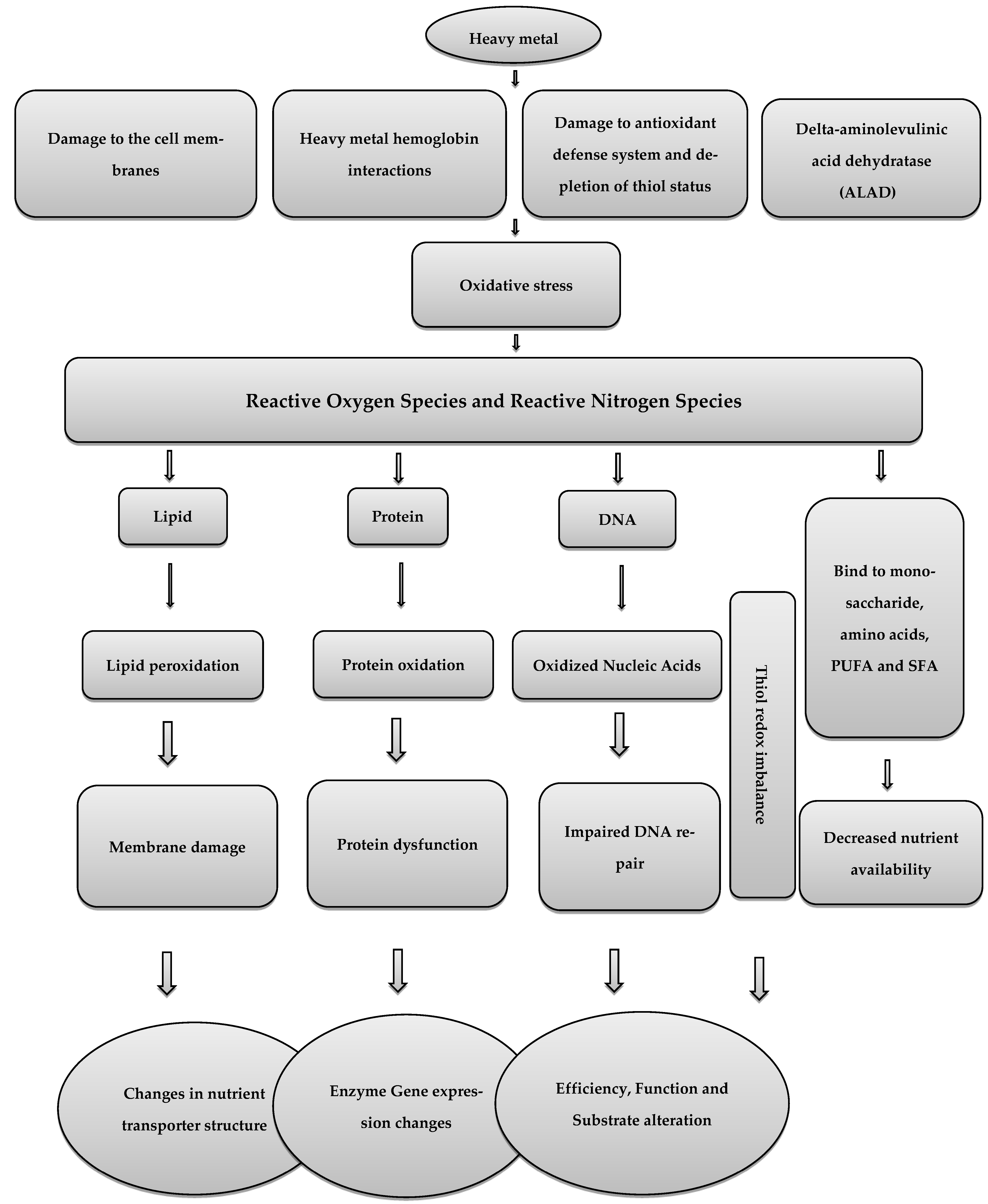
3. Heavy Metals and Oxidative Stress in Poultry

3.1. Pb

Because of Pb’s non-biodegradable nature and the ongoing use of items related to it, there are severe challenges to global food security [20]. Poultry is one of the most important agricultural sectors, and each year, more than 50 billion birds are raised for food [21]. On broiler growth, even 1.0 mg/kg of trace Pb concentration in feed can have a significant effect, including linear decreases in body weight gain, linear decrease in delta-aminolevulinic acid dehydratase (ALAD) activity and dose-related increase in Pb in the blood, kidney, liver and tibia in broiler chickens [22]. Two healthy elements of eggs, ovalbumin and phosvitin, have been proven to bind a variety of metals [23], increasing the risk of heavy metal accumulation in eggs. The rate of Pb transmission from female birds to eggs was assumed to be highly associated with the quantities in livers [24]. A previous investigation suggested that Pb may alter the way Ca is digested, making it easier for Pb to be incorporated into eggshells [25]. Therefore, egg production may offer a means for female birds to eliminate Pb other than through urine or feces [26]. A study showed that Pb can lead to oxidative stress in chickens and hasten biological component oxidation processes by generating free radicals [27]. The key contributor to the ionic mechanism of Pb poisoning, which eventually interferes with cell metabolism, is the ability of Pb metal ions to replace other bivalent cations such as Ca2+, Mg2+, and Fe2+ as well as monovalent cations such as Na+. The ionic mechanism of Pb poisoning significantly affects a wide range of biological activities, including cell adhesion, intracellular and intercellular communication, protein folding, maturation, apoptosis, ionic transport, enzyme control, and neurotransmitter release. Protein kinase C, a gene that regulates neuronal excitation and memory storage, is impacted by the fact that Pb can replace calcium even in picomolar quantities [28]. Pb ions may alter the morphology and operation of the mitochondria, which would cause cell death. The induction of the mitochondrial permeability transition pore (mPTP) is significantly influenced by the production of ROS [29]. The accumulation of ROS brought on by Pb, however, has been shown to impact energy metabolism and alter DNA through point mutations, rearrangements, and fragmentation [29]. It has also proposed that the disruptive effects of Pb ions on mitochondrial respiratory complexes are the root cause of Pb (II)-induced liver damage. The release of cytochrome C and opening the permeability transition pore (PTP), which results in mitochondrial dysfunction and possibly cell death signaling, is what causes the toxicity [30]. Overall, Pb can cause oxidative damage in various organs by having a direct impact on the peroxidation of membrane lipids and lowering antioxidant properties [31]. The antioxidant parameters GPx, CAT, SOD, glutathione S—Transferase (GST), and glutathione (GSH) are considerably decreased by exposure to Pb, whereas the oxidative parameters MDA and H2O2 are increased [32]. In Pb poisoning, caspase-3 is activated once mitochondrial cytochrome C is released, which inhibits Bcl-2 and causes apoptosis [33]. Additionally, blocking the release of neurotransmitters and inhibiting the N-methyl-D-aspartate (NMDA) receptor may cause Pb neurotoxicity and cognitive impairment [34].

3.2. Cd and As

Hazardous heavy metal Cd is widely present in nature and has a variety of adverse effects for human/animal health. The creation of nickel-cadmium batteries, electroplating, burning of fossil fuels, and mining waste are all examples of human activities that contribute to the widespread occurrence of Cd [9]. Due of these factors, almost everything we ingest, such as food, drinks, and air, includes Cd [10]. Continuous exposure to low levels of Cd causes daily buildup in several tissues and adverse effects on various organs [8]. Due to its chemical similarity, it can mimic and substitute some nutritional metals in a range of biological structures [35]. Recent research suggests that Cd may be dangerous to health even at low dosages because of its propensity to accumulate [36]. For animal husbandry in some countries, Cd contamination has been a problem, and in some instances, the amount of Cd in manure and animal feed can even approach 130 mg/kg [37]. Long-term exposure to Cd can result in hepatotoxicity, renal failure, and neurotoxicity [38]. The rapid impacts of Cd exposure in vivo include the generation of ROS in the mitochondrial electron transfer chain being stimulated, NADPH oxidase activity in the plasma being inhibited, and physiological antioxidants such as glutathione being depleted. The buildup of Cd in tissues could slow the pace of growth [39]. The kidney and liver are the main tissue targets for Cd toxicity. Cd produces oxidative stress by increasing the formation of free radicals. Increased ROS can lead to lipid peroxidation, DNA oxidation, sulfhydryl depletion, and a disruption of calcium homoeostasis [40]. The specific mechanism of Cd poisoning is unknown, although its effects on cells are widely known [41]. Cd concentration increases by a factor of 3000 when it binds to metallothionein or other proteins with a high cysteine content. After causing hepatotoxicity in the liver, the cysteine–metallothionein complex builds up in the renal tissue of the kidney and causes nephrotoxicity. Aspartate, glutamate, histidine, and cysteine are also among the ligands that Cd can bind, and this can cause an iron deficiency [42]. Due to their similar oxidation states, Cd and zinc could have been substituted in metallothionein to prevent it from acting as a cell’s free radical scavenger. Cd and Pb both have the capacity to mimic significant metals and/or replace them if necessary. These ions attach to calmodulin [43], protein kinase C [44], troponin C, and synaptic proteins [45] that include magnesium-, zinc-, and calcium-specific binding sites. The most stable complexes are created when these so-called “soft metals” join forces with the mixed N-S donor atom ligand. Recent research on mouse renal tubular epithelial cells has shown that Cd can trigger the release of Ca (II) from ER reserves through the phospholipase C (PLC)-IP3 pathway, which participates in the formation of ROS [46]. Cd can bind to lipids, proteins, and nucleic acids after entering the body. Thiol (-SH) groups are frequently used for binding to enzymes and proteins, and they modify cysteine residues in proteins. This type of protein inactivation has the power to disturb the intracellular redox balance. As a result, liver harm develops as a result of an unbalanced antioxidant defense [7]. Additionally, it has been thought that Cd indirectly produces ROS. The antioxidant defense of cells could be overwhelmed by O2, hydroxyl (OH), and the NO radicals that Cd could indirectly produce [47]. This might be the result of cellular proteins containing more Cd than iron and copper. The buildup of too much Fe and Cu is the cause of the oxidative stress. Additionally, replacing the necessary minerals throws off the cellular metabolism of the cell. Alternately, Cd might interfere with glutathione, causing oxidative stress to develop [48].
Long-term exposure to arsenic has been demonstrated to be harmful to the liver, lung epithelial transformed cells, and skin, and it has also been shown that arsenic alters multiple cellular pathways, including cytokine expression, apoptosis promotion and resistance, and increased oxidative stress [49]. These alterations result in the manifestation of disease [50]. In chicken hearts, subchronic arsenism-induced oxidative stress is also thought to trigger inflammation, and it is believed that ROS overproduction activates the NF-B pathway, which in turn causes an increase in the expression of pro-inflammatory mediators such as TNF, prostaglandin E synthase, cyclooxygenase-2, and inducible nitric oxide synthase [50]. Arsenic pathogenesis is characterized by oxidative damage caused by ROS. Arsenic also causes morphological abnormalities in the mitochondria’s structural integrity. Cells are more susceptible to the harmful effects of arsenic as a result of glutathione-depleting substances paired with cascade mechanisms of free radical generation resulting from the superoxide radical. The formation of ROS/RNS, including peroxyl radicals, the superoxide radical, singlet oxygen, hydroxyl radicals via the Fenton reaction, hydrogen peroxide, the dimethylarsenic radical, the dimethylarsenic peroxyl radical, and/or oxidant-induced DNA damage, is increased in both humans and animals exposed to As [51].

3.3. Mitigation of Oxidative Stress in Poultry

Dietary antioxidants help to keep the intestinal mucosa healthy while lowering intestinal free radicals [17]. Numerous studies indicate that oxidative stress rids birds of various pathogenic and welfare problems [52]. Therefore, the poultry industry must design a practical strategy to prevent oxidative stress [4]. To reduce oxidative distress in poultry [53], a variety of dietary therapies are available based on the best supplementation of antioxidative vitamins (E, A, C, and B2) and micronutrients (Se, Cu, and Zn). Researchers have recently become interested in the chemicals discussed below because of their chelating and growth-stimulating functions, as well as the antioxidant qualities of useful plant components.
The liver is known to be protected by polyphenolic compounds from a variety of xenobiotics, including Pb and diethyl nitrosamine, which can cause hepatotoxicity [54]. Over the past ten years, many in vitro and in vivo studies have suggested that tea and tea polyphenols have potent antioxidant activity as well as a variety of other potentially useful medicinal properties, including the capacity to inhibit tumor growth, metastasis and carcinogenesis in various animals. The main polyphenolic components in tea are catechins. Tea catechins are effective scavengers of superoxide, hydrogen peroxide, hydroxyl radicals, and nitric oxide in various compounds. They were also able to bind with metals, thanks to their catechol structure [55]. The protective effect tea catechins exert on oxidative damage in HepG2 cells exposed to Pb may be due to their capacity to bind metal ions and scavenge free radicals [56]. According to [57], experiments are being conducted to find ways to lessen the harmful effects of Cd and Pb on the body by chelating these metals with nutrients, which reduces their absorption by tissues or boosts the body’s oxidative capacity. However, there are presently no effective techniques to reduce the levels of Cd and Pb in food and hence lessen the risk of oxidative stress being induced in internal organs. Supplemental lycopene and fucoxanthin shield rat kidney, bone, and brain tissue against the effects of Cd-mediated oxidative stress [58].
Recent studies have used astaxanthin, a red carotenoid pigment found in some marine species and a potent antioxidant without provitamin-A activity, to improve rooster sperm quality [59]. According to [60], at a concentration of 25 mg/kg, astaxanthin nanoparticles act as a potent antioxidant to shield rooster testes from the oxidative stress caused by Cd injection and maintain the post-thawing quality of rooster sperm. As with other carotenoids, astaxanthin has a low bioavailability despite being a highly lipophilic molecule. As a result, an astaxanthin nano preparation that is more stable and bioavailable has been developed. However, the current study investigates how astaxanthin, a lipid-soluble carotenoid, protects against Cd-induced damage to rooster testis and decreased sperm quality. Additionally, it has been noted in the literature that astaxanthin has around 100 times the antioxidant activity of alpha-tocopherol and about 10 times the antioxidant activity of other carotenoids, including zeaxanthin, lutein, canthaxanthin, and beta-carotene [61].
It has been demonstrated that the polyphenolic molecule resveratrol has strong antioxidant properties that can protect against hydroxyl and superoxide radicals produced by heavy metals [62]. Additionally, it may increase the activity of GSH-Px, CAT, GST, SOD, and nicotinamide adenine while activating the key transcription factors that regulate the response to antioxidants (erythroid-derived nuclear factor) [63]. It might also maintain glutathione in its reduced state by blocking the creation of glutathione disulfide. This would prevent the oxidation of macromolecules, inhibit the peroxidation of the apolipoprotein B protein, and protect cells from the onslaught of free radicals [64]. Resveratrol has also been demonstrated to lessen oxidative stress and increase antioxidant status in chickens when provided as a dietary supplement [65]. Additionally, yucca’s resveratrol and other phenolic components may reduce lipid peroxidation and stop the generation of blood platelet free radicals (LPO) [66]. It might also maintain glutathione in its reduced state by stopping the manufacture of glutathione disulfide [67]. Chelation therapy, antioxidant therapy, and the consumption of natural food components are among the therapeutic and preventive approaches available to combat As toxicity. Natural dietary substances and medications with a plant-based origin provide effective and progressive treatment from As-mediated toxicity without causing any distinct side effects. Due in significant part to their robust antioxidant properties, bioactive compounds have generated considerable interest in their potential advantages [68]. In a recent study, 34 medicinal plants and 14 natural compounds, largely in preclinical trials and a few in clinical research, demonstrated considerable protection against As toxicity [69]. According to [70], Allium sativum, Curcuma longa, Silybum marianum, as well as various herbal fibers and algae, are the most effective medicinal plants for treating As toxicity. Organosulfur-containing vegetables are helpful in removing arsenic from the liver. Dialyl sulphide, an organosulfur natural substance present in garlic (Allium sativum), has been shown to reduce toxicity and As-induced mitochondrial dysfunction in mice [71]. Crude extracts of Viscum album and Allium sativum were investigated for their ability to counteract in vivo experimentally generated As toxicity [72]. Experimental evidence of the antioxidant properties of members of the Lamiaceae family was found in Ocimum sanctum leaf extract [73]. Due to its antioxidant activity, Silybum marianum’s flavonolignan, silibinin, exerts beneficial effects on rats exposed to As [74]. Table 1 summarizes the key findings of some prior research on the use of several phytomedicinal plants against oxidative stress caused by Pb and Cd. The following paragraphs explain ways that medicinal plants can be used as protective agents and lessen Pb toxicity by preventing intoxication or even being used for treatment (Table 1).

4. Pb and Phytogenic Plants

4.1. Allium sativum (Garlic)

Garlic (Allium sativum) has been used successfully in traditional medicine to treat a number of conditions and also improve broiler performance [109]. This effectiveness may be explained by the mixture’s high content of lipid-soluble molecules, S-allyl cysteine, and organic sulfur compounds [75]. Garlic is utilized extensively to lessen the toxicity brought on by metals. According to new supportive studies, garlic compounds are able to detoxify methyl mercury, phenyl mercury, arsenic, Pb, and Cd. Consuming garlic dramatically reduces heavy metals’ clastogenic effects, and also significantly reduces the lethal effects of oxidative stress and mitochondrial damage brought on by the body’s metal load. The therapeutic benefits of garlic were superior to those of D-penicillamine and 2,3-mercapto-1-propanol, N-acetyl DL-penicillamine and 2,3-dimercaptosuccinic acid, and the current therapies. It is generally known that broiler chicks are susceptible to Pb toxicity, which indicates that dietary Pb concentrations of (1.0 mg/kg) can considerably impair broiler growth and persistently lower blood levels of the erythrocyte enzyme known as d-aminolaevulinic acid dehydratase [22]. Garlic administration is protective against Pb toxicity in chickens [76] with the exception of feed conversion rate modifications brought on by Pb poisoning (100 mg/kg). Broiler chickens were fed garlic (1, 2 or 4 percent Allium sativum) to improve growth performance and significantly raise blood variable values [110]. The three main factors that contribute to garlic’s protective qualities against Pb toxicity are its ability to chelate Pb, which is provided by compounds with free amino and carboxyl groups and amino acids containing sulfur, as well as its antioxidative role, which is provided by organo-sulfur components such as diallyl tetra sulfide [111].

4.2. Yucca schidigera

There are only a few of Yucca schidigera advantageous properties, which include growth-promoting, hypocholesterolemia, hypoglycemia, antioxidant, immunostimulatory, anticancer, and anti-inflammatory properties [63]. It also provides resveratrol, saponins, numerous enzymes, and antioxidants for use in the poultry industry [63]. In chicken farms, Yucca can sustain ammonia emission and lower its level, and lower blood urea levels [80]. Hazardous volatile chemicals such as hydrogen sulfide and ammonia are easily absorbed by Yucca [81]. Recently, Yucca showed a possible ability to modulate the effects of Pb-induced inhibition on the reproductive and productive characteristics of Japanese quails [63]. The authors claim that Yucca schidigera therapy, especially at a dose of 200 mg/kg diet, may reverse Pb-induced modifications as well as increase immunohistochemistry and histomorphometric abnormalities. These beneficial effects may be due to its antioxidant properties [82]; NO, vascular endothelial growth factor (VEGF), tumor necrosis factor-alpha (TNF-α), and transforming growth factor-1 (TGF-1) levels; and declining levels of these inflammatory markers. Quails that were also subjected to Yucca schidigera extract displayed significantly better control of glucose homeostasis [16].

4.3. Coriandrum sativum (Coriander)

Coriander (Coriandrum sativum), a common member of the “Umbelliferae” plant family, has a number of beneficial therapeutic properties, which have revealed that Coriandrum sativum supplements are beneficial for a variety of animal species both nutritionally and therapeutically [84]. Because coriandrin and isocoumarines are this herb’s main chemical constituents, it is utilized as a potent antioxidant [112]. In the field of complementary medicine, coriander is extremely beneficial for treating stomach ulcers and other abdominal problems. It was discovered that Coriandrum sativum may have antioxidant qualities and may worsen the oxidative state in male rats after Pb overdose in the same environment [113]. Additionally, following treatment with the hydroalcoholic seed extract of Coriandrum sativum, the tissue-specific oxidative stress caused by Pb was reduced. Lower Pb concentrations and the lessening of histopathologic liver damage were seen in rat tissues after coriander treatment [114]. According to a recent study by [85], coriander leaf extract ingestion increases resistance to oxidative stress in the kidney, potentially through lower levels of heavy metals.

4.4. Garcinia Kola Heckel (Bitter cola)

Garcinia kola (Bitter kola) is beginning to be utilized in toxicological research as a therapy for numerous dangerous heavy metals and medications due to the presence of carbazole alkaloid, phenolic, organic, and inorganic compounds, which are responsible for its antioxidant benefits [86]. In recent studies using Wistar rats as experimental models, [87] evaluated the effectiveness of Garcinia kola aqueous extracts to reduce chronic Pb acetate poisoning. This study confirms therapeutic potential of coated and uncoated Bitter cola for treating Pb toxicity and serving as an alternate antibacterial. Additionally, Garcinia kola has two beneficial effects for treating Pb-related poisoning: first, reducing Pb toxicity, and second, limiting infections.

5. Cd and Phytogenic Plants

5.1. Garlic (Allium sativum L.)

A study by [91] confirmed the potent anti-Cd properties of Allium sativum and suggested using it in any form as a regular source to fend off the negative effects of Cd in food and drink. They claimed that the difference in body weight between the Cd-plus-garlic group and the Cd-alone group proved the value of garlic in reducing the effects of Cd poisoning.

5.2. Milk Thistle (Silybum marianum)

High levels of the antioxidants silibinin, silidianin, and silichristin can be found in the well-known herbal medicine Silybum marianum. This antioxidant property has been extensively used to reduce the potentially hazardous in vivo effects of numerous oxidative agents, in addition to its use in the treatment of cancer and liver disease. The efficacy of Silybum marianum against Cd-induced DNA damage in human blood cells has been studied in the past [92].

5.3. Ginger (Zingiber officinale)

For many years, people have safely treated a stomach upset using Zingiber officinale. In addition, various studies have focused on the antioxidant properties of ginger. Ginger’s antioxidant and hepatorenal protective effects against Cd-induced oxidative stress have previously been investigated in rat and fetal tissues [93]. Cd administration could have an impact on the immunohistochemistry expression of Caspase3 and tumor marker MKI67, which indicated that ginger had a protective effect against Cd poisoning in the kidney and liver of rabbits.

5.4. Winter Cherry (Withania somnifera)

The reduction and eradication of Cd toxicity in broiler chickens has been widely studied using a common medicinal plant called Withania somnifera. Oral administration of winter cherry root prevented the Cd-induced peroxidation of chicken tissues [95]. According to some studies [98,103], the effects of Cd-induced oxidative stress and hepatorenal damage in broilers can be mitigated by Withania somnifera. The plant’s high content of alkaloids, steroidal lactones, tropine, withanolides, and withaferin may account for these effects [97].

5.5. Tulsi Leaf (Ocimum sanctum)

Occimum sanctum has been used for medicinal and spiritual purposes. Numerous research studies have looked at Ocimum sanctum’s ameliorating effects on Cd-induced oxidative stress and tissue damage [99]. Ocimum sanctum has an important role in tissue repair, it acts as an antioxidant, and it has anti-metal bioaccumulation properties. Additionally, it enhances oxidative state after Cd poisoning [99].

5.6. Indian Gooseberry (Emblica officinalis)

The Indian medicinal herb Emblica officinalis has been used for centuries to enhance mental and physical health. Prior studies using rats and mice showed that Emblica officinalis has antioxidant properties and the ability to remove ROS from the environment [106] and broiler chickens [113], which has been exploited to mitigate the negative effects of the heavy metal Cd on physiological functioning [105]. It has been discovered that ellagic and gallic acids play the main part in their antioxidant properties [115].

5.7. Rosemary (Rosmarinus officinalis)

The rosemary plant’s significant anti-inflammatory and antibacterial capabilities are due to the presence of flavonoids and phenolic acids. Additionally, the extracted rosmarinic acid from the plant possesses strong antioxidant properties that guard against a variety of oxidative diseases. Studies looked at the benefits of Rosmarinus officinalis in vivo against the hepatic, renal, and oxidative stress brought on by Cd chloride [107,116].

5.8. Extract of Korean Ginseng (Panex ginseng)

Panax ginseng, a well-known species of the ginseng plant family, is frequently and effectively used to increase immunity and minimize the side effects of several risky medical operations. Furthermore, it was successful in halting tissue oxidation and the damage brought on by Cd. The antioxidant and anti-apoptotic properties of Panax ginseng depend on its ginsenosides [117]. According to earlier research, Panax ginseng shielded rats and mice against the negative effects of Cd [118].

5.9. Other Different Types of Herbal Plants

More varieties of herbal plants [103] have been studied. Some researchers described the effectiveness of using various herbal adaptogens against Cd-induced oxidative stress and toxicity in broiler chicks. For example, regarding tulasi (O. sanctum), shatavari, holy basil, satavar or shatamull (Asperagus recemosus), kalmegh (Andrographis paniculata), or shilajith (Asphaltum panjabinum), it is possible to conclude that using such adaptogens decreases Cd bioaccumulation in the kidney and liver, which in turn decreases oxidative damage.

6. Conclusions

Environmental contamination has significantly increased over the past few decades, offering potential risks to all biological systems, including the poultry sector. In addition to exposure from the general environment, industrial pollution, and agricultural processes, chickens are also exposed to heavy metals such as Cd and Pb. Heavy metal toxicity has been demonstrated to have negative effects on biological macromolecules, lipid peroxidation, protein oxidation, and cell structure, and increases oxidative stress and the formation of ROS/RNS. In the last two decades, phytobiotics have been shown to exert multiple effects, including anti-inflammatory, antimicrobial, antioxidative, and metabolic-modulating effects. According to the probable effect of heavy metals on the development of Pb-/Cd-induced oxidative stress, some plant secondary metabolites with antioxidant and chelating properties may assist with and modify Pb-/Cd-induced toxicity. Their use could be available to relevant specialists as a nutritional strategy to ensure the health of animal protein products in the human food chain.

Author Contributions

Conceptualization, M.S. and R.E.; resources, M.S. and M.E.; writing—original draft preparation, R.E.; writing—review and editing, M.S. and M.E. All authors have read and agreed to the published version of the manuscript.

Funding

This research received no external funding.

Institutional Review Board Statement

Not applicable.

Informed Consent Statement

Not applicable.

Data Availability Statement

Not applicable.

Conflicts of Interest

The authors declare no conflict of interest.

References

  1. Bacou, E.; Walk, C.; Rider, S.; Litta, G.; Perez-Calvo, E. Dietary oxidative distress: A review of nutritional challenges as models for poultry, swine and fish. Antioxidants 2021, 10, 525. [Google Scholar] [CrossRef] [PubMed]
  2. Shakeri, M.; Oskoueian, E.; Le, H.H.; Shakeri, M. Strategies to combat heat stress in broiler chickens: Unveiling the roles of selenium, vitamin E and vitamin C. Vet. Sci. 2020, 7, 71. [Google Scholar] [CrossRef]
  3. Shakeri, M.; Le, H.H. Deleterious Effects of Heat Stress on Poultry Production: Unveiling the Benefits of Betaine and Polyphenols. Poultry 2022, 1, 147–156. [Google Scholar] [CrossRef]
  4. Mishra, B.; Jha, R. Oxidative stress in the poultry gut: Potential challenges and interventions. Front. Vet. Sci. 2019, 6, 60. [Google Scholar] [CrossRef] [PubMed] [Green Version]
  5. Shakeri, M.; Cottrell, J.J.; Wilkinson, S.; Zhao, W.; Le, H.H.; McQuade, R.; Furness, J.B.; Dunshea, F.R. Dietary betaine improves intestinal barrier function and ameliorates the impact of heat stress in multiple vital organs as measured by evans blue dye in broiler chickens. Animals 2019, 10, 38. [Google Scholar] [CrossRef] [PubMed] [Green Version]
  6. Sharma, R.K.; Agrawal, M. Biological effects of heavy metals: An overview. J. Environ. Biol. 2005, 26, 301–313. [Google Scholar]
  7. Balali-Mood, M.; Naseri, K.; Tahergorabi, Z.; Khazdair, M.R.; Sadeghi, M. Toxic mechanisms of five heavy metals: Mercury, lead, chromium, cadmium, and arsenic. Front. Pharmacol. 2021, 12, 643972. [Google Scholar] [CrossRef]
  8. Khafaga, A.F.; El-Hack, A.; Mohamed, E.; Taha, A.E.; Elnesr, S.S.; Alagawany, M. The potential modulatory role of herbal additives against Cd toxicity in human, animal, and poultry: A review. Environ. Sci. pollut. Res. 2019, 26, 4588–4604. [Google Scholar] [CrossRef]
  9. Nai, G.A.; Marin, F.F.; Queiroz, L.M.M.; Estrella, M.P.S. Respiratory tract cadmium-induced injuries—poisoning via intake and water pH could influence their genesis? An experimental study in rats. Comp. Clin. Path. 2017, 26, 997–1002. [Google Scholar] [CrossRef]
  10. Schaefer, H.R.; Dennis, S.; Fitzpatrick, S. Cadmium: Mitigation strategies to reduce dietary exposure. J. Food Sci. 2020, 85, 260–267. [Google Scholar] [CrossRef] [Green Version]
  11. Papanikolaou, N.C.; Hatzidaki, E.G.; Belivanis, S.; Tzanakakis, G.N.; Tsatsakis, A.M. Lead toxicity update. A brief review. Med. Sci. Monit. 2005, 11, RA329. [Google Scholar] [PubMed]
  12. Korish, M.A.; Attia, Y.A. Evaluation of heavy metal content in feed, litter, meat, meat products, liver, and table eggs of chickens. Animals 2020, 10, 727. [Google Scholar] [CrossRef] [PubMed] [Green Version]
  13. Baloš, M.Ž.; Jakšić, S.; Pelić, D.L. The role, importance and toxicity of arsenic in poultry nutrition. Worlds Poult. Sci. J. 2019, 75, 375–386. [Google Scholar] [CrossRef]
  14. Cadet, J.; Davies, K.J. Oxidative DNA damage & repair: An introduction. Free Radic. Biol. Med. 2017, 107, 2–12. [Google Scholar]
  15. Rehman, Z.U.; Meng, C.; Sun, Y.; Safdar, A.; Pasha, R.H.; Munir, M.; Ding, C. Oxidative stress in poultry: Lessons from the viral infections. Oxid. Med. Cell. Longev. 2018, 2018, 5123147. [Google Scholar] [CrossRef] [Green Version]
  16. Farag, M.R.; Alagawany, M.; Abd El-Hack, M.E.; El-Sayed, S.A.; Ahmed, S.Y.; Samak, D.H. Yucca schidigera extract modulates the lead-induced oxidative damage, nephropathy and altered inflammatory response and glucose homeostasis in Japanese quails. Ecotoxicol. Environ. Saf. 2018, 156, 311–321. [Google Scholar] [CrossRef]
  17. Surai, P.F.; Kochish, I.I.; Fisinin, V.I.; Kidd, M.T. Antioxidant defence systems and oxidative stress in poultry biology: An update. Antioxidants 2019, 8, 235. [Google Scholar] [CrossRef] [PubMed] [Green Version]
  18. Bottje, W.; Wang, S.; Kelly, F.J.; Dunster, C.; Williams, A.; Mudway, I. Antioxidant defenses in lung lining fluid of broilers: Impact of poor ventilation conditions. Poult. Sci. 1998, 77, 516–522. [Google Scholar] [CrossRef]
  19. Xiong, Y.; Yin, Q.; Li, J.; He, S. Oxidative stress and endoplasmic reticulum stress are involved in the protective effect of alpha lipoic acid against heat damage in chicken testes. Animals 2020, 10, 384. [Google Scholar] [CrossRef] [Green Version]
  20. Zheng, S.; Wang, Q.; Yuan, Y.; Sun, W. Human health risk assessment of heavy metals in soil and food crops in the Pearl River Delta urban agglomeration of China. Food Chem. 2020, 316, 126213. [Google Scholar] [CrossRef] [PubMed]
  21. Okoye, P.; Ajiwe, V.; Okeke, O.; Ujah, I.; Asalu, U.; Okeke, D. Estimation of heavy metal levels in the muscle, gizzard, liver and kidney of broiler, layer and local (cockerel) chickens raised within Awka metropolis and its environs, Anambra state, south eastern Nigeria. J. Environ. Prot. 2015, 6, 609. [Google Scholar] [CrossRef] [Green Version]
  22. Bakalli, R.; Pesti, G.; Ragland, W. The magnitude of lead toxicity in broiler chickens. Vet. Hum. Toxicol. 1995, 37, 15–19. [Google Scholar]
  23. Dobrzański, Z.; Gorecki, H.; Chojnacka, K.; Gorecka, H.; Synowiec, M. Effect of dietary humic preparations on the content of trace elements in hens’ eggs. Am. J. Agric. Biol. Sci. 2007, 2, 234–240. [Google Scholar]
  24. Abduljaleel, S.A.; Shuhaimi-Othman, M. Metals concentrations in eggs of domestic avian and estimation of health risk from eggs consumption. J. Biol. Sci. 2011, 11, 448–453. [Google Scholar] [CrossRef] [Green Version]
  25. Hunton, P. Research on eggshell structure and quality: An historical overview. Braz. J. Poult. Sci. 2005, 7, 67–71. [Google Scholar] [CrossRef] [Green Version]
  26. Dauwe, T.; Janssens, E.; Bervoets, L.; Blust, R.; Eens, M. Heavy-metal concentrations in female laying great tits (Parus major) and their clutches. Arch. Environ. Contam. Toxicol. 2005, 49, 249–256. [Google Scholar] [CrossRef] [PubMed]
  27. Ebrahimi, R.; Faseleh Jahromi, M.; Liang, J.B.; Soleimani Farjam, A.; Shokryazdan, P.; Idrus, Z. Effect of dietary lead on intestinal nutrient transporters mRNA expression in broiler chickens. BioMed Res. Int. 2015, 2015, 149745. [Google Scholar] [CrossRef] [PubMed] [Green Version]
  28. Flora, S.; Mittal, M.; Mehta, A. Heavy metal induced oxidative stress & its possible reversal by chelation therapy. Indian J. Med. Res. 2008, 128, 501. [Google Scholar] [PubMed]
  29. Ye, F.; Li, X.; Li, F.; Li, J.; Chang, W.; Yuan, J.; Chen, J. Cyclosporin A protects against Lead neurotoxicity through inhibiting mitochondrial permeability transition pore opening in nerve cells. Neurotoxicology 2016, 57, 203–213. [Google Scholar] [CrossRef]
  30. Ma, L.; Liu, J.-Y.; Dong, J.-X.; Xiao, Q.; Zhao, J.; Jiang, F.-L. Toxicity of Pb2+ on rat liver mitochondria induced by oxidative stress and mitochondrial permeability transition. Toxicol. Res. 2017, 6, 822–830. [Google Scholar] [CrossRef] [Green Version]
  31. Gurer-Orhan, H.; Sabır, H.U.; Özgüneş, H. Correlation between clinical indicators of lead poisoning and oxidative stress parameters in controls and lead-exposed workers. Toxicology 2004, 195, 147–154. [Google Scholar] [CrossRef] [PubMed]
  32. Wang, J.; Zhu, H.; Yang, Z.; Liu, Z. Antioxidative effects of hesperetin against lead acetate-induced oxidative stress in rats. Indian J. Pharmacol. 2013, 45, 395. [Google Scholar]
  33. Liu, C.-M.; Ma, J.-Q.; Sun, Y.-Z. Puerarin protects rat kidney from lead-induced apoptosis by modulating the PI3K/Akt/eNOS pathway. Toxicol. Appl. Pharmacol. 2012, 258, 330–342. [Google Scholar] [CrossRef]
  34. Neal, A.P.; Guilarte, T.R. Molecular neurobiology of lead (Pb2+): Effects on synaptic function. Mol. Neurobiol. 2010, 42, 151–160. [Google Scholar] [CrossRef] [PubMed] [Green Version]
  35. Zhu, M.; Li, H.; Bai, L.; Wang, L.; Zou, X. Histological changes, lipid metabolism, and oxidative and endoplasmic reticulum stress in the liver of laying hens exposed to cadmium concentrations. Poult. Sci. 2020, 99, 3215–3228. [Google Scholar] [CrossRef] [PubMed]
  36. Branca, J.J.V.; Morucci, G.; Pacini, A. Cadmium-induced neurotoxicity: Still much ado. Neural Regen. Res. 2018, 13, 1879. [Google Scholar]
  37. Li, Y.-x.; Xiong, X.; Chun-ye, L.; Feng-song, Z.; Wei, L.; Wei, H. Cadmium in animal production and its potential hazard on Beijing and Fuxin farmlands. J. Hazard. Mater. 2010, 177, 475–480. [Google Scholar] [CrossRef]
  38. Fan, R.; Hu, P.-c.; Wang, Y.; Lin, H.-y.; Su, K.; Feng, X.-s.; Wei, L.; Yang, F. Betulinic acid protects mice from cadmium chloride-induced toxicity by inhibiting cadmium-induced apoptosis in kidney and liver. Toxicol. Lett. 2018, 299, 56–66. [Google Scholar] [CrossRef]
  39. Akyolcu, M.; Ozcelik, D.; Dursun, S.; Toplan, S.; Kahraman, R. Accumulation of cadmium in tissue and its effect on live performance. In Proceedings of the Journal de Physique IV (Proceedings), Porto, Portugal, 8–10 September 2003; pp. 33–36. [Google Scholar]
  40. Stohs, S.J.; Bagchi, D. Oxidative mechanisms in the toxicity of metal ions. Free Radic. Biol. Med. 1995, 18, 321–336. [Google Scholar] [CrossRef] [PubMed] [Green Version]
  41. Patrick, L. Toxic metals and antioxidants: Part II. The role of antioxidants in arsenic and cadmium toxicity. Altern. Med. Rev. 2003, 8, 106–128. [Google Scholar]
  42. Castagnetto, J.M.; Hennessy, S.W.; Roberts, V.A.; Getzoff, E.D.; Tainer, J.A.; Pique, M.E. MDB: The metalloprotein database and browser at the Scripps Research Institute. Nucleic Acids Res. 2002, 30, 379–382. [Google Scholar] [CrossRef]
  43. Kern, M.; Wisniewski, M.; Cabell, L.; Audesirk, G. Inorganic lead and calcium interact positively in activation of calmodulin. Neurotoxicology 2000, 21, 353–363. [Google Scholar] [PubMed]
  44. Sun, X.; Tian, X.; Tomsig, J.L.; Suszkiw, J.B. Analysis of differential effects of Pb2+ on protein kinase C isozymes. Toxicol. Appl. Pharmacol. 1999, 156, 40–45. [Google Scholar] [CrossRef] [PubMed]
  45. Marchetti, C. Role of calcium channels in heavy metal toxicity. Int. Sch. Res. Not. 2013, 2013, 184360. [Google Scholar] [CrossRef] [Green Version]
  46. Gu, J.; Dai, S.; Liu, Y.; Liu, H.; Zhang, Y.; Ji, X.; Yu, F.; Zhou, Y.; Chen, L.; Tse, W.K.F. Activation of Ca2+-sensing receptor as a protective pathway to reduce Cadmium-induced cytotoxicity in renal proximal tubular cells. Sci. Rep. 2018, 8, 1–13. [Google Scholar] [CrossRef] [Green Version]
  47. Sarkar, A.; Ravindran, G.; Krishnamurthy, V. A brief review on the effect of cadmium toxicity: From cellular to organ level. Int. J. Biotechnol. Res. 2013, 3, 17–36. [Google Scholar]
  48. Liu, J.; Qu, W.; Kadiiska, M.B. Role of oxidative stress in cadmium toxicity and carcinogenesis. Toxicol. Appl. Pharmacol. 2009, 238, 209–214. [Google Scholar] [CrossRef] [PubMed] [Green Version]
  49. Flora, S.J. Arsenic-induced oxidative stress and its reversibility. Free Radic. Biol. Med. 2011, 51, 257–281. [Google Scholar] [CrossRef] [PubMed]
  50. Zhao, H.; He, Y.; Li, S.; Sun, X.; Wang, Y.; Shao, Y.; Hou, Z.; Xing, M. Subchronic arsenism-induced oxidative stress and inflammation contribute to apoptosis through mitochondrial and death receptor dependent pathways in chicken immune organs. Oncotarget 2017, 8, 40327. [Google Scholar] [CrossRef] [PubMed] [Green Version]
  51. Jomova, K.; Jenisova, Z.; Feszterova, M.; Baros, S.; Liska, J.; Hudecova, D.; Rhodes, C.; Valko, M. Arsenic: Toxicity, oxidative stress and human disease. J. Appl. Toxicol. 2011, 31, 95–107. [Google Scholar] [CrossRef] [PubMed]
  52. Estévez, M. Oxidative damage to poultry: From farm to fork. Poult. Sci. 2015, 94, 1368–1378. [Google Scholar] [CrossRef] [PubMed]
  53. Lauridsen, C. From oxidative stress to inflammation: Redox balance and immune system. Poult. Sci. 2019, 98, 4240–4246. [Google Scholar] [CrossRef] [PubMed]
  54. McGill, M.R.; Du, K.; Weemhoff, J.L.; Jaeschke, H. Critical review of resveratrol in xenobiotic-induced hepatotoxicity. Food Chem. Toxicol. 2015, 86, 309–318. [Google Scholar] [CrossRef] [PubMed] [Green Version]
  55. Rice-Evans, C. Flavonoid antioxidants. Curr. Med. Chem. 2001, 8, 797–807. [Google Scholar] [CrossRef] [PubMed]
  56. Chen, L.; Yang, X.; Jiao, H.; Zhao, B. Tea catechins protect against lead-induced cytotoxicity, lipid peroxidation, and membrane fluidity in HepG2 cells. Toxicol. Sci. 2002, 69, 149–156. [Google Scholar] [CrossRef] [Green Version]
  57. Winiarska-Mieczan, A. Protective effect of tea against lead and cadmium-induced oxidative stress—A review. Biometals 2018, 31, 909–926. [Google Scholar] [CrossRef] [Green Version]
  58. Mężyńska, M.; Brzóska, M.M.; Rogalska, J.; Piłat-Marcinkiewicz, B. Extract from Aronia melanocarpa L. berries prevents cadmium-induced oxidative stress in the liver: A study in a rat model of low-level and moderate lifetime human exposure to this toxic metal. Nutrients 2018, 11, 21. [Google Scholar] [CrossRef] [Green Version]
  59. Fang, Y.; Zhong, R.; Chen, L.; Feng, C.; Sun, H.; Zhou, D. Effects of astaxanthin supplementation on the sperm quality and antioxidant capacity of ram semen during liquid storage. Small Rumin. Res. 2015, 130, 178–182. [Google Scholar] [CrossRef]
  60. Najafi, D.; Taheri, R.A.; Najafi, A.; Shamsollahi, M.; Alvarez-Rodriguez, M. Effect of astaxanthin nanoparticles in protecting the post-thawing quality of rooster sperm challenged by cadmium administration. Poult. Sci. 2020, 99, 1678–1686. [Google Scholar] [CrossRef] [PubMed]
  61. Miki, W. Biological functions and activities of animal carotenoids. Pure Appl. Chem. 1991, 63, 141–146. [Google Scholar] [CrossRef]
  62. Rubiolo, J.; Vega, F. Resveratrol protects primary rat hepatocytes against necrosis induced by reactive oxygen species. Biomed. Pharmacother. 2008, 62, 606–612. [Google Scholar] [CrossRef] [PubMed]
  63. Alagawany, M.; El-Hack, A.; Mohamed, E.; El-Kholy, M.S. Productive performance, egg quality, blood constituents, immune functions, and antioxidant parameters in laying hens fed diets with different levels of Yucca schidigera extract. Environ. Sci. pollut. Res. 2016, 23, 6774–6782. [Google Scholar] [CrossRef] [PubMed]
  64. Chakravarthi, S.; Jessop, C.E.; Bulleid, N.J. The role of glutathione in disulphide bond formation and endoplasmic-reticulum-generated oxidative stress. EMBO Rep. 2006, 7, 271–275. [Google Scholar] [CrossRef] [Green Version]
  65. Wang, C.; Zhao, F.; Li, Z.; Jin, X.; Chen, X.; Geng, Z.; Hu, H.; Zhang, C. Effects of resveratrol on growth performance, intestinal development, and antioxidant status of broilers under heat stress. Animals 2021, 11, 1427. [Google Scholar] [CrossRef] [PubMed]
  66. Olas, B.; Wachowicz, B.; Stochmal, A.; Oleszek, W. Inhibition of oxidative stress in blood platelets by different phenolics from Yucca schidigera Roezl. bark. Nutrition 2003, 19, 633–640. [Google Scholar] [CrossRef]
  67. Nurdiana, S.; Goh, Y.M.; Ahmad, H.; Dom, S.M.; Syimal’ain Azmi, N.; Noor Mohamad Zin, N.S.; Ebrahimi, M. Changes in pancreatic histology, insulin secretion and oxidative status in diabetic rats following treatment with Ficus deltoidea and vitexin. BMC Complement. Altern. Med. 2017, 17, 1–17. [Google Scholar] [CrossRef] [Green Version]
  68. Krishnaiah, D.; Sarbatly, R.; Nithyanandam, R. A review of the antioxidant potential of medicinal plant species. Food Bioprod. Process. 2011, 89, 217–233. [Google Scholar] [CrossRef]
  69. Bhattacharya, S. Medicinal plants and natural products in amelioration of arsenic toxicity: A short review. Pharm. Biol. 2017, 55, 349–354. [Google Scholar] [CrossRef] [Green Version]
  70. Mehrandish, R.; Rahimian, A.; Shahriary, A. Heavy metals detoxification: A review of herbal compounds for chelation therapy in heavy metals toxicity. J. Herbmed Pharmacol. 2019, 8, 69–77. [Google Scholar] [CrossRef] [Green Version]
  71. Das, B.; Chaudhuri, K. Amelioration of sodium arsenite induced toxicity by diallyl disulfide, a bioactive component of garlic: The involvement of antioxidants and the chelate effect. RSC Adv. 2014, 4, 20964–20973. [Google Scholar] [CrossRef]
  72. Adegboyega, A.; Odunola, O. The modulatory effects of aqueous extracts of Viscum album and garlic on sodium arsenite induced toxicity in Wistar albino rat. J. Chem. Pharm. Res. 2012, 4, 4698–4701. [Google Scholar]
  73. Sharmila Banu, G.; Kumar, G.; Murugesan, A. Effect of ethanolic leaf extract of Trianthema portulacastrum L. on aflatoxin induced hepatic damage in rats. Indian J. Clin. Biochem. 2009, 24, 414–418. [Google Scholar] [CrossRef] [Green Version]
  74. Bjørklund, G.; Rahaman, M.S.; Shanaida, M.; Lysiuk, R.; Oliynyk, P.; Lenchyk, L.; Chirumbolo, S.; Chasapis, C.T.; Peana, M. Natural dietary compounds in the treatment of arsenic toxicity. Molecules 2022, 27, 4871. [Google Scholar] [CrossRef] [PubMed]
  75. Arreola, R.; Quintero-Fabián, S.; López-Roa, R.I.; Flores-Gutiérrez, E.O.; Reyes-Grajeda, J.P.; Carrera-Quintanar, L.; Ortuño-Sahagún, D. Immunomodulation and anti-inflammatory effects of garlic compounds. J. Immunol. Res. 2015, 2015, 401630. [Google Scholar] [CrossRef] [PubMed] [Green Version]
  76. Ibrahim, D.; Ismail, T.A.; Khalifa, E.; Abd El-Kader, S.A.; Mohamed, D.I.; Mohamed, D.T.; Shahin, S.E.; Abd El-Hamid, M.I. Supplementing Garlic Nanohydrogel Optimized Growth, Gastrointestinal Integrity and Economics and Ameliorated Necrotic Enteritis in Broiler Chickens Using a Clostridium perfringens Challenge Model. Animals 2021, 11, 2027. [Google Scholar] [CrossRef]
  77. Morales-González, J.A.; Madrigal-Bujaidar, E.; Sánchez-Gutiérrez, M.; Izquierdo-Vega, J.A.; Valadez-Vega, M.d.C.; Álvarez-González, I.; Morales-González, Á.; Madrigal-Santillán, E. Garlic (Allium sativum L.): A brief review of its antigenotoxic effects. Foods 2019, 8, 343. [Google Scholar] [CrossRef] [PubMed] [Green Version]
  78. Obioha, U.E.; Suru, S.M.; Ola-Mudathir, K.F.; Faremi, T.Y. Hepatoprotective potentials of onion and garlic extracts on cadmium-induced oxidative damage in rats. Biol. Trace Elem. Res. 2009, 129, 143–156. [Google Scholar] [CrossRef] [PubMed]
  79. Ro, J.-H.; Liu, C.-C.; Lin, M.-C. Resveratrol mitigates cerebral ischemic injury by altering levels of trace elements, toxic metal, lipid peroxidation, and antioxidant activity. Biol. Trace Elem. Res. 2021, 199, 3718–3727. [Google Scholar] [CrossRef] [PubMed]
  80. Piacente, S.; Pizza, C.; Oleszek, W. Saponins and phenolics of Yucca schidigera Roezl: Chemistry and bioactivity. Phytochem. Rev. 2005, 4, 177–190. [Google Scholar] [CrossRef]
  81. Saeed, M.; Arain, M.A.; Naveed, M.; Alagawany, M.; El-Hack, A.; Ezzat, M.; Bhutto, Z.A.; Bednarczyk, M.; Kakar, M.U.; Abdel-Latif, M. Yucca schidigera can mitigate ammonia emissions from manure and promote poultry health and production. Environ. Sci. pollut. Res. 2018, 25, 35027–35033. [Google Scholar] [CrossRef]
  82. Taha, H.S.; Abdelnour, S.A.; Alagawany, M. Growth performance, biochemical, cytological and molecular aspects of rabbits exposed to lead toxicity. J. Anim. Physiol. Anim. Nutr. 2019, 103, 747–755. [Google Scholar] [CrossRef] [PubMed]
  83. Fatemi, H.; Pour, B.E.; Rizwan, M. Isolation and characterization of lead (Pb) resistant microbes and their combined use with silicon nanoparticles improved the growth, photosynthesis and antioxidant capacity of coriander (Coriandrum sativum L.) under Pb stress. Environ. Pollut. 2020, 266, 114982. [Google Scholar] [CrossRef]
  84. Velaga, M.K.; Yallapragada, P.R.; Williams, D.; Rajanna, S.; Bettaiya, R. Hydroalcoholic seed extract of Coriandrum sativum (Coriander) alleviates lead-induced oxidative stress in different regions of rat brain. Biol. Trace Elem. Res. 2014, 159, 351–363. [Google Scholar] [CrossRef]
  85. Nishio, R.; Tamano, H.; Morioka, H.; Takeuchi, A.; Takeda, A. Intake of heated leaf extract of Coriandrum sativum contributes to resistance to oxidative stress via decreases in heavy metal concentrations in the kidney. Plant Foods Hum. Nutr. 2019, 74, 204–209. [Google Scholar] [CrossRef] [PubMed]
  86. Daramola, B.; Adegoke, G. Bitter kola (Garcinia kola) seeds and health management potential. In Nuts and Seeds in Health and Disease Prevention; Elsevier: Amsterdam, The Netherlands, 2011; pp. 213–220. [Google Scholar]
  87. Osemwegie, O.O.; Nwonuma, C.O.; Oluyori, A.P.; Abraham, P.O.; Akanbi, A.A.; Opaleke, D.O.; Alejolowo, O.O. In vitro antimicrobial and in vivo lead acetate poison abatement study of Garcinia kola Heckel. J. Taibah Univ. Sci. 2017, 11, 883–894. [Google Scholar] [CrossRef] [Green Version]
  88. Tandon, S.; Singh, S.; Prasad, S. Influence of garlic on the disposition and toxicity of lead and cadmium in the rat. Pharm. Biol. 2001, 39, 450–454. [Google Scholar] [CrossRef]
  89. Mohamed, M.; Mohamed, A.H. Protective role of garlic against cadmium toxicity in rats: Clinicopathological and histopathological studies. Egypt. J. Comp. Path. Clin. Path 2009, 22, 114–140. [Google Scholar]
  90. Eteng, M.U.; Onwuka, F.C.; Akpanyung, E.O.; Osuchukwu, N.C.; Bassey, S.C.; Nwankpa, P. Reversal of cadmium induced toxicity following dietary supplementation with garlic, ginger and cabbage in male Wistar rats. J. Nat. Prod. Plant. Res. 2012, 2, 169–174. [Google Scholar]
  91. Andleeb, S.; Shaukat, S.; Ara, C. Protection against cadmium-induced abnormalities and hepatotoxicity in ovo by Allium sativum. Punjab Univ. J. Zool. 2018, 33, 34–41. [Google Scholar] [CrossRef]
  92. Borges, F.F.V.; e Silva, C.R.; Goes, W.M.; Godoy, F.R.; Franco, F.C.; Véras, J.H.; Bailão, E.F.L.C.; e Silva, D.d.M.; Cardoso, C.G.; da Cruz, A.D. Protective effects of silymarin and silibinin against DNA damage in human blood cells. BioMed Res. Int. 2018, 2018, 6056948. [Google Scholar] [CrossRef] [Green Version]
  93. Egwurugwu, J.; Ufearo, C.; Abanobi, O.; Nwokocha, C.; Duruibe, J.; Adeleye, G.; Ebunlomo, A.; Odetola, A.; Onwufuji, O. Effects of ginger (Zingiber officinale) on cadmium toxicity. Afr. J. Biotechnol. 2007, 6. [Google Scholar] [CrossRef]
  94. Genchi, G.; Sinicropi, M.S.; Lauria, G.; Carocci, A.; Catalano, A. The effects of cadmium toxicity. Int. J. Environ. Res. Public Health 2020, 17, 3782. [Google Scholar] [CrossRef] [PubMed]
  95. Mishra, B.; Singh Sangwan, N. Amelioration of cadmium stress in Withania somnifera by ROS management: Active participation of primary and secondary metabolism. Plant Growth Regul. 2019, 87, 403–412. [Google Scholar] [CrossRef]
  96. Yan, L.-J.; Allen, D.C. Cadmium-induced kidney injury: Oxidative damage as a unifying mechanism. Biomolecules 2021, 11, 1575. [Google Scholar] [CrossRef] [PubMed]
  97. Ganguly, B.; Mrigesh, M.; Chauhan, P.; Rastogi, S.K. Dietary supplementation with Withania somnifera root powder ameliorates experimentally induced Infectious Bursal Disease in chicken. Trop. Anim. Health Prod. 2020, 52, 1195–1206. [Google Scholar] [CrossRef]
  98. Yadav, S.N.; Batra, M. Effect of Withania Somnífera Root Powder Administration on Immune Responses in Cadmium-Treated Chickens. Indian J. 2017, 19, 36–41. [Google Scholar] [CrossRef]
  99. Ramesh, B.; Satakopan, V. Antioxidant activities of hydroalcoholic extract of Ocimum sanctum against cadmium induced toxicity in rats. Indian J. Clin. Biochem. 2010, 25, 307–310. [Google Scholar] [CrossRef] [Green Version]
  100. Park, J.H.; Lee, B.M.; Kim, H.S. Potential protective roles of curcumin against cadmium-induced toxicity and oxidative stress. J. Toxicol. Environ. Health Part B 2021, 24, 95–118. [Google Scholar] [CrossRef] [PubMed]
  101. Prabu, M.; Selvarajan, N.; Hemalatha, S.; Rameshkumar, T. Hepatoprotective effect of Andrographis paniculata against cadmium induced toxicity in male Wistar rats. Toxicol. Int. 2008, 15, 21. [Google Scholar]
  102. Ivanova, J.; Gluhcheva, Y.; Tsanova, D.; Piskova, A.; Djaleva, R.; Mokresheva, S.; Kamenova, D.; Mitewa, M. On the effect of chelating agents and antioxidants on cadmium-induced organ toxicity. An overview. Eur. J. Chem. 2013, 4, 74–84. [Google Scholar] [CrossRef]
  103. Bharavi, K.; Reddy, A.G.; Rao, G.; Kumar, P.R.; Kumar, D.S.; Prasadini, P.P. Prevention of cadmium bioaccumulation by herbal adaptogens. Indian J. Pharmacol. 2011, 43, 45. [Google Scholar] [CrossRef] [Green Version]
  104. Karadeniz, A.; Cemek, M.; Simsek, N. The effects of Panax ginseng and Spirulina platensis on hepatotoxicity induced by cadmium in rats. Ecotoxicol. Environ. Saf. 2009, 72, 231–235. [Google Scholar] [CrossRef]
  105. Swapna, G.; Reddy, A.G.; Reddy, A.R. Cadmium-induced oxidative stress and evaluation of Embilica officinalis and stressroak in broilers. Toxicol. Int. 2010, 17, 49. [Google Scholar] [PubMed] [Green Version]
  106. Mehrzadi, S.; Mehrabani, M.; Malayeri, A.R.; Bakhshayesh, M.; Kalantari, H.; Goudarzi, M. Ellagic acid as a potential antioxidant, alleviates methotrexate-induced hepatotoxicity in male rats. Acta Chir. Belg. 2019, 119, 69–77. [Google Scholar] [CrossRef] [PubMed]
  107. Promy, V.; Mai, E.; Sherifa, H.; Zainab, K.; Maha, A.-A.; Maha, D.S.O.; Eibtisam, A.; Muzammil, I.S.; Nada, M.M. Ameliorative effects of Embilica officinalis and Rosmarinus officinalis on cadmium-induced oxidative stress in Wistar rats. J. Med. Plant Res. 2013, 7, 805–818. [Google Scholar]
  108. Park, S.J.; Lee, J.R.; Jo, M.J.; Park, S.M.; Ku, S.K.; Kim, S.C. Protective effects of Korean red ginseng extract on cadmium-induced hepatic toxicity in rats. J. Ginseng Res. 2013, 37, 37. [Google Scholar] [CrossRef] [Green Version]
  109. SHAKERI, M.; SHAKERI, M.; OMIDI, A. Effect of Garlic Supplementation to Diet on Performance and Intestinal Morphology of Broiler Chickens under High Stocking Density. İstanbul Üniversitesi Vet. Fakültesi Derg. 2014, 41, 212–217. [Google Scholar]
  110. Hossain, M.; Akanda, M.; Mostofa, M.; Awal, M. Ameliorative effects of dried garlic powder (Allium sativum) on hematological parameters against lead (Pb) intoxication in broiler chickens. Pharmacologia 2014, 5, 110–119. [Google Scholar] [CrossRef] [Green Version]
  111. Flora, S.J. Structural, chemical and biological aspects of antioxidants for strategies against metal and metalloid exposure. Oxid. Med. Cell. Longev. 2009, 2, 191–206. [Google Scholar] [CrossRef] [Green Version]
  112. Tang, E.L.; Rajarajeswaran, J.; Fung, S.Y.; Kanthimathi, M. Antioxidant activity of Coriandrum sativum and protection against DNA damage and cancer cell migration. BMC Complement. Altern. Med. 2013, 13, 1–13. [Google Scholar] [CrossRef] [Green Version]
  113. Sharma, V.; Kansal, L.; Sharma, A. Prophylactic efficacy of Coriandrum sativum (Coriander) on testis of lead-exposed mice. Biol. Trace Elem. Res. 2010, 136, 337–354. [Google Scholar] [CrossRef] [PubMed]
  114. Téllez-López, M.Á.; Mora-Tovar, G.; Ceniceros-Méndez, I.M.; García-Lujan, C.; Puente-Valenzuela, C.O.; del Carmen Vega-Menchaca, M.; Serrano-Gallardo, L.B.; Garza, R.G.; Morán-Martínez, J. Evaluation of the chelating effect of methanolic extract of Coriandrum sativum and its fractions on wistar rats poisoned with lead acetate. J. Tradit. Complement. Altern. Med. 2017, 14, 92–102. [Google Scholar] [CrossRef] [Green Version]
  115. Song, J.; Jiao, L.; Xiao, K.; Luan, Z.; Hu, C.; Shi, B.; Zhan, X. Cello-oligosaccharide ameliorates heat stress-induced impairment of intestinal microflora, morphology and barrier integrity in broilers. Anim. Feed Sci. Technol. 2013, 185, 175–181. [Google Scholar] [CrossRef]
  116. Mężyńska, M.; Brzóska, M.M. Review of polyphenol-rich products as potential protective and therapeutic factors against cadmium hepatotoxicity. J. Appl. Toxicol. 2019, 39, 117–145. [Google Scholar] [CrossRef] [PubMed] [Green Version]
  117. Goto, K.; Watanabe, S. Large-billed crows (Corvus macrorhynchos) have retrospective but not prospective metamemory. Anim. Cogn. 2012, 15, 27–35. [Google Scholar] [CrossRef]
  118. Shukla, R.; Kumar, M. Role of Panax ginseng as an antioxidant after cadmium-induced hepatic injuries. Food Chem. Toxicol. 2009, 47, 769–773. [Google Scholar] [CrossRef] [PubMed]
Figure 1. The effect of oxidative stress caused by heavy metals on the enterocytes of poultry.
Poultry 02 00019 g001
Table 1. Main conclusions from previous research on the effects of phytogenic plants on Cd/Pb-induced toxicities.
PlantsNameMain IngredientsMechanisms of ActionHeavy MetalReferences
Allium sativumGarlic• Water-soluble sulfur compounds• AntioxidantPb[75,76]
• S-allyl cysteine• Antiallergic
• Lipid soluble compounds• Immunostimulatory
• Enzymes and volatile oils• The impact of specific enzyme inhibitors
• Antioxidant activities
• Chelating capability
• Preventing intestinal absorption of Pb, by its amino acids containing sulfur groups such as S-allyl mercaptocysteine and S-allyl cysteine
• Reduce mitochondrial damage [77]
• Lessen apoptosis in tissue culture models [78]
Yucca schidigeraYucca• Resveratrol• HypocholesterolemicPb[79]
• Saponins• Hypoglycemic [63]
• Several enzymes•Antioxidant [63]
• Antioxidants agents• Immunostimulatory [16]
• Preserve ammonia emission and reduce its level in poultry farms [80]
• Decrease blood urea contents
• Significant capacity for absorbing volatile chemicals that can be harmful, such as hydrogen sulfide and ammoniaPb[81]
• Potential modulation of Pb-induced inhibitory effects on the reproductive and productive characteristics of Japanese quails [16]
• Pb-induced histomorphometry and immunohistochemical alterations that are more pronounced [82]
• Nitric oxide (NO), vascular endothelial growth factor (VEGF), tumor necrosis factor-alpha (TNF-), and transforming growth factor-1 (TGF-1) levels that are declining [16]
• Helps co-exposed quails’ glucose homeostasis [16]
Coriandrum sativumCoriander• Coriandrin• Effective antioxidantPb[83]
• Isocoumarines• Stomach ulcer treatment and other abdominal challenges [84]
• Magnify the oxidative condition in rats treated upon Pb intoxication
• Reduce renal oxidative stress, perhaps by lowering heavy metal levels. [85]
Garcinia kolaHeckel (bitter cola) • Antioxidant activityPb[86]
• Animal studies on the effectiveness of antihepatotoxic drugs against ethanol, galactosamine, and tetrachloride [87]
• Make long-term Pb acetate poisoning more incapacitating
Allium sativum L.Garlic• Diallyl sulfide, diallyl disulfide, and diallyl trisulfideReduce the harmful effects of metal deficiency that cause tissue zinc and copper to rise when Cd is presentCd[88]
• Improvement of antioxidant and metal-chelating capabilities [89]
• Reduces the oxidative stress caused by Cd [90,91]
Silybum marianumMilk thistle, Carduus marianus, silymarin• Silibinin, silidianin, and silichristin• Offers mitigative effectiveness for DNA damage and survivabilityCd[92]
Zingiber officinaleGinger• Gingerol, shogaol, citral, pyrogallol• By decreasing bioavailability and metal removal, it alleviates the hepatotoxicity brought on by CdCd[93]
• Reduces the oxidative stress brought on by Cd [90]
• Reduces the harmful effects of Cd on the liver and renal tissues of fetuses and mothers [94]
Withania somniferaAshwagandha, Indian ginseng, winter cherry• Alkaloids, steroidal lactones, tropine, cuscohygrine, withanolides, withaferin A• Significant improvement in body weights, liver and kidney functioning, and recovery of oxidative damage caused by CdCd[95]
• Has antioxidant protective efficacy against oxidative stress and Cd-induced liver and kidney damage [96]
• Significant improvement of blood biochemical parameters in Cd-intoxicated chicks, including protein, albumin, globulin, ALT, AST, uric acid, and creatinine [97,98]
Ocimum. sanctumHoly basil, tulasi• Eugenol• Reduction in hepatic and renal Cd accumulationCd[99]
Asparagus recemosusSatavar, shatavari, or shatamull• Saponins, isoflavones, asparagine, racemosol, polysaccharides, mucilage• Decrease in Cd-induced tissue oxidative damageCd[100]
Andrographis paniculataGreen chiretta, kalmegh• Andrographolide• Scavenger action of antioxidants against oxygen free radicalsCd[101]
Asphaltum panjabinumShilajith• Asphaltum punjabinum• Chelating and elimination activity against CdCd[102]
Spirulina platensisSpirulina• Cyclic peptides, alkaloids, and lipopolysaccharides•Antioxidant protective effect against oxidative stress and Cd-induced liver and kidney damageCd[103,104]
Emblica OfficinalisIndian gooseberry•Lannins (gallic acid, ellagic acid)•Antioxidant effect against Cd-induced toxicityCd[105,106]
Ocimum sanctumTulsi leaf • Significant enhancement in body weight
• Protection against tissue oxidative damage and Cd bioaccumulation
Cd[103]
Rosmarinus officinalisRosemary• Rosmarinic acid, ursolic acid, and oleanolic acid• Reduces tissue damage and oxidative stress brought on by CdCd[92,107]
Zingiber officinaleGinger• Gingerol, shogaol, citral, pyrogallol• Relieves the hepatotoxicity caused by Cd by reducing bioavailability and metal eliminationCd[93]
• Ameliorates Cd-induced oxidative stress [90]
• Reduces the harmful effects of Cd on the liver and renal tissues of fetuses [94]
Panex ginsengKorean red ginseng extract• Ginsenosides• Reduces the toxicity of Cd on the liver by possessing antioxidative and antiapoptotic effectsCd[108]
Disclaimer/Publisher’s Note: The statements, opinions and data contained in all publications are solely those of the individual author(s) and contributor(s) and not of MDPI and/or the editor(s). MDPI and/or the editor(s) disclaim responsibility for any injury to people or property resulting from any ideas, methods, instructions or products referred to in the content.

