Review

Phototherapy-Based Treatment for Sexually Transmitted Infections—Shining Light into Unexplored Territory

1
L2CM, Université de Lorraine, CNRS, F-54000 Nancy, France
2
Laser Research Centre, University of Johannesburg, Doornfontein 2028, South Africa
3
Faculté de Pharmacie, 7 Avenue de la Foret de Haye, F-54505 Nancy, France
4
Department of Infectious Disease, University Hospital of Nancy, University of Lorraine, F-54052 Nancy, France
5
Laboratoire de Virologie, CHRU de Nancy Brabois, F-54500 Nancy, France
*
Author to whom correspondence should be addressed.
Venereology 2022, 1(2), 170-186; https://doi.org/10.3390/venereology1020012
Submission received: 14 March 2022 / Revised: 13 June 2022 / Accepted: 27 June 2022 / Published: 4 July 2022

Abstract

:
New therapeutic strategies are urgently needed to overcome drawbacks in the treatment of some infections, particularly sexually transmitted infections (STI). STIs are easily spread by the transmission of various bacteria, viruses, and parasites with some of the infections being incurable or even lethal, leading to a serious impact on reproductive health worldwide. Phototherapy (PT) is a major therapeutic approach based on the controlled administration of light in the visible, near infrared, or UV spectrum, with or without the application of an external photosensitizer. Despite the fact that PT has not been explored to its full potential in the control of STIs, it has already demonstrated good clinical response rates and lower recurrence rates in genital infections. For instance, increasing evidence has demonstrated that 5-aminolevulinic acid photodynamic therapy (5-ALA-PDT) is effective in the treatment of condyloma acuminatum (CA), by eliminating the causative latent human papillomavirus (HPV) infection, and also in the antiviral treatment of recurrent genital herpes simplex virus (HSV) infections. The clinical application of PDT is a new treatment for oral fungal infection caused by Candida albicans in adult acquired immune deficiency syndrome (AIDS) patients, with human immunodeficiency virus (HIV), and could also be used for genital fungal infections. Another antimicrobial PT strategy, water-filtered infrared A combined with visible light irradiation, has been shown to be effective against genital Chlamydia trachomatis bacterial infection, and an optical nano-genosensor has been designed for the diagnosis of trichomoniasis, a parasitic Trichomonas vaginalis infection. This review aims to summarize the published evidence for the effectiveness of PT in the treatment of STIs, and for the suppression of STI-related pathogens of various types.

1. Introduction

Sexually transmitted infections (STIs) are a major public health problem throughout the world [1]. They can be either ancient or emerging infections, with an increasing likelihood of being resistant to treatment [2]. Three types of viruses are often responsible for STIs [3]. First and foremost are human papillomavirus (HPV) infections, which have recently been brought under control by the development of highly effective vaccines [4]. Herpes virus infections (HSV) are characterized by high prevalence and morbidity [5]. No effective vaccine against viruses of the herpes group is currently available. This is also the case with HIV (human immunodeficiency virus) infections [6]. STIs can also be caused by non-viral pathogens such as bacteria, parasites, or fungi. Examples of these are Chlamydia trachomatis, Neisseria gonorrhoeae, Treponema pallidum, Mycoplasma genitalium and Trichomonas vaginalis [7]. While the site of infection is mainly urogenital, it can also be pharyngeal and/or anorectal [2]. Although pharyngeal infections with C. trachomatis and N. gonorrhoeae often resolve spontaneously, they can be transmitted by oral sex, suggesting a large oral reservoir of germs as a source of contamination [8,9]. Genital and extra-genital co-infection is far from rare and could explain the occurrence of re-infection and the sustainability of an epidemic [8]. The clinical presentation could be an erosive or ulcerative anorectal or genital lesion, more or less painful or raised [10]. The context is important to confirm the diagnosis, and possible co-infections should be systematically sought out because they are frequent, and this is essential in HIV-positive patients [11].
Moreover, the emergence of antimicrobial resistance in sexually acquired infection pathogens is an important global public health threat. There is an urgent need for novel STI treatment and prevention strategies to tackle the rising incidence of STIs in high-income settings, and the static incidence in low- and middle-income settings over the past decade [12]. Both the treatment and the prevention of these infections are often complex. Recent clinical studies have made it possible to use new light-based therapeutic approaches to treat skin lesions caused by sexually transmitted viral infections [13].
The primary care therapeutic approaches to treat benign skin lesions associated with HPV infection are quite diverse, such as topical therapy with agents such as podophyllin, trichloroacetic acid, salicylic acid, or 5-fluorouracil. However, 5-fluorouracil has the disadvantage of inducing a strong local inflammatory response, which makes its use on mucous membranes difficult [14]. Similar treatments have been used in cases of vaginal intraepithelial neoplasia with high-risk HPV infection, and other sites of this infection, including excision by surgery and intravaginal radiotherapy or chemotherapy [15]. Some physical methods constitute an alternative approach employed in current practice [16,17]. Several different approaches could be considered: cryotherapy [18]; photodynamic therapy (PDT) [19]; thermocoagulation [20]; CO2 laser ablation [21]. However, chemotherapy has certain disadvantages and side effects which can vary according to the individual patient, the drugs used, the doses, and the combination treatments. In some cases chemotherapy is performed during concomitant radiochemotherapy [22]. This therapeutic approach does not have the same side effects as chemotherapy alone, and it may be more effective than chemotherapy alone, because it targets the tissue with malignant cells in a spatially confined manner [23,24]. The undesirable effects often include digestive disorders, a drop in immune cells, red blood cells, and platelets, oral mucositis, and the appearance of skin disorders [25,26]. Locally applied laser and radiation therapies could improve the cure rate while avoiding the damage caused by surgery [27], but they could also cause damage to the vaginal mucosa resulting in scarring or vaginal stenosis, and other adverse effects such as burning sensations, pain, and dyspareunia [28,29]. Moreover, the development of therapeutic HPV vaccines could be used for the treatment of persistent infections, and to prevent the progression of HPV-associated cancers [6]. There are three vaccines called Cervarix (GlaxoSmithKline, London, United Kingdom), Gardasil and Gardasil9 (Merck Sharp & Dohme Corp, Whitehouse Station, Township of Readington, New Jersey, United States). Cervarix can protect against oncogenic HPV types HPV16 and HPV18. Gardasil provides protection against oncogenic types HPV16 and 18, as well as low-risk HPV types 6 and 11. Gardasil9 provides protection against a broad panel of HPV strains, protecting against the seven most common oncogenic HPV types (HPV16, 18, 31, 33, 45, 52 and 58) and the two low risk types (HPV6 and 11) [30,31]. For HSV lesions, the primary therapeutic arsenal consists of anti-herpesviridae antiviral drugs such as aciclovir, valaciclovir, or famciclovir [32]. The use of topical aciclovir still has a strong following. However, new approaches are in the evaluation phase, but do not yet present an established management plan in current practice [33].
For infectious diseases caused by C. trachomatis, the first-line treatment is doxycycline in the European and American guidelines [34]. However, in the event of an allergy to doxycycline or in pregnant women, a single dose of oral azithromycin or erythromycin (500 mg) has been used [35]. Exceptional treatment failure has been reported with this treatment, which could then be controlled with moxifloxacin. Due to the frequent co-infection with gonococcus bacteria, treatment with ceftriaxone is often added to this regimen. Nevertheless, the notable limitations of these antibiotics, highlight the need for further, updated research in this area, particularly for low- and middle-income settings [36]. Furthermore, azithromycin (500 mg) and doxycycline (100 mg) are also recommended for the treatment of Mycoplasma genitalium. The persistence of this infection requires treatment with pristinamycin (1 g) [37]. To date, all findings suggest that doxycycline is inefficient for the eradication of M. genitalium. Although azithromycin was not significantly less efficient than extended dosage doxycycline, it was associated with the selection of macrolide-resistant M. genitalium strains. The monitoring of M. genitalium macrolide resistance should be encouraged.
In the case of gonococcal infections, the WHO Global Gonococcal Antimicrobial Surveillance Program (WHO GASP) reported that between 2009 and 2014, there emerged a persistent and widespread resistance to penicillin, tetracycline, ciprofloxacin, and azithromycin. This was accompanied by a decrease in sensitivity to broad-spectrum cephalosporins, especially cefixime [38]. To date, three strains of gonococci with a high resistance to ceftriaxone have been reported in France, Japan and Spain [39,40]. There is a need to develop and implement a national sentinel program for gonococcal antimicrobial susceptibility and to develop new therapeutic strategies to combat this scourge.
STIs can also be caused by a parasitic infection, particularly Trichomonas vaginalis. The standard treatment for trichomoniasis is metronidazole. This molecule is an antibiotic and an antiparasitic drug. Treatment adherence should be followed closely to avoid re-infection [41]. Therapeutic failure of this protocol sometimes occurs, and then the treatment dose is increased. Because metronidazole can cause leukopenia, an antabuse effect (similar to disulfiram) or a candida secondary infection, there is a relative contraindication in early pregnancy [42].
Drug resistance is increasingly observed in pathogens causing STIs. Clinical trials have used some physical therapy approaches that do not cause resistance. In this review, we are particularly interested in the treatment of STIs by phototherapy (PT). PT is a major therapeutic approach based on the controlled administration of light in the visible, near infrared, or UV spectrum, with or without the application of an external photosensitizer. When the light is combined with a photosensitizer, it is called photodynamic therapy (PDT). PDT is a physicochemical method initially developed to treat cancer and tumors [19]. It was first approved in the 1980s, as a new approach intended for cancer patients who could not be treated by surgery or radiotherapy [43]. During phototherapy, the non-toxic photosensitizer agents can be activated by light irradiation to induce cell death without causing much damage to normal tissues [44]. A successful clinical PDT involves complex procedures, but can lead to the eradication of tumor or infected tissue with lower toxicity due to more limited light penetration compared to PT [44]. The three key components of the PDT are photosensitizer, oxygen and light, with light dosimetry being a key factor. The photosensitizer should have some selective affinity for the target cells. Since the 1980s, three generations of photosensitizers have emerged. The first generation derivatives were the first photosensitizers to have been used clinically, hematoporphyrin and its derivatives (HpD) [45]. HpD is difficult to obtain pure and contains oligomeric compounds responsible for photocytotoxicity [44]. Photofrin (first generation) is a purified form of HpD. Photofrin can persist in normal skin for long periods of time [46,47], meaning that patients should be protected from strong light sources for several weeks after Photofrin administration. This problem was minimized when new photosensitizing agents were identified with shorter persistence [46]. The second generation photosensitizers (e.g., Visudyne, Foscan, 5-aminolevulinic acid (5-ALA) …) have been developed to overcome some of these drawbacks [48]. They are pure compounds; mostly absorbing strongly in the red part of the spectrum, and have a high quantum yield of singlet oxygen formation. Rapid elimination of the photosensitizer from the body is also desirable in order to limit residual phototoxicity after treatment. The third generation photosensitizers have been modified by conjugation or by encapsulation (e.g., liposomes and nanoparticles) of the second generation photosensitizers in order to allow passive or active targeting of neoplastic cells, and thus improve their selectivity for the lesion to be treated [49]. Basic studies made it possible to establish the mechanistic principles of PDT, which relies on the absorption of light by a photosensitizer molecule or dye, leading to a photochemical reaction to produce reactive oxygen species that can kill cancer cells or microorganisms [46]. Furthermore, it is only relatively recently that PDT has been studied as a treatment for various types of localized infections. This resurgence of interest has been partly motivated by the alarming increase in drug resistance amongst bacteria and other pathogens. The clinical application of antimicrobial PDT to localized viral infections caused by herpes or papilloma viruses, or non-viral dermatological infections such as acne, yeast, fungal, and bacterial skin infections has been validated. PDT has been used to treat bacterial infections in brain abscesses and non-healing ulcers [50]. Figure 1 schematically illustrates the applications of PT and PDT to treat a variety of STIs.

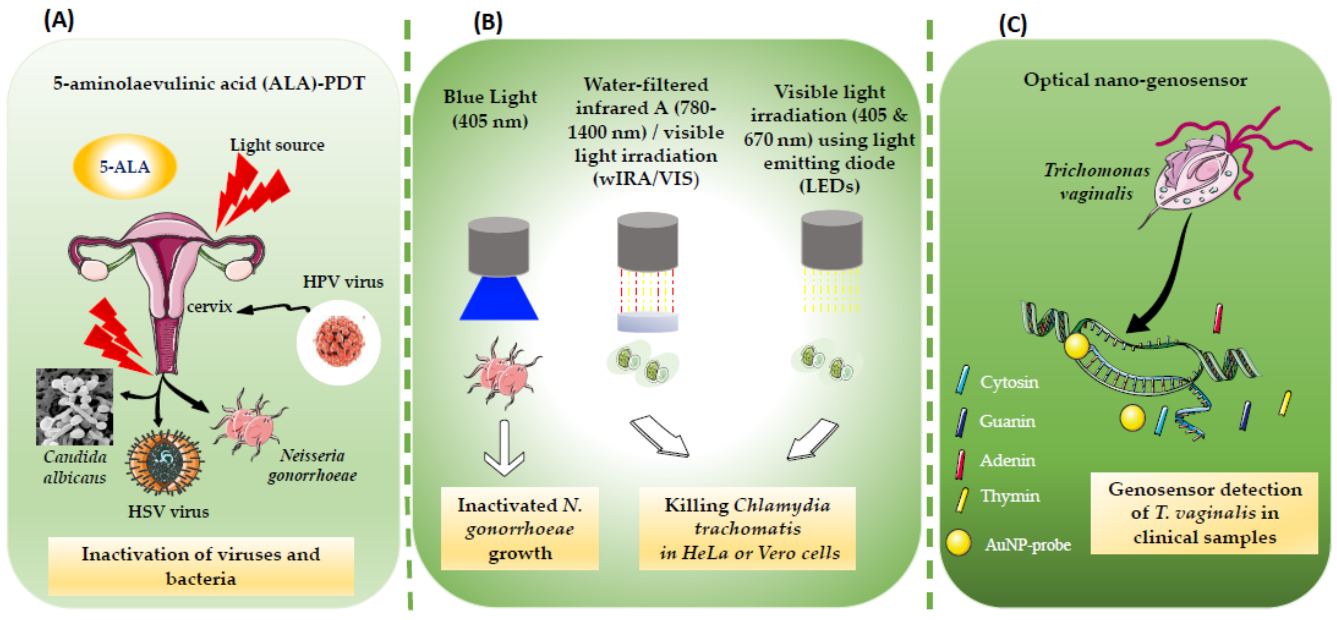
2. PT for Viral STIs

Human papilloma virus (HPV) infection, the most common sexually transmitted disease in the world and the main cause of genital warts, infects millions of people worldwide every year [51], with an estimated 291 million HPV-positive women worldwide in 2007 [52]. Among diseases caused by HPV, condyloma acuminata refers to an anogenital infection caused by HPV strains 6 and 11 [53,54]. Successful treatment can still be challenging. Traditional forms of treatment for condyloma acuminata that are effective at removing wart tissue including topical approaches, carbon dioxide laser (CO2 laser), cryotherapy, or electrosurgery [55], or medical treatment with topical agents (imiquimod, podofilox, podophyllin, trichloroacetic acid, sinecatechins ointment) [56,57].
However, these therapies are often ineffective with a high recurrence rate, as they cannot eliminate subclinical latent HPV infection. Multiple reports have shown that 5-aminolaevulinic acid (ALA)-PDT can be effective in treating HPV lesions [16,58]. A real benefit of PDT lies in its ability to treat multifocal disease without tissue loss [59]. Treatment with 5-ALA-PDT can destroy the warts and cause selective and specific destruction of subclinical virus infected areas [58]. The cure rate and viral clearance rate are significantly higher after ALA-PDT therapy compared to pharmacotherapy or physically destructive therapies. However, clinical trials have demonstrated the feasibility of applying topical ALA cream for photodiagnosis (PD) and PDT of condylomas caused by HPV. Women receiving PDT in this clinical trial had often failed with conventional treatment [56]. Conventional treatment can often be painful and sometimes disfiguring, and often results in high recurrence rates, which reinforces the need for new approaches [56,60]. One study evaluated the persistence or clearance of HPV infection after ALA-PDT in patients suffering from genital warts. The data was analyzed between January 2019 and December 2020 at Nanfang Hospital and Dermatology Department, Hospital of Southern Medical University in China, and showed that different variables such as multiple sexual partners, a history of recurrent infection, and severe pain during PDT affected the overall outcomes of PDT treatment. The authors suggested that the patients may need additional PDT sessions. Interestingly, PDT appears from this study to be effective against single strain HPV infections [61].
In one patient with condyloma acuminata covering the glans penis, a case study reported a patient who had a single large lesion. ALA-PDT was used as a therapeutic approach to reduce the risk of recurrence and minimize the trauma caused by traditional methods such as CO2 laser therapy. The choice of therapy by ALA-PDT is dictated by the fact that ALA accumulates the warts and reduce the rate of recurrence in the surrounding tissue with subclinical infection [55]. ALA-PDT is an effective, safe and curative alternative to the conventional treatment of genital warts [55]. ALA-PDT-based treatment was also effective against urethral condyloma acuminata; in this case, the authors suggested that the dynamic monitoring of the HPV viral load could objectively demonstrate the effectiveness and guide the treatment of PDT [62]. These results were reinforced by a randomized controlled clinical trial carried out recently. The objective was to compare the use of PDT with the application of trichloracetic acid (TAA) in the treatment of HPV condyloma in the perianal and vulval regions. A total of 16 patients was treated with PDT using the prodrug methyl aminolevulinate incubated for 3 h and irradiation at 630 nm (100 J/cm2). Fifteen patients were treated with TAA, received acid using a cotton swab. The results of these experimentations revealed that the PDT-based treatment appeared to be effective in the treatment of lesions due to the physical destruction of condyloma and subclinical lesions. A complete response rate was evaluated at 63% for PDT versus 60% (10 had a complete response and 6 had a partial response) for TAA (9 patients had a complete response in the elimination of lesions, 3 had a partial response), and a recurrence rate of 0% for PDT versus 33% for TAA. In addition, treatment with PDT led to complete clearance in an area with many warts; the authors suggested that PDT may be more beneficial for patients with recurrent HPV warts [63].
Another therapeutic strategy based on a combination of CO2 laser and ALA-PDT was tested to treat condyloma acuminata in 98 adult patients (male and female). Firstly, patients were treated by CO2 laser to remove the visible warts. Secondly, the patients had the ALA-PDT treatment immediately after the laser exposition. The ALA surface application was performed for 3 h at light irradiation of 100–150 J/cm2. The ALA-PDT was continued once a week for three weeks. A combination of CO2 laser and ALA-PDT has been shown to be feasible and effective in the treatment of condyloma acuminata. The cure rate was high at 93.8% (92/98) [64]. Shi et al., (2013) tested a different treatment strategy on 361 patients diagnosed with condyloma acuminata. Patients were divided into three groups according to the maximum diameter of their lesion (A < 0.5, B = 0.5–2.0, and C > 2.0–4.0 cm). Five treatments were compared in each group (cryotherapy, CO2 laser, ALA-PDT alone, ALA-PDT plus CO2 laser, ALA-PDT plus cryotherapy). The clinical outcomes evaluated during follow up after each treatment showed that the ALA-PDT was best if the maximum lesion diameter was <0.5 cm, while ALA-PDT plus cryotherapy was better for lesions 0.5–2.0 cm. ALA-PDT treatment, after either cryotherapy or CO2 laser was effective for lesions >2.0–4.0 cm, which should be the first choice. They suggested that all treatments could be effective, but the choice depended on the size of the condyloma lesion [65].
However, a study by Szeimies et al. (2009) reported a different result. CO2 laser ablation followed by ALA-PDT was investigated in a phase III prospective randomized bicenter double-blind clinical trial to prevent recurrence of condyloma acuminata. One hundred seventy-five patients with condyloma acuminata received CO2 laser vaporization plus adjuvant ALA-PDT or adjuvant placebo-PDT. Results showed no statistically significant difference between the groups with regard to recurrence rates up to 12 months after treatment. No major complications were observed [66].
Persistent HPV infection can lead to the development of malignant lesions in the vaginal and cervical epithelium. Indeed, HPV infection represents the main cause of cervical intraepithelial neoplasia [15]. The clinical treatments recommended for cervical intraepithelial neoplasia have already been mentioned, and mainly include the topical application of certain drugs or surgical excision, as well as irradiation by intravaginal radiotherapy or ablation by a laser [67]. PDT is a new therapeutic tool which has been used mainly in HPV infections causing condyloma acuminata, or in the case of non-melanoma skin tumors [58,68]. PDT has become a promising therapeutic method used in the treatment of various tumors including cervical intraepithelial neoplasia, cervical HPV infection, and vaginal intraepithelial neoplasia [69]. One meta-analysis revealed that out of 77 patients with cervical HPV infection included in four randomized controlled trials who received PDT, 48 of them showed complete remission. The complete remission rate ranged from 53.5 to 94.4% [69]. The authors of this meta-analysis concluded that PDT was effective for HPV clearance, particularly high-risk HPV genotypes. In addition, the study showed that out of 120 patients with cervical intraepithelial neoplasia treated by PDT, 77 patients achieved full primary remission by the end of the 3-month follow-up. Interestingly, the complete remission rate ranged from 31.3 to 100% [69]. Clinically, PDT is becoming increasingly employed to treat malignant HPV viral infection, including cervical intraepithelial neoplasia and cervical HPV infection, and has shown complete remission and local eradication of the virus. Another systematic review also demonstrated the efficacy of PDT for the treatment of cervical intraepithelial neoplasia. Analysis revealed that the complete remission rate of PDT for cervical intraepithelial neoplasia ranged from 0 to 100%, with an HPV eradication rate varying from 53.4 to 80% [70]. These results were based on analysis of the data published in several studies [71,72]. The effectiveness of PDT for the treatment of cervical intraepithelial neoplasia and for the eradication of HPV has also been confirmed in several studies by Li et al. [73], Cang et al. [74], Wu et al. [75], Su et al. [76] and Zang et al. [15]. Furthermore, the efficacy of PDT for the treatment of cervical cancer has also recently been demonstrated. At three months after PDT, complete elimination of HPV was detected in more than 90% of patients with early stage cervical cancer [19].
Herpes simplex viruses, HSV-1 and HSV-2, are the most common cause of mucocutaneous herpes lesions with a chronic or recurrent course. Approximately 20% of people infected with HSV have clinical manifestations that recur, especially during periods of weakened immunity [1]. HSV-1 is mainly transmitted by oral-to-oral contact causing oral herpes, but can also cause genital herpes. The World Health Organization has estimated that 3.7 billion people under age 50 (67%) have HSV-1 infection globally. HSV-2 is a sexually transmitted infection that causes genital herpes and has been estimated to affect 491 million people aged 15–49 (13%) worldwide [77]. HSV infections cause discomfort (itching, pain) associated with the eruption of vesicles and development of erosions. Lesion recurrence and aesthetic discomfort significantly reduce patient quality of life [78]. To improve the quality of life of women affected by genital HSV, several therapeutic approaches are being developed. Conventional therapy with nucleotide analogue antiviral drugs inhibits viral replication and shortens the duration of symptoms, but does not prevent recurrence. PDT is selective, non-invasive, not harmful to the patient, and can be used in parallel with other therapies, even in immunocompromised subjects (e.g., transplantation or oncology patients). Recent studies have shown the effectiveness of PDT in the inactivation of different types of virus in vitro and in vivo [79]. The HSV strains VR-3 and MS were used to infect Vero cell cultures and the antiherpetic effect was evaluated after PDT and laser irradiation treatments (Figure 1A). The results showed a significant reduction in virus load (100–1000 times) [79]. Nevertheless, a study determining the effect of ALA-PDT on the recurrence of herpes simplex showed that one patient irradiated with a higher dose of light (630 ± 20 nm; 120 J/cm2) developed an acute inflammation, accompanied by the appearance of prolonged scabs [80].
Published case reports regarding the treatment of recurrent herpes with phototherapy (including lasers) sometimes combined with synthetic dyes have focused mainly on reducing the duration of HSV infection symptoms and its accompanying pain, reducing viral titers, and the acceleration of wound healing, in cases of herpes labialis and genital herpes [80]. Donnarumma et al. demonstrated that laser phototherapy applied in HSV infection could also act on the immune response, restoring the expression of proinflammatory cytokines, tumor necrosis factor α (TNFα), interleukin (IL)-1β, and IL-6 suppressed by the virus, and limiting the viral spread from cell to cell [81]. A case report by Ferreira et al., (2011) used low-level laser therapy on a patient (50-year-old heterosexual female), with recurrent episodes of labial herpes over the preceding 5 years. Follow up showed that the patient remained symptom free for 17 months. Low-level laser therapy showed promising clinical results as a long-lasting suppression therapy in a single patient, but its wider efficacy for long-term suppression has yet to be established [82]. HSV infection can cause hyperpigmentation of the skin which may be present even when the infection is latent. Hyperpigmentation is linked to an inflammatory process in the tissues. Staining can cause people aesthetic inconvenience with a sense of shame. Hyperpigmentation can be treated topically with alpha-hydroxy acids (AHAs), hydroquinone, azelaic acid, retinoic acid, ascorbic acid, kojic acid can be effective alone or in combination with others therapies such as phototherapy or laser therapy [83]. A multitude of different lasers are currently available for the treatment of cutaneous hyperpigmentation. Five major classes of dermatological lasers are currently used: ablative and non-ablative lasers in their fractionated and non-fractionated forms as well as radio frequency technologies. Non-ablative lasers are gentler on the skin and allow faster healing, while harsher ablative lasers tend to be more effective. Fractionating increases the number of treatments but minimizes downtime and complications [84]. In addition, given the absorption spectrum of melanin (250–1200 nm), visible and near-infrared lasers can be used successfully to target excess melanin in the skin in the case of postinflammatory hyperpigmentation [85]. The Q-switched Nd-YAG laser, the fractional thulium laser (via tranexamic acid) and the picosecond alexandrite laser are the most used in the treatment of hyperpigmentation of the skin [86,87].

3. PT for Bacterial STIs

Neisseria gonorrhoeae (also known as gonococcus) is a Gram-negative bacterial species that can be sexually transmitted. N. gonorrhoeae infects the urogenital tract, causing dysuria with a penile discharge in men, and a vaginal mucopurulent discharge, along with severe pelvic pain in women [88]. Antimicrobial resistance is emerging, which limits antibiotic treatment for N. gonorrhoeae. Novel treatment strategies are urgently needed, according to Klausner et al. (2021) [89].
To combat antibiotic resistance in sexually transmitted bacteria, PDT has been tested against N. gonorrhoeae, including antibiotic-resistant strains cocultured with human vaginal epithelial cells in vitro. No PDT-induced genotoxicity to vaginal epithelial cells was observed upon delivering a sufficient radiant exposure to inactivate N. gonorrhoeae. PDT effectively inactivated N. gonorrhoeae, which had attached and invaded vaginal epithelial cells in their co-culture model [90]. In addition, studies have demonstrated that blue light (405 nm) used alone has an intrinsic antimicrobial activity against N. gonorrhoeae. This wavelength of light inactivated N. gonorrhoeae cells, including antibiotic-resistant strains, without causing epithelial cell cytotoxicity. Blue light was still effective against these bacteria after 15 successive cycles of exposure [90,91].
The use of water-filtered infrared A (wIRA), a short wavelength band of infrared radiation with a spectrum from 780–1400 nm, which is combined with visible light VIS (wIRA/VIS) showed efficacy in killing intracellular and extracellular Chlamydia strains (C. pecorum, C. trachomatis serovar E) in two different cell lines (Vero and HeLa). Irradiation of the infected cells (HeLa and Vero) neither affected cell viability nor induced any molecular markers of cytotoxicity. However, a single exposure to wIRA/VIS at 40 h post infection (hpi) led to a significant reduction in the frequency of C. pecorum incorporation in Vero cells, and C. trachomatis in HeLa cells (Figure 1B). Three sessions of irradiation (24, 36, 40 hpi) during the course of C. trachomatis infection further reduced the chlamydial incorporation frequency in HeLa cells [92].
In another study carried out in vitro, Wasson et al. (2021) investigated the effects of visible light irradiation (405 and 670 nm) using light emitting diodes (LEDs) on Chlamydial growth in HeLa cells (Figure 1B). The results demonstrated a significant dose-dependent inhibitory effect and a diminished bacterial load during both active and persistent infection, following irradiation [93].

4. PT for Parasitic STIs

Trichomoniasis is a condition caused by Trichomonas vaginalis, and is a very common vaginal infection that, in some women, manifests as a vaginal discharge, with a disagreeable smell and a yellowish or greenish color. T. vaginalis is usually transmitted sexually [94]. Recently, a new diagnostic approach based on an optical nano-genosensor was designed by conjugation of gold nanoparticles to a specific oligonucleotide that recognized a T. vaginalis gene sequence (AuNP-probe) for specific and sensitive PCR diagnosis. An investigation was performed using the AuNP-probe with different concentrations of a synthetic complementary sequence as a standard for T. vaginalis. Complete hybridization was detected by adding acid to the medium and observing the changes in the color and the spectroscopic absorption spectrum. The results confirmed the accurate function of the genosensor for the detection of T. vaginalis in clinical samples (Figure 1C). This new diagnosis strategy used a photo-genosensor for T. vaginalis detection [95]. PDT has also been used for T. vaginalis inactivation. The trophozoites (JT and CDC 085 strains) were exposed to PDT using methylene blue as a photosensitizer. The degree of parasite inhibition was significant, with 80.21% ± 7.11 for the JT strain and 91.13% ± 2.31 for the CDC 085 strain. This study confirmed that PDT using methylene blue could inhibit parasite multiplication and therefore could possibly reduce infection [96].

5. PDT for Fungal STIs

There was a pilot clinical trial carried out by Du et al. (2021), in which they investigated the effectiveness of a PDT-based treatment on 21 adult AIDS patients with C. albicans oral candidiasis. After two consecutive PDT treatments the clinical symptoms of oral candidiasis in adult AIDS patients were improved [97]. This success suggests that PDT might also be effective in genital thrush caused by C. albicans. In addition, one case report has shown that treatment with methylene blue (660 nm red laser) for PDT of vulvovaginal candidiasis significantly decreased fungal infection in the vaginal canal. This report claimed that this therapy improved the quality of life of patients who reported a reduction in symptoms [98]. Moreover, antifungal blue light therapy (400–470 nm) has been reported to be effective in decreasing vaginal candidiasis. A viability study using both C. albicans and human vaginal epithelial cells was performed. These two models were irradiated at different wavelength ranges of 405, 415 and 450 nm. The experimental data showed that an inhibition rate of approximately 80% of C. albicans was achieved, while the epithelial cells had a survival rate which varied according to the wavelength: 0.6700, 0.7748, and 0.6027, respectively, for treatment with light of wavelength 405, 415 and 450 nm. Additionally, 415 nm light showed a more effective antifungal effect with less damage to epithelial cells compared to 405 nm or 450 nm light [99].
The efficacy of PDT was studied against yeast cells in a mouse model of vaginal infection. PDT was carried out using two photosensitizers, methylene blue and protoporphyrin IX. The study was carried out on mice with persistent vaginitis evoked by an intravaginal inoculation of C. albicans. PDT was performed 5 days after fungal inoculation using both photosensitizers. The irradiation was carried out using two custom-made LED devices at 660 and 630 nm. The results showed that PDT reduced fungal colony-forming units. After a follow-up time, the results did not change and the colonies did not increase from the initial value immediately after PDT. The use of PDT as a therapy to reduce fungal infection in a model of vaginal candidiasis produced a significant reduction in C. albicans while causing no damage to the vaginal mucosa [100]. Another mouse model study testing the effects of PDT on vaginal candidiasis using methylene blue and red (660 nm) laser irradiation, demonstrated that PDT significantly reduced C. albicans colonies. In addition, this study also demonstrated that the percentage of inflammatory cells per unit surface area was significantly reduced after only two sessions of PDT [101].
Table 1 summarizes the published reports of PT or PDT to treat a variety of STIs, either in vitro, in animal models, or in humans.

6. Conclusions

So far, despite the range of treatment options available, no light-based approach has been able to achieve satisfactory results against all pathogens that cause STIs. In addition, to date and to our knowledge, there are no randomized controlled clinical trials evaluating the efficacy of phototherapy-based treatment against each pathogen causing a specific infection and comparing this efficacy to a reference treatment. Nevertheless, controlled trials comparing treatments are currently being optimized. In the case of HPV infection, no current PDT-based treatment completely eradicates the HPV virus. However, the variables that could be considered include (but are not limited to) the morphology of the lesions such as thickness and size, quantity, anatomic location, and HPV strain. The viral replication cycle and stage of infection appear to play a role in the response of the infection. It would be difficult at present for this type of treatment to be implicated as a specific mode of treatment (e.g., phototherapy) and could cover a wide variety of biologically unrelated pathogens that belonged only to STIs. PDT approaches should be further developed against genital HSV infection, and against STIs with bacterial and parasitic causes (C. trachomatis, N. gonorrhoeae, Treponema pallidum, Mycoplasma genitalium and T. vaginalis). The development of nanotechnology over the past decades has led to the incorporation of nanomedicine into PT for cancer treatment. The merging of nanomedicine into PT has allowed the continuous refinement of PDT approaches. With the careful design of phototherapeutic agents and good control of light illumination at the site of the lesions, effective PDT can be achieved, with reductions in the systemic toxicity associated with traditional chemotherapy and radiotherapy. However, we hope that this localized therapy could also be used in the case of STIs, especially in view of the rise in drug resistance observed in most types of pathogens.

Author Contributions

The manuscript was written through the contributions of all authors, as follows: conceptualization, M.V.; authors of chapters: N.M., P.R., M.R.H. and M.V.; writing—review and editing, N.M., P.R., M.R.H., M.V. and L.B.; supervision, M.V.; project administration: M.V.; funding acquisition, M.V. on behalf of the French team of OEMONOM. All authors have read and agreed to the published version of the manuscript.

Funding

This open-access review paper was supported by the Erasmus+ Programme of the European Union, Key Action 2: Strategic Partnerships, Project No. 2020-1-CZ01-KA203-078218. The authors also acknowledge the support of the CNRS and the Université de Lorraine. M.R.H. was supported by US NIH Grants R01AI050875 and R21AI121700.

Institutional Review Board Statement

Not applicable.

Informed Consent Statement

Not applicable.

Data Availability Statement

This is a literature review citing results already published in primary scientific articles. No data collection was performed from patients in this review.

Acknowledgments

All authors have consented to the acknowledgement and would like to express their gratitude to Open Access Educational Materials on Naturally Occurring Molecules (https://portal.faf.cuni.cz/OEMONOM/EN/ (accessed on 14 march 2022), to ADRESSE association, and to Erguestine Andriamahatahitry for the careful proofreading of the manuscript, and for useful discussion and comments on the work.

Conflicts of Interest

M.R.H. declares the following potential conflicts of interest. Scientific Advisory Boards: Transdermal Cap Inc., Cleveland, OH, USA; Hologenix Inc., Santa Monica, CA, USA; Vielight, Toronto, ON, Canada; JOOVV Inc., Minneapolis–St. Paul, MN, USA; Consulting; USHIO Corp., Japan; Sanofi-Aventis Deutschland GmbH, Frankfurt am Main, Germany. Stockholding: Niraxx Light Therapeutics, Inc., Irvine, CA, USA; JelikaLite Corp., New York, NY, USA. The other authors declare no conflict of interest.

Abbreviations

HPV: human papilloma virus; 5-ALA: 5-aminolevulinic acid; CO2 laser: carbon diode laser; LED: light-emitting diode; YAG-OPO laser: laser pumped optical parametric oscillator; HAL: hexaminolevulinate; PDT: photodynamic therapy; HSV: virus herpes simplex; DMC: direct machining control; N. gonorrhoeae: Neisseria gonorrhoeae; Chlamydia pecorum: C. pecorum; Chlamydia trachomatis: C. trachomatis; wIRA/VIS: water-filtered infrared A combined with visible light; WARP: Warfighter Accelerated Recovery by Photobiomodulation; HIV/AIDS: human immunodeficiency virus/acquired immunodeficiency syndrome; C. albicans: Candida albicans; MAC: scar acceleration method. NI: not indicated.

References

  1. Sexually Transmitted Infections (STIs). Available online: https://www.who.int/westernpacific/health-topics/sexually-transmitted-infections (accessed on 19 November 2021).
  2. Williamson, D.A.; Chen, M.Y. Emerging and Reemerging Sexually Transmitted Infections. N. Engl. J. Med. 2020, 382, 2023–2032. [Google Scholar] [CrossRef] [PubMed]
  3. Smith, L.; Angarone, M.P. Sexually Transmitted Infections. Urol. Clin. N. Am. 2015, 42, 507–518. [Google Scholar] [CrossRef] [PubMed]
  4. Ciccarese, G.; Herzum, A.; Pastorino, A.; Dezzana, M.; Casazza, S.; Mavilia, M.G.; Copello, F.; Parodi, A.; Drago, F. Prevalence of Genital HPV Infection in STI and Healthy Populations and Risk Factors for Viral Persistence. Eur. J. Clin. Microbiol. Infect. Dis. 2021, 40, 885–888. [Google Scholar] [CrossRef] [PubMed]
  5. Cole, S. Herpes Simplex Virus: Epidemiology, Diagnosis, and Treatment. Nurs. Clin. N. Am. 2020, 55, 337–345. [Google Scholar] [CrossRef]
  6. Kardani, K.; Basimi, P.; Fekri, M.; Bolhassani, A. Antiviral Therapy for the Sexually Transmitted Viruses: Recent Updates on Vaccine Development. Expert Rev. Clin. Pharmacol. 2020, 13, 1001–1046. [Google Scholar] [CrossRef]
  7. Juliana, N.C.A.; Deb, S.; Ouburg, S.; Chauhan, A.; Pleijster, J.; Ali, S.M.; Morré, S.A.; Sazawal, S.; Ambrosino, E. The Prevalence of Chlamydia Trachomatis and Three Other Non-Viral Sexually Transmitted Infections among Pregnant Women in Pemba Island Tanzania. Pathogens 2020, 9, 625. [Google Scholar] [CrossRef]
  8. Gannon-Loew, K.E.; Holland-Hall, C. A Review of Current Guidelines and Research on the Management of Sexually Transmitted Infections in Adolescents and Young Adults. Ther. Adv. Infect. Dis. 2020, 7, 2049936120960664. [Google Scholar] [CrossRef]
  9. Doernberg, S.B.; Komarow, L.; Tran, T.T.T.; Sund, Z.; Pandori, M.W.; Jensen, D.; Tsalik, E.L.; Deal, C.D.; Chambers, H.F.; Fowler, V.G.; et al. Simultaneous Evaluation of Diagnostic Assays for Pharyngeal and Rectal Neisseria Gonorrhoeae and Chlamydia Trachomatis Using a Master Protocol. Clin. Infect. Dis. 2020, 71, 2314–2322. [Google Scholar] [CrossRef]
  10. Yarbrough, M.L.; Burnham, C.-A.D. The ABCs of STIs: An Update on Sexually Transmitted Infections. Clin. Chem. 2016, 62, 811–823. [Google Scholar] [CrossRef] [Green Version]
  11. Lin, K.-Y.; Sun, H.-Y.; Lee, T.-F.; Chuang, Y.-C.; Wu, U.-I.; Liu, W.-C.; Chang, S.-Y.; Chen, Y.-J.; Hung, C.-C.; Chang, S.-C. High Prevalence of Sexually Transmitted Coinfections among At-Risk People Living with HIV. J. Formos. Med. Assoc. 2021, 120, 1876–1883. [Google Scholar] [CrossRef]
  12. Tien, V.; Punjabi, C.; Holubar, M.K. Antimicrobial Resistance in Sexually Transmitted Infections. J. Travel. Med. 2020, 27, taz101. [Google Scholar] [CrossRef]
  13. Williams, E.; Fairley, C.K.; Williamson, D. Novel Strategies for Prevention and Treatment of Antimicrobial Resistance in Sexually-Transmitted Infections. Curr. Opin. Infect. Dis. 2021, 34, 591–598. [Google Scholar] [CrossRef]
  14. Li, Y.; Yu, T.; Yan, H.; Li, D.; Yu, T.; Yuan, T.; Rahaman, A.; Ali, S.; Abbas, F.; Dian, Z.; et al. Vaginal Microbiota and HPV Infection: Novel Mechanistic Insights and Therapeutic Strategies. Infect. Drug Resist. 2020, 13, 1213–1220. [Google Scholar] [CrossRef]
  15. Zhang, T.; Hu, R.; Tang, Y.; Zhang, Y.; Qin, L.; Shen, Y.; Wang, B.; Zhang, L.; Cao, L.; Zhou, Y.; et al. The Effect of Local Photodynamic Therapy with 5-Aminolevulinic Acid in the Treatment of Vaginal Intraepithelial Lesions with High-Risk HPV Infection. Photodiagn. Photodyn. Ther. 2022, 37, 102728. [Google Scholar] [CrossRef]
  16. Fathi, R.; Tsoukas, M.M. Genital Warts and Other HPV Infections: Established and Novel Therapies. Clin. Dermatol. 2014, 32, 299–306. [Google Scholar] [CrossRef]
  17. Schnürch, H.-G.; Ackermann, S.; Alt-Radtke, C.D.; Angleitner, L.; Barinoff, J.; Beckmann, M.W.; Böing, C.; Dannecker, C.; Fehm, T.; Gaase, R.; et al. Diagnosis, Therapy and Follow-up of Vaginal Cancer and Its Precursors. Guideline of the DGGG and the DKG (S2k-Level, AWMF Registry No. 032/042, October 2018). Geburtshilfe Und Frauenheilkd. 2019, 79, 1060–1078. [Google Scholar] [CrossRef] [Green Version]
  18. Chumworathayi, B.; Thinkhamrop, J.; Blumenthal, P.D.; Thinkhamrop, B.; Pientong, C.; Ekalaksananan, T. Cryotherapy for HPV Clearance in Women with Biopsy-Confirmed Cervical Low-Grade Squamous Intraepithelial Lesions. Int. J. Gynaecol. Obstet. 2010, 108, 119–122. [Google Scholar] [CrossRef]
  19. Afanasiev, M.D.D.S.M.S.; Dushkin, M.D.A.D.; Grishacheva, D.S.T.G.; Afanasiev, M.D.D.S.S.S.; Karaulov Academician Ras, M.D.D.S.A.V. Photodynamic Therapy for Early-Stage Cervical Cancer Treatment. Photodiagn. Photodyn. Ther. 2021, 37, 102620. [Google Scholar] [CrossRef]
  20. Viviano, M.; Kenfack, B.; Catarino, R.; Tincho, E.; Temogne, L.; Benski, A.-C.; Tebeu, P.-M.; Meyer-Hamme, U.; Vassilakos, P.; Petignat, P. Feasibility of Thermocoagulation in a Screen-and-Treat Approach for the Treatment of Cervical Precancerous Lesions in Sub-Saharan Africa. BMC Womens Health 2017, 17, 2. [Google Scholar] [CrossRef] [Green Version]
  21. Gutierrez, P.; Garza, J.; Gandhi, K.; Voice, A.; Stout, E.; Ventolini, G. Carbon Dioxide (CO2) Laser Ablation Treatment of a Peri-Urethral Genital Wart: A Case Report. Case Rep. Womens Health 2020, 27, e00226. [Google Scholar] [CrossRef]
  22. Cosper, P.F.; McNair, C.; González, I.; Wong, N.; Knudsen, K.E.; Chen, J.J.; Markovina, S.; Schwarz, J.K.; Grigsby, P.W.; Wang, X. Decreased Local Immune Response and Retained HPV Gene Expression during Chemoradiotherapy Are Associated with Treatment Resistance and Death from Cervical Cancer. Int. J. Cancer 2020, 146, 2047–2058. [Google Scholar] [CrossRef]
  23. Foster, C.C.; Lee, A.Y.; Furtado, L.V.; Hart, J.; Alpert, L.; Xiao, S.-Y.; Hyman, N.H.; Sharma, M.R.; Liauw, S.L. Treatment Outcomes and HPV Characteristics for an Institutional Cohort of Patients with Anal Cancer Receiving Concurrent Chemotherapy and Intensity-Modulated Radiation Therapy. PLoS ONE 2018, 13, e0194234. [Google Scholar] [CrossRef]
  24. Chera, B.S.; Amdur, R.J.; Green, R.; Shen, C.; Gupta, G.; Tan, X.; Knowles, M.; Fried, D.; Hayes, N.; Weiss, J.; et al. Phase II Trial of De-Intensified Chemoradiotherapy for Human Papillomavirus-Associated Oropharyngeal Squamous Cell Carcinoma. J. Clin. Oncol. 2019, 37, 2661–2669. [Google Scholar] [CrossRef]
  25. Sindhu, S.K.; Bauman, J.E. Current Concepts in Chemotherapy for Head and Neck Cancer. Oral. Maxillofac. Surg. Clin. N. Am. 2019, 31, 145–154. [Google Scholar] [CrossRef]
  26. Bonomi, M.; Ahmed, T.; Warner, D.; Waltonen, J.; Sullivan, C.; Porosnicu, M.; Batt, K.; Ruiz, J.; Cappellari, J. Human Papillomavirus-Related Small Cell Carcinoma of the Oropharynx: A Case Report and Literature Review. Cancers Head Neck 2017, 2, 3. [Google Scholar] [CrossRef] [PubMed] [Green Version]
  27. Perrone, A.M.; Tesei, M.; Ferioli, M.; De Terlizzi, F.; Della Gatta, A.N.; Boussedra, S.; Dondi, G.; Galuppi, A.; Morganti, A.G.; De Iaco, P. Results of a Phase I-II Study on Laser Therapy for Vaginal Side Effects after Radiotherapy for Cancer of Uterine Cervix or Endometrium. Cancers 2020, 12, 1639. [Google Scholar] [CrossRef] [PubMed]
  28. Bogani, G.; Ditto, A.; Martinelli, F.; Mosca, L.; Chiappa, V.; Rossetti, D.; Leone Roberti Maggiore, U.; Sabatucci, I.; Lorusso, D.; Raspagliesi, F. LASER Treatment for Women with High-Grade Vaginal Intraepithelial Neoplasia: A Propensity-Matched Analysis on the Efficacy of Ablative versus Excisional Procedures. Lasers Surg. Med. 2018, 50, 933–939. [Google Scholar] [CrossRef] [PubMed]
  29. Boonlikit, S. Recurrence of High-Grade Vaginal Intraepithelial Neoplasia after Various Treatments. Curr. Probl. Cancer 2022, 46, 100792. [Google Scholar] [CrossRef] [PubMed]
  30. Farmer, E.; Cheng, M.A.; Hung, C.-F.; Wu, T.-C. Vaccination Strategies for the Control and Treatment of HPV Infection and HPV-Associated Cancer. Recent Results Cancer Res. 2021, 217, 157–195. [Google Scholar] [CrossRef]
  31. Dadar, M.; Chakraborty, S.; Dhama, K.; Prasad, M.; Khandia, R.; Hassan, S.; Munjal, A.; Tiwari, R.; Karthik, K.; Kumar, D.; et al. Advances in Designing and Developing Vaccines, Drugs and Therapeutic Approaches to Counter Human Papilloma Virus. Front. Immunol. 2018, 9, 2478. [Google Scholar] [CrossRef]
  32. Taylor, M.; Gerriets, V. Acyclovir. In StatPearls; StatPearls Publishing: Treasure Island, FL, USA, 2021. [Google Scholar]
  33. Feng, E.; Balint, E.; Vahedi, F.; Ashkar, A.A. Immunoregulatory Functions of Interferons during Genital HSV-2 Infection. Front. Immunol. 2021, 12, 724618. [Google Scholar] [CrossRef]
  34. Lau, A.; Kong, F.Y.S.; Fairley, C.K.; Templeton, D.J.; Amin, J.; Phillips, S.; Law, M.; Chen, M.Y.; Bradshaw, C.S.; Donovan, B.; et al. Azithromycin or Doxycycline for Asymptomatic Rectal Chlamydia Trachomatis. N. Engl. J. Med. 2021, 384, 2418–2427. [Google Scholar] [CrossRef]
  35. Zofkie, A.C.; Fomina, Y.Y.; Roberts, S.W.; McIntire, D.D.; Nelson, D.B.; Adhikari, E.H. Effectiveness of Chlamydia Trachomatis Expedited Partner Therapy in Pregnancy. Am. J. Obstet. Gynecol. 2021, 225, 325.e1–325.e7. [Google Scholar] [CrossRef]
  36. Adachi, K.N.; Nielsen-Saines, K.; Klausner, J.D. Chlamydia Trachomatis Screening and Treatment in Pregnancy to Reduce Adverse Pregnancy and Neonatal Outcomes: A Review. Front. Public Health 2021, 9, 531073. [Google Scholar] [CrossRef]
  37. Gnanadurai, R.; Fifer, H. Mycoplasma Genitalium: A Review. Microbiology 2020, 166, 21–29. [Google Scholar] [CrossRef]
  38. Unemo, M.; Lahra, M.M.; Cole, M.; Galarza, P.; Ndowa, F.; Martin, I.; Dillon, J.-A.R.; Ramon-Pardo, P.; Bolan, G.; Wi, T. World Health Organization Global Gonococcal Antimicrobial Surveillance Program (WHO GASP): Review of New Data and Evidence to Inform International Collaborative Actions and Research Efforts. Sex. Health 2019, 16, 412–425. [Google Scholar] [CrossRef] [Green Version]
  39. Poncin, T.; Fouere, S.; Braille, A.; Camelena, F.; Agsous, M.; Bebear, C.; Kumanski, S.; Lot, F.; Mercier-Delarue, S.; Ngangro, N.N.; et al. Multidrug-Resistant Neisseria Gonorrhoeae Failing Treatment with Ceftriaxone and Doxycycline in France, November 2017. Eurosurveillance 2018, 23, 1800264. [Google Scholar] [CrossRef]
  40. Salmerón, P.; Viñado, B.; Arando, M.; Alcoceba, E.; Romero, B.; Menéndez, B.; Bernal, S.; Idigoras, P.; Colomina, J.; Martin-Saco, G.; et al. Neisseria Gonorrhoeae Antimicrobial Resistance in Spain: A Prospective Multicentre Study. J. Antimicrob. Chemother. 2021, 76, 1523–1531. [Google Scholar] [CrossRef]
  41. Bouchemal, K.; Bories, C.; Loiseau, P.M. Strategies for Prevention and Treatment of Trichomonas Vaginalis Infections. Clin. Microbiol. Rev. 2017, 30, 811–825. [Google Scholar] [CrossRef] [Green Version]
  42. Graves, K.J.; Novak, J.; Secor, W.E.; Kissinger, P.J.; Schwebke, J.R.; Muzny, C.A. A Systematic Review of the Literature on Mechanisms of 5-Nitroimidazole Resistance in Trichomonas Vaginalis. Parasitology 2020, 147, 1383–1391. [Google Scholar] [CrossRef]
  43. Lam, S. Photodynamic Therapy of Lung Cancer. Semin. Oncol. 1994, 21, 15–19. [Google Scholar] [CrossRef] [Green Version]
  44. Kessel, D. Photodynamic Therapy: Critical PDT Theory. Photochem. Photobiol. 2022. [Google Scholar] [CrossRef]
  45. Dougherty, T.J. Hematoporphyrin Derivative for Detection and Treatment of Cancer. J. Surg. Oncol. 1980, 15, 209–210. [Google Scholar] [CrossRef]
  46. Kou, J.; Dou, D.; Yang, L. Porphyrin Photosensitizers in Photodynamic Therapy and Its Applications. Oncotarget 2017, 8, 81591–81603. [Google Scholar] [CrossRef] [Green Version]
  47. Zhang, J.; Jiang, C.; Figueiró Longo, J.P.; Azevedo, R.B.; Zhang, H.; Muehlmann, L.A. An Updated Overview on the Development of New Photosensitizers for Anticancer Photodynamic Therapy. Acta. Pharm. Sin. B 2018, 8, 137–146. [Google Scholar] [CrossRef]
  48. Bellnier, D.A.; Greco, W.R.; Loewen, G.M.; Nava, H.; Oseroff, A.R.; Dougherty, T.J. Clinical Pharmacokinetics of the PDT Photosensitizers Porfimer Sodium (Photofrin), 2-[1-Hexyloxyethyl]-2-Devinyl Pyropheophorbide-a (Photochlor) and 5-ALA-Induced Protoporphyrin IX. Lasers Surg. Med. 2006, 38, 439–444. [Google Scholar] [CrossRef]
  49. Mfouo-Tynga, I.S.; Dias, L.D.; Inada, N.M.; Kurachi, C. Features of Third Generation Photosensitizers Used in Anticancer Photodynamic Therapy: Review. Photodiagn. Photodyn. Ther. 2021, 34, 102091. [Google Scholar] [CrossRef]
  50. Kharkwal, G.B.; Sharma, S.K.; Huang, Y.-Y.; Dai, T.; Hamblin, M.R. Photodynamic Therapy for Infections: Clinical Applications. Lasers Surg. Med. 2011, 43, 755–767. [Google Scholar] [CrossRef] [Green Version]
  51. Kombe Kombe, A.J.; Li, B.; Zahid, A.; Mengist, H.M.; Bounda, G.-A.; Zhou, Y.; Jin, T. Epidemiology and Burden of Human Papillomavirus and Related Diseases, Molecular Pathogenesis, and Vaccine Evaluation. Front. Public Health 2020, 8, 552028. [Google Scholar] [CrossRef]
  52. De Sanjosé, S.; Diaz, M.; Castellsagué, X.; Clifford, G.; Bruni, L.; Muñoz, N.; Bosch, F.X. Worldwide Prevalence and Genotype Distribution of Cervical Human Papillomavirus DNA in Women with Normal Cytology: A Meta-Analysis. Lancet Infect. Dis. 2007, 7, 453–459. [Google Scholar] [CrossRef]
  53. Pennycook, K.B.; McCready, T.A. Condyloma Acuminata. In StatPearls; StatPearls Publishing: Treasure Island, FL, USA, 2021. [Google Scholar]
  54. Hu, Z.; Zheng, H.; Zeng, K. Patterns of Multiple Human Papillomavirus Clearance during 5-Aminolevulinic Acid-Based Photodynamic Therapy in Patients with Genital Warts. Photodiagn. Photodyn. Ther. 2021, 35, 102454. [Google Scholar] [CrossRef] [PubMed]
  55. Yin, G.; Zhang, Y.; Geng, M.; Cai, B.; Zheng, Y. Cure of Condyloma Acuminata Covering the Glans Penis Using Aminolevulinic Acid/Photodynamic Therapy. Photodiagn. Photodyn. Ther. 2020, 30, 101658. [Google Scholar] [CrossRef] [PubMed]
  56. Inada, N.M.; Buzza, H.H.; Carbinatto, F.M.; Blanco, K.C.; de Andrade, C.T.; Vollet-Filho, J.D.; Bagnato, V.S.; Allison, R.R. Optical Techniques for the Diagnosis and Treatment of Lesions Induced by the Human Papillomavirus—A Resource Letter. Photodiagn. Photodyn. Ther. 2018, 23, 106–110. [Google Scholar] [CrossRef] [PubMed]
  57. Owczarek, W.; Slowinska, M.; Walecka, I.; Ciazynska, M.; Nowicka, D.; Walczak, L.; Paluchowska, E. Correlation of the ALA-PDT Treatment Efficacy and the HPV Genotype Profile of Genital Warts after Cryotherapy Failure and Podophyllotoxin Therapy in Male Patients. Life 2021, 11, 146. [Google Scholar] [CrossRef]
  58. Kechichian, E.; Helou, E.; Sarkis, J.; Hayek, C.; Labaki, C.; Nemr, E.; Tomb, R. The Place of 5-Aminolaevulinic Acid-Photodynamic Therapy in the Treatment Landscape of Urethral Warts: A Systematic Review. Photodiagn. Photodyn. Ther. 2021, 33, 102204. [Google Scholar] [CrossRef]
  59. Stern, P.L.; van der Burg, S.H.; Hampson, I.N.; Broker, T.R.; Fiander, A.; Lacey, C.J.; Kitchener, H.C.; Einstein, M.H. Therapy of Human Papillomavirus-Related Disease. Vaccine 2012, 30 (Suppl. S5), F71–F82. [Google Scholar] [CrossRef] [Green Version]
  60. Mistrangelo, M.; Dal Conte, I.; Volpatto, S.; DI Benedetto, G.; Testa, V.; Currado, F.; Morino, M. Current Treatments for Anal Condylomata Acuminata. Minerva Chir. 2018, 73, 100–106. [Google Scholar] [CrossRef]
  61. Hu, Z.; Zheng, H.; Zeng, K. Predictors of Human Papillomavirus Persistence or Clearance after 5-Aminolevulinic Acid-Based Photodynamic Therapy in Patients with Genital Warts. Photodiagn. Photodyn. Ther. 2021, 35, 102431. [Google Scholar] [CrossRef]
  62. Xie, J.; Ao, C.; Li, J.; Jiang, L.; Liu, H.; Zeng, K. 5-Aminolevulinic Acid Photodynamic Therapy for Condyloma Acuminatum of Urethral Meatus. J. Dermatolog. Treat. 2019, 30, 714–717. [Google Scholar] [CrossRef]
  63. Buzzá, H.H.; Stringasci, M.D.; de Arruda, S.S.; Crestana, R.H.S.; de Castro, C.A.; Bagnato, V.S.; Inada, N.M. HPV-Induced Condylomata Acuminata Treated by Photodynamic Therapy in Comparison with Trichloroacetic Acid: A Randomized Clinical Trial. Photodiagn. Photodyn. Ther. 2021, 35, 102465. [Google Scholar] [CrossRef]
  64. Hu, S.; Yang, Y.; Jiang, B.; Su, D.; Zhang, L.; Huang, Z.; Zhang, F. Treatment of Condyloma Acuminatum Using the Combination of Laser Ablation and ALA-PDT. Photodiagn. Photodyn. Ther. 2019, 25, 193–196. [Google Scholar] [CrossRef]
  65. Shi, H.; Zhang, X.; Ma, C.; Yu, N.; Wang, J.; Xia, L.; Ge, X.; Liu, M.; Duan, A. Clinical Analysis of Five Methods Used to Treat Condylomata Acuminata. Dermatology 2013, 227, 338–345. [Google Scholar] [CrossRef]
  66. Szeimies, R.-M.; Schleyer, V.; Moll, I.; Stocker, M.; Landthaler, M.; Karrer, S. Adjuvant Photodynamic Therapy Does Not Prevent Recurrence of Condylomata Acuminata after Carbon Dioxide Laser Ablation-A Phase III, Prospective, Randomized, Bicentric, Double-Blind Study. Dermatol. Surg. 2009, 35, 757–764. [Google Scholar] [CrossRef]
  67. Kim, J.-H.; Kim, J.; Kim, K.; No, J.H.; Kim, Y.B.; Suh, D.H. Risk Factor and Treatment of Vaginal Intraepithelial Neoplasia After Hysterectomy for Cervical Intraepithelial Neoplasia. J. Low Genit. Tract. Dis. 2022, 26, 147–151. [Google Scholar] [CrossRef]
  68. Hampson, L.; Martin-Hirsch, P.; Hampson, I.N. An Overview of Early Investigational Drugs for the Treatment of Human Papilloma Virus Infection and Associated Dysplasia. Expert. Opin. Investig. Drugs 2015, 24, 1529–1537. [Google Scholar] [CrossRef]
  69. Zhang, W.; Zhang, A.; Sun, W.; Yue, Y.; Li, H. Efficacy and Safety of Photodynamic Therapy for Cervical Intraepithelial Neoplasia and Human Papilloma Virus Infection. Medicine 2018, 97, e10864. [Google Scholar] [CrossRef]
  70. Tao, X.H.; Guan, Y.; Shao, D.; Xue, W.; Ye, F.S.; Wang, M.; He, M.H. Efficacy and Safety of Photodynamic Therapy for Cervical Intraepithelial Neoplasia: A Systemic Review. Photodiagn. Photodyn. Ther. 2014, 11, 104–112. [Google Scholar] [CrossRef]
  71. Bodner, K.; Bodner-Adler, B.; Wierrani, F.; Kubin, A.; Szölts-Szölts, J.; Spängler, B.; Grünberger, W. Cold-Knife Conization versus Photodynamic Therapy with Topical 5-Aminolevulinic Acid (5-ALA) in Cervical Intraepithelial Neoplasia (CIN) II with Associated Human Papillomavirus Infection: A Comparison of Preliminary Results. Anticancer. Res. 2003, 23, 1785–1788. [Google Scholar]
  72. Wang, J.; Xu, J.; Chen, J.; He, Q.; Xiang, L.; Huang, X.; Ding, G.; Xu, S. Successful Photodynamic Therapy with Topical 5-Aminolevulinic Acid for Five Cases of Cervical Intraepithelial Neoplasia. Arch. Gynecol. Obstet. 2010, 282, 307–312. [Google Scholar] [CrossRef]
  73. Li, D.; Zhang, F.; Shi, L.; Lin, L.; Cai, Q.; Xu, Y. Treatment of HPV Infection-Associated Low Grade Cervical Intraepithelial Neoplasia with 5-Aminolevulinic Acid-Mediated Photodynamic Therapy. Photodiagn. Photodyn. Ther. 2020, 32, 101974. [Google Scholar] [CrossRef]
  74. Cang, W.; Gu, L.; Hong, Z.; Wu, A.; Di, W.; Qiu, L. Effectiveness of Photodynamic Therapy with 5-Aminolevulinic Acid on HPV Clearance in Women without Cervical Lesions. Photodiagn. Photodyn. Ther. 2021, 34, 102293. [Google Scholar] [CrossRef] [PubMed]
  75. Wu, A.; Li, Q.; Ling, J.; Gu, L.; Hong, Z.; Di, W.; Qiu, L. Effectiveness of Photodynamic Therapy in Women of Reproductive Age with Cervical High-Grade Squamous Intraepithelial Lesions (HSIL/CIN2). Photodiagn. Photodyn. Ther. 2021, 36, 102517. [Google Scholar] [CrossRef] [PubMed]
  76. Su, Y.; Zhang, Y.; Tong, Y.; Zhang, L.; Li, P.; Zhang, H.; Zhang, X.; Tang, Y.; Qin, L.; Shen, Y.; et al. Effect and Rational Application of Topical Photodynamic Therapy (PDT) with 5-Aminolevulinic Acid (5-ALA) for Treatment of Cervical Intraepithelial Neoplasia with Vaginal Intraepithelial Neoplasia. Photodiagn. Photodyn. Ther. 2022, 37, 102634. [Google Scholar] [CrossRef] [PubMed]
  77. Herpes Simplex Virus. Available online: https://www.who.int/news-room/fact-sheets/detail/herpes-simplex-virus (accessed on 26 November 2021).
  78. Fisman, D.N. Health Related Quality of Life in Genital Herpes: A Pilot Comparison of Measures. Sex. Transm. Infect. 2005, 81, 267–270. [Google Scholar] [CrossRef] [Green Version]
  79. Makarov, O.V.; Khashukaeva, A.Z.; Svitich, O.A.; Markova, È.A.; Khlynova, S.A.; Labzhinov, P.A.; Zverev, V.V. Anti-herpetic effect of photodynamic action in an in vitro experiment. Zhurnal Mikrobiol. Epidemiol. Immunobiol. 2014, 1, 48–55. [Google Scholar]
  80. Osiecka, B.J.; Nockowski, P.; Kwiatkowski, S.; Szepietowski, J.C. Photodynamic Therapy with Red Light and 5-Aminolaevulinic Acid for Herpes Simplex Recurrence: Preliminary Results. Acta. Derm. Venereol. 2017, 97, 1239–1240. [Google Scholar] [CrossRef] [Green Version]
  81. Donnarumma, G.; De Gregorio, V.; Fusco, A.; Farina, E.; Baroni, A.; Esposito, V.; Contaldo, M.; Petruzzi, M.; Pannone, G.; Serpico, R. Inhibition of HSV-1 Replication by Laser Diode-Irradiation: Possible Mechanism of Action. Int. J. Immunopathol. Pharmacol. 2010, 23, 1167–1176. [Google Scholar] [CrossRef]
  82. Ferreira, D.C.; Reis, H.L.B.; Cavalcante, F.S.; Santos, K.R.N.D.; Passos, M.R.L. Recurrent Herpes Simplex Infections: Laser Therapy as a Potential Tool for Long-Term Successful Treatment. Rev. Soc. Bras. Med. Trop. 2011, 44, 397–399. [Google Scholar] [CrossRef]
  83. Davis, E.C.; Callender, V.D. Postinflammatory Hyperpigmentation. J. Clin. Aesthet. Dermatol. 2010, 3, 20–31. [Google Scholar]
  84. Preissig, J.; Hamilton, K.; Markus, R. Current Laser Resurfacing Technologies: A Review That Delves Beneath the Surface. Semin. Plast. Surg. 2012, 26, 109–116. [Google Scholar] [CrossRef] [Green Version]
  85. Kaufman, B.P.; Aman, T.; Alexis, A.F. Postinflammatory Hyperpigmentation: Epidemiology, Clinical Presentation, Pathogenesis and Treatment. Am. J. Clin. Dermatol. 2018, 19, 489–503. [Google Scholar] [CrossRef]
  86. Barrett, T.; de Zwaan, S. Picosecond Alexandrite Laser Is Superior to Q-Switched Nd:YAG Laser in Treatment of Minocycline-Induced Hyperpigmentation: A Case Study and Review of the Literature. J. Cosmet. Laser Ther. 2018, 20, 387–390. [Google Scholar] [CrossRef]
  87. Wang, J.V.; Christman, M.P.; Feng, H.; Ferzli, G.; Jeon, H.; Geronemus, R.G. Laser-Assisted Delivery of Tranexamic Acid for Melasma: Pilot Study Using a Novel 1927 Nm Fractional Thulium Fiber Laser. J. Cosmet. Dermatol. 2021, 20, 105–109. [Google Scholar] [CrossRef]
  88. Unemo, M.; Ross, J.; Serwin, A.B.; Gomberg, M.; Cusini, M.; Jensen, J.S. Background Review for the “2020 European Guideline for the Diagnosis and Treatment of Gonorrhoea in Adults”. Int. J. STD AIDS 2021, 32, 108–126. [Google Scholar] [CrossRef]
  89. Klausner, J.D.; Bristow, C.C.; Soge, O.O.; Shahkolahi, A.; Waymer, T.; Bolan, R.K.; Philip, S.S.; Asbel, L.E.; Taylor, S.N.; Mena, L.A.; et al. Resistance-Guided Treatment of Gonorrhea: A Prospective Clinical Study. Clin. Infect. Dis. 2021, 73, 298–303. [Google Scholar] [CrossRef]
  90. Wang, Y.; Ferrer-Espada, R.; Baglo, Y.; Goh, X.S.; Held, K.D.; Grad, Y.H.; Gu, Y.; Gelfand, J.A.; Dai, T. Photoinactivation of Neisseria Gonorrhoeae: A Paradigm-Changing Approach for Combating Antibiotic-Resistant Gonococcal Infection. J. Infect. Dis. 2019, 220, 873–881. [Google Scholar] [CrossRef] [Green Version]
  91. Wang, Y.; Ferrer-Espada, R.; Gu, Y.; Dai, T. Antimicrobial Blue Light: An Alternative Therapeutic for Multidrug-Resistant Gonococcal Infections? MOJ Sol. Photoenergy Syst. 2017, 1, 00009. [Google Scholar] [CrossRef]
  92. Marti, H.; Koschwanez, M.; Pesch, T.; Blenn, C.; Borel, N. Water-Filtered Infrared a Irradiation in Combination with Visible Light Inhibits Acute Chlamydial Infection. PLoS ONE 2014, 9, e102239. [Google Scholar] [CrossRef] [Green Version]
  93. Wasson, C.J.; Zourelias, J.L.; Aardsma, N.A.; Eells, J.T.; Ganger, M.T.; Schober, J.M.; Skwor, T.A. Inhibitory Effects of 405 Nm Irradiation on Chlamydia Trachomatis Growth and Characterization of the Ensuing Inflammatory Response in HeLa Cells. BMC Microbiol. 2012, 12, 176. [Google Scholar] [CrossRef] [Green Version]
  94. Kissinger, P. Trichomonas Vaginalis: A Review of Epidemiologic, Clinical and Treatment Issues. BMC Infect. Dis. 2015, 15, 307. [Google Scholar] [CrossRef] [Green Version]
  95. Ilbeigi, S.; Dehdari Vais, R.; Sattarahmady, N. Photo-Genosensor for Trichomonas Vaginalis Based on Gold Nanoparticles-Genomic DNA. Photodiagn. Photodyn. Ther. 2021, 34, 102290. [Google Scholar] [CrossRef]
  96. Silva Fonseca, T.H.; Alacoque, M.; Silva Oliveira, F.M.; Soares, B.M.; Leite, H.V.; Caliari, M.V.; Gomes, M.A.; Busatti, H. Photodynamic Therapy as a New Approach to Trichomonas Vaginalis Inactivation. Photodiagn. Photodyn. Ther. 2018, 22, 91–95. [Google Scholar] [CrossRef]
  97. Du, M.; Xuan, W.; Zhen, X.; He, L.; Lan, L.; Yang, S.; Wu, N.; Qin, J.; Zhao, R.; Qin, J.; et al. Antimicrobial Photodynamic Therapy for Oral Candida Infection in Adult AIDS Patients: A Pilot Clinical Trial. Photodiagn. Photodyn. Ther. 2021, 34, 102310. [Google Scholar] [CrossRef]
  98. Pinto, M.V.M.; Baron, M.; Corrêa, M.; Berton, J.; de Mattos, C.O.C.; Pieczaki, M.; Ronis, A.; Fortuny, E.; Padilha, M.R. Use of the Scar Acceleration Method—Mac® in the Treatment of Vulvovaginal Candidiasis: A Proposal for Treatment in Public Health in Sus, Brazil. Open J. Appl. Sci. 2020, 10, 758–765. [Google Scholar] [CrossRef]
  99. Wang, T.; Dong, J.; Yin, H.; Zhang, G. Blue Light Therapy to Treat Candida Vaginitis with Comparisons of Three Wavelengths: An in Vitro Study. Lasers Med. Sci. 2020, 35, 1329–1339. [Google Scholar] [CrossRef]
  100. De Santi, M.E.S.O.; Prates, R.A.; França, C.M.; Lopes, R.G.; Sousa, A.S.; Ferreira, L.R.; Bussadori, S.K.; Fernandes, A.U.; Deana, A.M. Antimicrobial Photodynamic Therapy as a New Approach for the Treatment of Vulvovaginal Candidiasis: Preliminary Results. Lasers Med. Sci. 2018, 33, 1925–1931. [Google Scholar] [CrossRef]
  101. Machado-de-Sena, R.M.; Corrêa, L.; Kato, I.T.; Prates, R.A.; Senna, A.M.; Santos, C.C.; Picanço, D.A.; Ribeiro, M.S. Photodynamic Therapy Has Antifungal Effect and Reduces Inflammatory Signals in Candida Albicans-Induced Murine Vaginitis. Photodiagn. Photodyn. Ther. 2014, 11, 275–282. [Google Scholar] [CrossRef]
  102. Ichimura, H.; Yamaguchi, S.; Kojima, A.; Tanaka, T.; Niiya, K.; Takemori, M.; Hasegawa, K.; Nishimura, R. Eradication and Reinfection of Human Papillomavirus after Photodynamic Therapy for Cervical Intraepithelial Neoplasia. Int. J. Clin. Oncol. 2003, 8, 322–325. [Google Scholar] [CrossRef]
  103. Wierrani, F.; Kubin, A.; Jindra, R.; Henry, M.; Gharehbaghi, K.; Grin, W.; Söltz-Szötz, J.; Alth, G.; Grünberger, W. 5-Aminolevulinic Acid-Mediated Photodynamic Therapy of Intraepithelial Neoplasia and Human Papillomavirus of the Uterine Cervix—A New Experimental Approach. Cancer Detect. Prev. 1999, 23, 351–355. [Google Scholar] [CrossRef]
  104. Soergel, P.; Wang, X.; Stepp, H.; Hertel, H.; Hillemanns, P. Photodynamic Therapy of Cervical Intraepithelial Neoplasia with Hexaminolevulinate. Lasers Surg. Med. 2008, 40, 611–615. [Google Scholar] [CrossRef]
  105. Yamaguchi, S.; Tsuda, H.; Takemori, M.; Nakata, S.; Nishimura, S.; Kawamura, N.; Hanioka, K.; Inoue, T.; Nishimura, R. Photodynamic Therapy for Cervical Intraepithelial Neoplasia. Oncology 2005, 69, 110–116. [Google Scholar] [CrossRef] [PubMed]
  106. Istomin, Y.P.; Lapzevich, T.P.; Chalau, V.N.; Shliakhtsin, S.V.; Trukhachova, T.V. Photodynamic Therapy of Cervical Intraepithelial Neoplasia Grades II and III with Photolon. Photodiagn. Photodyn. Ther. 2010, 7, 144–151. [Google Scholar] [CrossRef] [PubMed]
  107. Wang, Y.; Ferrer-Espada, R.; Baglo, Y.; Gu, Y.; Dai, T. Antimicrobial Blue Light Inactivation of Neisseria Gonorrhoeae: Roles of Wavelength, Endogenous Photosensitizer, Oxygen, and Reactive Oxygen Species. Lasers Surg. Med. 2019, 51, 815–823. [Google Scholar] [CrossRef] [PubMed]
Figure 1. (A) Treatment of multifocal disease based on 5-aminolaevulinic acid (ALA)-PDT, effective in treating HPV lesions. The PDT can be also used for selective and specific destruction of subclinical bacteria and fungui-infected areas, as in the case of N. gonorrhoeae and C. albicans. (B) The combination of water-filtered infrared A (wIRA) with visible light VIS (wIRA/VIS) is used for killing intracellular and extracellular Chlamydia trachomatis strains in Vero and HeLa cell lines. Visible light irradiation (405 and 670 nm) also leeds to dose-dependent inhibitory effect on Chlamydia. (C) Diagnostic approach based on an optical nano-genosensor and gold nanoparticles conjugated to a specific oligonucleotide for T. vaginalis PCR detection.
Venereology 01 00012 g001
Table 1. Selected studies of PT or PDT treatment for sexually transmitted infections.
Type of DiseaseType of PhotosensitizerLight SourceLight ParametersReferences
Condyloma acuminata (penis)5-ALAHelium-neon laserWavelength
635 nm
Power density 100 mW/cm2
[55]
Genital warts and
Subclinical Human papilloma virus (HPV)
5-aminolevulinic acid (ALA)
Carbon diode (CO2) laserWavelength
635 nm
Fluence
100 J/cm2
Power density 100 mW/cm2
[61]
Condyloma acuminata
(urethral)
5-ALASemiconductor laserWavelength
635 nm
Power density
100 mW/cm2
Fluence
100 J/cm2
[62]
HPV Condyloma (perianal and vulval regions)Methyl aminolevulinateLEDsWavelength
630 nm
Power density 80 mW/cm2
Fluence
100 J/cm2
[63]
Condyloma acuminata5-ALACO2 laser
light
Fluence 100–150 J/cm2
Power density 60–100 mW/cm2
[64]
Condyloma acuminata5-ALACylindrical laser fiber
High energy narrow-band red light therapy equipment
Urethral meatus with a spot size
<3 cm:
Wavelength
630 ± 5 nm
Fluence 100–150 J/cm2
Power density
150–300 mW/cm2
Size ≥ 3 cm
Wavelength
633 ± 5 nm
Fluence 105 J/cm2
[65]
Condyloma acuminata5-ALACO2 laser
red light
Wavelength
600–740 nm
Fluence
100 J/cm2
Power density
100 mW/cm2
[66]
Cervical intraepithelial neoplasiaPolyhematoporphyrin ether/esterYAG-OPO laser (laser pumped optical parametric oscillator)Wavelength
630 nm
Fluence
100 J/cm2
[102]
Cervical intraepithelial neoplasia5-ALAThermal light source emitting a broadband red light
Illuminate the cervical canal
150 W halogen lamp
Power density
90 mW/cm2
Fluence
100 J/cm2
Power density
300 mW/cm2
Fluence
50 J/cm2
[103]
Cervical intraepithelial neoplasiaHexaminolevulinate (HAL)Red coherent laser and a special light catheterWavelength
633 nm
[104]
Cervical intraepithelial neoplasiaPhotofrinExcimer dye laser
Or
YAG-OPO laser
Wavelength
630 nm
Fluence 100 J/cm2
[105]
Cervical intraepithelial neoplasiaPhotolon
(a combination of chlorin e6 potassium salt and low-weight polyvinylpyrrolidone)
Therapeutic laser device “LD680-2000”Wavelength
670 nm
Power density
200 mW/cm2
Fluence 100 J/cm2
[106]
Cervical intraepithelial neoplasia5-ALASemiconductor laserWavelength
635 nm
Power density
100–150 mW/cm2
Fluence 100 J/cm2
[72]
Cervical intraepithelial neoplasia
combined with high-risk HPV
5-ALARed laserWavelength
633 nm
Fluence 80 J/cm2
[73]
High-risk HPV without cervical lesions5-ALACylindrical semiconductor laser fiberWavelength
635 nm
Power density
100 mW/cm2
Fluence 100 J/cm2
[74]
Cervical intraepithelial neoplasia
combined with high-risk HPV
5-ALALEDWavelength
635 nm
Fluence 100 J/cm2
[75]
Cervical intraepithelial neoplasia with vaginal intraepithelial neoplasia5-ALALED optical fibers
Semiconductor
laser
Wavelength
635 nm
Fluence
80–120 J/cm2
[76]
Early stage cervical cancerPhotoran E6 FotoditazinLED
Flexible cylindrical diffuser
Wavelength
400 nm
Power
1–1.2 W
Fluence
400 J/cm2
[19]
HSV (HSV-1 (VR-3 strain) and HSV-2 (MS strain)) infected Vero cell cultureFotoditazin preparation (chlorin E6 derivative)NINI[79]
Genital and oral herpes5-ALARed light from a halogen lampWavelength
630 ± 20 nm
Power density
100 mW/cm2
Fluence
120 J/cm2
[80]
HSV-I strain infected human epithelial cellNIDiode laserWavelength
830 nm
[81]
Hyperemic lesions
labial herpes
NILow intensity red laser
Direct machining control (DMC) Photon Laser II
Wavelength
660 nm
Power density
100 mW/cm2
Fluence
30 J/cm2
[82]
Neisseria gonorrhoeae (N. gonorrhoeae)
(ATCC 700825)
4 clinical N. gonorrhoeae isolates
NILEDWavelength
405 nm
Power density
60 mW/cm2
[90]
N. gonorrhoeae
(ATCC 700825)
one multidrug-resistant clinical strain of N. gonorrhoeae
NIBlue lightWavelength
405 nm
Fluence
54 J/cm2
[91]
N. gonorrhoeae (ATCC 700825)NILEDWavelength
405 nm, 470 nm
Power density
60 mW/cm2
[107]
Chlamydia (C.) pecorum 1710S
C. trachomatis serovar E
NIWater-filtered infrared A combined with visible light (wIRA/VIS)Wavelengths
380 nm up to 1400 nm
Power density
3700 W/m2
[92]
C. trachomatis serovar ENIWarfighter Accelerated Recovery by Photobiomodulation (WARP) 10 LEDWavelengths
405 nm, 670 nm
Power density
60 mW/cm2
Fluence 5 J/cm2
[93]
Trichomonas vaginalis
CDC 085 strain (ATCC 50143)
Methylene blueLED monochromatic light sourceWavelength
630 nm
Power
300 mW
[96]
HIV/AIDS, co-infected with Candida (C.) albicans in the oral cavity Methylene blueLEDWavelength
633 nm
Power density
20.72 mW/cm2
Fluence
37.29 J/cm2
[97]
Vulvovaginal candidiasis (C. albicans)Methylene bluePDT using the MAC Scar Acceleration Method
Red laser
Wavelength
660 nm
Power
100 mW
[98]
C. albicansNIBlue LED light sourcesWavelength
415 nm
Power density
50 mW/cm2
[99]
Vaginal candidiasis C. albicans (mouse model)Methylene blue and protoporphyrin IXLEDsWavelengths
660 nm, 630 nm Power
800 mW
[100]
C. albicans
(mouse model)
Methylene blueRed laserWavelength
660 nm
Power density 100 mW/cm2
Fluence 18 J/cm2,
36 J/cm2
[101]
Publisher’s Note: MDPI stays neutral with regard to jurisdictional claims in published maps and institutional affiliations.

