Article

Design, Manufacturing, and Trial of a 3D Printed Customized Finger Splint for Patients with Rheumatoid Arthritis

1
Centre for Biomedical Engineering, Indian Institute of Technology (IIT), Delhi 110016, India
2
Indian Spinal Injuries Center-Institute of Rehabilitation Sciences, Delhi 110037, India
3
Department of Physiotherapy, Vikas College of Physiotherapy, Mangalore 575008, India
4
Department of Biomedical Engineering, All India Institute of Medical Science (AIIMS), Delhi 110029, India
*
Author to whom correspondence should be addressed.
Rheumato 2023, 3(1), 51-62; https://doi.org/10.3390/rheumato3010004
Submission received: 9 November 2022 / Revised: 19 December 2022 / Accepted: 30 December 2022 / Published: 4 January 2023

Abstract

:
Rheumatoid arthritis has become one of the most common inflammatory diseases and plays a major role in the disability of the population affected by it. The prevalence of finger deformities in the upper extremity caused by rheumatoid arthritis is increasing day by day, especially in low and middle-income countries such as India. For the management of these finger deformities, the splinting options are either customized or prefabricated. The performance and success of finger splinting depend on several factors, including precision, aesthetics, patient acceptance, comfort, the convenience of usage, effects, price, and side effects. However, to date, customized splints are high-cost and usually fabricated by conventional production techniques, which dominantly work on approximation. This study focused on the development of a novel finger splint through computational optimization and 3D printing for the management of boutonniere and swan neck deformity caused by rheumatoid arthritis. Twenty subjects with finger deformities were recruited, and the performance of the 3D-printed splint was characterized. The results were assessed using the nine-hole peg test and QUEST 2.0, which showed positive effects of the splint, including achievement of corrected joint positions, finger dexterity, and comfort. Such a low-cost and effective splint, with further acceptability testing, is anticipated to be a better line of conservative management for patients affected by rheumatoid arthritis.

Graphical Abstract

1. Introduction

Rheumatoid arthritis (RA) is distinguished by continual synovitis, the presence of autoantibodies, and systemic inflammation [1]. It is characterized as an autoimmune disease that affects 0.5% to 1% of the entire population. In the Indian scenario, the prevalence of rheumatoid arthritis is estimated to be approximately 2.8 to 7 per 1000 [2]. Patients with rheumatoid arthritis frequently face the impairment of hand function due to the complications that occur in their joints [3]. Rheumatoid arthritis-related finger malformation often leads to swan neck and boutonniere deformities in the affected population (Figure 1). These deformities are defined by different movements at different joints present in the finger itself. In swan neck deformity, there is a movement of reciprocal flexion at the metacarpophalangeal joint, hyperextension at the proximal inter-phalangeal joint, and flexion at the distal interphalangeal joint [4,5]. Whereas in the boutonniere deformity, these movements are in the reverse order, i.e., extension at the metacarpal-phalangeal joint, hyperflexion of the proximal inter-phalangeal joint (PIPJ), and extension of the distal inter-phalangeal joint (DIPJ) [6]. These exaggerated movements occur due to an extensor or flexor apparatus dominance that is not counteracted by the opposite movement forces. This can lead to a loss of capacity to actively flex and extend the PIPJ and DIPJ, resulting in diminished dexterity [7].
RA-based finger deformities can be treated by using several management options, including surgical and non-surgical options. There is a variety of non-surgical management options, such as orthotic management combined with passive stretching, movement-blocking splints, progressive splinting, and splinting combined with surgery [4]. Conservative management can also be used post-operatively and during the rehabilitation period of the patient. During the initial period of the rehabilitation, the patients are immobilized in plaster splints and then prescribed active flexion and extension exercises. Later, the patients are advised for static splinting options. Amongst the various conservative management options available, finger splinting is one of the most popular options. As per the literature, several studies have shown that finger splints can improve hand function in people with RA and swan neck or boutonniere abnormalities [7,8,9].
The finger splints can be broadly categorized as prefabricated or customized [10]. Evidence of better performance of the customized splints, when compared with prefabricated ones, is found in the literature [11,12]. Both these types of splints can be fabricated using different types of materials available such as plaster, fiberglass, metal, foam, thermoplastics, carbon fiber, and other materials [13,14]. Also, there are different types of fabrication techniques that are used to produce these splints [14]. Customized splints can be fabricated using casting or scanning methods for measurements. The current developments in fabrication techniques allow the use of additive manufacturing for finger splints [15,16,17]. It was found in the literature that the application of 3D printing widens the scope of minor measurements and adjustments, allowing for better acceptance of finger splints. Also, the choice of designs and quick prototyping exceeds in 3D printing compared to the conventional customization options [18].
The performance and success of finger splinting depend on several factors, including aesthetics, patient acceptance, comfort, the convenience of usage, effects, and side effects. Therefore, the splints should be fabricated very precisely and as per the patient’s requirements. Consequently, this study investigates the use of 3D-printed finger splints in patients with rheumatoid arthritis with swan neck and boutonniere deformities. The aim of the study was to customize the finger splints according to the patient’s requirements using 3D printing and to make them more aesthetic, therefore limiting the chances of non-acceptance of the finger splints.

2. Materials and Methods

2.1. Demographics

The study was conducted at the Indian Spinal Injuries Centre (ISIC) in New Delhi, India. Twenty patients diagnosed with rheumatoid arthritis were recruited from the Out-patient Rheumatology Department of the hospital. It should be mentioned that the number of subjects was limited for this pilot trial, in line with a similar work conducted by Gupta et al. [14], where a 3D finger splint was fabricated for mallet fingers and tested on 20 volunteers.

2.2. Geometrical Analysis of Splints

The geometric model of the finger was built according to the dimensions taken from the fingers of the patients. The measurements taken for the fabrication of the digital model in SolidWorks software (Dassault Systèmes, Vélizy-Villacoublay, France) included some circumferential, oblique, and linear dimensions (M1, M2, M3, and Ѳ) as shown in Figure 2. These measurements were taken using a Vernier Calliper and measuring tape.
In Figure 3, the five splint designs that were tested computationally for optimal rigidity are shown. These models of the splints shown were designed using the 3D modeling CAD software Solidworks. Each design consisted of a different structure and number of bars, generating different degrees of rigidity. The angulations between the two elliptical designs were based on the measurements of the subjects. The splints consisted of elliptical shapes, which were generated as per the measurements. These splints were designed keeping in mind the aim of realigning the finger joints to correct the deformities. The design extended from mid proximal phalange to mid distal phalange. Depending upon the bending restriction and for enhancing the overall structural strength, five designs were drafted by adding different types of bars. The angulation between the ellipses was calculated with the measurements, and the ellipses were assembled accordingly. These designs were then further tested using finite element methods (FEM).
A structural analysis module was used to evaluate the various designs of splints in ANSYS Workbench (ANSYS Inc. Southpointe, 275 Technology Drive, Canonsburg, PA, USA). Standard parameters were used to determine the mechanical properties of PLA using Type I ASTM D638 test standards, such as Young’s modulus, the thickness of the layer, Poisson’s ratio, and the width of the raster [19,20]. The material properties of all splints were defined in terms of Poisson’s ratio of 0.25 and Young’s modulus of 2850 MPa [10]. A maximum pressure of 0.64 MPa was applied on one of the boundaries, keeping the other boundary fixed (Figure 4). Figure 4 illustrates the load and boundary conditions applied to perform the computational finite element modeling. A pressure of 0.64 MPa was applied by keeping one end of the splint fixed. These pressure conditions depict the actual loading conditions observed due to the bending of the fingers while wearing such types of splints [10]. These boundary conditions depict the actual loading conditions observed due to the bending of the fingers while wearing such types of splints.
Figure 5 illustrates a gold standard mesh convergence study which is used to determine an optimal mesh with high accuracy for computational simulations. For the accuracy and precision of the computational model, five different types of meshes with elements in regular intervals, namely very coarse (865 elements), coarse (4288 elements), fine (14,769 elements), and superfine (64,147 elements) models, were used (Figure 5), similar to previous studies [21,22,23]. The mesh which produced stress values within the 5% error, as compared to adjacent meshes, will be considered the optimal mesh.

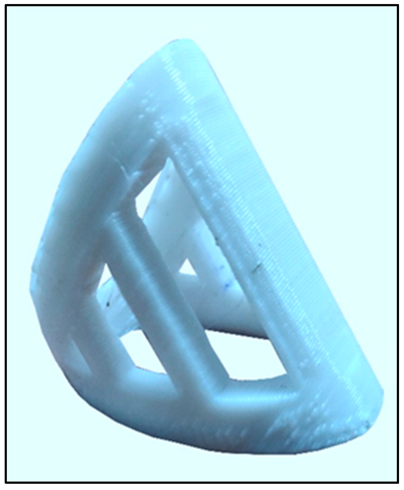
2.3. Fabrication of Splint

As per the computational analysis, splints 2 and 5 showed better results in comparison with other splints. The splint 5 was chosen for fabrication as per its performance on both the stress and deformation analysis [22,24,25]. The designs of the 3D printed splints were based on the three-point pressure system used to straighten the joints, which is a major design principle followed in the fabrication of various orthoses. The measurements of each patient were applied to the digital splint design obtained with the help of SolidWorks and Mesh mixer (Autodesk, San Rafael, CA, USA). The splint was fabricated using Polylactic Acid (PLA) material with the help of Creality Ender 3 (Shenzhen, China) 3D printer [19,26]. The splints were rapidly smoothed by placing them in the vicinity of ethyl acetate vapors, and the supports were manually removed. The splints were padded for cushioning between the subject’s skin and PLA material using pelite material of 2 mm thickness [26]. They were fitted in a slight flexion position, which corrected the hyperextension in other joints. All the correctable joints in all the fingers were fitted with the splints. The splints were fitted after several trials and after optimal fit was achieved using properly fitted splints. The subjects were prescribed the splints for more than four hours daily.

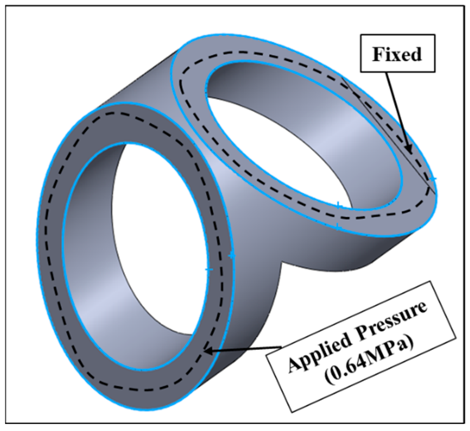
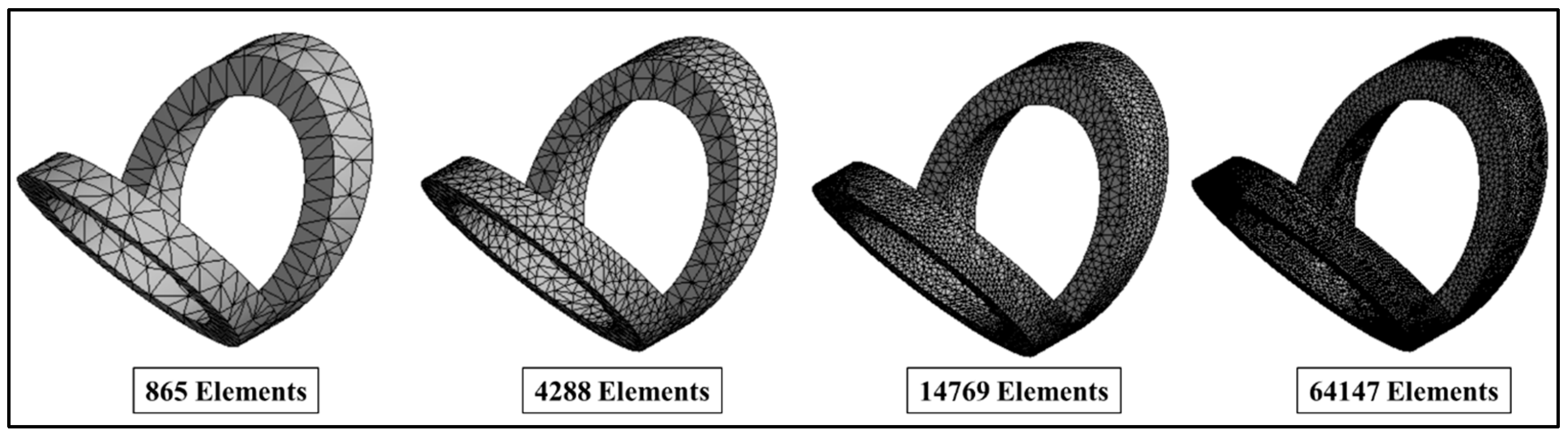
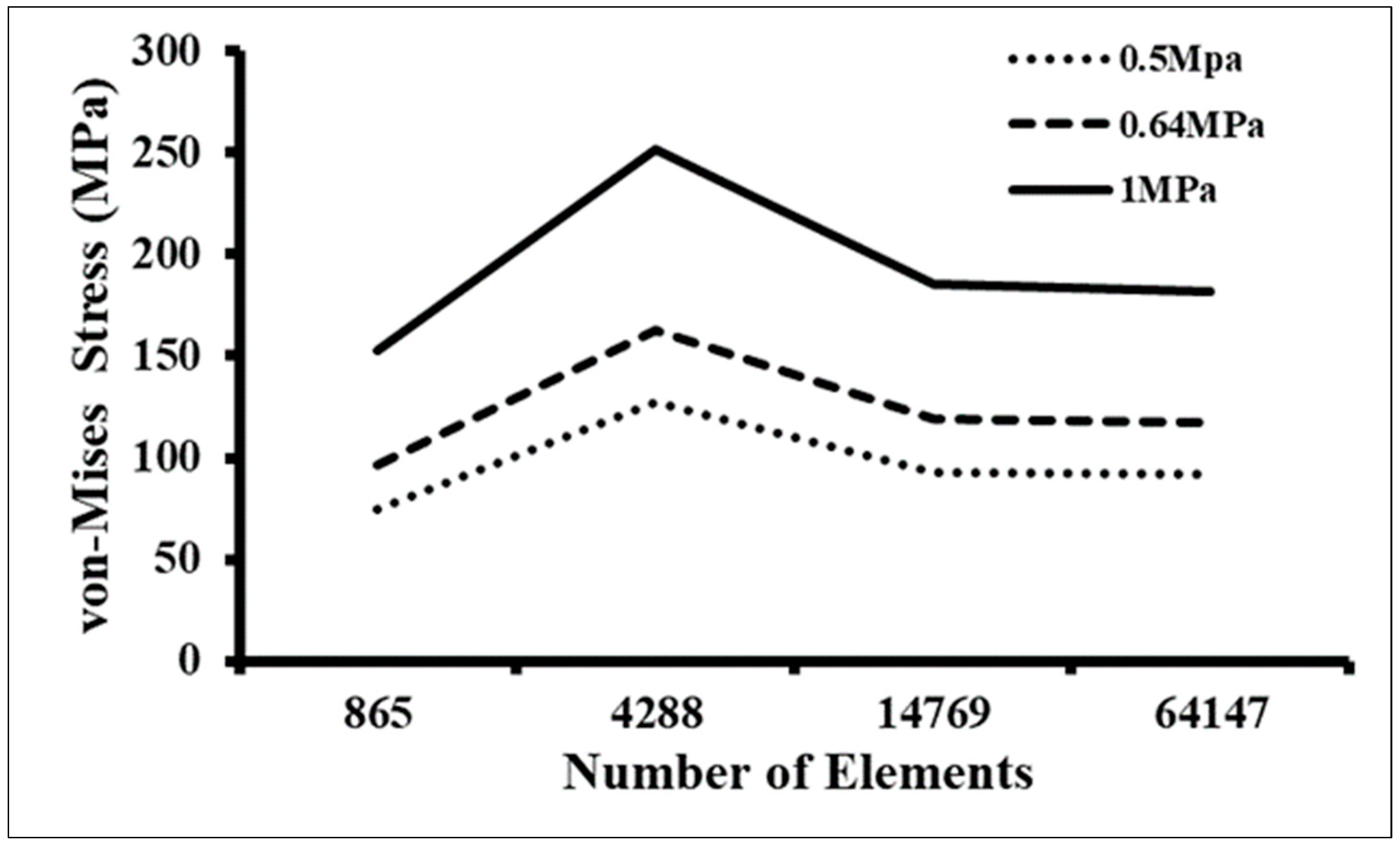
2.4. Testing of Splints

The study used two measures to estimate the performance of the 3D-printed finger splint. The first assessment was through a questionnaire QUEST 2.0 (Quebec User Evaluation of Satisfaction with Assistive Technology) [27]. It is a 12-item instrumentation that is used to evaluate the performance of any assistive technology by asking several questions to the user to assess the characteristics of the assistive device used and the service provided for the follow-up. The second assessment was an objective assessment using the Nine Hole Peg Test (NHPT) that aims to measure the finger dexterity of the affected patients [28]. The score in this test is administered depending upon the time taken by the patient to place the pegs into the holes present on the board (Figure 6). The time starts as soon as the subject touches the first peg and ends at the placement of the last peg. The measurements were taken with and without the splints. Both quantitative measurements were used to analyze the data obtained from the patient’s response.

2.5. Statistical Analysis

The data were analyzed using the SPSS 26.0 software (SPSS inc., Chicago, IL, USA). Mean and standard deviation was computed for each study variable. The outcome variables used for analysis were scores obtained from QUEST 2.0 and time values obtained from NHPT. A t-test was used to analyze the difference in the NHPT time values of each subject. An Independent t-test was used to analyze the pre-test and post-data within the subjects. The hypothesis was tested at a significant level of p ˂ 0.05.

3. Results and Discussion

The subjects recruited for the study had a mean age of 45.70 ± 10.57 years. The mean height and weight of the subjects were 157.05 ± 9.24 cm and 66.05 ± 16.03 kg, respectively. All the subjects meeting the inclusion criteria were asked to take part in the study after signing a consent form. Most of the patients had finger deformities in both the dominant and non-dominant hands. Out of twenty patients, seventeen patients had deformities in all the fingers of both hands.

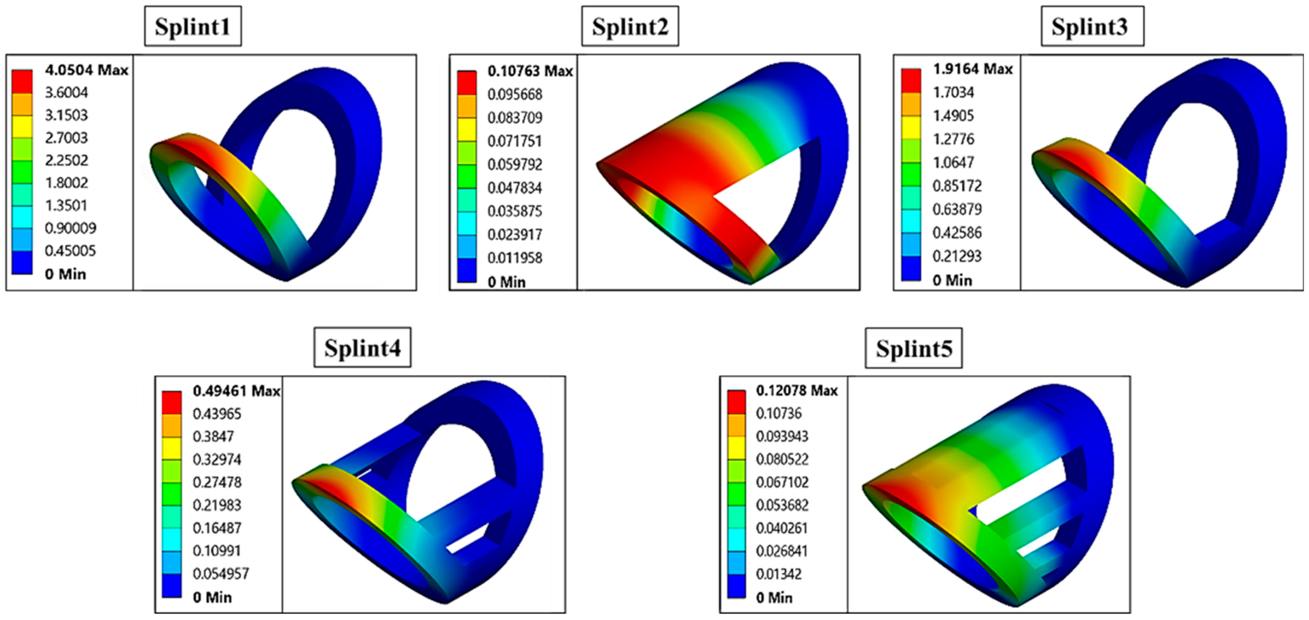
3.1. Computational Analysis of Splints

To conduct the mesh convergence at different loads, pressure values of 0.5 MPa, 0.64 MPa, and 1 MPa were applied on the splint surface, as mentioned in the boundary conditions section. A maximum pressure of 1 MPa was applied to all the meshes for the analysis. The applied pressure simulated a realistic loading scenario. Figure 7 shows the mesh convergence results where the mesh with 14,769 elements showed less than 5% variations in the outcome parameters [29]. Hence, this mesh was considered the optimal mesh and was used throughout the computational analysis across all the splint designs.
The total deformation analysis of designed splints is shown in Figure 8. The maximum and minimum deformation was observed in splint 1 (4.05 mm) and splint 2 (0.10 mm). The deformation values of other splints lay between splint 1 and splint 2, respectively. The deformation values of splint 2 and splint 5 showed similar values, i.e., 0.10 mm ± 5%. Overall, amongst all splints, splint 2 and splint 5 showed the minimum deformation, thus, indicating that these splints could perform well under a standard deformation value.
Since there were fewer differences seen in the deformation values, the stress analysis was also performed. The total von-Mises stress of designed splints is illustrated in Figure 9. The maximum and minimum stress were induced in splint 1 (118.97 MPa) and splint 5 (12.57 MPa), respectively.
The stress values of other splints ranged between splint 1 and splint 5. After deformation and stress analysis, two splints were found to exhibit better performance than the others. Further, a finite element modeling study was performed to assess the reliability of the splints. They were analyzed under high stress and pressure to ensure their rigidity in real-time when the splints were worn by the subjects. The splint 5 that performed the best in the stress and pressure analysis was chosen for the pilot study (Figure 10). The designs varied depending upon the addition of bars for better reliability and rigidity against the pressure created in the affected finger joints.
A total of 185 splints were prescribed to the subjects for an intervention period of four weeks. The most common joint involved was the proximal interphalangeal joints in most of the patients. A brief description of the subject demographics and splints is described in Table 1. The post-splinting period constantly had followed-up services through phone calls and emails, and modifications were done if needed. The subjects were constantly monitored for splint use and its effects on their deformities. The measures used to evaluate the performance of these splints were performed after four weeks. The results estimated are discussed below in the further subsections.

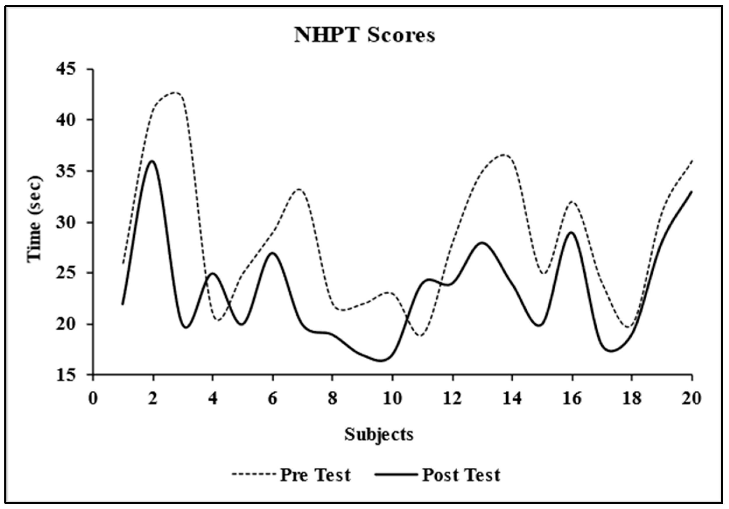
3.2. Nine Hole Peg Test
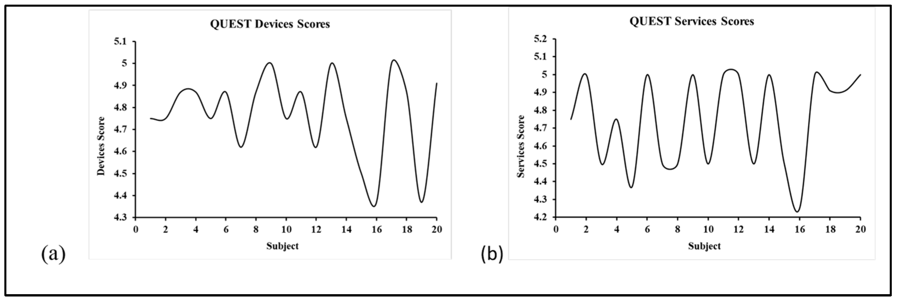
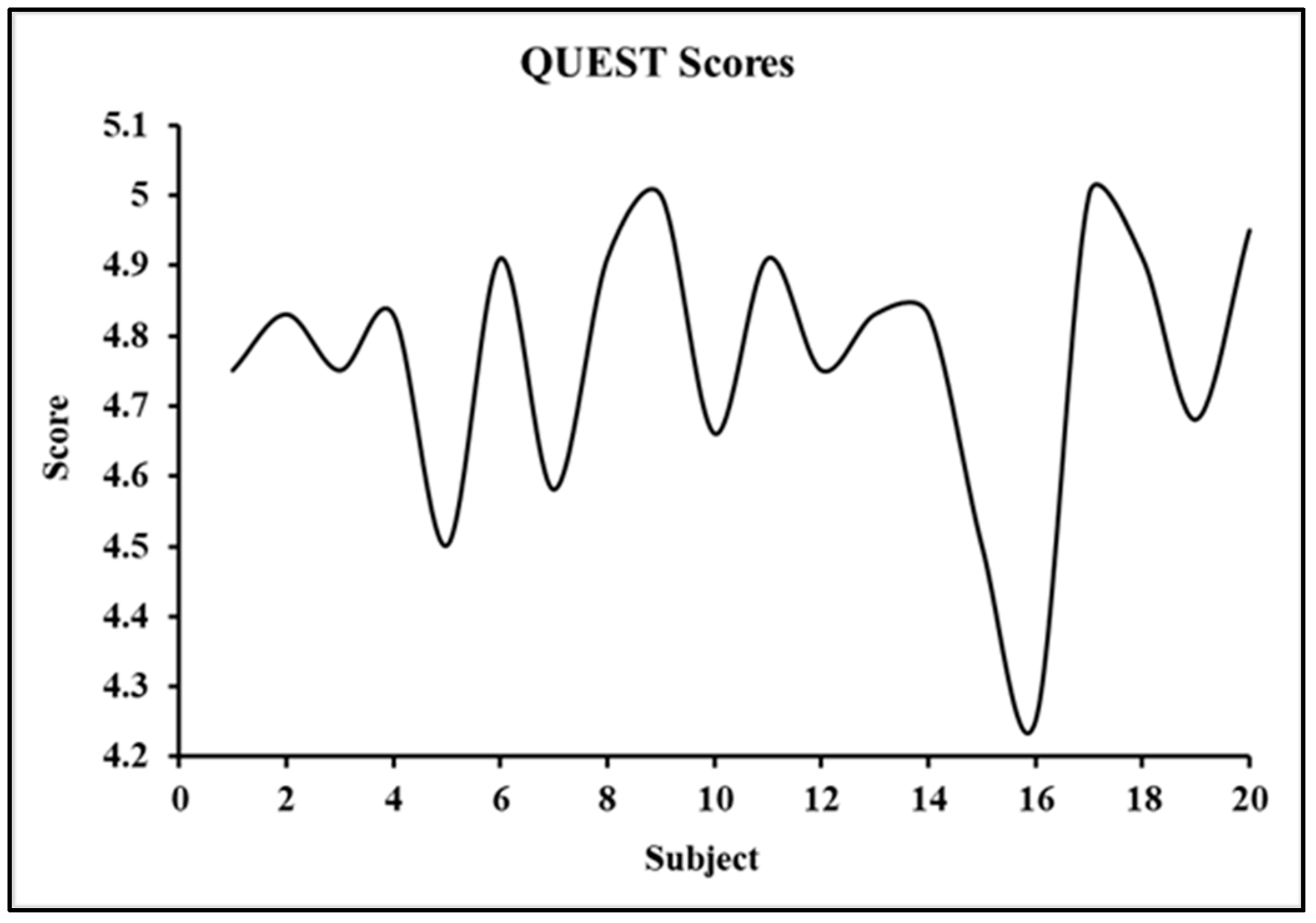
The nine-hole peg test was performed after an intervention time of four weeks. This test was used to assess finger dexterity in rheumatoid arthritis subjects. The test was performed in two phases, first without the splint and the second after the application of the splint. The NHPT values before the application of the splint, which can also be referred to as pre-test NHPT scores, were 28 ± 6.94. The post-test scores that are after the application of the device were 23.50 ± 5.32. The values were tested at a 95% confidence interval and a p-value ˃ 0.05, i.e., 0.001, as per the calculations by SPSS 26.0 software (Figure 11).
The improvements in the NHPT scores were approximately the same, with a maximum difference of 22 s to a minimum improvement of two seconds. There were two subjects whose NHPT scores were higher with splints rather than without. However, the differences in the scores were small.

3.3. QUEST

The scores assessed through this questionnaire were calculated using paired sample test in SPSS software. The scores obtained were under two categories, i.e., devices and services scores. The quantified value for the devices section was 4.76 ± 0.04 (Figure 12a), and for the services section was 4.74 ± 0.06 (Figure 12b); both the values were at p-value ˃ 0.005. The total scores obtained were 4.75 ± 0.04 (Figure 13) at a confidence interval of 95% and p-value ˃ 0.005. The questionnaire also had a subsection of 12 categories, out of which the subjects had to decide the three most satisfactory categories. The top three selections in aggregate were weight, ease to use, and effectiveness.
The QUEST scores were used to assess satisfaction with the performance of the splint and the services provided for follow-up. The scoring was based on scoring from one to five, where one stated not satisfied at all to very satisfied at five. The results obtained in this study were more than four, which implies the quite satisfactory performance of the splint and follow-up.

4. Conclusions

As per the literature, there are different varieties of finger splints available for patients with rheumatoid arthritis, including silver ring splints (SRS), prefabricated thermoplastic splints (PTS), and custom-made thermoplastic splints (CTS) [3,30,31,32], but there is a lack of customized and comfortable splints for prolonged uses. In our work, a novel customized finger splint design was optimized using computational analysis, fabricated using 3D printing, and tested on 20 patients to compare its effectiveness with respect to other costly splints. The splints were assessed using FEM for their rigidity and strength. The option of 3D printing provided more options for customizability. As compared to the commercially available splints, the cost of 3D-printed splints was less. The subjects recruited for this study were diagnosed with rheumatoid arthritis and prescribed custom-made 3D-printed splints fabricated with PLA material for correction of finger deformities such as swan neck and boutonniere. Overall, the splint was successful in providing better finger alignment and achieving corrected joint positions throughout the fingers, could be applied both ways, and was effective in the correction of both swan neck and boutonniere deformities. In conclusion, 3D printed PLA finger splint was found to be a cost-effective and good option for improving finger dexterity after the application of the splint. The performance of the splint was also found to be satisfactory, be it with respect to the ease of use, the weight, or the effectiveness of the splint, thus allowing prolonged wearing. In the future, a few limitations of this study will be addressed, including the use of more precise measurements to accommodate different joint sizes and its testing on a greater sample size for better patient assessment. In addition, future studies will also focus on the level of activities of every subject and set a criterion for the selection of the same. Also, the developed splint will be compared with the existing designs for a better assessment of their performance.

Author Contributions

K.C.: Methodology; Validation; Investigation; Formal Analysis; Writing—Original Draft; Writing—Review and Editing. S.G.: Methodology; Data Curation; Formal Analysis; Investigation. S.S. (Sakshi Saharawat): Data Curation; Investigation; Formal Analysis. S.S. (Shruti Sarkar): Data Curation; Investigation; Formal Analysis. A.C.: Conceptualization; Methodology; Formal Analysis; Supervision; Writing-Review and Editing. All authors have read and agreed to the published version of the manuscript.

Funding

This research received no external funding.

Institutional Review Board Statement

The study was approved by the Ethical Committee of the Indian Spinal Injuries Centre, New Delhi, (ISIC/RP/2022/024) on 13 July 2022.

Informed Consent Statement

The subject provided a signed consent form before the study was conducted.

Data Availability Statement

The datasets generated during and/or analyzed during the current study are not publicly available due to large dataset but are available from the corresponding author on reasonable request.

Conflicts of Interest

The authors declare no conflict of interest.

References

  1. Scott, D.L.; Wolfe, F.; Huizinga, T.W.J. Rheumatoid arthritis. Lancet 2010, 376, 1094–1108. [Google Scholar] [CrossRef] [PubMed]
  2. Handa, R.; Rao, U.; Lewis, J.F.M.; Rambhad, G.; Shiff, S.; Ghia, C.J. Literature review of rheumatoid arthritis in India. Int. J. Rheum. Dis. 2016, 19, 440–451. [Google Scholar] [CrossRef] [PubMed]
  3. van der Giesen, F.J.; Nelissen, R.G.H.H.; van Lankveld, W.J.; Kremers-Selten, C.; Peeters, A.J.; Stern, E.B.; le Cessie, S.; Vlieland, T.P.M.V. Swan neck deformities in rheumatoid arthritis: A qualitative study on the patients’ perspectives on hand function problems and finger splints. Musculoskelet. Care 2010, 8, 179–188. [Google Scholar] [CrossRef] [PubMed]
  4. Lane, R.; Nallamothu, S.V. Swan-Neck Deformity. StatPearls, June. 2022. Available online: https://www.ncbi.nlm.nih.gov/books/NBK525970/ (accessed on 20 October 2022).
  5. Nalebuff, E.A. The Rheumatoid Swan-Neck Deformity. Hand Clin. 1989, 5, 203–214. [Google Scholar] [CrossRef]
  6. To, P.; Watson, J.T. Boutonniere Deformity. J. Hand Surg. 2011, 36, 139–142. [Google Scholar] [CrossRef]
  7. Van Der Giesen, F.J.; Van Lankveld, W.J.; Kremers-Selten, C.; Peeters, A.J.; Stern, E.B.; le Cessie, S.; Nelissen, R.; Vlieland, T.V. Effectiveness of two finger splints for swan neck deformity in patients with rheumatoid arthritis: A randomized, crossover trial. Arthritis Care Res. 2009, 61, 1025–1031. [Google Scholar] [CrossRef]
  8. Zijlstra, T.R.; Heijnsdijk-Rouwenhorst, L.; Rasker, J.J. Silver Ring Splints improve dexterity in patients with rheumatoid arthritis. Arthritis Care Res. 2004, 51, 947–951. [Google Scholar] [CrossRef]
  9. Knipping, A.; ter Schegget, M. A Study Comparing Use and Effects of Custom-Made versus Prefabricated Splints for Swan Neck Deformity in Patients with Rheumatoid Arthritis. Br. J. Hand Ther. 2016, 5, 101–107. [Google Scholar] [CrossRef]
  10. Zolfagharian, A.; Gregory, T.M.; Bodaghi, M.; Gharaie, S.; Fay, P. Patient-specific 3D-Printed Splint for Mallet Finger Injury. Int. J. Bioprinting 2020, 6, 259. [Google Scholar] [CrossRef]
  11. Baradaran, A.; Ebrahimzadeh, M.H.; Kachooei, A.R.; Rivlin, M.; Beredjiklian, P. Comparison of Custom-made Versus Prefabricated Thumb Splinting for Carpometacarpal Arthrosis: A Systematic Review and Meta-analysis. Arch. Bone Jt. Surg. 2018, 6, 478–485. [Google Scholar] [CrossRef]
  12. Frye, S.K.; Geigle, P.R. A comparison of prefabricated and custom made resting hand splints for individuals with cervical spinal cord injury: A randomized controlled trial. Clin. Rehabil. 2021, 35, 861–869. [Google Scholar] [CrossRef] [PubMed]
  13. Papavasiliou, T.; Shah, R.K.; Chatzimichail, S.; Uppal, L.; Chan, J.C. Three-dimensional Printed Customized Adjustable Mallet Finger Splint: A Cheap, Effective, and Comfortable Alternative. Plast. Reconstr. Surg.-Glob. Open 2021, 9, e3500. [Google Scholar] [CrossRef] [PubMed]
  14. Gupta, A.; Chaturvedi, S.; Bhat, A.K.; Samheel, M.; Saxena, A. Design and Manufacture of Customizable Finger Immobilizer and Mallet Finger Splints. In Proceedings of the 2019 International Conference on Biomedical Innovations and Applications, Varna, Bulgaria, 8–9 November 2019; pp. 1–4. [Google Scholar] [CrossRef]
  15. Elzinga, K.; Chung, K.C. Managing Swan Neck and Boutonniere Deformities. Clin. Plast. Surg. 2019, 46, 329–337. [Google Scholar] [CrossRef] [PubMed]
  16. Waldburger, L.; Schaller, R.; Furthmüller, C.; Schrepfer, L.; Schaefer, D.J.; Kaempfen, A. 3D-Printed Hand Splints versus Thermoplastic Splints: A Randomized Controlled Pilot Feasibility Trial. Int. J. Bioprinting 2022, 8, 128–138. [Google Scholar] [CrossRef]
  17. Li, J.; Tanaka, H. Rapid customization system for 3D-printed splint using programmable modeling technique–a practical approach. 3D Print. Med. 2018, 4, 5. [Google Scholar] [CrossRef]
  18. Wong, J.Y. On-Site 3D Printing of Functional Custom Mallet Splints for Mars Analogue Crewmembers. Aerosp. Med. Hum. Perform. 2015, 86, 911–914. [Google Scholar] [CrossRef]
  19. Singh, G.; Gupta, V.; Chanda, A. Mechanical Characterization of Rotating Triangle Shaped Auxetic Skin Graft Simulants. Facta Univ. Ser. Mech. Eng. 2020, 1–16. [Google Scholar] [CrossRef]
  20. Singh, G.; Gupta, V.; Chanda, A. Artificial skin with varying biomechanical properties. Mater. Today Proc. 2022, 62, 3162–3166. [Google Scholar] [CrossRef]
  21. Gupta, V.; Chanda, A. Finite element analysis of a hybrid corrugated hip implant for stability and loading during gait phases. Biomed. Phys. Eng. Express 2022, 8, 035028. [Google Scholar] [CrossRef]
  22. Gupta, V.; Chanda, A. Expansion potential of skin grafts with alternating slit based auxetic incisions. Forces Mech. 2022, 7, 100092. [Google Scholar] [CrossRef]
  23. Gupta, S.; Singh, G.; Chanda, A. Prediction of diabetic foot ulcer progression: A computational study. Biomed. Phys. Eng. Express 2021, 7, 065020. [Google Scholar] [CrossRef] [PubMed]
  24. Gupta, S.; Gupta, V.; Chanda, A. Biomechanical modeling of novel high expansion auxetic skin grafts. Int. J. Numer. Methods Biomed. Eng. 2022, 38, e3586. [Google Scholar] [CrossRef] [PubMed]
  25. Gupta, V.; Gupta, S.; Chanda, A. Expansion potential of skin grafts with novel rotating-triangle-shaped auxetic incisions. Emerg. Mater. Res. 2022, 11, 406–414. [Google Scholar] [CrossRef]
  26. Chhikara, K.; Gupta, S.; Chanda, A. Development of a novel foot orthosis for plantar pain reduction. Mater. Today Proc. 2022, 62, 3532–3537. [Google Scholar] [CrossRef]
  27. Demers, L.; Monette, M.; Lapierre, Y.; Arnold, D.L.; Wolfson, C. Reliability, validity, and applicability of the Quebec User Evaluation of Satisfaction with assistive Technology (QUEST 2.0) for adults with multiple sclerosis. Disabil. Rehabil. 2002, 24, 21–30. [Google Scholar] [CrossRef] [PubMed]
  28. Johansson, G.M.; Häger, C.K. A modified standardized nine hole peg test for valid and reliable kinematic assessment of dexterity post-stroke. J. Neuroeng. Rehabil. 2019, 16, 8. [Google Scholar] [CrossRef]
  29. Singh, G.; Gupta, S.; Chanda, A. Biomechanical modelling of diabetic foot ulcers: A computational study. J. Biomech. 2021, 127, 110699. [Google Scholar] [CrossRef]
  30. Rizio, L.; Belsky, M.R. Finger Deformities in Rheumatoid Arthritis. Hand Clin. 1996, 12, 531–540. [Google Scholar] [CrossRef]
  31. Arulmozhi, R.S.; Vaidya, M.; Poojalakshmi, M.G.; Kumar, A.; Anuraag, K. 3D Design and Printing of Custom-Fit Finger Splint. Biomed. Eng. Appl. Basis Commun. 2018, 30, 1850032. [Google Scholar] [CrossRef]
  32. Egan, M.; Brosseau, L.; Farmer, M.; Ouimet, M.-A.; Rees, S.; Tugwell, P.; A Wells, G. Splints and Orthosis for treating rheumatoid arthritis. Cochrane Database Syst. Rev. 2001, 2010, CD004018. [Google Scholar] [CrossRef]
Figure 1. Swan neck and boutonniere deformities in Rheumatoid Arthritis.
Rheumato 03 00004 g001
Figure 2. Dimensions recorded in the fingers (A) Lateral view showing circumferential and oblique measurements, (B) Palmar view showing linear measurements.
Rheumato 03 00004 g002
Figure 3. Different types of splint designs used for structural analysis.
Rheumato 03 00004 g003
Figure 4. Boundary conditions of the splint.
Rheumato 03 00004 g004
Figure 5. Different meshes, from very coarse to superfine, with the number of elements.
Rheumato 03 00004 g005
Figure 6. Subject performing the Nine-hole peg test.
Rheumato 03 00004 g006
Figure 7. Mesh convergence study.
Rheumato 03 00004 g007
Figure 8. Deformation results of different splint models at 0.64 MPa pressure.
Rheumato 03 00004 g008
Figure 9. von-Mises stress distribution of different splint models at 0.64MPa pressure.
Rheumato 03 00004 g009
Figure 10. 3D printed splint.
Rheumato 03 00004 g010
Figure 11. The pre and post-test scores of the Nine Hole Peg Test without and with splinting.
Rheumato 03 00004 g011
Figure 12. QUEST (a) devices and (b) services scores.
Rheumato 03 00004 g012
Figure 13. QUEST total scores.
Rheumato 03 00004 g013
Table 1. Subject demographics and splint details.
SubjectAge
(in Years)
HandsModifications and Follow-Up
Left HandRight Hand
LRMITTIMRL
145PPDPDPD PDPDPDPModified the dimensions
241 PDPPDD PPPDPD
338PP P DPDPUncomfortable in cooking
460 PDPP PPPP
545 PPDP PDPDDP
656 PDPPD PPDPDD
745PDPDDPDDDPD P
822 P D PPD
945PPDPDPD PDP Poor fit despite adjustments
1046PPPP PPDPD
1142PPD P D DP
1265PPDPPDDP PDPDUnsatisfactory
1356PDPD PDPPDP
1440 PDPD D PD D
1555 PPPDDPPPPSplints reduced after 2 weeks due to discomfort
1657 PPPD PPP
1726PPDPDP PDPDPDP
1834 DP PPP
1950 PD PP PSplints added afterward
2046PDDPPD DPDDDArthritis increased
Fingers; L-Little, R-Ring, M-Middle, I-Index, T-Thumb Joints; D-Distal Interphalangeal Joint, P-Proximal Interphalangeal Joint.
Disclaimer/Publisher’s Note: The statements, opinions and data contained in all publications are solely those of the individual author(s) and contributor(s) and not of MDPI and/or the editor(s). MDPI and/or the editor(s) disclaim responsibility for any injury to people or property resulting from any ideas, methods, instructions or products referred to in the content.

