Case Report

Unusual Case Presentation of Systemic Lupus Erythematosus in a Young Woman

Rheumatology and Clinical Immunology Department, Tishreen Hospital, Damascus TH 34890, Syria
*
Author to whom correspondence should be addressed.
Rheumato 2022, 2(4), 93-97; https://doi.org/10.3390/rheumato2040013
Submission received: 19 September 2022 / Revised: 6 October 2022 / Accepted: 11 October 2022 / Published: 14 October 2022

Abstract

:
Systemic Lupus Erythematosus (SLE) is a chronic multisystem autoimmune disease. Serositis occurs in 16% of SLE patients, and while cardiac tamponade and acute peritonitis with ascites can occur during the course of the disease, they are rare as the first presentation. A 25-year-old woman presented to the emergency department in Tishreen Hospital with complaints of dyspnea, fever, chills, and chest and abdominal pain. Two months prior, she suffered from musculoskeletal pain, fatigue, anorexia, weight loss of about 15 kg, severe hair loss, and recurrent oral aphthous. On clinical examination, the patient was pale and tired with dyspnea and pitting edema (grade 3–4). Pericardiocentesis was emergently performed because there were signs of cardiac tamponade. Three days later, the patient developed an acute surgical abdomen due to acute peritonitis and ascites. Later, the patient was diagnosed with SLE after excluding malignant and infectious diseases. Consequently, methylprednisolone pulses, azathioprine, and hydroxychloroquine 200 mg/day were introduced immediately. The clinical status of the patient dramatically improved, and three months later, the patient was symptom-free with normal laboratory tests. In conclusion, although cardiac tamponade and acute surgical abdomen because of acute peritonitis and ascites as the initial presentation of SLE are very rare, they can occur coincidently.

1. Introduction

Systemic Lupus Erythematosus (SLE) is a chronic multisystem autoimmune disease characterized by very protean clinical manifestations [1]. It usually affects women of childbearing age, and the great variability in the presentation and severity of SLE is a major challenge for accurate diagnosis [2]. The most common presenting manifestations are constitutional symptoms and cutaneous and articular manifestations [3]. Each of these manifestations presents in about 50% of lupus patients at the time of diagnosis [3]. The other clinical features of SLE are much less likely to be presenting manifestations, although any of them can be the first clue to the diagnosis. However, these manifestations develop over time as the disease progress [4]. Serositis occurs in 16% of SLE patients; while pericarditis can occur, cardiac tamponade is rare, and as the first presentation, it is rarer. In addition, ascites and acute peritonitis can occur during the disease, but it is rare as the first manifestation [5,6]. Here, we present a case of a young woman who presented with both cardiac tamponade and acute surgical abdomen because of acute peritonitis and ascites as the first presentation of SLE.

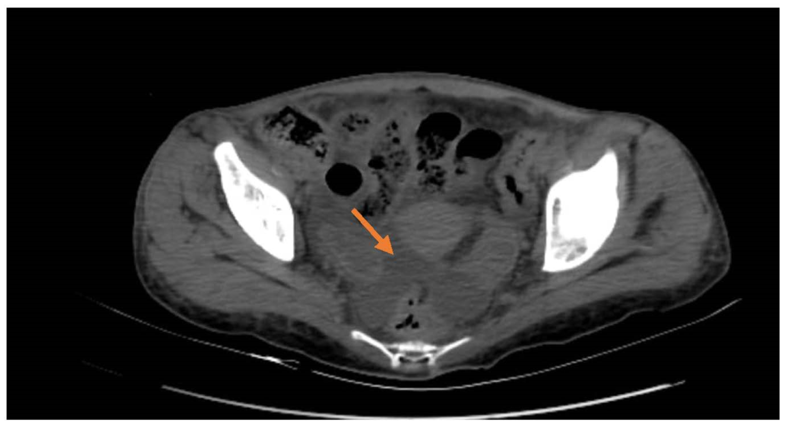
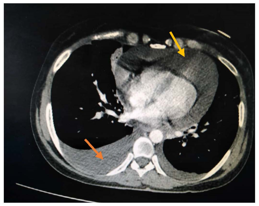
2. Case Presentation

A 25-year-old woman presented to the emergency department in Tishreen Hospital with complaints of dyspnea, fever, chills, chest, and abdominal pain. The complaints of the patient started two months before admission, as she suffered from musculoskeletal pain, fatigue, anorexia, weight loss of about 15 kg, severe hair loss, and recurrent oral aphthous. In addition, she mentioned a history of blood transfusions due to anemia. Regarding medical history, the patient had ITP five years prior and hypothyroidism 18 months prior, which was treated with Eltroxin 100 mg/day. She denied any familial history of rheumatic diseases. No prior meds were reported except Eltroxin 100 mg/day.
On presentation, her blood pressure was 90/70 mmHg, her temperature was 39 °C, her heart rate was 140 p/m, and her SpO2 was 93%.
On clinical examination, the patient was pale and tired with dyspnea and pitting edema (grade 3–4); her jugular venous pressure was raised at 5 cm with visible carotid pulsations and pulsus paradoxus. Auscultation of the heart revealed diminished heart sounds. There was reduced air entry bilaterally on examination of the chest.
Additional investigations were performed, and the results were as follows: a chest X-ray revealed increased cardiopulmonary index with pleural effusion, and an electrocardiogram showed sinus tachycardia. Neck, chest, and abdomen computed tomography showed mild bilateral pleural effusion, large pericardial effusion (20–22 mm), hepatomegaly > 18 cm, moderate ascites, bilateral axillary, and inguinal lymph nodes enlargement < 15 mm (Figure 1 and Figure 2). The echocardiogram showed signs of cardiac tamponade.
Therefore, pericardiocentesis was emergently performed, and the pericardial fluid contained a leukocyte count of 1500, a lymphocyte percentage of 70%, a protein level of 4 g/dL, and an LDH level of 2519; in the bacterial culture, the tuberculosis PCR was negative, and cytology for malignancy was negative. The patient was admitted to the coronary care unit, and additional investigations were performed to exclude infectious and malignant diseases. The COVID-19 PCR was negative, and the breast mammography and bone biopsy were normal. The laboratory tests are presented in Table 1. Three days after admission to the coronary care unit, the patient had severe abdominal pain, and on physical examination, the abdomen was distended with generalized tenderness and abdominal wall spasm, while abdominal echography revealed only moderate ascites. Exploratory laparotomy was performed following a surgeon consultation. The findings of exploratory laparotomy revealed a moderate amount of clear fluid without any signs of perforation or other abnormal pathologies. The peritoneal fluid analysis found a leukocyte count of 3000, a lymphocyte percentage of 67%, a protein level of 3.2 g/dL, an albumin level of 1.2 g/dL, and an SAAG score of 0.8 BK; bacteriology and cytology for malignancy were negative. Peritoneal biopsy revealed nonspecific inflammation. After that, a rheumatology consultation was performed to exclude systemic autoimmune disease.
On the basis of a thorough clinical examination, medical history, and laboratory findings, SLE was suspected, and additional tests were performed (Table 1).
Microscopic urinalysis revealed proteinuria and microscopic hematuria.
A diagnosis of SLE is made using the 1997 American College of Rheumatology (ACR) criteria, which require that four or more of the 11 criteria are present. In our case, the patient met five of the 11 criteria for a definite diagnosis of SLE, namely serositis, proteinuria, positive antinuclear antibody and anti-dsDNA, lymphopenia, and thrombocytopenia. In addition, she met the criteria of hemolytic anemia with reticulocytosis and elevated lactate dehydrogenase (LDH) and indirect bilirubin with a positive direct Coombs test.
Consequently, methylprednisolone pulses (1 g × three consequently days) were introduced immediately in addition to hydroxychloroquine 200 mg/day and azathioprine 100 mg/day.
After the initiation of corticosteroids, the clinical status of the patient dramatically improved, including the disappearance of abdominal pain, relief of musculoskeletal pain, a decrease in ESR and CRP values, and improvement in hemoglobin values.
Follow-up: Three months later, the patient was symptom-free with normal laboratory tests, and she was on prednisolone 10 mg/day as a maintenance dose, hydroxychloroquine 200 mg/day, azathioprine 100 mg/day, and Eltroxin 100 mg/day.

3. Discussion

Our patient had severe serositis as an initial presentation of SLE that presented as cardiac tamponade and acute peritonitis with moderate ascites and small pleural effusion.
SLE is a chronic multisystem autoimmune disease with a remitting and relapsing course [2].
The association between SLE and thyroid disease has been mentioned in many papers, and our patient had hypothyroidism before the SLE diagnosis [3].
The 1997ACR classification criteria for SLE include serositis as pleural and pericardial effusion, but they do not mention peritoneal serositis [7].
Pericardial effusion can be seen in up to 50% of SLE patients [8]. It tends to be mild and asymptomatic and is typically detected on echocardiography performed for another indication, but it can rarely enlarge to cause cardiac tamponade [8]. Cardiac tamponade occurs in less than 1% of SLE patients, and as an initial presentation, it is rarer; large pericardial effusion should be investigated to exclude malignancy and infection [5]. In addition, cardiac tamponade can occur in patients with known SLE and patients with renal disease, anemia, pleuritis, lower levels of complement, and higher ESR [9].
Regarding ascites, it is seen in 10% of SLE patients [6]. Moreover, it was thought to occur only in nephrotic syndrome, Budd–Chiari syndrome, and protein-losing enteropathy [10]. However, ascites that accompany acute peritonitis as the initial presentation for lupus are extremely rare, and to our knowledge, there have only been a few cases in the literature that have mentioned this presentation in juvenile patients, while other related cases have also occurred in well-known SLE patients [10,11]. In addition, the occurrence of cardiac tamponade and acute peritonitis with ascites at the same time had not been previously described.
As the patient presented with life-threatening manifestations that required emergency intervention, along with leukopenia, anemia, severe hair loss, and weight loss, malignancy was the first differential diagnosis; thus, she underwent a thorough study with negative results for any hematologic or solid malignancies. In addition, infections, especially COVID-19 and tuberculosis, were excluded. Finally, the diagnosis was confirmed after the rheumatology consultation.
Seungwon Ryu et al. studied the immunological and clinical associations between peritonitis and pericarditis, and they found that proteinuria, hemolytic anemia, lymphadenopathy, and positive anti-Sm were predictive of pericarditis [9], while gastrointestinal infarction and pulmonary fibrosis were predictive of peritonitis [9]. Our patient had hemolytic anemia and proteinuria in addition to lymphadenopathy.
Generally, the prognosis of lupus serositis is good; it responds well to corticosteroids and immunosuppressants. However, relapse and progression to fibrosis are uncommon [7].
This case is an important lesson, especially for non-rheumatologists, because this young woman was quite ill for two months with typical SLE manifestations before she presented with generalized serositis that necessitated pericardial drainage and later led to the unnecessary surgical exploration of the abdomen.

4. Conclusions

SLE can present with variable manifestations that are not mentioned in the ACR classification criteria for SLE; one of these presentations is peritoneal effusion [1,7]. Moreover, although cardiac tamponade and acute surgical abdomen because of acute peritonitis and ascites as the initial presentation of SLE are very rare, they can coincidently occur, so clinicians should keep them in mind as the first clue to SLE diagnosis.

Author Contributions

S.A. was the first author of this article and was the inpatient doctor; H.W., K.K. and H.Q. performed the writing and editing of the manuscript. All authors have read and agreed to the published version of the manuscript.

Funding

This research received no external funding.

Institutional Review Board Statement

This research was approved by the local ethical committee for medical research at Tishreen Hospital (N: 23600) in accordance with the ethical standards of the Helsinki Declaration.

Informed Consent Statement

Written informed consent was obtained from the patient to publish this paper.

Conflicts of Interest

The authors declare no conflict of interest.

References

  1. Man, B.L.; Mok, C.C. Serositis related to systemic lupus erythematosus: Prevalence and outcome. Lupus 2005, 14, 822–826. [Google Scholar] [CrossRef] [PubMed]
  2. Martinez, D.; Lowe, R. Case report: Systemic lupus erythematosus (SLE) serositis mimicking acute cholecystitis. Clin. Radiol. 1991, 44, 434–435. [Google Scholar] [CrossRef]
  3. Almousa, S.; Ahmad, T.; Hamdan, S.; Katt, N. Lupus erythematosus tumidus (LET) with autoimmune thyroid dysfunction (AITD) as the first presentation of systemic lupus erythematosus: A case report and review of the literature. Egypt. Rheumatol. 2020, 42, 245–250. [Google Scholar] [CrossRef]
  4. Hammami, S.; Bdioui, F.; Ouaz, A.; Loghmari, H.; Mahjoub, S.; Saffar, H. Successful treatment of massive ascites due to lupus peritonitis with hydroxychloroquine in old- onset lupus erythematosus. Pan. Afr. Med. J. 2014, 18, 165. [Google Scholar] [CrossRef] [PubMed]
  5. Cheng, W.; Balachandar, R.; Mistry, P. Cardiac tamponade: An initial presentation of SLE. BMJ Case Rep. 2013, 2013, bcr2013200011. [Google Scholar] [CrossRef] [PubMed] [Green Version]
  6. Cook, S.G. Ascites as the Presenting Sign of Systemic Lupus Erythematosus. Cureus 2022, 14, e23231. [Google Scholar] [CrossRef] [PubMed]
  7. Liang, Y.; Leng, R.-X.; Pan, H.-F.; Ye, D.-Q. The prevalence and risk factors for serositis in patients with systemic lupus erythematosus: A cross-sectional study. Rheumatol. Int. 2016, 37, 305–311. [Google Scholar] [CrossRef] [PubMed]
  8. Tian, D.G.; Jacobs, A.K.; Benjamin, E.J. A 19-year-old female with tamponade and systemic lupus erythematosus (SLE). Int. J. Cardiol. 2015, 179, 1–2. [Google Scholar] [CrossRef] [PubMed]
  9. Ryu, S.; Fu, W.; Petri, M.A. Associates and predictors of pleurisy or pericarditis in SLE. Lupus Sci. Med. 2017, 4, e000221. [Google Scholar] [CrossRef] [PubMed]
  10. Rodriguez, E.A.; Rodriguez, V.R.; Otero, D.; Larsen, M. SLE Ascites: An Uncommon Initial Presentation: 954. Am. J. Gastroenterol. 2015, 110, S408. [Google Scholar] [CrossRef]
  11. Lepore, L.; Facchini, S.; Codrich, D.; Pelizzo, G.; Messineo, A.; Ventura, A. Acute Abdomen: The Presenting Sign of Systemic Lupus Erythematosus in Childhood. J. Pediatr. Gastroenterol. Nutr. 2002, 35, 570–572. [Google Scholar] [CrossRef] [PubMed]
Figure 1. Moderate ascites (arrow).
Rheumato 02 00013 g001
Figure 2. Large pericardial effusion (yellow arrow), small pleural effusion (orange arrow).
Rheumato 02 00013 g002
Table 1. Laboratory results on admission and discharge.
ParameterAdmissionDischargeReference Value
WBC150049304–9 C/uL
Lymphocytes0.31.41.18–3.74 C/uL
Hemoglobin7.79.711.2–15.7 G/dL
Platelets224281182–370 C/uL
ESR6743Up to 15 mm/1 h
CRP84.30.40–5 mg/L
Creatinine4742.545–120 umol/L
Alt20.5205–40 U/I
Ast23.1215–40 U/I
LDH712400230–460 U/L
Indirect bilirubin2.50.70.2–0.8 mg/dL
Reticulocytes7.980.70.5–1.5%
Coombs directPositive
ANAHomogeneous 1/320 Less than 1/100
Anti-dsDNA250 Up to 25 U/mL
Protein 24 h urine562 0–150 mg/24 h
TSH11.544.70.35–5.1 uIU/mL
HBsAgNegative
Anti HCVNegative
C40.04 0.15–0.45 g/L
C30.28 0.8–2 g/L
ENA screenNegative Negative
APL antibodiesNegative Negative
WBC: White blood cells. ESR: Erythrocyte sedimentation rate. CRP: C-reactive protein. LDH: Lactate dehydrogenase. ANA: Antinuclear antibodies. Anti-dsDNA: Anti-double-stranded DNA.TSH: Thyroid stimulating hormone. C3, C4: complement. ENA: Extracted nuclear antibodies. APL: Antiphospholipid.
Publisher’s Note: MDPI stays neutral with regard to jurisdictional claims in published maps and institutional affiliations.

