Article

Effects of Coexisting Anions on the Formation of Hematite Nanoparticles in a Hydrothermal Process with Urea Hydrolysis and the Congo Red Dye Adsorption Properties

1
Department of Chemical Engineering, Osaka Prefecture University, Osaka 599-8531, Japan
2
Department of Chemical Engineering, Osaka Metropolitan University, Osaka 599-8531, Japan
*
Author to whom correspondence should be addressed.
Powders 2023, 2(2), 338-352; https://doi.org/10.3390/powders2020020
Submission received: 13 January 2023 / Revised: 2 March 2023 / Accepted: 4 May 2023 / Published: 8 May 2023
(This article belongs to the Special Issue Feature Papers in Powders)

Abstract

:
Crystalline hematite nanoparticles as adsorbents for anionic Congo red dye were prepared by a hydrothermal process using urea hydrolysis. To examine the effects of coexisting anions in a solution on the formation of hematite nanoparticles, different iron(III) salts, including iron chloride hexahydrate, iron nitrate nonahydrate, iron sulfate n-hydrate, ammonium iron sulfate dodecahydrate, and basic ferric acetate, were employed as iron-ion sources. After the hydrothermal treatment of the solution, consisting of an iron salt and urea at 423 K for 20 h, a single phase of hematite was formed from the iron-nitrate solution. The results suggested that the hydrothermal formation of hematite depended on the stability of iron complexes formed in the starting solution. The average crystallite size and median diameter of hematite nanoparticles also depended on the coexisting anions, suggesting that the appropriate selection of the coexisting anions in the starting solution can allow for control of the crystallite size and particle diameter of hematite nanoparticles. The Congo red adsorption kinetics and isotherms of the hematite nanoparticles were described by the Elovich model and Langmuir model, respectively. The adsorption thermodynamics parameters were estimated, which suggested an exothermic and spontaneous process. The results demonstrated good adsorption properties for Congo red adsorption.

1. Introduction

Elemental iron is an abundant natural resource contained in the earth’s crust and is conventionally and widely used in many industries as a basic material in metallic iron, steel, alloys, oxides, and hydroxides because of its low cost, high usability, and nontoxicity [1]. Among iron-containing materials, iron oxides, such as FeO, α-, β-, γ-, and ε-Fe2O3, and Fe3O4, have simple crystal structures and excellent properties, resulting in many practical uses in various industrial products, such as colorants, catalysts, and magnetic materials [2,3]. In particular, hematite (α-Fe2O3) fine powder has been traditionally used as a pigment; in recent years, hematite nanoparticles have been actively studied for advanced applications, e.g., as anode materials in lithium-ion batteries [4,5,6,7,8], adsorbents for removal of heavy metals and dyes [8,9,10,11,12], sensors [13,14,15,16,17], and photocatalysts [18,19,20,21].
Specific crystallinity and particle size are required for each application of α-Fe2O3 nanoparticles and are achievable through various methods [22,23]. Among them, synthesis processes using liquid phase reactions via formation of ferric hydroxides as precursors and/or intermediates, e.g., sol–gel and hydrothermal processes with/without postcalcination, have been industrially employed due to their simplicity of operation [10,13,17,24,25]. In addition, these methods can be used to control the crystallinity and particle size of products by adjusting reaction conditions, such as temperature, concentration, and additives. Accordingly, the liquid-phase processes are advantageous in the industrial production of α-Fe2O3 nanoparticles.
As an effective method to provide homogeneous metal-oxide nanoparticles, hydrothermal treatments of hydroxides as precursors/intermediates formed by urea hydrolysis in an aqueous phase have attracted much attention; the hydroxide-ion concentration increases uniformly at temperatures higher than approximately 90 °C [26,27,28], resulting in the formation of homogeneous hydroxides. Some researchers have employed this method for the synthesis of α-Fe2O3 nanoparticles [29,30,31]. In the processes, before urea hydrolysis, ferric ions can be converted to iron complexes with coexisting anions as ligands in a solution; this may affect the formation of ferric hydroxide [32,33]. This paper first reports a systematic study on the effects of coexisting anions on the formation of α-Fe2O3 nanoparticles in a hydrothermal process with urea hydrolysis. To vary the coexisting anions, iron(III) salts with different anions were used as the ferric-ion source, and the hydrothermal formation of homogeneous α-Fe2O3 nanoparticles was investigated.
As an application of α-Fe2O3 nanoparticles, removal of harmful pollutants from wastewaters by adsorption has been conducted using α-Fe2O3 nanoparticles as adsorbents [34,35]. For example, large amounts of wastewaters containing toxic organic dyes, which can have a serious impact on aquatic flora and fauna as well as human beings, have been generated in various dye industries such as dye manufacturing, textile dyeing, and printing, and discharged after proper treatments. Adsorption onto α-Fe2O3 nanoparticles has often been employed as an effective treatment method to remove the pollutants from aqueous solutions [10,36,37,38]. Anionic Congo red dye, which is widely used in not only the textile and optoelectronics industries but also amyloidosis studies [39], can be toxic because of its carcinogenic nature due to its degradation products such as benzidine [39,40]. Although Congo-red-containing wastewaters have been purified by various methods (e.g., degradation [41,42,43,44], flocculation [45,46]), adsorption has been attracting much attention because of its simplicity and efficiency [47,48]. In recent years, α-Fe2O3-based adsorbents with controlled structures have been developed for effective removal of Congo red [49,50]; however, few studies on the adsorption properties of pure α-Fe2O3 nanoparticles without additional functionalization have been reported [51], although they can be simply prepared under environmentally friendly conditions and are expected to have good adsorption performance. In particular, the thermodynamic analysis of the adsorption using an appropriate adsorption equilibrium constant [52] is insufficient. Therefore, we studied the adsorption removal of Congo red dye from aqueous solutions using simply synthesized α-Fe2O3 nanoparticles, and the adsorption properties were carefully investigated.

2. Materials and Methods

2.1. Hydrothermal Synthesis and Characterization of α-Fe2O3 Nanoparticles

All reagents were used without further purification. As iron(III) ion sources, iron chloride hexahydrate (FeCl3·6H2O), iron nitrate nonahydrate (Fe(NO3)3·9H2O), iron sulfate n-hydrate (Fe2(SO4)3·nH2O, n = 7.3), and ammonium iron sulfate dodecahydrate (FeNH4(SO4)2·12H2O), purchased from FUJIFILM Wako Pure Chemicals (Osaka, Japan), and basic ferric acetate (Fe(OH)(CH3COO)2; Kishida Chemical, Osaka, Japan) were used. In a typical synthesis, 100 mL of a solution containing 5 mmol of iron(III) ions were prepared by dissolving an iron salt in deionized water, and a predetermined amount of urea ((NH2)2CO; FUJIFILM Wako Pure Chemicals, Osaka, Japan) was added to the solution. The resulting solution was hydrothermally treated at 423 K for 20 h under autogenous pressure (approximately 0.6 MPa) in a Teflon-lined stainless steel autoclave (custom-made high-pressure vessel) with a capacity of 500 cm3. The solution occupied approximately 20% of the vessel capacity. During the hydrothermal treatment, α-Fe2O3 nanoparticles were formed according to the following possible reactions.
(NH2)2CO + 3H2O → 2NH4+ + 2OH + CO2
Fe3+ + 3OH → Fe(OH)3
Fe(OH)3 → α-FeOOH + H2O
2α-FeOOH → α-Fe2O3 + H2O
Thus, the overall reaction for the formation of α-Fe2O3 was expressed from Equation (1) to Equation (4) as follows.
Fe3+ + (3/2)(NH2)2CO + (9/2)H2O → (1/2)α-Fe2O3 + 3NH4+ + (3/2)CO2 + (3/2)H2O
The amount of urea in the starting solution was determined to be 37.5 mmol, which was 5 times the stoichiometric ratio stated in Equation (5). The obtained precipitate was washed with deionized water several times and dried at 353 K overnight in air. To examine the effects of coexisting anions on α-Fe2O3 formation, the hydrothermal synthesis was conducted in the presence of either sodium nitrate (NaNO3) or sodium sulfate (Na2SO4) purchased from FUJIFILM Wako Pure Chemicals (Osaka, Japan). Furthermore, to confirm the effects of urea hydrolysis on α-Fe2O3 formation, an ammonia solution (FUJIFILM Wako Pure Chemicals, Osaka, Japan) was used instead of urea at the same ammonium ion concentration. In the experiment, 5 mmol of Fe(NO3)3, as the iron source, was dissolved in 25 mL of deionized water. Then, 75 mL of 1 mol/L ammonia solution were added dropwise to the Fe(NO3)3 solution under vigorous stirring at room temperature. The resulting suspension was hydrothermally treated for 20 h at 423 K.
The phase evolution of the samples was measured with an X-ray diffractometer (XRD-6100, Shimadzu, Kyoto, Japan) using Cu–Kα (30 kV, 30 mA) at 1°/min. The morphology and particle size distribution were determined for samples with a single α-Fe2O3 phase by scanning electron microscopy (JSM-6700F, JEOL, Tokyo, Japan) and dynamic light scattering (Zetasizer Nano ZS, Malvern Panalytical, Malvern, UK), respectively. The zeta potential was also measured for the dilute suspensions with different pH values using the same analyzer (Zetasizer Nano ZS).

2.2. Batch Adsorption Studies for Congo Red

Congo red (Nacalai Tesque, Kyoto, Japan, Figure 1) was used as adsorbate. The α-Fe2O3 nanoparticles synthesized using Fe(NO3)3·9H2O as the iron source were employed as the adsorbent. Typically, 10 mg of the adsorbent was added to 10 mL of an aqueous solution of Congo red with an initial concentration of 100 mg/L (pH = 7.3 without adjustment). The suspension was sonicated for 10 min and then kept at 293 K under static conditions for a predetermined amount of time (described later). After that, the adsorbent was centrifuged and the absorbance of the supernatant was measured with a spectrophotometer (U-2900, Hitachi High-Technologies, Tokyo, Japan) at a wavelength of 498 nm. The concentration Ct (mg/L) at contact time t (h) was determined based on the absorbance. The removal efficiency R (%) and the adsorbed amount qt (mg/g) were calculated by Equations (6) and (7), respectively.
R = C 0 C t C 0 × 100
q t = C 0 C t V m
where C0 (mg/L) is the initial concentration, V (L) is the volume of solution, and m (mg) is the mass of the adsorbent. The equilibrium adsorption capacity qe (mg/g) was calculated from the equilibrium concentration Ce (mg/L) obtained after contact for more than 12 h using Equation (7).
To examine the effect of adsorption conditions on the removal efficiency, the dosage of adsorbent and the initial pH of aqueous solution were varied between 5 mg and 25 mg and between 3.3 and 10.7, respectively. The pH was adjusted by using 0.1 mol/L HCl or 0.1 mol/L NaOH. In the adsorption kinetics analysis, the contact time was varied from 1 h to 168 h under fixed conditions of 10 mg of adsorbent and 10 mL of 100 mg/L solution. For the adsorption isotherm analysis, the initial concentration was varied between 10 mg/L and 100 mg/L. Furthermore, to analyze thermodynamically the adsorption process, the temperature was changed from 293 K to 313 K.

3. Results and Discussion

3.1. Hydrothermal Synthesis of α-Fe2O3 Nanoparticles with Urea Hydrolysis

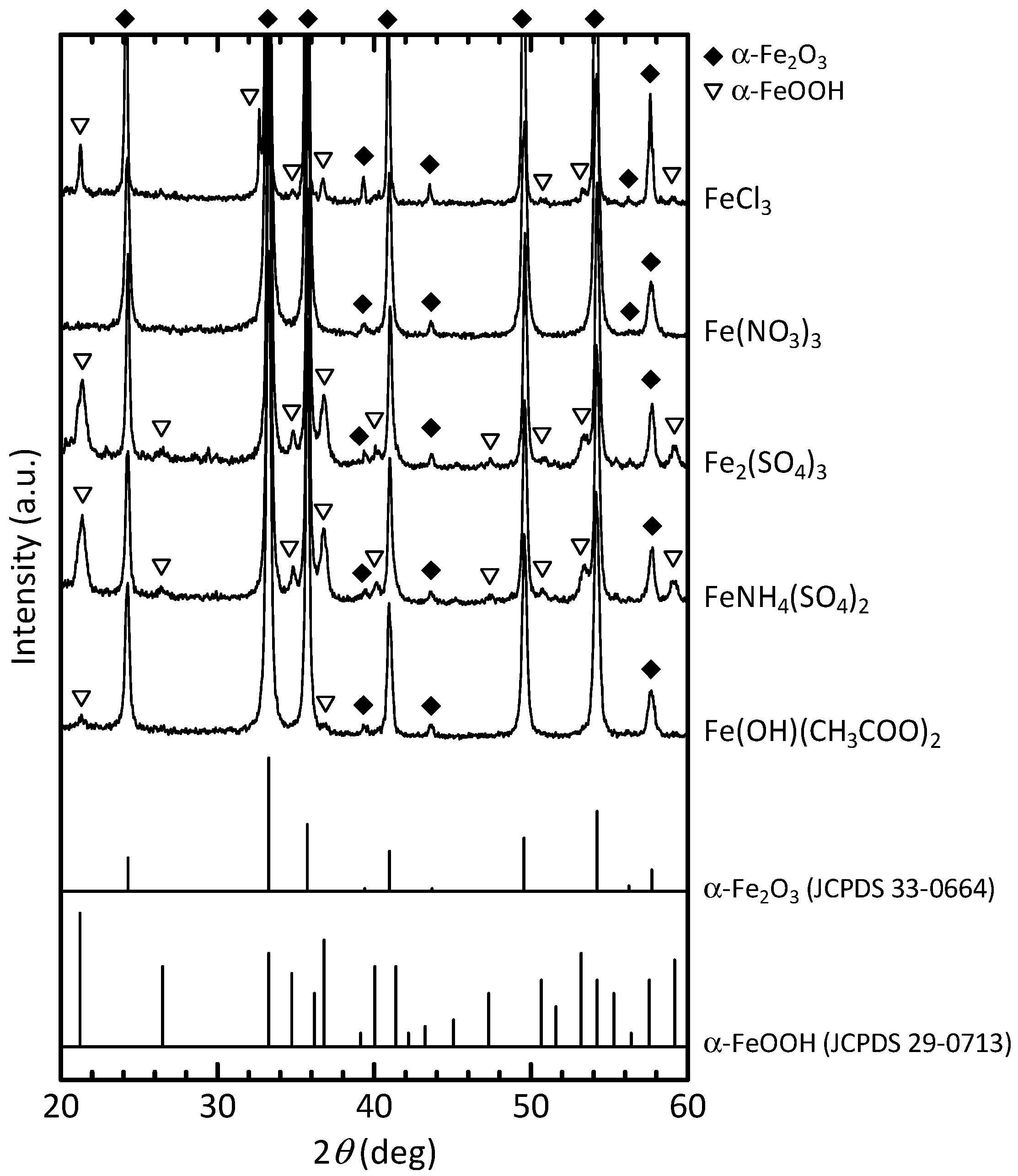
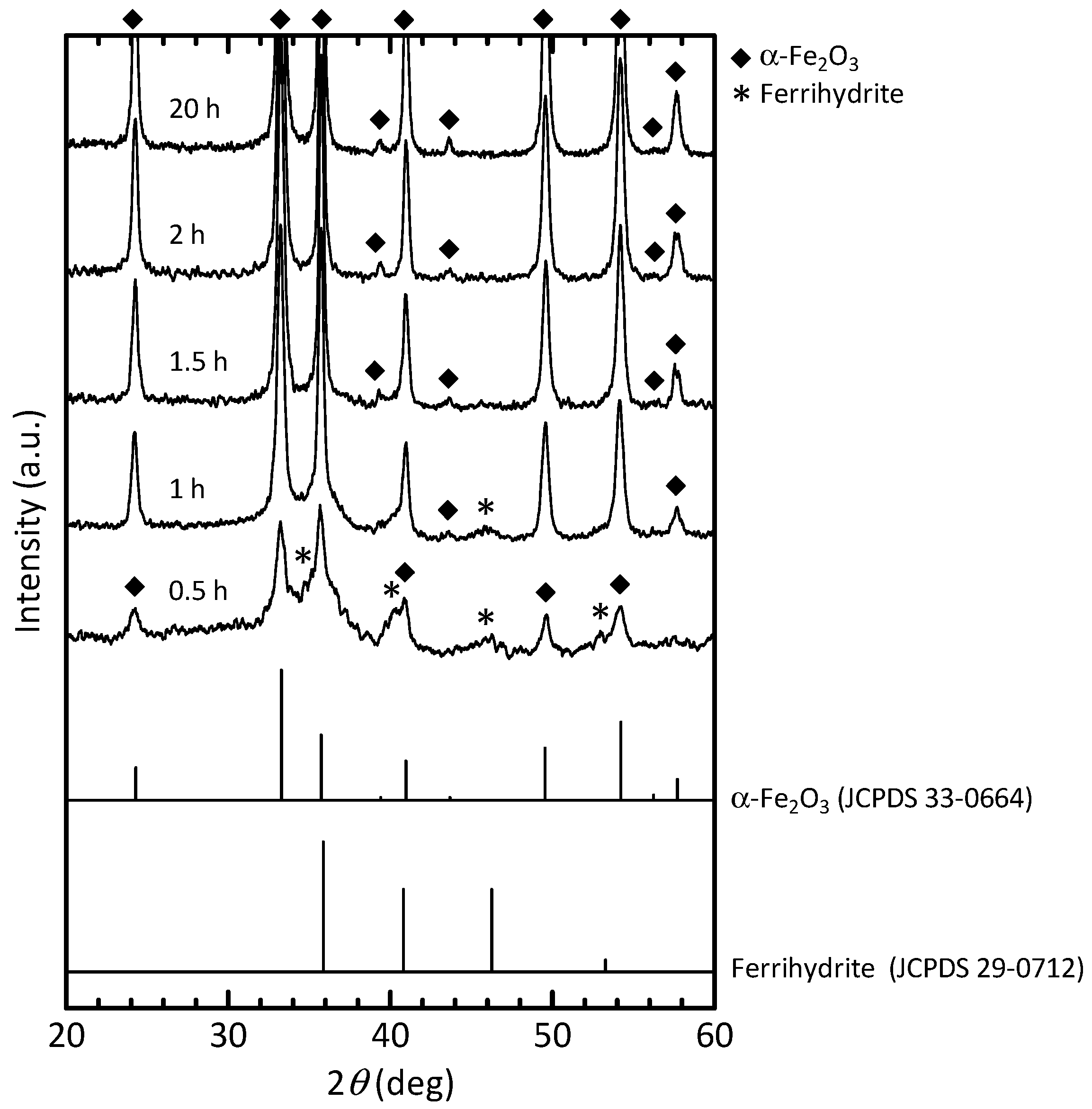
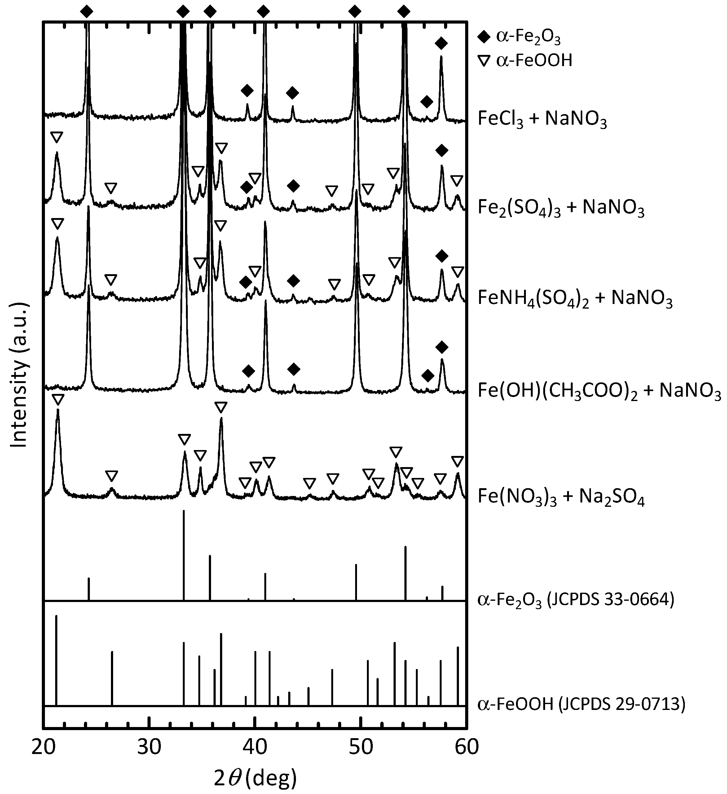
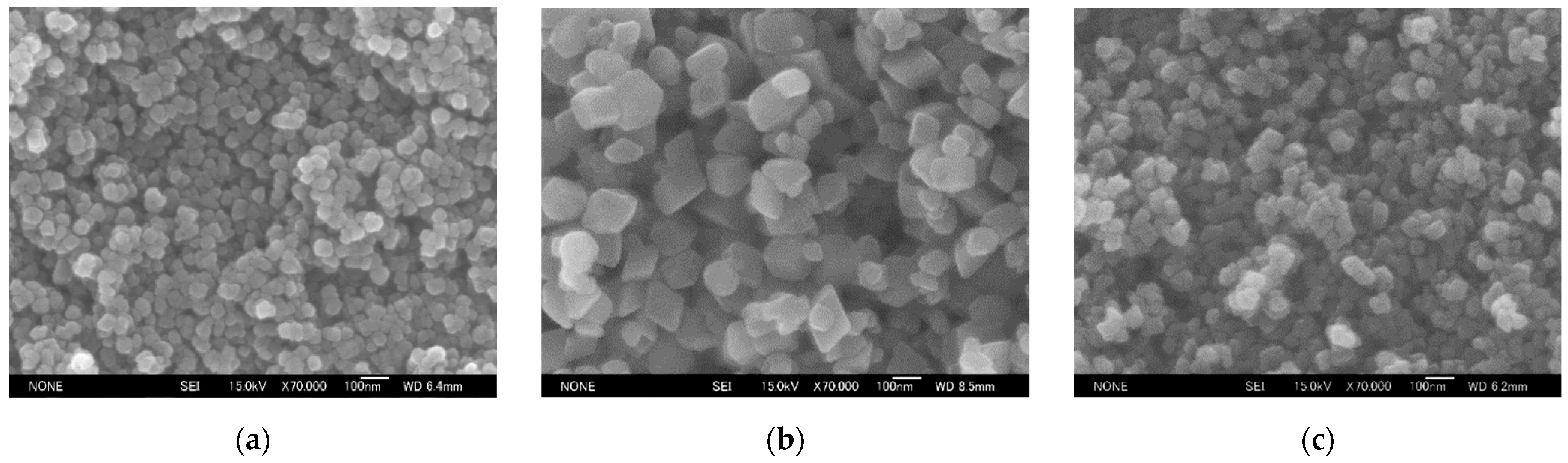
3.1.1. Effects of the Reactants on the α-Fe2O3 Formation

The X-ray diffraction (XRD) patterns of the samples prepared with the different iron salts are shown in Figure 2. The phase evolution in the hydrothermal process strongly depended on the iron salts employed, i.e., the anions in solution; however, the pH of the solutions after hydrothermal treatment was 9.2 ± 0.1 regardless of the iron salts that were present. When Fe(NO3)3 was used, single-phase crystalline α-Fe2O3 was obtained. However, samples prepared with the other iron salts contained not only α-Fe2O3 but also the intermediate α-FeOOH. The results suggested that NO3 ions promoted α-Fe2O3 formation. The hydrothermal synthesis using Fe(NO3)3 confirmed the complete α-Fe2O3 formation reaction in a relatively short period of time (approximately 2 h), as shown in Figure 3. In this case, instead of α-FeOOH, 6-line ferrihydrite (Fe5HO8·4H2O) intermediate [53] was detected at the early stages, which may have contributed toward the rapid formation of α-Fe2O3. The results revealed that Fe(NO3)3 may be a suitable iron source for the hydrothermal process with urea hydrolysis.
SEM images and particle size distributions of the samples prepared with urea and the ammonia solution are shown in Figure 4. While the XRD analysis confirmed that a uniform α-Fe2O3 phase was also obtained when using the ammonia solution, the particle size of α-Fe2O3 nanoparticles prepared with urea was relatively uniform compared with the case using the ammonia solution. The results demonstrated that the hydrothermal process using urea hydrolysis was effective for the preparation of uniformly sized α-Fe2O3 nanoparticles.

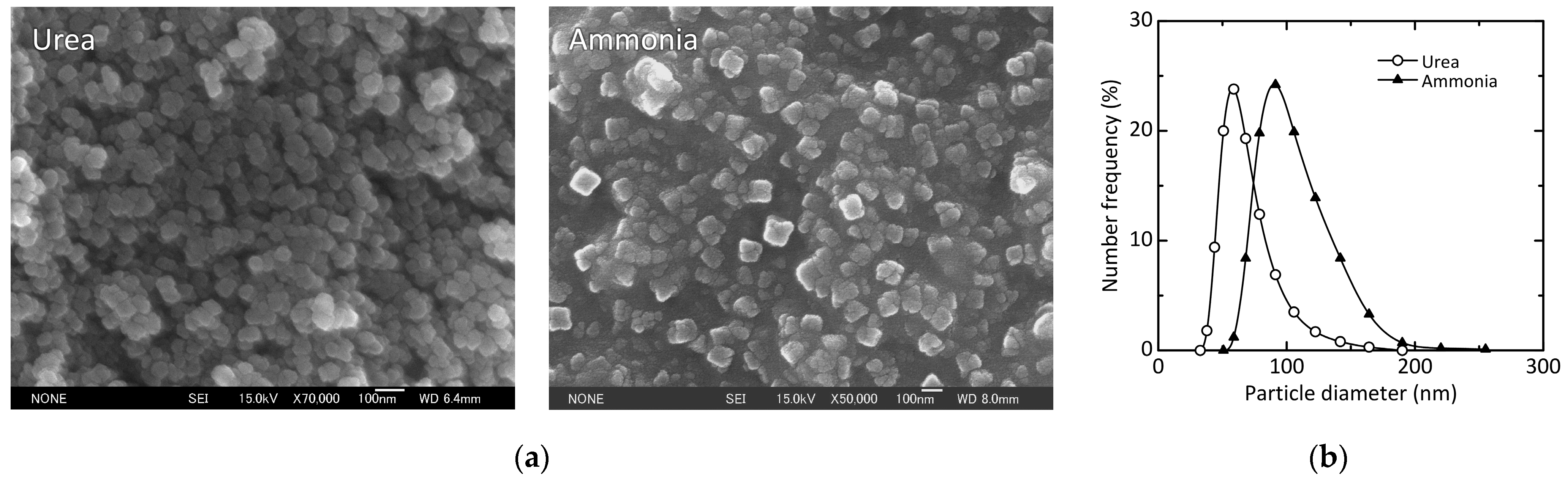
3.1.2. Change in the Crystallite Size and Particle Diameter with Coexisting Anion

To examine the reaction-promoting effect of NO3 ions, 75 mmol of NaNO3, i.e., five-times the amount of NO3 ions contained in the starting Fe(NO3)3 solution, were added to the starting solutions prepared with the iron salts except for Fe(NO3)3; hydrothermal treatment was then performed under the same conditions. The XRD patterns of the samples prepared in the presence of NO3 ions are shown in Figure 5. When the NaNO3-added FeCl3 and Fe(OH)(CH3COO)2 solutions were used, a single α-Fe2O3 phase was obtained; in particular, high-crystalline α-Fe2O3 was formed from the NaNO3-added FeCl3 solution. In contrast, the addition of NaNO3 to the Fe2(SO4)3 and FeNH4(SO4)2 solutions negligibly improved the hydrothermal formation of α-Fe2O3.
However, when the Fe(NO3)3 solution contained 1.5 mmol of Na2SO4, in which the molar ratio of SO42−/NO3 in the solution was 0.1, the obtained sample comprised mostly α-FeOOH, as shown in Figure 5, indicating that SO42− ions may have considerably reduced the reaction rate of α-Fe2O3 formation. In particular, the reactivity and stability of intermediate ferric hydroxide may be varied by coexisting SO42− ions [32,33]. Accordingly, the coexisting anions in the solution were found to greatly affect the hydrothermal formation of α-Fe2O3. NO3 and SO42− ions can especially promote and inhibit the α-Fe2O3 formation reaction in this process, respectively. In addition, the effects of SO42− ions may be stronger than those of NO3 ions. The results suggested that the stability of iron complexes in the solution plays an important role in the hydrothermal formation of α-Fe2O3. However, the detailed mechanism remains unknown and requires further study.
The crystallite sizes of samples with a single α-Fe2O3 phase prepared using Fe(NO3)3, NaNO3-added FeCl3, and NaNO3-added Fe(OH)(CH3COO)2 solutions were calculated using the Scherrer equation. The average values of crystallite size, which were calculated using the diffraction intensity peaks observed at 2θ ≈ 24.1°, 33.2°, 35.6°, 40.9°, 49.5°, 54.1°, 62.4°, and 64.0° corresponding to the (012), (104), (110), (113), (024), (116), (214), and (300) crystal planes of α-Fe2O3, respectively, the crystallinity index CI (%), which is defined by Equation (8),
C I = I t I a I t × 100
where It (−) is the total intensity for α-Fe2O3 (110) plane and ferrihydrite (110) plane at 2θ ≈ 35.7° and Ia (−) is the intensity for ferrihydrite (113) plane at 2θ ≈ 46.3° as amorphous phase, and the median diameter of number-basis distribution of particle sizes are summarized in Table 1. The SEM images are also shown in Figure 6. Although the crystallinity index was almost the same (98–99%), the crystallite size and particle diameter varied depending on the coexisting anions in the solution, suggesting that their control is possible by using the coexisting anions as an operating factor.

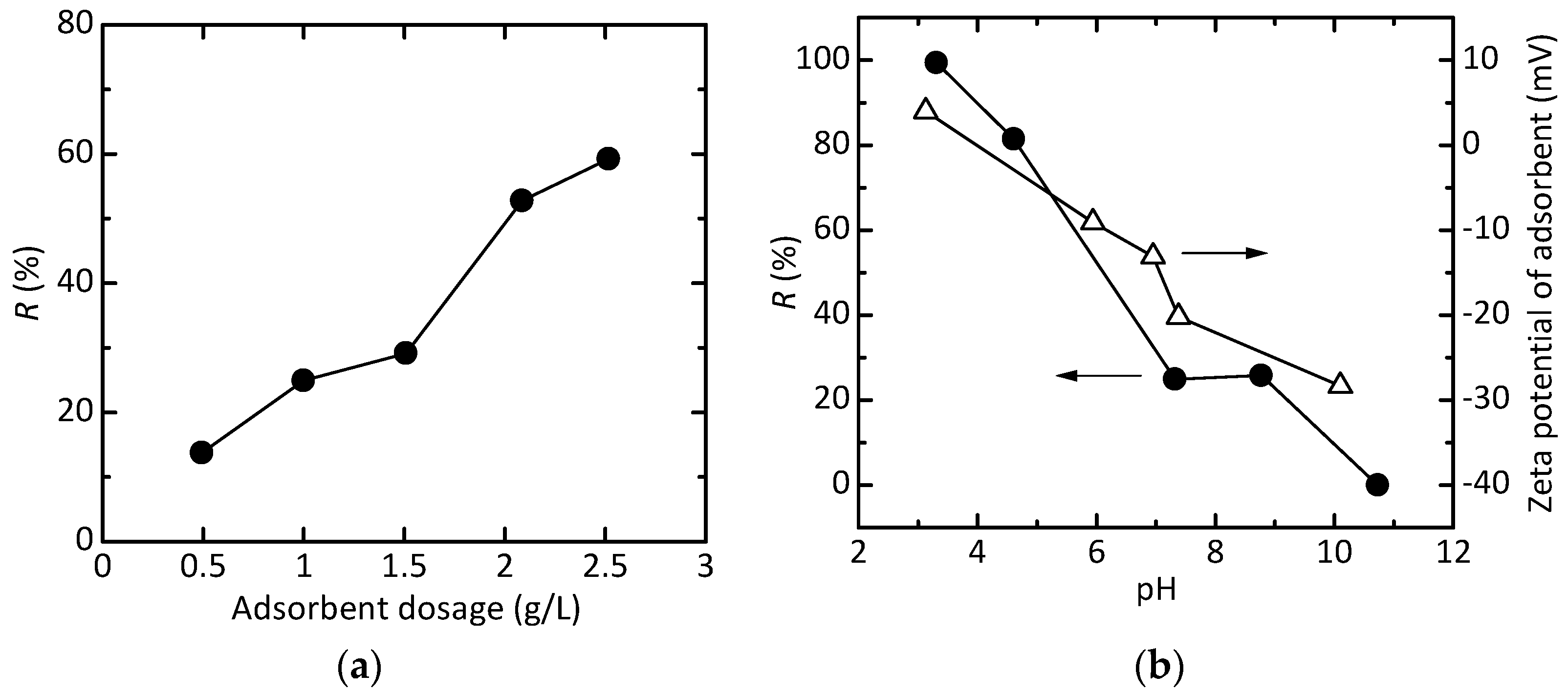
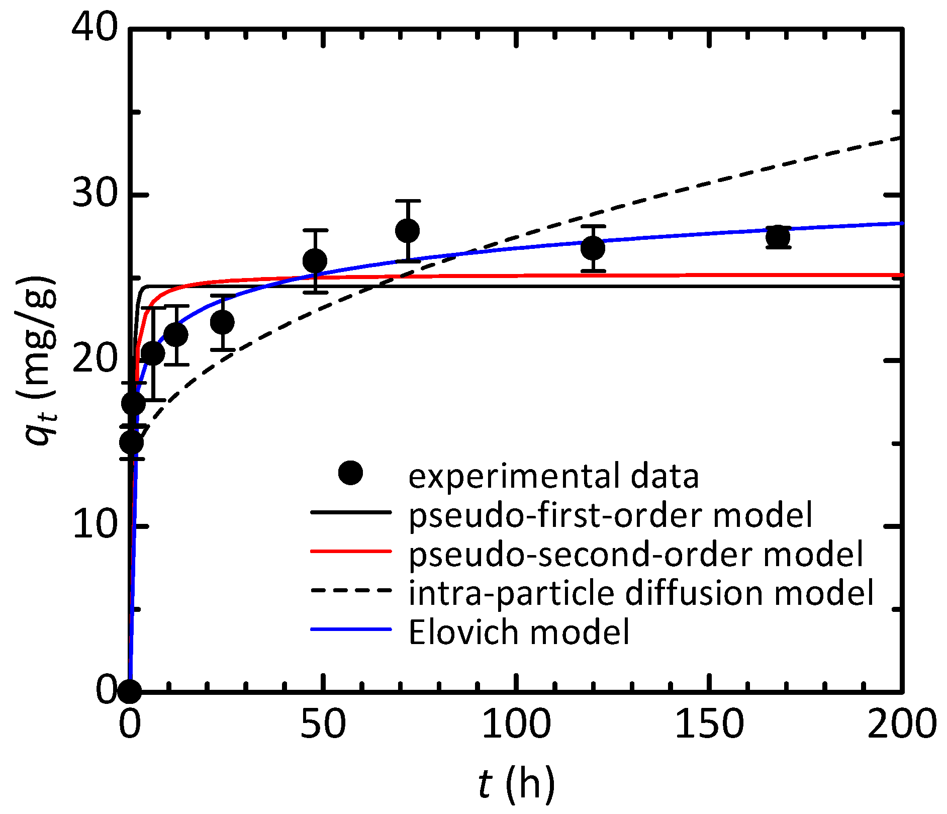
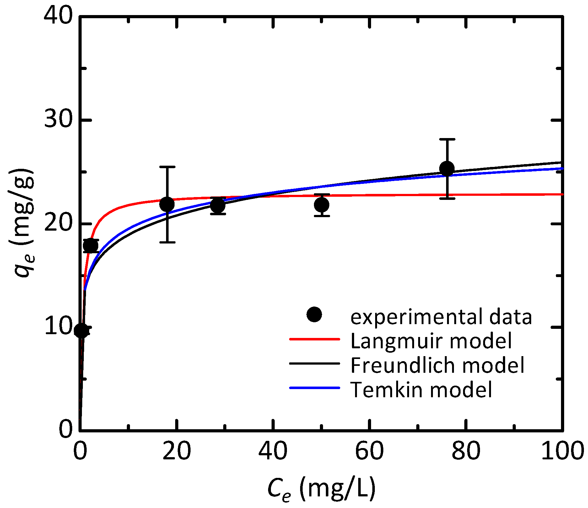
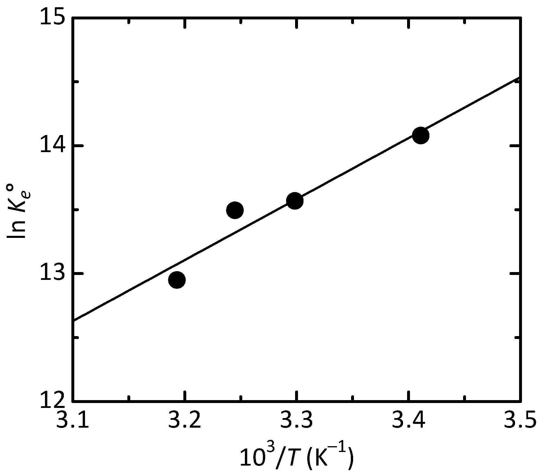
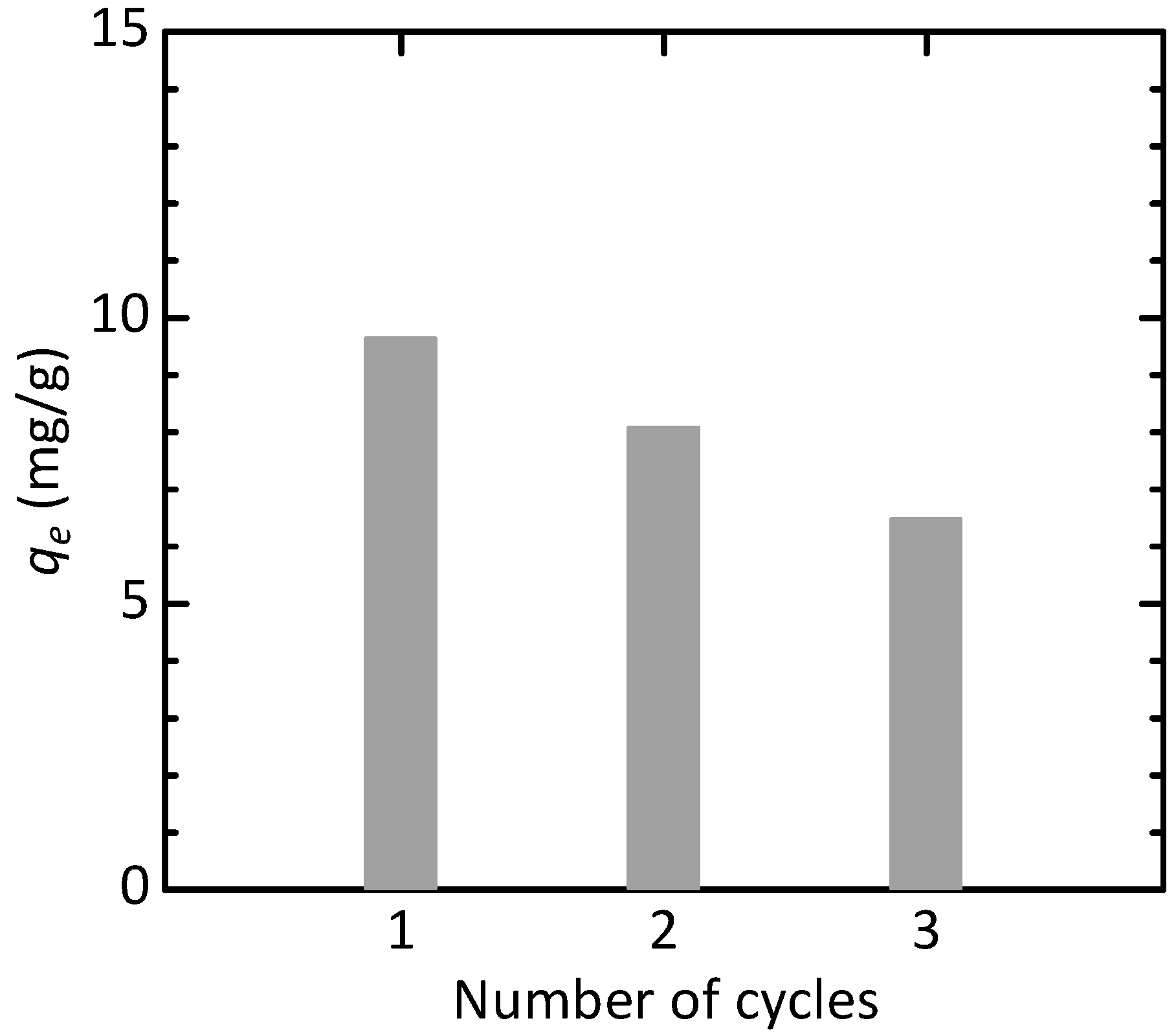
3.2. Adsorption Studies of Congo Red

As shown in Figure 7a, the removal efficiency increased with increasing adsorbent dosage due to an increase in the number of active adsorption sites. In contrast, with a decrease in pH, the removal efficiency increased (Figure 7b), which was similar to the results reported in the literature [51]. In particular, when pH ≤ 4.6, the removal efficiency reached above 80% and the adsorbent showed good adsorption performance. This may be attributed to electrostatic interaction between the adsorbent and the anionic dye molecules due to the positive surface potential of adsorbent [51], as seen in the pH dependence of zeta potential (Figure 7b).
The change in the adsorbed amount qt with contact time is illustrated in Figure 8. The adsorbed amount rapidly increased in the initial stage and approximately reached equilibrium after several hours. To analyze the adsorption kinetics, several representative models, i.e., pseudo-first-order model, pseudo-second-order model, intra-particle diffusion model, and Elovich model [54], which are expressed by Equations (9), (10), (11), and (12), respectively, were applied to the experimental data shown in Figure 7.
q t = q e 1 e k 1 t
q t = k 2 q e 2 t 1 + k 2 q e t
q t = k i t 0.5 + c
q t = 1 β ln α β t
k1 (h−1), k2 (g/(mg·h)), and ki (mg/(g·h0.5)) are the rate constants of the pseudo-first-order, pseudo-second-order, and intra-particle diffusion models, respectively. c (mg/g) is a constant. α (mg/(g·h)) and β (g/mg) are the initial sorption rate and the constant related to the extent of surface coverage and activation energy for chemisorption, respectively. The nonlinear fitting results using Equations (9)–(12) and the determined parameters, which were obtained by the Solver function of Microsoft Excel 2016 (Microsoft Corporation, Redmond, WA, USA), are shown in Figure 8 and Table 2, respectively. The experimental data fit well with the Elovich model, which suggest chemical adsorption process and the activation energy increased with adsorption time [55].
Using the relationship between qe and Ce shown in Figure 9, the adsorption isotherm was analyzed using the Langmuir model, the Freundlich model, and the Temkin model, which are described by Equations (13), (14), and (15), respectively.
q e = q m K L C e 1 + K L C e
q e = K F C e 1 / n
q e = q T ln A T C e
where qm (mg/g) is the maximum monolayer adsorption capacity and KL (L/mg) is the Langmuir equilibrium constant. KF (mg/(g·(mg/L)1/n)) and n (−) are the Freundlich constants related to the adsorption capacity and the intensity of adsorption, respectively. AT (L/mg) and qT (mg/g) are the adsorption equilibrium constant of solute on solid surface [56] and the surface capacity for contaminant adsorption per unit binding energy [57], respectively. As shown in Figure 9 and Table 3, the experimental data were well-described by the Langmuir model, which suggested the homogeneous monolayer adsorption. Here, the comparison of the maximum monolayer adsorption capacities with some literature values is summarized in Table 4. Although no additional treatments for enhancing the adsorption performance such as surface modification were performed, our hematite nanoparticles, which were synthesized by the simple process using inexpensive raw materials, have relatively good adsorption properties and can be a promising candidate as an adsorbent for Congo red.
The adsorption process was thermodynamically analyzed using the following equations [52]:
Δ G ° = R T ln K e °
ln K e ° = Δ S ° R Δ H ° R T
where ΔG° (J/mol), ΔS° (J/(mol·K)), and ΔH° (J/mol) are the Gibbs free energy, the standard entropy, and the standard enthalpy, respectively. Ke° (−) is the thermodynamic equilibrium constant and is obtained by Equation (18) [52].
K e ° = K L M c a d
where KL (L/mg) is the Langmuir equilibrium constant of adsorption, which was determined from the isotherm data obtained at different temperatures; M (mg/mol) is the molecular weight of adsorbate; and cad (mol/L) is the standard concentration of adsorbate, which is defined as 1 mol/L. The results of linear fitting with Equation (17) are shown in Figure 10 and Table 5. Although it is difficult to evaluate the adsorption performance of an adsorbent based on the magnitude of the values of ΔG°, ΔS°, and ΔH°, it is noteworthy that their values were negative, which indicates that the adsorption process is feasible and spontaneous, decreases in randomness at the adsorbent/dye interface, and is exothermic. In general, when such requirements are satisfied, the adsorption can be favorable. Therefore, using our α-Fe2O3 nanoparticles as the adsorbent for removal of Congo red is practically applicable.
For investigating the reusability of the adsorbent, the regeneration experiment was performed. After adsorption for more than 12 h under the typical conditions except for the initial concentration (10 mg/L in this investigation), the spent adsorbent was isolated from the dye solution and heated at 673 K in air for 2 h [63] as an example. The adsorption–regeneration was repeated three times under the same conditions. As shown in Figure 11, relatively large adsorption capacities were observed for the spent adsorbent even after the regeneration, although it had a tendency to decrease with increasing the cycle number. The result suggests that the adsorbent is reusable after the conditions for regeneration are optimized for maintaining the adsorption performance in the recycling.

4. Conclusions

In the hydrothermal synthesis of crystalline α-Fe2O3 nanoparticles via urea hydrolysis, the anions contained in the starting solution greatly affected the rate of α-Fe2O3 formation. In particular, NO3 ion promoted the formation reaction, which may be due to the formation of a 6-line ferrihydrite intermediate. This can contribute to the rapid synthesis of α-Fe2O3 nanoparticles. In contrast, when using FeCl3, Fe2(SO4)3, FeNH4(SO4)2, or Fe(OH)(CH3COO)2 as the iron source, the products contained the intermediate α-FeOOH in addition to α-Fe2O3. The addition of NaNO3 to the FeCl3 and Fe(OH)(CH3COO)2 solutions provided a single α-Fe2O3 phase. However, the addition of NaNO3 to the Fe2(SO4)3 and FeNH4(SO4)2 solutions resulted in no changes in phase evolution. Accordingly, SO42− ions tended to inhibit the reaction, which may be due to the formation of a relatively stable iron complex. Furthermore, the effects of SO42− ions may be stronger than those of NO3 ions. The average crystallite size and median diameter of α-Fe2O3 nanoparticles prepared using the Fe(NO3)3, NaNO3-added FeCl3, and NaNO3-added Fe(OH)(CH3COO)2 solutions also depended on the coexisting anions. Therefore, selecting the coexisting anions appropriately can contribute to controlling the growth of α-Fe2O3 nanoparticles.
The α-Fe2O3 nanoparticles prepared with Fe(NO3)3 were used as the adsorbent for removal of Congo red dye. The adsorption kinetics and isotherm followed the Elovich and Langmuir models, respectively. The adsorption thermodynamic study revealed that the adsorption process was feasible and practically applicable. The results demonstrated that the α-Fe2O3 nanoparticles synthesized by the simple process using the inexpensive raw materials have relatively good adsorption properties without additional functionalization, such as surface modification, and are a promising candidate as adsorbents for Congo red.

Author Contributions

Conceptualization, T.I.; methodology, T.I.; validation, T.O. and T.I.; formal analysis, T.O. and T.I.; investigation, T.O., M.F. and T.I.; data curation, T.O., M.F. and T.I.; writing—original draft preparation, T.O. and T.I.; writing—review and editing, T.I.; visualization, T.O. and T.I.; supervision, T.I.; project administration, T.I. All authors have read and agreed to the published version of the manuscript.

Funding

This research received no external funding.

Institutional Review Board Statement

Not applicable.

Informed Consent Statement

Not applicable.

Data Availability Statement

Not applicable.

Conflicts of Interest

The authors declare no conflict of interest.

References

  1. Davis, J.R.; Mills, K.M.; Lampman, S.R. Properties and Selection: Irons, Steels, and High-Performance Alloys. In ASM Handbook, 10th ed.; ASM International: Materials Park, OH, USA, 1990; Volume 1, p. 1063. [Google Scholar]
  2. Cornell, R.M.; Schwertman, U. The Iron Oxides: Structure, Properties, Reactions, Occurrence and Uses, 2nd ed.; Wiley-VCH: Weinheim, Germany, 2003. [Google Scholar]
  3. Faivre, D. Iron Oxides: From Nature to Applications; Wiley-VCH: Weinheim, Germany, 2016. [Google Scholar]
  4. Wang, L.; Ding, K.; Wei, B.; Li, C.; Shi, X.; Zhang, Y.; He, X. Leaf-like α-Fe2O3 micron-particle: Preparation and its usage as anode materials for lithium ion batteries. Mater. Chem. Phys. 2018, 207, 58–66. [Google Scholar] [CrossRef]
  5. Pan, Y.; Yin, L.; Li, M. Submicron-sized α-Fe2O3 single crystals as anodes for high-performance lithium-ion batteries. Ceram. Int. 2019, 45, 12072–12079. [Google Scholar] [CrossRef]
  6. Balasingam, S.K.; Kundu, M.; Balakrishnan, B.; Kim, H.-J.; Svensson, A.M.; Jayasayee, K. Hematite microdisks as an alternative anode material for lithium-ion batteries. Mater. Lett. 2019, 247, 163–166. [Google Scholar] [CrossRef]
  7. Yan, Z.; Sun, Z.; Li, A.; Liu, H. Three-dimensional porous flower-like S-doped Fe2O3 for superior lithium storage. Adv. Compos. Hybrid Mater. 2021, 4, 716–724. [Google Scholar] [CrossRef]
  8. Luo, Y.; Liu, L.; Qiao, W.; Liu, F.; Zhang, Y.; Tan, W.; Qiu, G. Facile crystal-structure-controlled synthesis of iron oxides for adsorbents and anode materials of lithium batteries. Mater. Chem. Phys. 2016, 170, 239–245. [Google Scholar] [CrossRef]
  9. Abd El Aal, S.A.; Abdelhady, A.M.; Mansour, N.A.; Hassan, N.M.; Elbaz, F.; Elmaghraby, E.K. Physical and chemical characteristics of hematite nanoparticles prepared using microwave-assisted synthesis and its application as adsorbent for Cu, Ni, Co, Cd and Pb from aqueous solution. Mater. Chem. Phys. 2019, 235, 121771. [Google Scholar] [CrossRef]
  10. Dehbi, A.; Dehmani, Y.; Omari, H.; Lammini, A.; Elazhari, K.; Abdallaoui, A. Hematite iron oxide nanoparticles (α-Fe2O3): Synthesis and modelling adsorption of malachite green. J. Environ. Chem. Eng. 2020, 8, 103394. [Google Scholar] [CrossRef]
  11. Panasenko, A.; Pirogovskaya, P.; Tkachenko, I.; Ivannikov, S.; Arefieva, O.; Marchenko, Y. Synthesis and characterization of magnetic silica/iron oxide composite as a sorbent for the removal of methylene blue. Mater. Chem. Phys. 2020, 245, 122759. [Google Scholar] [CrossRef]
  12. Mpelane, S.; Mketo, N.; Bingwa, N.; Nomngongo, P.N. Synthesis of mesoporous iron oxide nanoparticles for adsorptive removal of levofloxacin from aqueous solutions: Kinetics, isotherms, thermodynamics and mechanism. Alex. Eng. J. 2022, 61, 8457–8468. [Google Scholar] [CrossRef]
  13. Hjiri, M. Highly sensitive NO2 gas sensor based on hematite nanoparticles synthesized by sol-gel technique. J. Mater. Sci. Mater. El. 2020, 31, 5025–5031. [Google Scholar] [CrossRef]
  14. Alizadeh, T.; Zargr, F. Highly selective and sensitive iodide sensor based on carbon paste electrode modified with nanosized sulfate-doped α-Fe2O3. Mater. Chem. Phys. 2020, 240, 122118. [Google Scholar] [CrossRef]
  15. Sangale, S.S.; Jadhav, V.V.; Shaikh, S.F.; Shinde, P.V.; Ghule, B.G.; Raut, S.D.; Tamboli, M.S.; Al-Enizi, A.M.; Mane, R.S. Facile one-step hydrothermal synthesis and room-temperature NO2 sensing application of α-Fe2O3 sensor. Mater. Chem. Phys. 2020, 246, 122799. [Google Scholar] [CrossRef]
  16. Deshmanea, V.V.; Patil, A.V. Cobalt oxide doped hematite as a petrol vapor sensor. Mater. Chem. Phys. 2020, 246, 122813. [Google Scholar] [CrossRef]
  17. Li, G.; Ma, Z.; Hu, Q.; Zhang, D.; Fan, Y.; Wang, X.; Chu, X.; Xu, J. PdPt nanoparticle-functionalized α-Fe2O3 hollow nanorods for triethylamine sensing. ACS Appl. Nano Mater. 2021, 4, 10921–10930. [Google Scholar] [CrossRef]
  18. Tong, G.; Guan, J.; Zhang, Q. Goethite hierarchical nanostructures: Glucose-assisted synthesis, chemical conversion into hematite with excellent photocatalytic properties. Mater. Chem. Phys. 2011, 127, 371–378. [Google Scholar] [CrossRef]
  19. Alharbi, A.; Abdelrahman, E.A. Efficient photocatalytic degradation of malachite green dye using facilely synthesized hematite nanoparticles from Egyptian insecticide cans. Spectrochim. Acta A 2020, 226, 117612. [Google Scholar] [CrossRef]
  20. Haider, S.; Shar, S.S.; Shakir, I.; Agboola, P.O. Visible light active Cu-doped iron oxide for photocatalytic treatment of methylene blue. Ceram. Int. 2022, 48, 7605–7612. [Google Scholar] [CrossRef]
  21. Harijan, D.K.L.; Gupta, S.; Ben, S.K.; Srivastava, A.; Singh, J.; Chandra, V. High photocatalytic efficiency of α-Fe2O3-ZnO composite using solar energy for methylene blue degradation. Phys. B Condens. Matter. 2022, 627, 413567. [Google Scholar] [CrossRef]
  22. Ruan, C.; Wang, J.; Gao, M.; Zhao, G. The influence of structural size on thermal stability in single crystalline hematite uniform nano/micro-cubes. Mater. Chem. Phys. 2016, 183, 158–164. [Google Scholar] [CrossRef]
  23. Fan, H.; Song, B.; Liu, J.; Yang, Z.; Li, Q. Thermal formation mechanism and size control of spherical hematite nanoparticles. Mater. Chem. Phys. 2005, 89, 321–325. [Google Scholar] [CrossRef]
  24. Tadic, M.; Panjan, M.; Tadic, B.V.; Lazovic, J.; Damnjanovic, V.; Kopani, M.; Kopanja, L. Magnetic properties of hematite (α-Fe2O3) nanoparticles synthesized by sol-gel synthesis method: The influence of particle size and particle size distribution. J. Electr. Eng. 2019, 70, 71–76. [Google Scholar] [CrossRef]
  25. Katsuki, H.; Choi, E.-K.; Lee, W.-J.; Hwang, K.-T.; Cho, W.-S.; Huang, W.; Komarneni, S. Ultrafast microwave-hydrothermal synthesis of hexagonal plates of hematite. Mater. Chem. Phys. 2018, 205, 210–216. [Google Scholar] [CrossRef]
  26. Yang, P.; Yu, J.; Wang, Z.; Liu, Q.; Wu, T. Urea method for the synthesis of hydrotalcites. React. Kinet. Catal. Lett. 2004, 83, 275–282. [Google Scholar] [CrossRef]
  27. Zeng, H.-Y.; Deng, X.; Wang, Y.-J.; Liao, K.-B. Preparation of Mg-Al hydrotalcite by urea method and its catalytic activity for transesterification. AIChE J. 2009, 55, 1229–1235. [Google Scholar] [CrossRef]
  28. Wang, J.; Li, D.; Yu, X.; Zhang, M.; Jing, X. Fabrication of layered double hydroxide spheres through urea hydrolysis and mechanisms involved in the formation. Colloid Polym. Sci. 2010, 288, 1411–1418. [Google Scholar] [CrossRef]
  29. Liu, Z.; Yu, R.; Dong, Y.; Li, W.; Zhou, W. Preparation of α-Fe2O3 hollow spheres, nanotubes, nanoplates and nanorings as highly efficient Cr(VI) adsorbents. RSC Adv. 2016, 6, 82854. [Google Scholar] [CrossRef]
  30. Tadic, M.; Trpkov, D.; Kopanja, L.; Vojnovic, S.; Panjan, M. Hydrothermal synthesis of hematite (α-Fe2O3) nanoparticle forms: Synthesis conditions, structure, particle shape analysis, cytotoxicity and magnetic properties. J. Alloys Compd. 2019, 792, 599–609. [Google Scholar] [CrossRef]
  31. Li, D.; Xu, R.; Jia, Y.; Ning, P.; Li, K. Controlled synthesis of α-Fe2O3 hollows from β-FeOOH rods. Chem. Phys. Lett. 2019, 731, 136623. [Google Scholar] [CrossRef]
  32. Mizutani, N.; Iwasaki, T.; Watano, S.; Yanagida, T.; Kawai, T. Size control of magnetite nanoparticles in hydrothermal synthesis by coexistence of lactate and sulfate ions. Curr. Appl. Phys. 2010, 10, 801–806. [Google Scholar] [CrossRef]
  33. Iwasaki, T.; Mizutani, N.; Watano, S.; Yanagida, T.; Kawai, T. Size control of magnetite nanoparticles by organic solvent-free chemical coprecipitation at room temperature. J. Exp. Nanosci. 2010, 5, 251–262. [Google Scholar] [CrossRef]
  34. Tan, W.; Liang, Y.; Xu, Y.; Wang, M. Structural-controlled formation of nano-particle hematite and their removal performance for heavy metal ions: A review. Chemosphere 2022, 306, 135540. [Google Scholar] [CrossRef] [PubMed]
  35. Selvaraj, R.; Pai, S.; Vinayagam, R.; Varadavenkatesan, T.; Kumar, P.S.; Duc, P.A.; Rangasamy, G. A recent update on green synthesized iron and iron oxide nanoparticles for environmental applications. Chemosphere 2022, 308, 136331. [Google Scholar] [CrossRef] [PubMed]
  36. Dissanayake, D.M.S.N.; Mantilaka, M.M.M.G.P.G.; Palihawadana, T.C.; Chandrakumara, G.T.D.; De Silva, R.T.; Pitawala, H.M.T.G.A.; Nalin De Silva, K.M.; Amaratunga, G.A.J. Facile and low-cost synthesis of pure hematite (α-Fe2O3) nanoparticles from naturally occurring laterites and their superior adsorption capability towards acid-dyes. RSC Adv. 2019, 9, 21249–21257. [Google Scholar] [CrossRef] [PubMed]
  37. Dehbi, A.; Dehmani, Y.; Omari, H.; Lammini, A.; Elazhari, K.; Abouarnadasse, S.; Abdallaoui, A. Comparative study of malachite green and phenol adsorption on synthetic hematite iron oxide nanoparticles (α-Fe2O3). Surf. Interfaces 2020, 21, 100637. [Google Scholar] [CrossRef]
  38. Onizuka, T.; Iwasaki, T. Low-temperature solvent-free synthesis of polycrystalline hematite nanoparticles via mechanochemical activation and their adsorption properties for Congo red. Solid State Sci. 2022, 129, 106917. [Google Scholar] [CrossRef]
  39. Oladoye, P.O.; Bamigboye, M.O.; Ogunbiyi, O.D.; Akano, M.T. Toxicity and decontamination strategies of Congo red dye. Groundw. Sustain. Dev. 2022, 19, 100844. [Google Scholar] [CrossRef]
  40. Chattopadhyay, D.P. Chemistry of Dyeing. In Handbook of Textile and Industrial Dyeing Principle, Processes and Types of Dyes, 1st ed.; Clark, M., Ed.; Woodhead Publishing: Cambridge, UK, 2011; Volume 1, pp. 150–183. [Google Scholar]
  41. Maruthupandy, M.; Muneeswaran, T.; Vennila, T.; Anand, M.; Cho, W.-S.; Quero, F. Development of chitosan decorated Fe3O4 nanospheres for potential enhancement of photocatalytic degradation of Congo red dye molecules. Spectrochim. Acta A Mol. Biomol. Spectrosc. 2022, 267, 120511. [Google Scholar] [CrossRef]
  42. Hitkari, G.; Chowdhary, P.; Kumar, V.; Singh, S.; Motghare, A. Potential of copper-zinc oxide nanocomposite for photocatalytic degradation of Congo red dye. Clean. Chem. Eng. 2022, 1, 100003. [Google Scholar] [CrossRef]
  43. Chen, H.; Wang, S.; Huang, L.; Zhang, L.; Han, J.; Ren, W.; Pan, J.; Li, J. Core-shell hierarchical Fe/Cu bimetallic Fenton catalyst with improved adsorption and catalytic performance for Congo red degradation. Catalysts 2022, 12, 1363. [Google Scholar] [CrossRef]
  44. Bencheqroun, Z.; Sahin, N.E.; Soares, O.S.G.P.; Pereira, M.F.R.; Zaitan, H.; Nawdali, M.; Rombi, E.; Fonseca, A.M.; Parpot, P.; Neves, I.C. Fe(III)-exchanged zeolites as efficient electrocatalysts for Fenton-like oxidation of dyes in aqueous phase. J. Environ. Chem. Eng. 2022, 10, 107891. [Google Scholar] [CrossRef]
  45. Han, G.; Du, Y.; Huang, Y.; Wang, W.; Su, S.; Liu, B. Study on the removal of hazardous Congo red from aqueous solutions by chelation flocculation and precipitation flotation process. Chemosphere 2022, 289, 133109. [Google Scholar] [CrossRef] [PubMed]
  46. Lu, Y.-F.; He, Y.-H.; Liang, J.-B.; Jin, Q.; Ou, Y.-C.; Wu, J.-Z. First cadmium coordination compound as an efficient flocculant for Congo red. Inorg. Chem. Commun. 2022, 141, 109544. [Google Scholar] [CrossRef]
  47. Liu, Y.; Qiu, G.; Liu, Y.; Niu, Y.; Qu, R.; Ji, C.; Wang, Y.; Zhang, Y.; Sun, C. Fabrication of CoFe-MOF materials by different methods and adsorption properties for Congo red. J. Mol. Liq. 2022, 360, 119405. [Google Scholar] [CrossRef]
  48. Wang, L.; Tao, Y.; Wang, J.; Tian, M.; Liu, S.; Quan, T.; Yang, L.; Wang, D.; Li, X.; Gao, D. A novel hydroxyl-riched covalent organic framework as an advanced adsorbent for the adsorption of anionic azo dyes. Anal. Chim. Acta 2022, 1227, 340329. [Google Scholar] [CrossRef]
  49. Tian, P.; Sun, C.; Zhu, P.; Pang, H.; Gong, W.; Ye, J.; Ning, G. Template-engaged synthesis of macroporous tubular hierarchical α-Fe2O3 and its ultrafast transfer performance. Chem. Phys. Lett. 2018, 706, 261–265. [Google Scholar] [CrossRef]
  50. Prajapati, A.K.; Mondal, M.K. Development of CTAB modified ternary phase α-Fe2O3-Mn2O3-Mn3O4 nanocomposite as innovative super-adsorbent for Congo red dye adsorption. J. Environ. Chem. Eng. 2021, 9, 104827. [Google Scholar] [CrossRef]
  51. Maiti, D.; Mukhopadhyay, S.; Devi, P.S. Evaluation of mechanism on selective, rapid, and superior adsorption of Congo red by reusable mesoporous α-Fe2O3 nanorods. ACS Sustain. Chem. Eng. 2017, 5, 11255–11267. [Google Scholar] [CrossRef]
  52. Lima, E.C.; Hosseini-Bandegharaei, A.; Moreno-Piraján, J.C.; Anastopoulos, I. A critical review of the estimation of the thermodynamic parameters on adsorption equilibria. Wrong use of equilibrium constant in the Van’t Hoof equation for calculation of thermodynamic parameters of adsorption. J. Mol. Liq. 2019, 273, 425–434. [Google Scholar] [CrossRef]
  53. Song, Y.; Bac, B.H.; Lee, Y.-B.; Kim, M.H.; Yoon, W.-S.; Kim, J.H.; Moon, H.-S. Ge-incorporation into 6-line ferrihydrite nanocrystals. CrystEngComm 2010, 12, 1997–2000. [Google Scholar] [CrossRef]
  54. Xu, H.; Zhu, S.; Lu, K.; Jia, H.; Xia, M.; Wang, F. Preparation of hierarchically floral ZIF-8 derived carbon@polyaniline@Ni/Al layered double hydroxides composite with outstanding removal phenomenon for saccharin. Chem. Eng. J. 2022, 450, 138127. [Google Scholar] [CrossRef]
  55. Wang, J.; Guo, X. Adsorption kinetic models: Physical meanings, applications, and solving methods. J. Hazard. Mater. 2020, 390, 122156. [Google Scholar] [CrossRef] [PubMed]
  56. Zhou, X.; Maimaitiniyazi, R.; Wang, Y. Some consideration triggered by misquotation of Temkin model and the derivation of its correct form. Arab. J. Chem. 2022, 15, 104267. [Google Scholar] [CrossRef]
  57. Chu, K.H. Revisiting the Temkin isotherm: Dimensional inconsistency and approximate forms. Ind. Eng. Chem. Res. 2021, 60, 13140–13147. [Google Scholar] [CrossRef]
  58. Wo, R.; Li, Q.-L.; Zhu, C.; Zhang, Y.; Qiao, G.; Lei, K.; Du, P.; Jiang, W. Preparation and characterization of functionalized metal–organic frameworks with core/shell magnetic particles (Fe3O4@SiO2@MOFs) for removal of Congo red and methylene blue from water solution. J. Chem. Eng. Data 2019, 64, 2455–2463. [Google Scholar] [CrossRef]
  59. Tabti, H.A.; Medjahed, B.; Boudinar, M.; Kadeche, A.; Bouchikhi, N.; Ramdani, A.; Taleb, S.; Adjdir, M. Enhancement of Congo red dye removal efficiency using Mg-Fe-layered double hydroxide. Res. Chem. Intermed. 2022, 48, 2683–2703. [Google Scholar] [CrossRef]
  60. Rathee, G.; Awasthi, A.; Sood, D.; Tomar, R.; Tomar, V.; Chandra, R. A new biocompatible ternary layered double hydroxide adsorbent for ultrafast removal of anionic organic dyes. Sci. Rep. 2019, 9, 16225. [Google Scholar] [CrossRef] [PubMed]
  61. Yang, L.; Zhang, Y.; Liu, X.; Jiang, X.; Zhang, Z.; Zhang, T.; Zhang, L. The investigation of synergistic and competitive interaction between dye Congo red and methyl blue on magnetic MnFe2O4. Chem. Eng. J. 2014, 246, 88–96. [Google Scholar] [CrossRef]
  62. Gautam, R.K.; Rawat, V.; Banerjee, S.; Sanroman, M.A.; Soni, S.; Singh, S.K.; Chattopadhyaya, M.C. Synthesis of bimetallic Fe–Zn nanoparticles and its application towards adsorptive removal of carcinogenic dye malachite green and Congo red in water. J. Mol. Liq. 2015, 212, 227–236. [Google Scholar] [CrossRef]
  63. Wang, J.; Sun, P.; Xue, H.; Chen, J.; Zhang, H.; Zhu, W. Red mud derived facile hydrothermal synthesis of hierarchical porous α-Fe2O3 microspheres as efficient adsorbents for removal of Congo red. J. Phys. Chem. Solids 2020, 140, 109379. [Google Scholar] [CrossRef]
Figure 1. Chemical structure of Congo red dye.
Powders 02 00020 g001
Figure 2. XRD patterns of samples prepared using FeCl3, Fe(NO3)3, Fe2(SO4)3, FeNH4(SO4)2, and Fe(OH)(CH3COO)2, as the iron source.
Powders 02 00020 g002
Figure 3. Effect of hydrothermal treatment time on phase evolution of samples prepared using Fe(NO3)3.
Powders 02 00020 g003
Figure 4. (a) SEM images and (b) particle-size distributions of samples prepared using urea and ammonia as the precipitating agents by hydrothermal treatment for 2 h.
Powders 02 00020 g004
Figure 5. Effects of addition of NO3 and SO42− ions to the starting solutions of different iron salts on the hydrothermal formation of α-Fe2O3.
Powders 02 00020 g005
Figure 6. SEM images of α-Fe2O3 nanoparticles prepared with (a) Fe(NO3)3, (b) NaNO3-added FeCl3, and (c) NaNO3-added Fe(OH)(CH3COO)2 solutions by hydrothermal treatment for 20 h.
Powders 02 00020 g006
Figure 7. Effect of (a) adsorbent dosage and (b) solution pH on the removal efficiency. The closed circles and open triangles in (b) indicate the removal efficiency R and the zeta potential, respectively.
Powders 02 00020 g007
Figure 8. Adsorption kinetics plots: pseudo-first-order model, pseudo-second-order model, intra-particle diffusion model, and Elovich model.
Powders 02 00020 g008
Figure 9. Adsorption isotherm of Congo red dye onto the α-Fe2O3 nanoparticles using the Langmuir model, the Freundlich model, and the Temkin model.
Powders 02 00020 g009
Figure 10. Plot of ln Ke° against 1/T for estimation of thermodynamics parameters (R2 = 0.928).
Powders 02 00020 g010
Figure 11. Equilibrium adsorption capacity of regenerated α-Fe2O3 nanoparticles.
Powders 02 00020 g011
Table 1. Average crystallite size, crystallinity index, and median diameter of α-Fe2O3 nanoparticles obtained after hydrothermal treatment for 20 h.
Starting MaterialAverage Crystallite Size (nm)Crystallinity Index (%)Median Diameter (nm)
Iron SourceAdditive
Fe(NO3)324.198.153.2
FeCl3NaNO346.899.0125.4
Fe(OH)(CH3COO)2NaNO330.898.655.5
Table 2. Values of parameters for adsorption kinetic models.
Kinetic ModelParameterValue
Pseudo-first-orderqe (mg/g)24.5
k1 (h−1)1.55
R20.902
Pseudo-second-orderqe (mg/g)25.2
k2 (g/(mg·h))0.0928
R20.934
Intra-particle diffusionc (mg/g)12.9
ki (mg/(g·h0.5))1.45
R20.607
Elovichα (mg/(g·h))4.55 × 103
β (g/mg)0.457
R20.959
Table 3. Values of parameters for adsorption isotherm models.
Isotherm ModelParameterValue
Langmuirqm (mg/g)23.0
KL (L/mg)1.86
R20.944
FreundlichKF (mg/(g·(mg/L)1/n))13.8
n (−)7.32
R20.885
TemkinAT (L/mg)221
qT (mg/g)2.53
R20.926
Table 4. Comparison of the maximum adsorption capacities calculated from Langmuir model.
Adsorbentqm (mg/g)Reference
Porous α-Fe2O3 nanorod57.2[51]
Fe3O4@SiO2@Zn–TDPAT17.7[58]
MgFeAl LDHs14.8[59]
NiFeTi LDHs30.0[60]
MnFe2O425.8[61]
Fe–Zn bimetallic nanoparticles28.6[62]
α-Fe2O3 nanoparticles23.0This work
Table 5. Thermodynamic parameters for Congo red adsorption on the α-Fe2O3 nanoparticles.
Temperature (K)ΔG° (kJ/mol)ΔS° (J/(mol·K))ΔH° (kJ/mol)
293−34.40−18.0−39.66
303−34.22
308−34.13
313−34.04
Disclaimer/Publisher’s Note: The statements, opinions and data contained in all publications are solely those of the individual author(s) and contributor(s) and not of MDPI and/or the editor(s). MDPI and/or the editor(s) disclaim responsibility for any injury to people or property resulting from any ideas, methods, instructions or products referred to in the content.

