
Short-Term Combined Intake of Vitamin B2 and Vitamin E Decreases Plasma Homocysteine Concentrations in Female Track Athletes
 , , , and
Abstract
:1. Introduction
2. Materials and Methods
2.1. Ethics of Human Research
2.2. Participants and Procedure
2.2.1. Participants
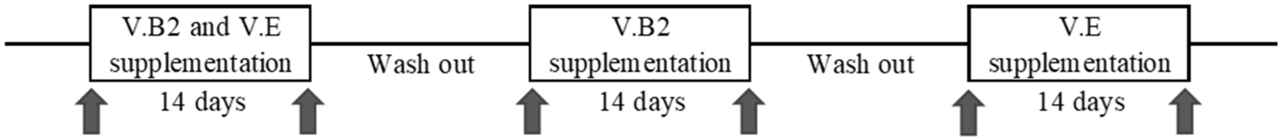
2.2.2. Procedure
2.3. Analysis of Saliva
2.4. Analysis of Blood Components
2.4.1. Plasma Homocysteine Analysis
2.4.2. Plasma V.E Analysis
2.4.3. Plasma Folate Analysis Using Enzyme-Linked Immune Sorbent Assay (ELISA) Kit
2.5. Statistical Analysis
3. Results
3.1. The C677T Polymorphism Status and Physical Characteristics of the Participants
3.2. Dietary Nutrient Intake
3.3. Serum Vitamin B2, Plasma Vitamin E, Folate, and Homocysteine Concentrations
4. Discussion
5. Conclusions
Author Contributions
Funding
Institutional Review Board Statement
Informed Consent Statement
Data Availability Statement
Acknowledgments
Conflicts of Interest
References
- Nattiv, A.; Loucks, A.B.; Manore, M.M.; Sanborn, C.F.; Sundgot-Borgen, J.; Warren, M.P.; American College of Sports Medicine. American College of Sports Medicine Position Stand. The female athlete triad. Med. Sci. Sports Exerc. 2007, 39, 1867–1882. [Google Scholar] [CrossRef] [PubMed]
- De Souza, M.J.; Nattiv, A.; Joy, E.; Misra, M.; Williams, N.I.; Mallinson, R.J.; Gibbs, J.C.; Olmsted, M.; Goolsby, M.; Matheson, G.; et al. 2014 Female Athlete Triad Coalition Consensus Statement on Treatment and Return to Play of the Female Athlete Triad: 1st International Conference held in San Francisco, California, May 2012 and 2nd International Conference held in San Francisco, California, May 2012 and 2nd International Conference held in Indianapolis, Indiana, May 2013. Br. J. Sports Med. 2014, 24, 96–119. [Google Scholar] [CrossRef] [Green Version]
- Mountjoy, M.; Sundgot-Borgen, J.; Burke, L.; Carter, S.; Constantini, N.; Lebrun, C.; Meyer, N.; Sherman, R.; Steffen, K.; Budgett, R.; et al. The IOC consensus statement: Beyond the Female Athlete Triad—Relative Energy Deficiency in Sport (RED-S). Br. J. Sports Med. 2014, 48, 491–497. [Google Scholar] [CrossRef] [PubMed]
- Hame, S.L.; Lafemina, J.M.; McAllister, D.R.; Schaadt, G.W.; Dorey, F.J. Fractures in the collegiate athlete. Am. J. Sports Med. 2004, 32, 446–451. [Google Scholar] [CrossRef] [PubMed]
- Blouin, S.; Thaler, H.W.; Korninger, C.; Schmid, R.; Hofstaetter, J.G.; Zoehrer, R.; Phipps, R.; Klaushofer, K.; Roschger, P.; Paschalis, E.P. Bone matrix quality and plasma homocysteine levels. Bone 2009, 44, 959–964. [Google Scholar] [CrossRef]
- Manolages, S.C. From estrogen-centric to aging and oxidative stress: A revised perspective of the pathogenesis of osteoporosis. Endocr. Rev. 2010, 31, 266–300. [Google Scholar] [CrossRef] [Green Version]
- McLean, R.R.; Hannan, M.T. B vitamins, homocysteine, and bone disease: Epidemiology and pathophysiology. Curr. Osteoporos. Rep. 2007, 5, 112–119. [Google Scholar] [CrossRef]
- Saito, M.; Fujii, K.; Marumo, K. Degree of mineralization-related collagen crosslinking in the femoral neck cancellous bone in cases of hip fracture and controls. Calcif. Tissue Int. 2006, 79, 160–168. [Google Scholar] [CrossRef]
- Saito, M.; Marumo, K. Collagen cross-links as a determinant of bone quality: A possible explanation for bone fragility in aging, osteoporosis, and diabetes mellitus. Osteoporos. Int. 2010, 21, 195–214. [Google Scholar] [CrossRef]
- Saito, M.; Marumo, K.; Soshi, S.; Kida, Y.; Ushiku, C.; Shinohara, A. Raloxifene ameliorates detrimental enzymatic and nonenzymatic collagen cross-links and bone strength in rabbits with hyperhomocysteinemia. Osteoporos. Int. 2010, 21, 655–666. [Google Scholar] [CrossRef]
- Boushey, C.J.; Beresford, S.A.; Omenn, G.S.; Motulsky, A.G. A Quantitative assessment of plasma homocysteine as a risk factor for vascular disease. probable benefits of increasing folic acid intakes. JAMA 1995, 274, 1049–1057. [Google Scholar] [CrossRef]
- Frosst, P.; Blom, H.J.; Milos, R.; Goyette, P.; Sheppard, C.A.; Matthews, R.G.; Boers, G.J.; den Heijer, M.; Kluijtmans, L.A.; van den Heuvel, L.P.; et al. A candidate genetic risk factor for vascular disease: A common mutation in methylenetetrahydrofolate reductase. Nat. Genet. 1995, 10, 111–113. [Google Scholar] [CrossRef]
- Rozen, R. Genetic predisposition to hyperhomocysteinemia: Deficiency of methylenetetrahydrofolate reductase (MTHFR). Thromb. Haemost. 1997, 78, 523–526. [Google Scholar] [CrossRef]
- Morita, H.; Taguchi, J.L.; Kurihara, H.; Kitaoka, M.; Kaneda, H.; Kurihara, Y.; Maemura, K.; Shindo, T.; Minamino, T.; Ohno, M.; et al. Genetic polymorphism of 5,10-methylenetetrahydrofolate reductase (MTHFR) as a risk factor for coronary artery disease. Circulation 1997, 95, 2032–2036. [Google Scholar] [CrossRef]
- Nishio, H.; Lee, M.J.; Fujii, M.; Kario, K.; Kayaba, K.; Shimada, K.; Matsuo, M.; Sumino, K. A common mutation in methylenetetrahydrofolate reductase gene among the Japanese population. Jpn. J. Hum. Genet. 1996, 41, 247–251. [Google Scholar] [CrossRef] [PubMed] [Green Version]
- Shiraki, M.; Urano, T.; Kuroda, T.; Saito, M.; Tanaka, S.; Miyao-Koshizuka, M.; Inoue, S. The synergistic effect of bone mineral density and methylenetetrahydrofolate reductase (MTHFR) polymorphism (C677T) on fractures. J. Bone Miner. Metab. 2008, 26, 595–602. [Google Scholar] [CrossRef]
- Yasui, K.; Kowa, H.; Nakaso, K.; Takeshima, T. Plasma homocysteine and MTHFR C677T genotype in levodopa-treated patients with PD. Neurology 2000, 55, 437–440. [Google Scholar] [CrossRef]
- Yang, J.; Hu, X.; Zhang, Q.; Cao, H.; Wang, J.; Liu, B. Homocysteine level and risk of fracture: A meta-analysis and systematic review. Bone 2012, 51, 376–382. [Google Scholar] [CrossRef]
- Hustad, S.; Ueland, P.M.; Vollset, S.E.; Zhang, Y.; Bjørke-Monsen, A.L.; Schneede, J. Riboflavin as a determinant of plasma total homocysteine: Effect modification by the methylenetetrahydrofolate reductase C677T polymorphism. Clin. Chem. 2000, 46 Pt 1, 1065–1071. [Google Scholar] [CrossRef] [Green Version]
- McKinley, M.C.; McNulty, H.; McPartlin, J.; Strain, J.J.; Scott, J.M. Effect of riboflavin supplementation on plasma homocysteine in elderly people with low riboflavin status. Eur. J. Clin. Nutr. 2002, 56, 850–856. [Google Scholar] [CrossRef]
- Bunout, D.; Garrido, A.; Suazo, M.; Kauffman, R.; Venegas, P.; de la Maza, P.; Petermann, M.; Hirsch, S. Effects of supplementation with folic acid and antioxidant vitamins on homocysteine levels and LDL oxidation in coronary patients. Nutrition 2000, 16, 107–110. [Google Scholar] [CrossRef]
- Sekine, M.; Kono, K.; Matsuya, K.; Sugahara, R.; Miyakoshi, Y.; Yano, T.; Ota, M. Screening parameters to evaluate risk for stress fracture in female long-distance runners. Clin. Funct. Nutr. 2015, 7, 42–46. (In Japanese) [Google Scholar]
- Chou, C.C.; Sung, Y.C.; Davison, G.; Chen, C.Y.; Liao, Y.H. Short-term high-dose vitamin C and E supplementation attenuates muscle damage and inflammatory responses to repeated taekwondo competitions: A randomized placebo-controlled trial. Int. J. Med. Sci. 2018, 15, 1217–1226. [Google Scholar] [CrossRef] [PubMed] [Green Version]
- Ministry of Health, Labour and Welfare. Dietary Reference Intakes for Japanese; Dai-Ichi SHUPPAN Co. Ltd.: Tokyo, Japan, 2015. [Google Scholar]
- Zempleni, J.; Galloway, J.R.; McCormick, D.B. Pharmacokinetics of orally and intravenously administered riboflavin in healthy humans. Am. J. Clin. Nutr. 1996, 63, 54–66. [Google Scholar] [CrossRef] [PubMed] [Green Version]
- Schoenen, J.; Lenaerts, M.; Bastings, E. High-Dose Riboflavin as a Prophylactic Treatment of Migraine: Results of an Open Pilot Study. Cephalalgia 1994, 14, 328–329. [Google Scholar] [CrossRef] [PubMed]
- Hu, J.J.; Roush, G.C.; Berwick, M.; Dubin, N.; Mahabir, S.; Chandiramani, M.; Boorstein, R. Effects of dietary supplementation of alpha-tocopherol on plasma glutathione and DNA repair activities. Cancer Epidemiol. Biomark. Prev. 1996, 5, 263–270. [Google Scholar]
- Kobayashi, S.; Honda, S.; Murakami, K.; Sasaki, S.; Okubo, H.; Hirota, N.; Notsu, A.; Fukui, M.; Date, C. Both comprehensive and brief self-administered diet history questionnaires satisfactorily rank nutrient intakes in Japanese adults. J. Epidemiol. 2012, 22, 151–159. [Google Scholar] [CrossRef] [Green Version]
- Kobayashi, S.; Murakami, K.; Sasaki, S.; Okubo, H.; Hirota, N.; Notsu, A.; Fukui, M.; Date, C. Comparison of relative validity of food group intakes estimated by comprehensive and brief-type self-administered diet history questionnaires against 16 d dietary records in Japanese adults. Public Health Nutr. 2011, 14, 1200–1211. [Google Scholar] [CrossRef] [Green Version]
- Okubo, H.; Sasaki, S.; Rafamantanantsoa, H.H.; Ishikawa-Tanaka, K.; Okazaki, H.; Tabata, I. Validation of self-reported energy intake by a self-administered diet history questionnaire using the doubly labeled water method in 140 Japanese adults. Eur. J. Clin. Nutr. 2008, 62, 1343–1350. [Google Scholar] [CrossRef]
- Castiglia, P.; Sanna, V.; Azara, A.; De Miglio, M.R.; Murgia, L.; Pira, G.; Sanges, F.; Fancellu, A.; Carru, C.; Bisail, M.; et al. Methylenetetrahydrofolate reductase (MTHFR) C677T and A1298C polymorphisms in breast cancer: A Sardinian preliminary case-control stury. Int. J. Med. Sci. 2019, 16, 10891095. [Google Scholar] [CrossRef] [Green Version]
- Goyette, P.; Rozen, R. The thermolabile variant 677C→T can further reduce activity when expressed in cis with severe mutations for human methylenetetrahydrofolate reductase. Hum Mutat. 2000, 16, 132–138. [Google Scholar] [CrossRef]
- Wu, Y.; Tomon, M.; Sumino, K. Methylenetetrahydrofolate reductase gene polymorphism and ischemic stroke: Sex difference in Japanese. Kobe J. Med. Sci. 2001, 47, 255–263. [Google Scholar]
- Jacques, P.F.; Bostom, A.G.; Wilson, P.W.; Rich, S.; Rosenberg, I.H.; Selhub, J. Eterminants of plasma total homocysteine concentration in the Framingham Offspring Cohort. Am. J. Clin. Nutr. 2001, 73, 613–621. [Google Scholar] [CrossRef]
- Lakshmi, R.; Lakshmi, A.V.; Bamji, M.S. Mechanism of impaired skin collagen maturity in riboflavin or pyridoxine deficiency. J. Biosci. 1990, 15, 289–295. [Google Scholar] [CrossRef]
- Boehnke, C.; Reuter, U.; Flach, U.; Schuh-Hofer, S.; Einhäupl, K.M.; Arnold, G. High Dose Riboflavin Treatment is efficacious in migraine prophylaxis: An open study in a tertiary care centre. Eur. J. Neurol. 2004, 11, 475–477. [Google Scholar] [CrossRef]
- McNulty, H.; Dowey, L.R.C.; Strain, J.J.; Dunne, A.; Ward, M.; Molloy, A.M.; McAnena, L.B.; Hughes, J.P.; Hannon-Fletcher, M.; Scott, J.M. Riboflavin lowers homocysteine in individuals homozygous for the MTHFR 677C→T polymorphism. Circulation 2006, 113, 74–80. [Google Scholar] [CrossRef] [Green Version]
- Cafolla, A.; Dragoni, F.; Girelli, G.; Tosti, M.E.; Costante, A.; De Luca, A.M.; Funaro, D.; Scott, C.S. Effect of folic acid and vitamin C supplementation on folate status and homocysteine level: A randomised controlled trial in Italian smoker-blood donors. Atherosclerosis 2002, 163, 105–111. [Google Scholar] [CrossRef]
- Magana, A.A.; Reed, R.L.; Koluda, R.; Miranda, C.L.; Maier, C.S.; Stevens, J.F. Vitamin C activates the folate-mediated one-carbon cycle in C2C12 myoblasts. Antioxidants 2020, 9, 217. [Google Scholar] [CrossRef] [Green Version]
- Meydani, M.; Evans, W.J.; Handelman, G.; Biddle, L.; Fielding, R.A.; Meydani, S.N.; Burrill, J.; Fiatarone, M.A.; Blumberg, J.B.; Cannon, J.G. Protective effect of vitamin e on exercise-induced oxidative damage in young and older adults. Am. J. Physiol. 1993, 264 Pt 2, R992–R998. [Google Scholar] [CrossRef]
- Sacheck, J.M.; Milbury, P.E.; Cannon, J.G.; Roubenoff, R.; Blumberg, J.B. Effect of Vitamin E and eccentric exercise on selected biomarkers of oxidative stress in young and elderly men. Free Radic. Biol. Med. 2003, 34, 1575–1588. [Google Scholar] [CrossRef]
- Ullegaddi, R.; Powers, H.J.; Gariballa, S.E. Antioxidant supplementation with or without B-group vitamins after acute ischemic stroke: A randomized controlled trial. JPEN J. Parenter. Enteral. Nutr. 2006, 30, 108–114. [Google Scholar] [CrossRef] [PubMed]


| Items | Equipment, Reagents, and Conditions for Hcy | Equipment, Reagents, and Conditions for V.E. |
|---|---|---|
| Pump | EP-700 (LIQUID CHROMATOGRAPH PUMP/Eicom) | EP-700 (LIQUID CHROMATOGRAPH PUMP/Eicom) |
| Auto Sampler | M-514 (Eicom) | AS-4050 (Eicom) |
| Detector | Eicom ECD-700 (Eicom) | FP-2025 Plus intelligent fluorescence detector (Jasco) |
| Working Electrode | Eicom WE-AU (Eicom) | Eicom WE-AU (Eicom) |
| Precolumn | COSMOSIL Guard Cartridge 5PFP 4.6ID × 10 mm (COSMOSIL) | Eicom PC-04 4.0 mmφ × 5 mm (Eicom) |
| Column | Eicom-3OSD 3.0φ × 150 nm (Eicom) | COSMOSIL Packed column 5PFP 4.6 mm I.D. × 250 mm (nacalai tesque) |
| Column Temp | 25 °C | 40 °C |
| Buffer | 99% 0.1 M Sodium phosphate buffer (pH 2.5), 1% Methanol, 170 mg/L Sodium octansulfonate, 5 mg/L EDTA-2Na | Methanol:MilliQ (v/v) = 9:1 |
| Flow rate | 500 µL/min | 700 µL/min |
| ID | Sex | Age (Years) | Height (cm) | Weight (kg) | BMI | Number of Stress Fractures | SNPs (MTHFR C677T) | Monthly Running Distance (km) | Competing in the Track and Field (Year) | Best Record (3000 m Race) (min) |
|---|---|---|---|---|---|---|---|---|---|---|
| A | F | 21 | 156.0 | 45.0 | 18.5 | 2 | CT | 425 | 9 | 9.34 |
| B | F | 21 | 165.2 | 49.5 | 18.1 | 1 | CT | 392 | 9 | 9.57 |
| C | F | 21 | 162.8 | 42.4 | 16.0 | 2 | CT | 416 | 10 | 9.43 |
| D | F | 21 | 150.0 | 42.0 | 18.7 | 0 | CT | 519 | 12 | 9.47 |
| E | F | 20 | 160.0 | 42.5 | 16.6 | 0 | CT | 804 | 5 | 9.25 |
| F | F | 20 | 160.0 | 46.5 | 18.2 | 0 | TT | 418 | 8 | 9.48 |
| G | F | 19 | 163.0 | 47.5 | 17.9 | 0 | CT | 500 | 7 | 9.36 |
| H | F | 19 | 158.3 | 38.5 | 15.4 | 0 | CT | 252 | 4 | 9.45 |
| I | F | 19 | 157.3 | 45.9 | 18.6 | 0 | CT | 517 | 7 | 9.49 |
| J | F | 19 | 163.5 | 53.0 | 19.8 | 0 | CT | 467 | 4 | 10.08 |
| K | F | 19 | 171.0 | 49.0 | 16.8 | 0 | CT | 525 | 7 | 9.29 |
| (Mean ± S.D.) | 19.9 ± 0.9 | 160.6 ± 5.2 | 45.6 ± 3.9 | 17.7 ± 1.3 | 0.5 ± 0.8 | 476 ± 128 | 7.5 ± 2.4 | 9.47 ± 21 | ||
| Calculated Values (Mean ± S.D.) | DRIs | ||
|---|---|---|---|
| Energy (kcal) | 2145 ± 572 | ||
| Protein (%) | 18.6 ± 2.4 | 13~20% | PFC energy ratio |
| Fat (%) | 28.5 ± 6.0 | 20~30% | |
| Carbohydrates (%) | 52.1 ± 7.6 | 50~65% | |
| V.B2 (mg/1000 kcal) | 0.9 ± 0.2 | 0.6 | RDA |
| V.B6 (mg/1000 kcal) | 1.0 ± 0.2 | 0.55 | RDA |
| V.B12 (μg/1000 kcal) | 9.1 ± 2.5 | 1.2 | RDA |
| Folic acid (μg/1000 kcal) | 252 ± 78 | 120 | RDA |
| V.C (mg/1000 kcal) | 95 ± 38 | 50 | RDA |
| V.A (μgRAE/1000 kcal) | 825 ± 310 | 325 | RDA |
| V.E (mg/1000 kcal) | 5.2 ± 1.4 | 2.5 | RDA |
| (n = 11) | V.E + V.B2 Period | V.B2 Period | V.E Period | |||
|---|---|---|---|---|---|---|
| Pre | Post | Pre | Post | Pre | Post | |
| Serum V.B2 (µg/dL) # | 20.3 ± 3.4 | 24.0 ± 2.6 ** | 17.4 ± 4.1 | 22.4 ± 2.4 *** | 23.5 ± 4.5 | 19.5 ± 2.5 * |
| Plasma V.E (µmol/L) # | 18.1 ± 3.1 | 20.7 ± 1.5 * | 25.2 ± 4.4 | 20.6 ± 3.9 *** | 19.5 ± 4.1 | 23.6 ± 4.0 * |
| Plasma folate (ng/mL) † | 9.5 ± 5.0 | 10.7 ± 3.7 | 8.0 ± 4.1 | 6.8 ± 3.8 | 7.7 ± 4.7 | 7.2 ± 4.2 |
Publisher’s Note: MDPI stays neutral with regard to jurisdictional claims in published maps and institutional affiliations. |
© 2022 by the authors. Licensee MDPI, Basel, Switzerland. This article is an open access article distributed under the terms and conditions of the Creative Commons Attribution (CC BY) license (https://creativecommons.org/licenses/by/4.0/).
Share and Cite
Shinagawa, A.; Serizawa, N.; Yamazaki, T.; Minematsu, A.; Miyakoshi, Y.; Yano, T.; Ota, M. Short-Term Combined Intake of Vitamin B2 and Vitamin E Decreases Plasma Homocysteine Concentrations in Female Track Athletes. Dietetics 2022, 1, 216-226. https://doi.org/10.3390/dietetics1030019
Shinagawa A, Serizawa N, Yamazaki T, Minematsu A, Miyakoshi Y, Yano T, Ota M. Short-Term Combined Intake of Vitamin B2 and Vitamin E Decreases Plasma Homocysteine Concentrations in Female Track Athletes. Dietetics. 2022; 1(3):216-226. https://doi.org/10.3390/dietetics1030019
Chicago/Turabian StyleShinagawa, Akiho, Naho Serizawa, Tomoki Yamazaki, Ayako Minematsu, Yuichi Miyakoshi, Tomohiro Yano, and Masako Ota. 2022. "Short-Term Combined Intake of Vitamin B2 and Vitamin E Decreases Plasma Homocysteine Concentrations in Female Track Athletes" Dietetics 1, no. 3: 216-226. https://doi.org/10.3390/dietetics1030019