Review

A Mechanistic Overview on Impact of Dietary Fibres on Gut Microbiota and Its Association with Colon Cancer

1
Department of Pharmacognosy and Phytochemistry, School of Pharmaceutical Sciences, Delhi Pharmaceutical Sciences and Research University, New Delhi 110017, India
2
School of Pharmaceutical Sciences, Lovely Professional University, Phagwara, Punjab 144411, India
*
Author to whom correspondence should be addressed.
Dietetics 2022, 1(3), 182-202; https://doi.org/10.3390/dietetics1030017
Submission received: 21 June 2022 / Revised: 12 August 2022 / Accepted: 22 August 2022 / Published: 2 November 2022

Abstract

:
Colorectal cancer (CRC) is an abnormal growth that occurs in the rectum or rectal portion. In 2020, an anticipated 104,610 new cases of colon illness and 43,340 new cases of rectal abnormal growth were expected in the United States. Red and processed meat, body bloatedness, belly fatness, and binge drinking expands the occurrence of colorectal disease. Dietary fibres contribute to faecal bulking, but they are break down by gut bacteria and produce metabolites such as short-chain fatty acids (SCFAs). SCFAs are chemical compounds that are mostly made up of acetate, propionate, and butyrate. Acetate and butyrate help to control mucus production and discharge, and thus, protect the gut mucosa. Reduced mucus secretion/increased bacterial catabolism, and fermentation of amino acids resulted in an increase of potentially detrimental metabolites such as branched-chain fatty acids, ammonia, amines, and N-nitroso complex components. Vital roles of fibres include reduction in the time that carcinogens encounter the intestinal lumen and promotion of healthy gut microbiota as well as modification of the host metabolism. The present review focuses on a brief introduction to various dietary fibres and specialised metabolites that can possess beneficial effect on CRC, as well as presenting our current, detailed understanding of various dietary fibres along with their potential effects on gut microbiota and its association with the colon cancer. A comprehensive discussion is also included, advocating the dietary fibre-enriched diet.

1. Introduction

Cancer is a serious public health issue that affects people globally. It is a disease in which abnormal cells expand uncontrollably, and is among the leading cause of death, with an estimated 10 million deaths projected by 2020 [1]. In 2022, the United States is predicted to have 1,918,030 new cancer cases and 609,360 cancer deaths, with around 350 deaths daily due to lung cancer [2]. The coronavirus disease 2019 (COVID-19) pandemic had a negative impact on cancer detection and therapy in 2020 [2,3].
Colorectal cancer (CRC) is the occurrence of tumour development in the colon or rectum. The colon, rectum (colorectum), and anus are the final sections of the gastrointestinal (GI) tract [1]. In the United States, an estimated 104,610 new cases of colon cancer and 43,340 new cases of rectal cancer were diagnosed in 2020. In the same year an estimated 53,200 people would die from CRC, with 3640 men and women below 50 years [4]. There is lack of concrete statistics on mortalities from colon and rectal cancers separately, due to the fact that around 40% of rectal cancer deaths are falsely claimed as colon cancer instead of rectal cancer on death certificates [4]. According to reports, due to ageing, human advancement, and population growth, the global number of new CRC cases is expected to reach 3.2 million in 2040 globally [5]. The increased prevalence of CRC is mostly caused by excessive exposure to environmental and genetic factors such as familial adenomatous polyposis, Lynch syndrome, MUTYH-associated polyposis, etc. [6]. This can also be associated with lifestyle changes [5].

Various Risk Factors of Colon Cancer

According to a recent report by the World Cancer Research Fund/American Institute for Cancer Research expert panel, eating a lot of red and processed meat, body fatness, belly fatness, and binge drinking raises the risk of CRC [7]. CRC may be prevented by drinking milk and eating full grains [7]. Intake of vegetables and possibly fruits have an indirect relationship with the risk of colon cancer. Male sex, age, race/ethnicity, body mass index (BMI), height, diabetes, dietary antioxidants, folate, calcium, postmenopausal hormone use, and smoking are all widely listed risk factors for CRC [8].

2. Dietary Fibres

Dietary fibres are defined as eatable carbohydrate polymers with three or more monosaccharide units that are resistant to action of endogenous digestive enzymes, and thus, are neither metabolised nor absorbed in the small intestine [9]. These are edible carbohydrate polymers present in foods such as fruits, vegetables, legumes, and cereals; (ii) edible raw food materials through physical, enzymatic, or chemical degradation and synthetic carbohydrate polymers with a proven biological benefit [9]. The World Health Organization (WHO) recommends consuming at least 25 g of fibre each day [10]. Various food and health groups recommend diets high in vegetables, fruit, and whole-grain cereals to satisfy the guidelines. For adults, most countries recommend 25–35 g of dietary fibre per day, with recommendations ranging from 18–38 g per day [9]. Although most national authorities use this definition, there are some differences among these definitions. These differences mainly concern (1) whether non-carbohydrates such as cellulose and other substances found in cell walls linked to polysaccharides are considered dietary fibres and (2) the required minimum quantity of carbohydrate monomers [9]. The present review provides a detailed understanding of various dietary fibres along with their potential effects on gut microbiota and their association with colon cancer.

2.1. Types and Characteristics of Dietary Fibres

Dietary fibres are classified as polysaccharides (non-starch polysaccharides (NSPs), resistant starch (RS), and resistant oligosaccharides (Ros)), soluble as well as insoluble in nature [9]. Fibres of an insoluble nature of the carbohydrate moiety, such as cellulose and hemicellulose, enter the colonic region and are gradually or not digested by gut bacteria and produce faecal bulking. Unlike ROs, most soluble NSPs, particularly polymers with a high molecular weight, such as guar gum, certain pectins, b-glucans, and psyllium, are viscous, and can form a gel structure in the intestinal tract. This delays glucose and lipid absorption and influences post-prandial metabolism [11].
Most soluble fibres do not facilitate to faecal bulking, but they are digested by gut bacteria and produce metabolites such as short-chain fatty acids (SCFAs) [11]. SCFAs are chemical compounds that are mostly made up of acetate, propionate, and butyrate. SCFAs are important regulators of the host’s metabolism, immunological system, and cell growth. SCFAs are abundant in the cecum and proximal colon, where they are utilised as an energy source by colonocytes (particularly butyrate), but they can also be transferred to the interstitial blood circulation via the portal vein and act on the hepatocytes and peripheral tissues [12]. Even though SCFA levels in the peripheral circulation are modest, it has become well known that they function as chemical messengers and regulate a variety of physiological functions in the host [12]. Soluble fibres increase food transit time in the intestine and are easily fermented into gases in the colon. Insoluble fibres can either be metabolically inert, providing bulk to intestinal contents, or they can be fermented in the large intestine [13]. Insoluble fibres have the effect of speeding up the passage of food through the intestine [13]. Soluble fibres bond to bile acids in the small intestine, preventing them from being absorbed. As a result, cholesterol levels of blood are reduced [14]. Soluble fibres also inhibit sugar absorption, normalise blood cholesterol levels, and create SCFA in the colon as by-products of fermentation, which have a wide range of physiological functions. Although insoluble fibres are linked to a lower incidence of diabetes, the exact mechanism remains uncertain [15].

2.2. Sources of Fibres

A promise that a food is high in fibre, or any claim likely to have the similar meaning for the consumer, may only be made if the product has at least 30 g of fibre per 500 g or at least 3 g of fibre per 100 kcal (418 kJ) [9]. In the human small intestine, dietary fibres are the indigestible matter of plant food. Poly/oligosaccharide cellulose and other plant-derived compounds make up dietary fibres [16]. Raw chickpea seeds have a total dietary fibre content (DFC) of 18–22 g per 100 g [17]. Soluble and insoluble DFC is around 4–8 and 10–18 g per 100 g of raw chickpea seed, respectively [16]. A study on total dietary fibre of various gram and peas seeds showed that soaking can significantly enhance total dietary fibre and soluble dietary fibres [13]. A summary on dietary fibre content and their energy in some widely used food is presented in Table 1 [18].

3. Imbalance of Gut Microbiota and Health Disorders

Firmicutes, Bacteroidetes, Actinobacteria, and Proteobacteria are the major groups in the human intestinal microbiome. Gut microbiotas digest indigestible food, produce nutrients such as vitamins, detoxify metabolites, influence the immune response, provide signals for epithelial cell renewal and mucosal integrity maintenance, and exude antimicrobial compounds [19]. The microbiome (which includes bacteria, viruses, fungus, and other microorganisms) governs health, and microbiota changes can lead to disease. Alteration in the intestinal microbiome allow environmental risk factors to develop and perpetuate CRC [20,21]. This could be because microbiome influence metabolism and immune function. Evidence suggests that the alterations in the gut microbiota occur early in CRC and can be used to identify those who are at risk for colorectal adenoma, a precursor lesion to CRC [22]. Thus, microbiota alteration can be utilised as biomarker for the early diagnosis of CRC [22]. There is a definite link between the GI microbiome and the progression of chronic non-communicable diseases, which shows the importance for development of strategies to enhance human health by targeting the GI microbiota.

4. Symbiotic Relationship between Dietary Fibres and Microbiota

4.1. Human Gut Microbiota

The human gut is home to trillions of bacteria from all kingdoms of life, all of which are necessary for the growth and physiology of the host. This “gut microbiota” is a complicated ecosystem to communicate with one another and with the human to influence vital biological function, such as regulating juvenile growth [23,23], maturation of the immune system, and protection against some infectious agents [24]. In addition, it helps in carbohydrate and lipid metabolism that regulate and maintains the host’s energy storage [25,26].

4.2. Understanding of the Host-Microbiota System

The microbiome helps to maintain homeostasis in all our body’s tissues. Symbiotic relationships exist between vertebrates and the large and complex microbial populations that populate their gastrointestinal tracts. The general interaction between people and their microbiota may be described as mutualistic [27], and the balance of gut microbial ecosystems (eubiosis) is a fundamental concept. Disturbance of gut microbiota results in the onset of a variety of chronic diseases [28]. Pathogenic changes in the microbiome’s profile and functions are referred to as dysbiosis. Changes in the number of good intestinal bacteria can result in long-term inflammation and the formation of carcinogenic chemicals, which can lead to neoplasia [19].

4.3. Bacterial Signaling for Virulence

The gut bacteria convert dietary and endogenous substances into metabolites that allow communication with host’s peripheral organs and tissues. If any changes in gut bacteria composition have been connected to specific illnesses, altering gut bacteria composition by dietary changes may be a potential therapy option [29]. Dietary fibre confers resistance against the colonisation of the gut with multidrug-resistant bacteria and increases the number of bacteria that convert fibre into SCFA [30]. The intestinal microbiota and the gut mucosal immune system have a homeostatic relationship, and disturbance of this interaction can lead to illnesses [31]. The Western lifestyle comprises a diet low in microbiota-accessible carbohydrates (MACs), which leads to h alter participation and functionality of microbioata as compared with the non-Westernised population, which includes a high fibre in their diet. Immunological dysregulation caused by interactions between resident microorganisms and the host could explain a number of disorders involving inflammation as a common cause [32].

5. Impact of Dietary Fibres on Gut Bacteria

Diet has a big influence on the gut microbiota’s composition, variety, and richness. Different aspects of a diet have a time-dependent effect on gut bacterial ecosystems [33]. Diet is the simplest therapeutic intervention, since it is the easiest to change [34]. Bacteroides, Prevotella, and Ruminococcus were found to be the most prevalent varieties, or “enterotypes”, in a study of gut microbial populations [34]. Variation in the ratio of Prevotella/Bacteroides was also seen across industrialised and unindustrialised human populations, implying that these bacterial communities are influenced by long-term dietary differences, for instance, mutton (which drives Bacteroides in Westernised populations) and dietary fibre (which drives Prevotella in non-Westernised populations) [35].
Prevotella plays a crucial role in nurturing healthy gut microbiota. The impact of nutrition on the microbiome was revealed by comparing the gut bacteria of children from rural (Burkina Faso (BF)) and urban (Italy) communities [35]. In both groups, the gut microbiota composition changed significantly after breastfeeding. In BF children, there was a considerable rise in the population of microbiota from the genera Prevotella and Xylanibacter, which was associated with greater amounts of faecal SCFAs, indicating their ability to break down complex carbohydrates [35]. These variations are irrespective of race, as the gut microbial populations of BF children residing in urban, rather than rural, parts of BF resembled those of Italian children. The modification in the gut microbiota population is linked with the fact that when people relocate to cities, they are introduced to a Westernised way of lifestyle, which includes availability to high-saturated fat and high-sugar foods. As a result, children in urbanised areas of BF have bacteria that are better suited to metabolise meat protein, fat, and sugar-rich foods, whereas children in rural areas have bacteria that are better suited to digest fibre and carbohydrate fermentation from vegetables (enrichment in Prevotella, Treponema, and Succinivibrio). Surprisingly, the microbiomes of BF children living in cities were equivalent to those of Italian children, indicating that nutrition had a significant impact independent of host genetics [35].
Hadza is a modern human community who reside in an important geographic region for human evolution research and hunt for resources [36]. It was found that Hadza hunter-gatherer people have a greater level of microbial abundance and biodiversity than Italian city dwellers. These people’s gut microbiota exhibited higher Bacteroidetes and lower Firmicutes species richness, and they almost completely lacked the Actinobacteria phylum, with minor amounts of Bifidobacterium. Interestingly, Hadza SCFA production was linked with higher propionate levels, whereas the Italian cohort had higher butyrate levels [36]. Environmental lifestyle changes, rather than host genetics, affected gut microbiota diversity, and industrialisation results in a considerable loss of species, according to these findings [37].
Antibiotic use, therapeutic practices (e.g., caesarean sections), and sanitation are the underlying causes for a decreasing variety of microbiota [32]. For instance, the gut microbiota was reported to be altered after caesarean section [38]. The fact that babies delivered naturally had greater gut bacterial counts at one month of age than babies delivered by caesarean section shows that natural birth promotes and starts the colonisation of the gut by microorganisms [39]. Poor personal hygiene and unsanitary living circumstances in poorer nations can contribute to the spread of infectious diseases. Circadian disorder caused by travel, shift work, or other factors affects gut health and changes the bacteria communities in the gut [39,40]. The long-term effects of various antibiotics include reduced microbial diversity, altered Bacteroidetes/firmicutes ratio, overgrowth of clostridium difficile, and increased expansion of opportunistic pathogens, such as Salmonella typhimurium, Escherichia, and Klebsiella species [41].

5.1. Probiotic-Prebiotic Relationship of Dietary Fibres

Probiotics, prebiotics, and synbiotics are all based on the same concept, i.e., to develop meals that increase “good” bacteria in the intestinal lumen after consumption [42]. This can be accomplished by adding either health-promoting “probiotic” bacteria or fermentable “prebiotic” carbohydrates that are indigestible but fermentable [42]. The functions of the probiotic bacteria added to food include the reduction of potentially pathogenic bacteria and/or toxic metabolites in the intestine, restoration of GI motility, and modulation of the immune response. Conversely, so-called prebiotic food components should support the growth of beneficial bacteria of the indigenous intestinal microbiota of humans, and also improve survival of probiotic bacteria which have been consumed at the same moment [43]. Prebiotic polysaccharides are dietary fibres that are fermented by the large intestinal microbiota rather than processed by human enzymes. As a result, they improve the health of the intestinal lumen mucosa by increasing biomass, faeces weight, and regulate frequency of defecation, reducing constipation and enhancing the health of the intestinal lumen mucosa [44,45].

5.2. Microbial Metabolism of Dietary Fibres and Functional Implication

Dietary fibres are a good source of energy for the bacteria that live in the cecum and colon. Under certain intestinal conditions, anaerobic bacteria activate their machinery, which includes important enzymes and metabolic pathways that may digest complex carbohydrates and produce metabolites such as SCFAs. SCFAs are chemical compounds that are mostly made up of acetate, propionate, and butyrate, which are necessary for the health of the intestinal mucosa and are otherwise unavailable in the diet. The colonic microbiota’s ability to ferment fibre for their own metabolic needs, leading in the generation of luminal SCFAs [12,46,47]. A study by Roediger [48] demonstrated that colonocytes choose butyrate over glucose as a source of energy, laying the groundwork for the now well-known symbiotic relationship between colonic microorganisms and colonic mucosal health.

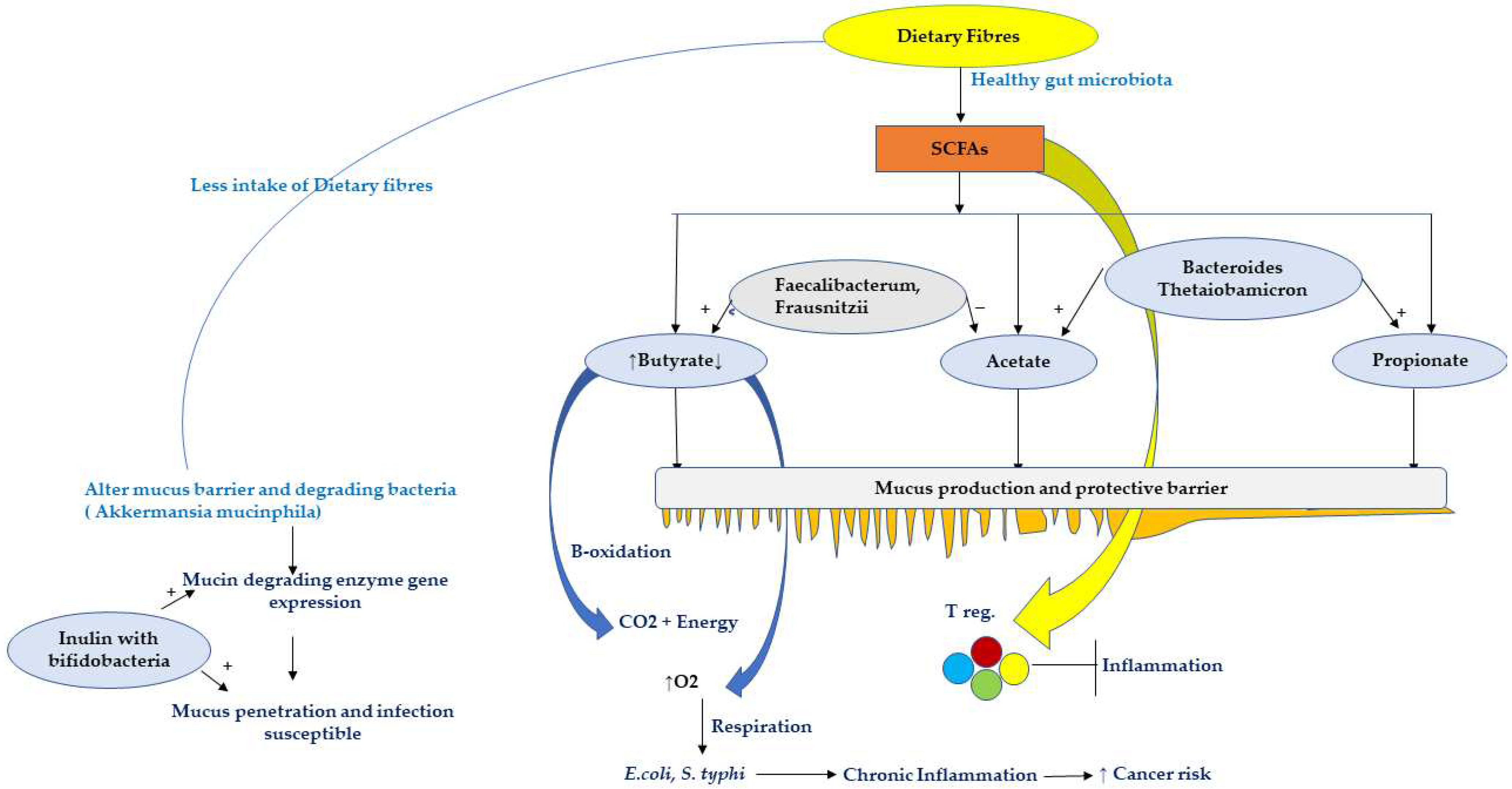
5.3. Effect of Dietary Fibres and SCFA on Host

Mucus generation and secretion are stimulated by dietary fibres and SCFAs. Both acetate and butyrate help to control mucus production and discharge. Acetate- and propionate-producing bacteria, Bacteroides thetaiotaomicron, enhance goblet cell development and mucin-related genes expression. On the other hand, Faecalibacterium prausnitzii, an acetate consumer and butyrate generator, reduces the influence of acetate on mucus and inhibits mucus overproduction, allowing the gut epithelium to maintain its correct shape and composition [49]. Moreover, dietary fibres can mechanically increase mucus secretion by the intestinal epithelium [50].
Long-term dietary fibre deficiency has been linked to an increase in mucin-degrading bacteria such as Akkermansia muciniphila, which affects the mucus barrier [51]. When the diet lacks dietary fibres, some gut bacteria adapt their metabolism to utilise mucin glycans by activating the gene expression of mucin-degrading enzyme [52].
A Western diet (low fibre content) given to mice increased the permeability of the inner mucosal layer and decreased the development rate, making the mucus penetrable and perhaps increasing infection susceptibility [29]. A study conducted on obese mice showed that a small inulin dose (1%) or Bifidobacterium longum (a prebiotic with bifidogenic properties) was beneficial in mucus problems. The permeability of the inner mucosal layer was restored by the supplementation of insulin, whereas the mucus growth rate defect was restored by B. longum supplementation, indicating that the two criteria are independent and may be governed by distinct mechanisms. Obese animals did not benefit from the administration of either 1% inulin or B. longum [53]. High inulin intake (20%), on the other hand, prevented microbial invasion, enhanced intestinal health, and contributed to the relief of low-grade inflammation in obese mice [53]. A previous study revealed that colonocytes caused beta-oxidation of butyrate aerobically, resulting in an anaerobic environment in the gut [54]. Because butyrate-producing bacteria are particularly sensitive to oxygen, their numbers are reduced even further, resulting in a reduction in butyrate production. This feedforward loop results in enhanced luminal oxygen levels, allowing proteobacteria such as Escherichia coli and Salmonella enterica serovar Typhimurium to flourish. This unique approach not only explains most of the diseases linked with a low-fibre diet, but also provides a molecular explanation for why lower microbiota richness is reported both in humans and mice having low-fibre diet.

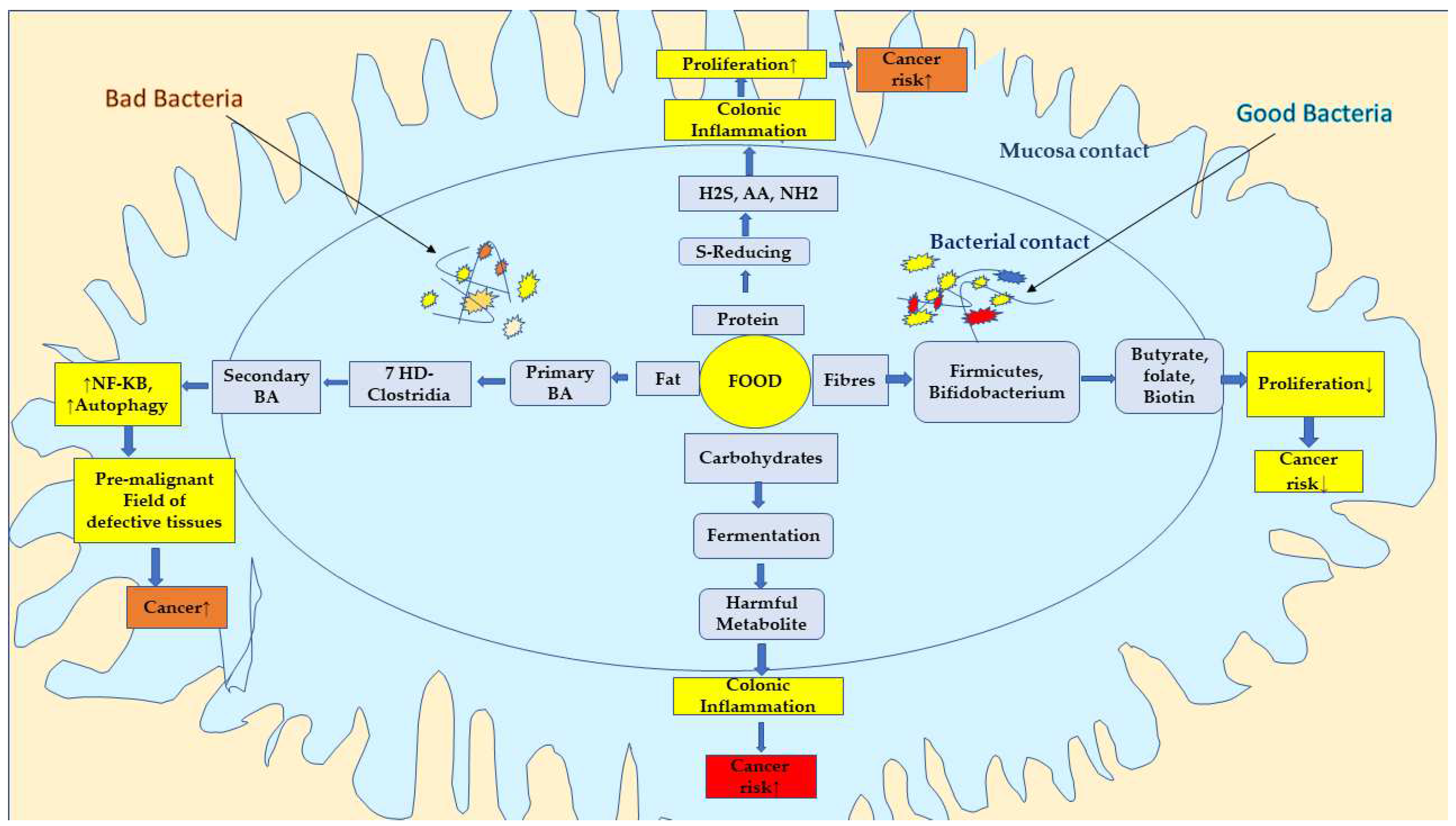
6. Diet Manipulation, Gut Microbiota, and Colon Cancer

Humans and their gut microbiota coexist as mutualists, but this connection can become pathological in some cases, such as obesity, diabetes, atherosclerosis, inflammatory bowel diseases (IBD), and cancer [33]. CRC is thought to be linked to localised inflammation. Though inflammation cannot initiate oncogenesis on its own, it is thought to be a crucial contributor. Previous research has revealed that gut inflammation and CRC oncogenesis can be modulated by food, gut microbiota, and gut environment, and these variables may be adjustable variables in modifying CRC outcomes [55]. SCFA is a useful metabolite that is produced under the action of local biota that ferments fibre and other indigestible starches as they move from the small intestine into the large colon. These SCFAs, in turn, have antineoplastic, anti-inflammatory, and physiological properties.

6.1. Causal Relationship between Microbiota, Dietary Fibre, and Colon Cancer

Low dietary fibre consumption reduces microbial diversity, SCFA generation, and shifting gut microbial metabolism to less favourable substrates, such as dietary and endogenously provided proteins [56,57]. This might promote mucus-degrading bacteria, which might harm the host [58]. The generation of total SCFAs and butyrate was dramatically reduced when human volunteers were fed a high-protein, low-carbohydrate diet [56,59]. However, the fermentation of amino acids resulted in an increase of potentially harmful metabolites such as branched-chain fatty acids, ammonia, N-nitroso compounds, p-cresol, sulphides, indoles compounds, and hydrogen sulphide [60]. These metabolites’ cytotoxic as well as pro-inflammatory properties contribute to the development of chronic illnesses, especially CRC. Butyrate-producing bacteria are lower in CRC patients than in healthy people, suggesting that a significant structural imbalance of the gut bacteria is produced by a decrease in butyrate bio-makers and an increase in pathogenic organisms [61]. Reduced dietary fibre intake is linked to a higher risk of CRC [62]. As a result, it is not surprising that low-fat diets high in fibre-rich grain products, vegetables, and fruits have health claims allowed by the FDA for a possible reduction in the risk of developing some types of cancer. According to the findings, increasing dietary fibre in African Americans modified the microbiota and enhanced butyrogenesis, resulting in lower cancer risk indicators is depicted in Figure 1 [63].

6.2. Effect of Dietary Fibre on Colon Cancer

CRC and ulcerative colitis are two common bowel illnesses that affect the descending distal colon, which is the principal location for fermentation of protein into their amino acids [60]. IBD can also lead to CRC [65], which is the third most common cancer [66]. CRC is linked to food habits, smoking, physical activity, and hereditary and environmental variables [66]. Americans consumed two to three times more animal protein and fat, while Africans consumed more carbohydrate and fibre, primarily in the form of resistant starch. African Americans had more polyps and greater rates of mucosal proliferation on colonoscopy when evaluated using Ki67 epithelial cell staining, showing its potential application as a cancer risk biomarker [63,63]. A plant-based cuisine includes vegetables, fruits, cereals, nuts, and legumes, most of which are made up of considerable amounts of olive oil, with moderate use of fish, seafood, or dairy, and limited intake of meat and alcohol (mostly red wine), which is the nutritional dimension of the Mediterranean lifestyle [67]. Indian meals are also beneficial to one’s health, because they have been linked to a lower prevalence of numerous ailments. Increased consumption of high-fibre meals or fibre supplements decreases blood pressure, improves blood glucose, assists weight loss, and reduces the risk of CRC [68].
These differences in the dietary pattern were associated to major changes in microbiota (the genus Bacteroides dominated in Americans, and the genus Prevotella in Africans) and metabolic phenotype derivatives [69]. Africans had larger quantities of starch break-down, carbohydrate fermenters, and butyrate makers, as well as their metabolites derivatives [69]. In Americans, there were more potentially pathogenic proteobacteria (Escherichia and Acinetobacter) and bile acid deconjugators and their derivatives [69]. The products of fibre fermentation, particularly butyrate, have been shown to be anti-inflammatory and antineoplastic in numerous studies [70,71,72] and secondary bile acids (BAs), the by-products of bacterial bile acid conjugation, were reported as carcinogenic [73].
Dietary fibres bind conjugated primary BAs may act as a medium for gut bacteria that produce non-conjugated BAs via the bile salt hydrolase (Bsh). These can attach to dietary fibres and then be processed by bacteria that have the activity of 7-alpha dehydroxylation, resulting in secondary BAs. Dietary fibre’s ability to bind secondary BAs indicates that they may be involved in controlling BA levels in the gut. This structural connection may influence host physiology by either avoiding the build-up of toxic BAs that can lead to polyps and CRC or by boosting the destruction of BAs that can trigger G protein-coupled receptor 5 (TGR5) and promote Glucagon-like peptide-1 (GLP-1) production [56]. GLP-1 has separate effects on cell growth and survival, and chronic activation of GLP-1 receptor (GLP-1R) signalling causes colon cancer in mice, which can be prevented by using a GLP-1R antagonist [74].
High-fibre food products, nuts, avocados, and eggs, which are high in monosaccharides, peptides, and amino acids, as well as monounsaturated and polyunsaturated fatty acids and SCFAs nutrients, appear to influence GLP-1 secretion, This might enhance associated beneficial outcomes in healthy individuals [75]. Furthermore, bacterial digestion of dietary fibres releases minerals and phenolic compounds that can be absorbed by the distal intestine [56]. This highlighted two potential mechanisms for diet-related cancer risk: dietary fibre’s preventive action in increasing butyrogenesis and dietary fat’s promotional effect in promoting bile acid production by the liver. According to the findings of two previously published human investigations, dietary changes in fibre and fat content have a significant influence on the gut microbes within 2–3 days [33,33,76]. Shifting African Americans to a high-fibre, low-fat meal for two weeks resulted in an extreme reduction in colonic mucosal inflammation and suppression of secondary bile acid synthesis. The association and role of bile acid, protein, and high fat in cancer progression is depicted in Figure 2.

6.3. Probiotic

Probiotic bacteria not only just influence the intestinal microbiota in the large intestine, but also have an impact on other organs, either by altering immunological parameters, intestinal permeability, and bacterial translocation, or by providing bioactive or regulatory metabolites [77,78,79]. Probiotics are “live microorganisms that, when administered in adequate amounts, confer a health benefit on the host”, according to the FAO/WHO 2001 [80,81]. Most of the health benefits associated to probiotic microorganisms are linked to the GI tract, either directly or indirectly (i.e., mediated by the immune system) [82,83]. This is not just because probiotics in food or therapeutically used microbes are usually taken by mouth [82,83] however, interactions with the host’s unique microbiota or immunocompetent cells of the intestinal lumen typically determine the mechanisms and potency of a probiotic effect. The gut (or the gut-associated lymphatic system (GALT)) is the largest immunologically competent organ, and the growth and composition of the indigenous microbiota determines the immune system’s maturation and optimal development from birth [31,84].

6.3.1. Probiotics and Their Role on Gut Microbiota

Probiotics may improve or prevent gut inflammation and other intestinal or systemic disease phenotypes by restoring the composition of the gut microbiome and introducing beneficial functionalities to gut microbial communities [79]. Many probiotic bacteria strains have been found to alter the microbiota of small intestine and/or limit pathogen colonisation of the gut; in addition, pathogenic bacteria translocation through intestinal lining and infection of other organs [85]. The mechanisms for these effects are unknown, including decreased intestinal pH, generation of bactericidal chemicals (e.g., organic acids, H2O2, and bacteriocines), agglutination of infective bacteria, and strengthening of the intestinal mucosa’s barrier function, which are some of the unproven mechanisms underlying these effects [86]. Competition for microbial fermentation substrates or receptors on the mucosal cellular surface, release of gut-protective compounds such as arginine, glutamine, SCFA, CLA, SCFA, and CLA [46,87,88,89,90]. In addition to their immunomodulatory features, probiotics’ potential utility in the prevention or treatment of diarrhoea or IBD has been investigated [91].
Antibiotic-associated diarrhoea traveller’s diarrhoea and diarrhoea caused by other causes are substantially reduced when probiotics are used [92] The most well-documented probiotic benefit is protection from immunostimulatory bacteria yeasts, or symptoms relief and shortening of acute infections [79]. A reduced frequency of diarrhoea, GI, and respiratory tract infections was reported after two months of daily administration of L. casei-bearing fermented milk and milk constitute a probiotic Lacticaseibacillus rhamnosus (Lactobacillus rhamnosus) [93]. The consequence of probiotic bacteria on traveller’s diarrhoea has yielded mixed results, possibly due to differences in probiotic strains, travelled countries, local microbiota, traveller’s specified (eating) habits, or the method of probiotic administration (before or during travel, as a capsule or a fermented dairy product). In some research, participants who consumed the probiotic had fewer or shorter episodes of diarrhoea [94].
In many studies, taking probiotic strains before and during antibiotic treatment reduced the duration and/or frequency of antibiotic-associated diarrhoea episodes, as well as the severity of symptoms [92,95]. Chemo- and radiotherapy frequently cause significant immune system and intestinal microbiota disruptions, as well as diarrhoea and/or increased fungus (Candida albicans) cell counts in the GI and other organs. Probiotic microorganisms were given before and during chemo side effects in rodents [96]. Although it has not been proven whether frequent consumption of probiotics has useful or helpful effects on HIV patients, it has been established that probiotic product are well digested by these individuals [97]. Similarly, in lactose malabsorbers, fermented milk products improve lactose digestion and prevent intolerance symptoms [98]. This action is mostly due to the presence of microbial galactosidase in fermented milk products containing live bacteria, which survives passage via or by way of the stomach before being freed in the small intestine to facilitate lactose hydrolysis [98].

6.3.2. Synbiotic Food

Synbiotics are probiotic and prebiotic blends that benefit the host by enhancing the survival and implant of live microbial dietary additives into the host’s GI tract. However, this phrase should be used only when there is actual “synergistic” mutual reinforcement. This condition is not met by most foods that include both probiotic microorganisms and prebiotic carbohydrates [99].

7. Cancer Prevention and Dietary Fibres

CRC is thought to be linked to localised inflammation. Previous research has revealed that gut inflammation and CRC oncogenesis can be modulated by food, gut microbiota, and gut environment, and they might serve as adjustable variables in modifying CRC outcomes [55]. Colonocytes, unlike the rest of our body’s cells, obtain their energy from butyrate and other SCFAs instead of glucose [100]. Chundakkattumalayil et al., in their study on male Balb/c mice, showed a decreased expression of beta-glucuronidase with increase butyrate production and improved colon motility and TNF- levels. The levels were found to be lower after using synbiotics and prebiotics at a dose of 1 gm/100 mL [101]. Another study on colon cancer through the Cantharellus cibarius crude polysaccharides showed COX-1 and COX-2 inhibition in the colon cancer cells proliferation at a dose of 100 µg/mL [102]. A resistant starch diet enhanced choline acetyltransferase expression along with butyrate synthesis and alleviate colon motility in rats emphasised the importance of SCFAs such as butyrate in colon health [103]. Furthermore, butyrate enemas reduced the levels of pro-inflammatory cytokines IL-1, IL-6, and TNF in peripheral blood, which was found to be interfered by butyrate-induced NF-kB transcription activity attenuation [104]. Hence, butyrate seems to be able to regulate both fundamental colon function and alter locoregional inflammation. In addition, the link between gut bacteria and butyrate levels, as well as their relationship to CRC, is gaining attention. Additional research has indicated that gut microbiota can help CRC cell models and patients have better outcomes. Lacticaseibacillus casei (Lactobacillus casei) and Lacticaseibacillus rhamnosus (Lactobacillus rhamnosus) supernatants have been found to suppress the aggression and progression of CRC in in vitro studies [105]. Though the exact mechanism is unknown, it is known that L. casei and L. rhamnosus supernatants reduce the activity of matrix metalloproteinase-9 (MMP-9) while increasing the tight junction protein zona occludens-1 levels (ZO-1). Both proteins govern colonocytes’ extracellular matrix (ECM), and MMP-9 is thought to be used by malignant colonocytes to breakdown the ECM to spread to nearby healthy group of cells. MMP-9 is, of-course, associated with nodal metastases in patients with CRC [105].
In adenomatous polyposis coli (APC) gene mutant in vivo mouse model studies, Lactiplantibacillus plantarum (Lactobacillus plantarum) has also been shown to reduce the progression of CRC. Over 80% of CRC cases contain a mutation in the APC gene. APC-mutated mice administered oral enemas of L. plantarum had a slower progression of CRC. The levels of IL-6, IL-17, and TNF in the blood were reduced after treatment with these enemas. Enema therapy also boosted gut microbial diversity and the abundance of Firmicutes and Actinobacteria, mimicking the gut ecology of wild type mice. Microbial variety was found to be less in the non-treated APC mouse model, with greater levels of Proteobacteria and Bacteroides [106]. Earlier, it was discovered that L. plantarum enema reduced inflammatory cytokines and beta-catenin protein production in colonocyte nuclei. The Wnt signalling pathway, which is a critical regulator in oncogenesis in APC-mutated CRC, is modulated by beta-catenin levels [3,100]. It has been established that Limosilactobacillus reuteri (Lactobacillus reuteri) inhibits nuclear factor-kappaB (NF-B)-dependent gene products that control cell survival and proliferation. It slowed down the proliferation of cancer cells by inhibiting NF-ĸB activation caused by tumour necrosis factors (TNF), including NF-ĸB-dependent reporter gene expression in a dose- and time-dependent manner [107,108].

In Vitro, In Vivo, and Clinical Studies of Dietary Fibres on Colon Cancer

In a previous study the count of total aberrant crypt foci, sialomucin-producing ACF (SIM-ACF), and mucin-depleted foci (MDF) in the distal colon were considerably reduced by 10% djulis (Chenopodium formosanum) plus 5 × 10⁶ cfu L. of acidophilus/g (DLA) and 10% djulis plus 5 × 10⁷ cfu L. of acidophilus/g (DHA). DLA and DHA also regulated apoptosis-related proteins and suppressed the expression of proliferating cell nuclear antigen (PCNA) and COX-2. The synbiotic as adjuvant of djulis with Lactobacillus crispatus (L. acidophilus) inhibited colon carcinogenesis [109]. After treating ApcMin/+ mice with Gynostemma pentaphyllum saponin (GpS), the polyps in ApcMin/+ mice were effectively reduced. In Bifidobacterium animalis culture, GpS increased the expression of a wide variety of genes encoding biogenesis and metabolic processes. Furthermore, colonisation with B. animalis significantly lowers the polyp load in ApcMin/+ mice [110].
In another study, Myrciaria jaboticaba seed extract inhibited amylase, glucosidase, and ACE-I activities and was more harmful to cancer cells than normal cells in a mice model. It was mixed into yoghurts at various concentrations, and total phenolic content, AA (amino acid), and EI increased in a dose-dependent manner and influenced the gut bacterial microbiota, acting as a prebiotic [111]. It was discovered that polysaccharides extracted from N. commune (NVPS) prevent mice from colitis-associated colon carcinogenesis. It significantly reduced the number and size of tumours and the incidence of intestinal tumours, by dramatically increasing the number of SCFA-yielding genera, such as butyric acid-producing genera (Butyricicoccus, Butyrivibrio, and Butyricimonas), and acetic acid-yielding genera (Butyricimonas) (Lachnospiraceae UCG 001, Lachnospiraceae UCG 006, and Blautia) [112].
On the basis of clinical data, twenty patients were given 20 g/day of Plantago ovata seeds orally for three months, which eradicated colorectal cancer and significantly increased butyrate (and acetate) production, as well as increased faecal butyrate concentrations by 42% in patients diminished for colonic cancer [112]. Another study showed that Yacón (Smallanthus sonchifolius) flour as a source of fructooligosaccharides lowered intraluminal pH, lactulose/mannitol ratio, (TNF-α)/interleukin (IL)-10 ratio, and secretory immunoglobulin A levels in rats with CRC. It could promote and protect gut health. A study conducted by Nowacka-Jechalke et al. on colon cancer through the Cantharellus cibarius crude polysacchrides showed inhibition of both COX-1 and COX-2 activity at 100 µg/mL dose [102].
Jujube polysaccharides were found to restore the gut microbiota composition, making them interesting prebiotic options for the prevention and treatment of colorectal cancer. It displayed prebiotic-like properties by positively modifying gut bacteria and influencing KEGG (Kyoto encyclopedia of genes and genomes) metabolic pathways and ATP-binding cassette (ABC) transporters that contribute to host health. It may help to prevent colon cancer by reversing the gut dysbiosis caused by colitis. There was also a considerable reduction in Firmicutes/Bacteroidetes [113]. The beneficial effect of soluble dietary fibre from Musa paradisiaca inflorescence (PIF)-fermented adjuvant with Lacticaseibacillus casei (Lactobacillus casei) and Bifidobacterium bifidum on HT29 colon cancer cells was also demonstrated [114]. The details of various in vitro and in vivo studies on colon cancer are presented in Table 2.

8. Mechanistic Attributes of Dietary Fibres on Cancer

Increased carbohydrate intake promotes bacterial cell growth, resulting in laxative effects and faster colon transit times. Protein degradation and the accumulation of potentially pathogenic chemicals such as ammonia, phenols, amines, and hydrogen sulphide in the colon are decreased as transit periods are shortened. Dietary fibre has several health benefits, including laxation, mineral absorption, anticancer, lipid metabolism, anti-inflammatory, and anticancer effects [124]. The chemical degradation of dietary fibre into SCFAs inside the colon is responsible for many of these health advantages. The colonic bacteria produce these SCFAs, and an equation describing total carbohydrate fermentation inside the colon was established [125,126]:
59 C6H12O6 + 38 H2O → 60 acetate + 22 propionates+ 18 butyrate + 96 CO2 + 256 H+
Dietary fibre improves viscosity, faecal bulking reduces proteolytic fermentation time and the amount of interaction between potential carcinogens and mucosal cells [127,128]. Furthermore, dietary fibre could bind/excrete possible luminal carcinogens (e.g., secondary bile acids) as well as lower faecal pH in the colon [124,129]. In addition, dietary fibre is a source of vitamins, minerals, and carbohydrates, and it produce digested energy [130]. Phytochemicals such as phenolics, carotenoids, lignans, beta-glucan, and inulin are linked to dietary fibre [130,131]. It has the potential to protect the GI tract from oxidative stress [132]. Bioactive compounds originating from dietary fibre sources can be found in considerably higher concentrations in the intestinal lumen than in plasma or other tissues, suggesting that these phytochemicals may help to prevent colon cancer.
SCFAs are ligands that bind to specific G-protein coupled receptors (GPCRs) on colonocytes and immune cells, in addition to providing energy [133]. As a result, they can serve as signalling molecules in the large intestine, reducing the production of proinflammatory cytokines and increasing the total number of regulatory T (Treg) cells [134]. The primary GPCRs that bind particularly to SCFAs are GPCR43 (FFAR2), GPCR41 (FFAR3), and GPCR109A. The GPCR109A appears to be more selective to butyrate than GPCR41 and GPCR43, which can bind to butyrate, propionate, and acetate [133]. These receptors are responsible for the majority of anticarcinogenic changes in the gut microbiome [135,136,137,138]. SCFAs, for instance, activate GPCR43 on (Treg) cells, causing them to expand and inhibit procarcinogenic inflammation [139]. A study conducted on mice treated with 50% chitin-glucan and 50% raw potato starch as a prebiotic in colon-specific polyposis revealed that the GPCR109A expression was enhanced, and the tumour numbers were reduced when butyrate levels increased [135].
The acetate, propionate, and butyrate are the three primaries colonic SCFAs, with total SCFA concentrations in colonic content exceeding 100 mmol/L [47]. The molar proportion of SCFA components is determined by the content of the diet intake and the gut microbiota. Acetate accounts for 60% to 75% of total SFCA and is produced by a variety of bacteria in the colon, with reductive acetogenesis accounting for around one-third of the total [140]. Propionate and butyrate are formed by specific bacterial groups that are of great interest due to their health benefits. The most important propionate-yielding microbiota in the human colon are still being discovered, and various metabolic routes for propionate production have been identified [141,142].
Cluster IV bacteria associated to Faecalibacterium prausnitzii and cluster XIVa bacteria related to Eubacterium rectal and Roseburia spp. account for 7% to 24% of total gut microbiota in healthy persons [143]. In rat colonic smooth muscle, acetate increases normal crypt cell proliferation but decreases the frequency of spontaneous longitudinal muscular contractions [144]. Through interactions with the GPCR43 and GPCR41 in adipose tissue and immune cells, acetate improves ileal motility, enhanced colonic blood flow, and plays a function in adipogenesis and the host immune system [145,146]. Acetate lowers lipopolysaccharide-stimulated (TNF), (IL)-6, and nuclear factor (NF)-ĸB levels while increasing peripheral blood antibody production in several organs [147]. Propionate decreases human colon cancer cell proliferation and differentiation by hyperacetylating histone proteins and stimulating apoptosis, which may protect against carcinogenesis [148,149]. Propionate also inhibits the formation of pro-inflammatory cytokines (TNF-α and NF-kB) in a variety of organs [150,151]. Butyrate has potent anti-inflammatory activities, which are likely due to reduction of TNF- α production, NF-kB activation, and IL-8, -10, and -12 expression in immunological and colonic epithelial cells [152,153].
Adenomatous polyposis coli (APC) gene interaction with beta-catenin was discovered, and loss of APC function led to excessive T-cell factor (TCF)4/beta-catenin signalling [154]. These studies proved that Wnt signalling and human CRC are directly related. In colorectal cancer, loss of APC is the primary regulator of Wnt signalling [155]. Along with APC, mutations in the R-spondin/Lgr5/RNF43 module have also been linked to the promotion of Wnt-dependent tumour growth. Only 19% of CRC cases have been reported to have harmful RNF43 mutations, which are exclusive of APC alterations [156]. Because RNF43 mutant CRC are highly dependent on Wnt secretion, they can be treated with drugs that specifically target Wnt secretion [3,155].

Effect of SCFAS on Cell Cycle and Colorectal Cancer

Butyrate restrict HDACs, allowing for histone hyperacetylation, which result into the transcription of a number of genes, including p21/Cip1 and cyclin D3 [157]. Butyrate inhibits migration and invasion of cancer cells by raising antimetastasis genes expression and reducing the activation of pro-metastatic genes at 0.5 or higher mmol/L concentrations [158,159]. Dietary fibre helps to prevent colon cancer in its early stages. Carbohydrates may prevent colonocytes against genotoxicity caused by high-protein, high-fat Western diets. As a result, resistant starch reduces DNA damage in colonocytes expressed by single strand breaks by 70% [160]. If this DNA damage is not repaired, colonic carcinogenesis can occur, and resistant starch protects against it [161]. The increased generation of SCFAs, as well as lower phenol and ammonia levels, may explain the preventive role of resistant starch against such DNA changes [160]. Butyrate, one of the SCFAs, has been shown to have a biological effect on neoplastic colonic cells [162]. Dietary sodium gluconate raises the level of butyrate and lowers the number of colon cancers [163]. Oral administration of bacteria that produce butyrate Butyrivibrio fibrisolvens increased the level of butyrate in the colon and rectum and decreased the production of aberrant crypt foci, an early colonic lesion [164].

9. Dietary Modification

Dietary modification can result in significant differences in the risks and occurrences of a variety of malignancies. Natural dietary compounds obtained from fruits, vegetables, and herbs have recently gained a lot of attention as chemo preventive and chemotherapeutic medicines all around the world [165]. Many researchers have endorsed the strategy of cancer prevention, employing non-toxic, new plant-derived medicines. Most plants contain flavonoids, which have a wide range of physiological actions, including antioxidant, anticarcinogenic, antibacterial, immune modulating, anti-inflammatory, and antiviral properties [165,166,167,168].
Fisetin (3,3′,4′,7-tetrahydroxyflavone) is a phytoconstituent which is a flavanol derivatives found in strawberries, apples, persimmons, grapes, onions, and cucumbers in concentrations ranging from 3 to 165 µg/g [166]. Antioxidants derived from food such as fisetin are being studied for their health-promoting properties, including their potential significance in cancer chemoprevention [165]. Camptothecins and their analogue are a group of terpene alkaloids derived from the Chinese plant Camptotheca acuminata (Nyssaceae), which have shown to be effective against colorectal cancer (CRC). CRC is a serious health problem that affects people all around the world. Despite major advances in treatment, this condition continues to cause severe morbidity and mortality [5]. Therefore, a diet rich in dietary fibres may possess a highly beneficial role in cancer prevention in general and CRC in particular.

10. Conclusions

Much literature suggests that dietary fibre consumption plays a crucial impact in general metabolic health, via key pathways such as T reg. cells, Wnt signalling pathway, and GPCRs. There are clear links between dietary fibre consumption and a variety of diseases, including cardiovascular disease, colon health, gut motility, and CRC risk. The gut microbiota plays a key role in the positive benefits of dietary fibre, including appetite management, metabolic activities, and chronic inflammatory pathways. The deficiency of dietary fibre in the typical Western diet is caused by several factors. A large population have been adapted to our modern surroundings, lifestyles, diets, eating habit, and consumption of ultra-processed meals. Our gut microbiota, similar to our bodies, did not evolve to embrace this nutritional maladaptive behaviour. Our diet serves as a predictor of general health and wellbeing, including various advantages mediated by our gut bacteria. Many features of our food are troubling (such as excessive consumption of carbohydrates and fat), and the evident lack of dietary fibre in our modern diet is particularly concerning. As food consumers, preference for high-fibre foods over fibre-poor ultra-processed foods is likely to have a significant positive impact on our future health and wellbeing, and will eventually influence food companies’ strategic commercial plans, with likely improvements in the fibre content of processed foods.

Funding

This research received no external funding.

Institutional Review Board Statement

Not applicable.

Informed Consent Statement

Not applicable.

Data Availability Statement

Not applicable.

Conflicts of Interest

The authors declare no conflict of interest.

References

  1. American Cancer Society. Colorectal Cancer Facts & Figures 2020–2022; American Cancer Society: Atlanta, GA, USA, 2020. [Google Scholar]
  2. Siegel, R.L.; Miller, K.D.; Fuchs, H.E.; Jemal, A. Cancer statistics, 2022. CA Cancer J. Clin. 2022, 72, 7–33. [Google Scholar] [CrossRef] [PubMed]
  3. Choudhary, N.; Collignon, T.E.; Tewari, D.; Bishayee, A. Hypericin and its anticancer effects: From mechanism of action to potential therapeutic application. Phytomedicine 2022, 105, 154356. [Google Scholar] [CrossRef]
  4. Daixin, Y.; Cyllene, R.M.; Janet, H.B.; Robert, R.G. Effect of Misclassified Underlying Cause of Death on Survival Estimates of Colon and Rectal Cancer. J. Natl. Cancer Inst. 2011, 103, 1130–1133. [Google Scholar]
  5. Xi, Y.; Xu, P. Global colorectal cancer burden in 2020 and projections to 2040. Transl. Oncol. 2021, 14, 101174. [Google Scholar] [CrossRef] [PubMed]
  6. Valle, L.; Vilar, E.; Tavtigian, S.V.; Stoffel, E.M. Genetic predisposition to colorectal cancer: Syndromes, genes, classification of genetic variants and implications for precision medicine. J. Pathol. 2019, 247, 574–588. [Google Scholar] [CrossRef] [PubMed]
  7. Vieira, A.R.; Abar, L.; Chan, D.S.M.; Vingeliene, S.; Polemiti, E.; Stevens, C.; Greenwood, D.; Norat, T. Foods and beverages and colorectal cancer risk: A systematic review and meta-analysis of cohort studies, an update of the evidence of the WCRF-AICR Continuous Update Project. Ann. Oncol. 2017, 28, 1788–1802. [Google Scholar] [CrossRef]
  8. Demb, J.; Earles, A.; Martínez, M.E.; Bustamante, R.; Bryant, A.K.; Murphy, J.D.; Liu, L.; Gupta, S. Risk factors for colorectal cancer significantly vary by anatomic site. BMJ Open Gastroenterol. 2019, 6, e000313. [Google Scholar] [CrossRef] [Green Version]
  9. Stephen, A.M.; Champ, M.M.; Cloran, S.J.; Fleith, M.; van Lieshout, L.; Mejborn, H.; Burley, V.J. Dietary fibre in Europe: Current state of knowledge on definitions, sources, recommendations, intakes and relationships to health. Nutr. Res. Rev. 2017, 30, 149–190. [Google Scholar] [CrossRef] [Green Version]
  10. World Health Organization. Diet, Nutrition and the Prevention of Chronic Diseases; WHO: Geneva, Switzerland, 2003. [Google Scholar]
  11. Deehan, E.C.; Duar, R.M.; Armet, A.M.; Perez-Muñoz, M.E.; Jin, M.; Walter, J. Modulation of the Gastrointestinal Microbiome with Nondigestible Fermentable Carbohydrates to Improve Human Health. Microbiol. Spectr. 2017, 5, 453–483. [Google Scholar] [CrossRef]
  12. Koh, A.; De Vadder, F.; Kovatcheva-Datchary, P.; Bäckhed, F. From Dietary Fiber to Host Physiology: Short-Chain Fatty Acids as Key Bacterial Metabolites. Cell 2016, 165, 1332. [Google Scholar] [CrossRef] [Green Version]
  13. Dhingra, D.; Michael, M.; Rajput, H.; Patil, R.T. Dietary fibre in foods: A review. J. Food Sci. Technol. 2012, 49, 255–266. [Google Scholar] [CrossRef] [PubMed]
  14. Naumann, S.; Schweiggert-Weisz, U.; Bader-Mittermaier, S.; Haller, D.; Eisner, P. Differentiation of Adsorptive and Viscous Effects of Dietary Fibres on Bile Acid Release by Means of In Vitro Digestion and Dialysis. Int. J. Mol. Sci. 2018, 19, 2193. [Google Scholar] [CrossRef] [PubMed] [Green Version]
  15. Chen, C.; Zeng, Y.; Xu, J.; Zheng, H.; Liu, J.; Fan, R.; Zhu, W.; Yuan, L.; Qin, Y.; Chen, S.; et al. Therapeutic effects of soluble dietary fiber consumption on type 2 diabetes mellitus. Exp. Ther. Med. 2016, 12, 1232–1242. [Google Scholar] [CrossRef] [PubMed] [Green Version]
  16. Jukanti, A.; Gaur, P.; Laxmipathi Gowda, C.; Chibbar, R. Nutritional Quality and Health Benefits of Chickpea (Cicer arietinum L.): A Review. Br. J. Nutr. 2012, 108 (Suppl. 1), S11–S26. [Google Scholar] [CrossRef] [Green Version]
  17. Tosh, S.M.; Yada, S. Dietary fibres in pulse seeds and fractions: Characterization, functional attributes, and applications. Food Res. Int. 2010, 43, 450–460. [Google Scholar] [CrossRef]
  18. Longvah, T.; Ananthan, R.; Bhaskarachary, K.; Venkaiah, K. Indian Food Composition Tables; Indian Council of Medical Research: New Delhi, India, 2017. [Google Scholar]
  19. Sun, J.; Chang, E.B. Exploring gut microbes in human health and disease: Pushing the envelope. Genes Dis. 2014, 1, 132–139. [Google Scholar] [CrossRef] [Green Version]
  20. Song, M.; Chan, A.T. Environmental Factors, Gut Microbiota, and Colorectal Cancer Prevention. Clin. Gastroenterol. Hepatol. Off. Clin. Pract. J. Am. Gastroenterol. Assoc. 2019, 17, 275–289. [Google Scholar] [CrossRef]
  21. Scott, A.J.; Alexander, J.L.; Merrifield, C.A.; Cunningham, D.; Jobin, C.; Brown, R.; Alverdy, J.; O’Keefe, S.J.; Gaskins, H.R.; Teare, J.; et al. International Cancer Microbiome Consortium consensus statement on the role of the human microbiome in carcinogenesis. Gut 2019, 68, 1624–1632. [Google Scholar] [CrossRef] [Green Version]
  22. Roy, S.; Trinchieri, G. Microbiota: A key orchestrator of cancer therapy. Nat. Rev. Cancer 2017, 17, 271–285. [Google Scholar] [CrossRef]
  23. Blanton, L.V.; Charbonneau, M.R.; Salih, T.; Barratt, M.J.; Venkatesh, S.; Ilkaveya, O.; Subramanian, S.; Manary, M.J.; Trehan, I.; Jorgensen, J.M.; et al. Gut bacteria that prevent growth impairments transmitted by microbiota from malnourished children. Science 2016, 351, aad3311. [Google Scholar] [CrossRef] [Green Version]
  24. Khoruts, A.M.; Dicksved, J.; Jansson, J.K.; Sadowsky, M.J. Changes in the Composition of the Human Fecal Microbiome After Bacteriotherapy for Recurrent Clostridium difficile-associated Diarrhea. J. Clin. Gastroenterol. 2010, 44, 354–360. [Google Scholar] [CrossRef] [PubMed]
  25. Watts, J.L.; Ristow, M. Lipid and Carbohydrate Metabolism in Caenorhabditis elegans. Genetics 2017, 207, 413–446. [Google Scholar] [CrossRef] [PubMed]
  26. Schoeler, M.; Caesar, R. Dietary lipids, gut microbiota and lipid metabolism. Rev. Endocr. Metab. Disords 2019, 20, 461–472. [Google Scholar] [CrossRef] [PubMed] [Green Version]
  27. Walter, J.; Britton, R.A.; Roosc, S. Host-microbial symbiosis in the vertebrategastrointestinal tract and the Lactobacillus reuteri paradigm. Proc. Natl. Acad. Sci. USA 2010, 108 (Suppl. 1), 4645–4652. [Google Scholar] [CrossRef] [PubMed] [Green Version]
  28. Hand, T.W.; Vujkovic-Cvijin, I.; Ridaura, V.K.; Belkaid, Y. Linking the Microbiota, Chronic Disease, and theImmune System. Trends Endocrinol. Metab. 2016, 1176, 1–13. [Google Scholar]
  29. Schroeder, B.O.; Bäckhed, F. Signals from the gut microbiota to distant organs in physiology and disease. Nat. Med. 2016, 22, 1059–1089. [Google Scholar] [CrossRef]
  30. Freedberg, D.E.; Messina, M.; Lynch, E.; Tess, M.; Miracle, E.; Chong, D.H.; Wahab, R.; Abrams, J.A.; Wang, H.H.; Munck, C. Impact of Fiber-Based Enteral Nutrition on the Gut Microbiome of ICU Patients Receiving Broad-Spectrum Antibiotics: A Randomized Pilot Trial. Crit. Care Explor. 2020, 2, e0135. [Google Scholar] [CrossRef]
  31. Zheng, D.; Liwinski, T.; Elinav, E. Interaction between microbiota and immunity in health and disease. Cell Res. 2020, 30, 492–506. [Google Scholar] [CrossRef]
  32. Sonnenburg, E.D.; Sonnenburg, J.L. Starving our Microbial Self: The Deleterious Consequences of a Diet Deficient in Microbiota-Accessible Carbohydrates. Cell Metab. 2014, 20, 779–786. [Google Scholar] [CrossRef] [Green Version]
  33. Wu, G.D.; Chen, J.; Hoffmann, C.; Bittinger, K.; Chen, Y.Y.; Keilbaugh, S.A.; Bewtra, M.; Knights, D.; Walters, W.A.; Knight, R.; et al. Linking long-term dietary patterns with gut microbial enterotypes. Science 2011, 334, 105–108. [Google Scholar] [CrossRef] [Green Version]
  34. Arumugam, M.; Raes, J.; Pelletier, E.; Le Paslier, D.; Yamada, T.; Mende, D.R.; Fernandes, G.R.; Tap, J.; Bruls, T.; Batto, J.M.; et al. Enterotypes of the human gut microbiome. Nature 2011, 473, 174–180. [Google Scholar] [CrossRef] [Green Version]
  35. Filippoa, C.D.; Cavalieria, D.; Paolab, M.D.; Ramazzottic, M.; Poulletd, J.B.; Massartd, S.; Collinib, S.; Pieraccinie, G.; Lionettib, P. Impact of diet in shaping gut microbiota revealed sby a comparative study in children from Europe and rural Africa. Proc. Natl. Acad. Sci. USA 2010, 107, 14691–14696. [Google Scholar] [CrossRef] [PubMed]
  36. Schnorr, S.L.; Candela, M.; Rampelli, S.; Centanni, M.; Consolandi, C.; Basaglia, G.; Turroni, S.; Biagi, E.; Peano, C.; Severgnini, M.; et al. Gut microbiome of the Hadza hunter-gatherers. Nat. Commun. 2014, 5, 3654. [Google Scholar] [CrossRef] [PubMed] [Green Version]
  37. Smits, S.A.; Leach, J.; Sonnenburg, E.D.; Gonzalez, C.G.; Lichtman, J.S.; Reid, G.; Knight, R.; Manjurano, A.; Changalucha, J.; Elias, J.E.; et al. Seasonal cycling in the gut microbiome of the Hadza hunter-gatherers of Tanzania. Science 2017, 357, 802–806. [Google Scholar] [CrossRef] [Green Version]
  38. Hoang, D.M.; Levy, E.I.; Vandenplas, Y. The impact of Caesarean section on the infant gut microbiome. Acta Paediatr. 2021, 110, 60–67. [Google Scholar] [CrossRef]
  39. Conlon, M.A.; Bird, A.R. The impact of diet and lifestyle on gut microbiota and human health. Nutrients 2014, 7, 17–44. [Google Scholar] [CrossRef]
  40. Voigt, R.M.; Forsyth, C.B.; Green, S.J.; Mutlu, E.; Engen, P.; Vitaterna, M.H.; Turek, F.W.; Keshavarzian, A. Circadian disorganization alters intestinal microbiota. PLoS ONE 2014, 9, e97500. [Google Scholar] [CrossRef]
  41. Dudek-Wicher, R.K.; Junka, A.; Bartoszewicz, M. The influence of antibiotics and dietary components on gut microbiota. Przegląd Gastroenterol. 2018, 13, 85–92. [Google Scholar] [CrossRef]
  42. Bottazzi, V. Food and feed production with microorganisms. Biotechnology 1983, 5, 315–363. [Google Scholar]
  43. Slavin, J. Fiber and prebiotics: Mechanisms and health benefits. Nutrients 2013, 5, 1417–1435. [Google Scholar] [CrossRef] [PubMed] [Green Version]
  44. Portalatin, M.; Winstead, N. Medical management of constipation. Clin. Colon Rectal Surg. 2012, 25, 12–19. [Google Scholar] [CrossRef] [PubMed] [Green Version]
  45. Bellini, M.; Tonarelli, S.; Barracca, F.; Rettura, F.; Pancetti, A.; Ceccarelli, L.; Ricchiuti, A.; Costa, F.; de Bortoli, N.; Marchi, S.; et al. Chronic Constipation: Is a Nutritional Approach Reasonable? Nutrients 2021, 13, 3386. [Google Scholar] [CrossRef] [PubMed]
  46. Parada Venegas, D.; De la Fuente, M.K.; Landskron, G.; González, M.J.; Quera, R.; Dijkstra, G.; Harmsen, H.J.M.; Faber, K.N.; Hermoso, M.A. Short Chain Fatty Acids (SCFAs)-Mediated Gut Epithelial and Immune Regulation and Its Relevance for Inflammatory Bowel Diseases. Front. Immunol. 2019, 10, 277. [Google Scholar] [CrossRef] [PubMed]
  47. den Besten, G.; van Eunen, K.; Groen, A.K.; Venema, K.; Reijngoud, D.J.; Bakker, B.M. The role of short-chain fatty acids in the interplay between diet, gut microbiota, and host energy metabolism. J. Lipid Res. 2013, 54, 2325–2340. [Google Scholar] [CrossRef] [Green Version]
  48. O’Keefe, S. Diet, microorganisms and their metabolites, and colon cancer. Nat. Rev. Gastroenterol. Hepatol. 2016, 13, 691–706. [Google Scholar] [CrossRef]
  49. Wrzosek, L.; Miquel, S.; Noordine, M.-L.; Bouet, S.; Chevalier-Curt, M.J.; Robert, V.; Philippe, C. Bacteroides thetaiotaomicron and Faecalibacterium prausnitzii influence the production of mucus glycans and the development of goblet cells in the colonic epithelium of a gnotobiotic model rodent. BMC Biol. 2013, 11, 61. [Google Scholar] [CrossRef] [Green Version]
  50. McRorie, J.W., Jr.; McKeown, N.M. Understanding the Physics of Functional Fibers in the Gastrointestinal Tract: An Evidence-Based Approach to Resolving Enduring Misconceptions about Insoluble and Soluble Fiber. J. Acad. Nutr. Diet. 2017, 117, 251–264. [Google Scholar] [CrossRef] [Green Version]
  51. Desai, M.S.; Seekatz, A.M.; Koropatkin, N.M.; Kamada, N.; Hickey, C.A.; Wolter, M.; Pudlo, N.A.; Kitamoto, S.; Terrapon, N.; Muller, A.; et al. A Dietary Fiber-Deprived Gut Microbiota Degrades the Colonic Mucus Barrier and Enhances Pathogen Susceptibility. Cell 2016, 167, 1339–1353.e21. [Google Scholar] [CrossRef] [PubMed] [Green Version]
  52. Tailford, L.E.; Crost, E.H.; Kavanaugh, D.; Juge, N. Mucin glycan foraging in the human gut microbiome. Front. Genet. 2015, 6, 81. [Google Scholar] [CrossRef] [Green Version]
  53. Zou, J.; Chassaing, B.; Singh, V.; Pellizzon, M.; Ricci, M.; Fythe, M.D.; Kumar, M.V.; Gewirtz, A.T. Fiber-Mediated Nourishment of Gut Microbiota Protects against Diet-Induced Obesity by Restoring IL-22-Mediated Colonic Health. Cell Host Microbe 2018, 23, 41–53. [Google Scholar] [CrossRef] [Green Version]
  54. Byndloss, M.X.; Olsan, E.E.; Rivera-Chávez, F.; Tiffany, C.R.; Cevallos, S.A.; Lokken, K.L.; Torres, T.P.; Byndloss, A.J.; Faber, F.; Gao, Y.; et al. Microbiota-activated PPAR-γ signaling inhibits dysbiotic Enterobacteriaceae expansion. Science 2017, 357, 570. [Google Scholar] [CrossRef] [PubMed] [Green Version]
  55. Cho, M.; Carter, J.; Harari, S.; Pei, Z. The interrelationships of the gut microbiome and inflammation in colorectal carcinogenesis. Clin. Lab. Med. 2014, 34, 699–710. [Google Scholar] [CrossRef] [PubMed] [Green Version]
  56. Makki, K.; Deehan, E.C.; Walter, J.; Bäckhed, F. The Impact of Dietary Fiber on Gut Microbiota in Host Health and Disease. Cell Host Microbe 2018, 23, 705–715. [Google Scholar] [CrossRef] [PubMed]
  57. Yusuf, K.; Saha, S.; Umar, S. Health Benefits of Dietary Fiber for the Management of Inflammatory Bowel Disease. Biomedicines 2022, 10, 1242. [Google Scholar] [CrossRef]
  58. Gazzaniga, F.S.; Kasper, D.L. Veggies and Intact Grains a Day Keep the Pathogens Away. Cell 2016, 167, 1161–1162. [Google Scholar] [CrossRef] [Green Version]
  59. Louis, P.; Flint, H.J. Formation of propionate and butyrate by the human colonic microbiota. Environ. Microbiol. 2017, 19, 29–41. [Google Scholar] [CrossRef] [Green Version]
  60. Windey, K.; de Preter, V.; Verbeke, K. Relevance of protein fermentation to gut health. Mol. Nutr. Food Res. Impact Factor 2012, 56, 184–196. [Google Scholar] [CrossRef]
  61. Wang, T.; Cai, G.; Qiu, Y.; Fei, N.; Zhang, M.; Pang, X.; Jia, W.; Cai, S.; Zhao, L. Structural segregation of gut microbiota between colorectal cancer patients and healthy volunteers. Int. Soc. Microb. Ecol. 2012, 6, 320–329. [Google Scholar] [CrossRef] [PubMed] [Green Version]
  62. Aune, D.; Keum, N.; Giovannucci, E.; Fadnes, L.T.; Boffetta, P.; Greenwood, D.C.; Tonstad, S.; Vatten, L.J.; Riboli, E.; Norat, T. Whole grain consumption and risk of cardiovascular disease, cancer, and all cause and cause specific mortality: Systematic review and dose-response meta-analysis of prospective studies. BMJ 2016, 353, 2716. [Google Scholar] [CrossRef] [Green Version]
  63. O’Keefe, S.J.; Li, J.V.; Lahti, L.; Ou, J.; Carbonero, F.; Mohammed, K.; Posma, J.M.; Kinross, J.; Wahl, E.; Ruder, E.; et al. Fat, fibre and cancer risk in African Americans and rural Africans. Nat. Commun. 2015, 6, 6342. [Google Scholar] [CrossRef] [Green Version]
  64. Morrison, D.J.; Preston, T. Formation of short chain fatty acids by the gut microbiota and their impact on human metabolism. Gut Microbes 2016, 7, 189–200. [Google Scholar] [CrossRef] [PubMed] [Green Version]
  65. 65. Beaugerie, L.; Steven, H.; Itzkowitz. Cancers Complicating Inflammatory Bowel. N. Engl. J. Med. 2015, 372, 1441–1452. [Google Scholar] [CrossRef] [PubMed]
  66. Johnson, C.M.; Wei, C.; Ensor, J.E.; Smolenski, D.J.; Amos, C.I.; Levin, B.; Berry, D.A. Meta-analyses of colorectal cancer risk factors. Cancer Causes Control 2013, 24, 207–1222. [Google Scholar] [CrossRef] [Green Version]
  67. Lăcătușu, C.-M.; Grigorescu, E.-D.; Floria, M.; Onofriescu, A.; Mihai, B.-M. The Mediterranean Diet: From an Environment-Driven Food Culture to an Emerging Medical Prescription. Int. J. Environ. Res. Public Health 2019, 16, 942. [Google Scholar] [CrossRef] [PubMed] [Green Version]
  68. Singh, A.; Singh, S. Dietary fiber content of indian diets. Asian J. Pharm. Clin. Res. 2015, 8, 58–61. [Google Scholar]
  69. Ou, J.; Carbonero, F.; Zoetendal, E.G.; DeLany, J.P.; Wang, M.; Newton, K.; Gaskins, H.R.; O’Keefe, S.J. Diet, microbiota, and microbial metabolites in colon cancer risk in rural Africans and African Americans. Am. J. Clin. Nutr. 2013, 98, 111–120. [Google Scholar] [CrossRef] [Green Version]
  70. Greer, J.B.; O’Keefe, S.J. Microbial induction of immunity, inflammation, and cancer. Microbiota Dis. 2011, 1, 168. [Google Scholar] [CrossRef] [Green Version]
  71. Vipperla, K.; O’Keefe, S.J. The Microbiota and Its Metabolites in Colonic Mucosal Health and Cancer Risk. Nutr. Clin. Pract. 2012, 27, 624–635. [Google Scholar] [CrossRef]
  72. Smith, P.M.; Howitt, M.R.; Panikov, N.; Michaud, M.; Gallini, C.A.; Bohlooly, Y.M.; Glickman, J.N.; Garrett, W.S. The Microbial Metabolites, Short-Chain Fatty Acids, Regulate Colonic Treg Cell Homeostasis. Science 2013, 341, 569–573. [Google Scholar] [CrossRef] [Green Version]
  73. Bernstein, C.; Holubec, H.; Bhattacharyya, A.K.; Nguyen, H.; Payne, C.M.; Zaitlin, B.; Bernstein, H. Carcinogenicity of deoxycholate, a secondary bile acid. Arch. Toxicol. 2011, 85, 863–871. [Google Scholar] [CrossRef] [Green Version]
  74. Koehler, J.A.; Kain, T.; Drucker, D.J. Glucagon-like peptide-1 receptor activation inhibits growth and augments apoptosis in murine CT26 colon cancer cells. Endocrinology 2011, 152, 3362–3372. [Google Scholar] [CrossRef] [PubMed]
  75. Bodnaruc, A.M.; Prud’homme, D.; Blanchet, R.; Giroux, I. Nutritional modulation of endogenous glucagon-like peptide-1 secretion: A review. Nutr. Metab. 2016, 13, 92. [Google Scholar] [CrossRef] [PubMed] [Green Version]
  76. David, L.A.; Maurice, C.F.; Carmody, R.N.; Gootenberg, D.B.; Button, J.E.; Wolfe, B.E.; Ling, A.V.; Devlin, A.S.; Varma, Y.; Fischbach, M.A.; et al. Diet rapidly and reproducibly alters the human gut microbiome. Nature 2014, 505, 559–563. [Google Scholar] [CrossRef] [PubMed] [Green Version]
  77. Schrezenmeir, J.; de Vrese, M. Probiotics, prebiotics, and synbiotics—Approaching a definition. Am. J. Clin. Nutr. 2001, 73, 361–364. [Google Scholar] [CrossRef]
  78. Markowiak, P.; Śliżewska, K. Effects of Probiotics, Prebiotics, and Synbiotics on Human Health. Nutrients 2017, 9, 1021. [Google Scholar] [CrossRef]
  79. Hemarajata, P.; Versalovic, J. Effects of probiotics on gut microbiota: Mechanisms of intestinal immunomodulation and neuromodulation. Ther. Adv. Gastroenterol. 2013, 6, 39–51. [Google Scholar] [CrossRef] [Green Version]
  80. Binda, S.; Hill, C.; Johansen, E.; Obis, D.; Pot, B.; Sanders, M.E.; Tremblay, A.; Ouwehand, A.C. Criteria to qualify microorganisms as “probiotic” in foods and dietary supplements. Front. Microbiol. 2022, 11, 1662. [Google Scholar] [CrossRef]
  81. Hill, C.; Guarner, F.; Reid, G.; Gibson, G.R.; Merenstein, D.J.; Pot, B.; Morelli, L.; Canani, R.B.; Flint, H.J.; Salminen, S.; et al. Expert consensus document. The International Scientific Association for Probiotics and Prebiotics consensus statement on the scope and appropriate use of the term probiotic. Nat. Rev. Gastroenterol. Hepatol. 2014, 11, 506–514. [Google Scholar] [CrossRef] [Green Version]
  82. Metchnikoff, E. Lactic acid as inhibiting intestinal putrefaction. In The Pro Longation of Life: Optimistic Studies; Heinemann: London, UK, 1907; pp. 161–183. [Google Scholar]
  83. Maldonado Galdeano, C.; Cazorla, S.I.; Lemme Dumit, J.M.; Vélez, E.; Perdigón, G. Beneficial Effects of Probiotic Consumption on the Immune System. Ann. Nutr. Metab. 2019, 74, 115–124. [Google Scholar] [CrossRef] [Green Version]
  84. Guarner, F.; Malagelada, J.R. Gut flora in health and disease. Lancet 2003, 361, 512–519. [Google Scholar] [CrossRef]
  85. Pickard, J.M.; Zeng, M.Y.; Caruso, R.; Núñez, G. Gut microbiota: Role in pathogen colonization, immune responses, and inflammatory disease. Immunol. Rev. 2017, 279, 70–89. [Google Scholar] [CrossRef] [PubMed]
  86. Pietrzak, B.; Tomela, K.; Olejnik-Schmidt, A.; Mackiewicz, A.; Schmidt, M. Secretory IgA in Intestinal Mucosal Secretions as an Adaptive Barrier against Microbial Cells. Int. J. Mol. Sci. 2020, 21, 9254. [Google Scholar] [CrossRef] [PubMed]
  87. Li, Z.; Quan, G.; Jiang, X.; Yang, Y.; Ding, X.; Zhang, D.; Wang, X.; Hardwidge, P.R.; Ren, W.; Zhu, G. Effects of Metabolites Derived from Gut Microbiota and Hosts on Pathogens. Front. Cell. Infect. Microbiol. 2018, 8, 314. [Google Scholar] [CrossRef] [PubMed] [Green Version]
  88. Walker, W.A. Role of Nutrients and Bacterial Colonization in the Development of Intestinal Host Defense. J. Pediatr. Gastroenterol. Nutr. 2000, 30, 2–7. [Google Scholar] [CrossRef]
  89. Mattar, A.; Teitelbaum, D.H.; Drongowski, R.; Yongyi, F.; Harmon, C.; Coran, A. Probiotics up-regulate MUC-2 mucin gene expression in a Caco-2 cell-culture model. Pediatr. Surg. Int. 2002, 18, 586–590. [Google Scholar]
  90. Tegegne, B.A.; Kebede, B. Probiotics, their prophylactic and therapeutic applications in human health development: A review of the literature. Heliyon 2022, 8, e09725. [Google Scholar] [CrossRef]
  91. Jakubczyk, D.; Leszczyńska, K.; Górska, S. The Effectiveness of Probiotics in the Treatment of Inflammatory Bowel Disease (IBD)-A Critical Review. Nutrients 2020, 12, 1973. [Google Scholar] [CrossRef]
  92. Hickson, M.; D’Souza, A.; Muthu, N.; Rogers, T.; Want, S.; Rajkumar, C.; Bulpitt, C. Use of probiotic Lactobacillus preparation to prevent diarrhoea associated with antibiotics: Randomised double blind placebo controlled trial. BMJ 2007, 335, 80. [Google Scholar] [CrossRef] [Green Version]
  93. Shida, K.; Sato, T.; Iizuka, R.; Hoshi, R.; Watanabe, O.; Igarashi, T.; Miyazaki, K.; Nanno, M.; Ishikawa, F. Daily intake of fermented milk with Lactobacillus casei strain Shirota reduces the incidence and duration of upper respiratory tract infections in healthy middle-aged office workers. Eur. J. Nutr. 2017, 56, 45–53. [Google Scholar] [CrossRef] [Green Version]
  94. Iqbal, Z.; Ahmed, S.; Tabassum, N.; Bhattacharya, R.; Bose, D. Role of probiotics in prevention and treatment of enteric infections: A comprehensive review. 3 Biotech 2021, 11, 242. [Google Scholar] [CrossRef]
  95. Tribble, D.R. Antibiotic Therapy for Acute Watery Diarrhea and Dysentery. Mil. Med. 2017, 182, 17–25. [Google Scholar] [CrossRef] [PubMed] [Green Version]
  96. O’Reilly, M.; Mellotte, G.; Ryan, B.; O’Connor, A. Gastrointestinal side effects of cancer treatments. Ther. Adv. Chronic Dis. 2020, 11, 2040622320970354. [Google Scholar] [CrossRef] [PubMed]
  97. D’Angelo, C.; Reale, M.; Costantini, E. Microbiota and Probiotics in Health and HIV Infection. Nutrients 2017, 9, 615. [Google Scholar] [CrossRef] [PubMed] [Green Version]
  98. Szilagyi, A.; Ishayek, N. Lactose Intolerance, Dairy Avoidance, and Treatment Options. Nutrients 2018, 10, 1994. [Google Scholar] [CrossRef] [Green Version]
  99. Pandey, K.R.; Naik, S.R.; Vakil, B.V. Probiotics, prebiotics and synbiotics- a review. J. Food Sci. Technol. 2015, 52, 7577–7587. [Google Scholar] [CrossRef] [PubMed] [Green Version]
  100. Roediger, W.E. Utilization of nutrients by isolated epithelial cells of the rat colon. Gastroenterology 1982, 83, 424–429. [Google Scholar] [CrossRef]
  101. Chundakkattumalayil, H.C.; Kumar, S.; Narayanan, R.; Thalakattil Raghavan, K. Role of L. plantarum KX519413 as Probiotic and Acacia Gum as Prebiotic in Gastrointestinal Tract Strengthening. Microorganisms 2019, 7, 659. [Google Scholar] [CrossRef] [Green Version]
  102. Nowacka-Jechalke, N.; Nowak, R.; Juda, M.; Malm, A.; Lemieszek, M.; Rzeski, W.; Kaczyński, Z. New biological activity of the polysaccharide fraction from Cantharellus cibarius and its structural characterization. Food Chem. 2018, 268, 355–361. [Google Scholar] [CrossRef]
  103. Soret, R.; Chevalier, J.; De Coppet, P.; Poupeau, G.; Derkinderen, P.; Segain, J.P.; Neunlist, M. Short-chain fatty acids regulate the enteric neurons and control gastrointestinal motility in rats. Gastroenterology 2010, 138, 1772–1782. [Google Scholar] [CrossRef]
  104. Chen, J.; Vitetta, L. The Role of Butyrate in Attenuating Pathobiont-Induced Hyperinflammation. Immune Netw. 2020, 20, e15. [Google Scholar] [CrossRef]
  105. Escamilla, J.; Lane, M.A.; Maitin, V. Cell-free supernatants from probiotic Lactobacillus casei and Lactobacillus rhamnosus GG decrease colon cancer cell invasion in vitro. Nutr. Cancer 2012, 64, 871–878. [Google Scholar] [CrossRef] [PubMed]
  106. Yue, Y.; Ye, K.; Lu, J.; Wang, X.; Zhang, S.; Liu, L.; Yang, B.; Nassar, K.; Xu, X.; Pang, X.; et al. Probiotic strain Lactobacillus plantarum YYC-3 prevents colon cancer in mice by regulating the tumour microenvironment. Biomed. Pharmacother. 2020, 127, 110159. [Google Scholar] [CrossRef] [PubMed]
  107. Zhong, L.; Zhang, X.; Covasa, M. Emerging roles of lactic acid bacteria in protection against colorectal cancer. World J. Gastroenterol. 2014, 20, 7878–7886. [Google Scholar] [CrossRef] [PubMed]
  108. Zheng, J.; Wittouck, S.; Salvetti, E.; Franz, C.; Harris, H.M.B.; Mattarelli, P.; O’Toole, P.W.; Pot, B.; Vandamme, P.; Walter, J.; et al. A taxonomic note on the genus Lactobacillus: Description of 23 novel genera, emended description of the genus Lactobacillus Beijerinck 1901, and union of Lactobacillaceae and Leuconostocaceae. Int. J. Syst. Evol. Microbiol. 2020, 70, 2782–2858. [Google Scholar] [CrossRef]
  109. Lee, C.-W.; Chen, H.-J.; Chien, Y.-H.; Hsia, S.-M.; Chen, J.-H.; Shih, C.-K. Synbiotic Combination of Djulis (Chenopodium formosanum) and Lactobacillus acidophilus Inhibits Colon Carcinogenesis in Rats. Nutrients 2020, 12, 103. [Google Scholar] [CrossRef] [PubMed]
  110. Liao, W.; Khan, I.; Huang, G.; Chen, S.; Liu, L.; Leong, W.K.; Li, X.A.; Wu, J.; Wendy Hsiao, W.L. Bifidobacterium animalis: The missing link for the cancer-preventive effect of Gynostemma pentaphyllum. Gut Microbes 2021, 13, 1847629. [Google Scholar] [CrossRef]
  111. Fidelis, M.; Santos, J.S.; Escher, G.B.; Rocha, R.S.; Cruz, A.G.; Cruz, T.M.; Marques, M.B.; Nunes, J.B.; do Carmo, M.A.V.; de Almeida, L.A.; et al. Polyphenols of jabuticaba [Myrciaria jaboticaba (Vell.) O.Berg] seeds incorporated in a yogurt model exert antioxidant activity and modulate gut microbiota of 1,2-dimethylhydrazine-induced colon cancer in rats. Food Chem. 2021, 334, 127565. [Google Scholar] [CrossRef]
  112. Guo, M.; Li, Z. Polysaccharides isolated from Nostoc commune Vaucher inhibit colitis-associated colon tumorigenesis in mice and modulate gut microbiota. Food Funct. 2019, 10, 6873–6881. [Google Scholar] [CrossRef]
  113. Ji, X.; Hou, C.; Gao, Y.; Xue, Y.; Yan, Y.; Guo, X. Metagenomic analysis of gut microbiota modulatory effects of jujube (Ziziphus jujuba Mill.) polysaccharides in a colorectal cancer mouse model. Food Funct. 2020, 11, 163–173. [Google Scholar] [CrossRef]
  114. Arun, K.B.; Madhavan, A.; Reshmitha, T.R.; Thomas, S.; Nisha, P. Short chain fatty acids enriched fermentation metabolites of soluble dietary fibre from Musa paradisiaca drives HT29 colon cancer cells to apoptosis. PLoS ONE 2019, 14, e0216604. [Google Scholar] [CrossRef]
  115. El-Garawani, I.; Hassab El-Nabi, S.; El Kattan, A.; Sallam, A.; Elballat, S.; Abou-Ghanima, S.; El Azab, I.H.; El-Seedi, H.R.; Khalifa, S.A.M.; El-Shamy, S. The Ameliorative Role of Acacia senegal Gum against the Oxidative Stress and Genotoxicity Induced by the Radiographic Contrast Medium (Ioxitalamate) in Albino Rats. Antioxidants 2021, 10, 221. [Google Scholar] [CrossRef] [PubMed]
  116. Razmovski-Naumovski, V.; Huang, T.H.-W.; Van Hoan Tran, G.; Qian, L.; Duke, C.C.; Roufogalis, B.D. Chemistry and pharmacology of Gynostemma pentaphyllum. Phytochem. Rev. 2005, 4, 197–219. [Google Scholar] [CrossRef]
  117. Fernández, J.; Moreno, F.J.; Olano, A.; Clemente, A.; Villar, C.J.; Lombó, F. A Galacto-Oligosaccharides Preparation Derived From Lactulose Protects Against Colorectal Cancer Development in an Animal Model. Front. Microbiol. 2018, 9, 2004. [Google Scholar] [CrossRef] [Green Version]
  118. Li, Z.; Guo, M. Healthy efficacy of Nostoc commune Vaucher. Oncotarget 2018, 9, 14669–14679. [Google Scholar] [CrossRef] [PubMed] [Green Version]
  119. Xie, W.; Li, J.; Li, H.M. Study on chemical constituents of Smallanthus sonchifolius]. Zhong Yao Cai = Zhongyaocai = J. Chin. Med. Mater. 2008, 31, 1510–1512. [Google Scholar]
  120. Verediano, T.A.; Viana, M.L.; das Graças Vaz Tostes, M.; de Oliveira, D.S.; de Carvalho Nunes, L.; Costa, N.M. Yacón (Smallanthus sonchifolius) prevented inflammation, oxidative stress, and intestinal alterations in an animal model of colorectal carcinogenesis. J. Sci. Food Agric. 2020, 100, 5442–5449. [Google Scholar] [CrossRef]
  121. Dos Santos Cruz, B.C.; da Silva Duarte, V.; Giacomini, A.; Corich, V.; de Paula, S.O.; da Silva Fialho, L.; Guimarães, V.M.; de Luces Fortes Ferreira, C.L.; Gouveia Peluzio, M.D.C. Synbiotic VSL#3 and yacon-based product modulate the intestinal microbiota and prevent the development of pre-neoplastic lesions in a colorectal carcinogenesis model. Appl. Microbiol. Biotechnol. 2020, 104, 8837–8857. [Google Scholar] [CrossRef]
  122. Yu, L.; Jiang, B.P.; Luo, D.; Shen, X.C.; Guo, S.; Duan, J.A.; Tang, Y.P. Bioactive components in the fruits of Ziziphus jujuba Mill. against the inflammatory irritant action of Euphorbia plants. Phytomed. Int. J. Phytother. Phytopharm. 2012, 19, 239–244. [Google Scholar] [CrossRef]
  123. Fernández, J.; Ledesma, E.; Monte, J.; Millán, E.; Costa, P.; de la Fuente, V.G.; García, M.T.F.; Martínez-Camblor, P.; Villar, C.J.; Lombó, F. Traditional Processed Meat Products Re-designed Towards Inulin-rich Functional Foods Reduce Polyps in Two Colorectal Cancer Animal Models. Sci. Rep. 2019, 9, 14783. [Google Scholar] [CrossRef] [Green Version]
  124. Zeng, H.; Lazarova, D.L.; Bordonaro, M. Mechanisms linking dietary fiber, gut microbiota and colon cancer prevention. World J. Gastrointest. Oncol. 2014, 6, 41–51. [Google Scholar] [CrossRef]
  125. Gibson, D.R.; Macfarlane, G.T. Human Colonic Bacteria: Role in Nutrition, Physiology, and Pathology; CRC Press: Boca Raton, FL, USA, 1995. [Google Scholar]
  126. Ríos-Covián, D.; Ruas-Madiedo, P.; Margolles, A.; Gueimonde, M.; de Los Reyes-Gavilán, C.G.; Salazar, N. Intestinal Short Chain Fatty Acids and their Link with Diet and Human Health. Front. Microbiol. 2016, 7, 185. [Google Scholar] [CrossRef] [PubMed] [Green Version]
  127. Lattimer, J.M.; Haub, M.D. Effects of dietary fiber and its components on metabolic health. Nutrients 2010, 2, 1266–1289. [Google Scholar] [CrossRef] [PubMed] [Green Version]
  128. Macfarlane, G.T.; Macfarlane, S. Bacteria, colonic fermentation, and gastrointestinal health. J. AOAC Int. 2012, 95, 50–60. [Google Scholar] [CrossRef] [PubMed]
  129. Dove-Edwin, I.; Thomas, H.J. Review article: The prevention of colorectal cancer. Aliment. Pharm. 2001, 15, 323–336. [Google Scholar] [CrossRef]
  130. Stevenson, L.; Phillips, F.; O’Sullivan, K.; Walton, J. Wheat bran: Its composition and benefits to health, a European perspective. Int. J. Food Sci. Nutr. 2012, 63, 1001–1013. [Google Scholar] [CrossRef] [Green Version]
  131. Liu, R.H. Health benefits of fruit and vegetables are from additive and synergistic combinations of phytochemicals. Am. J. Clin. Nutr. 2003, 78, 517S–520S. [Google Scholar] [CrossRef] [Green Version]
  132. Halliwell, B.; Zhao, K.; Whiteman, M. The gastrointestinal tract: A major site of antioxidant action? Free Radic. Res. 2000, 33, 819–830. [Google Scholar] [CrossRef]
  133. Sivaprakasam, S.; Prasad, P.D.; Singh, N. Benefits of short-chain fatty acids and their receptors in inflammation and carcinogenesis. Pharmacol. Ther. 2016, 164, 144–151. [Google Scholar] [CrossRef] [Green Version]
  134. Vieira, A.T.; Vinolo, M.A.R. Chapter 9—Regulation of Immune Cell Function by Short Chain Fatty Acids and Their Impact on Arthritis, 2nd ed.; Academic Press: Cambridge, MA, USA, 2019. [Google Scholar]
  135. Bishehsari, F.; Engen, P.A.; Preite, N.Z.; Tuncil, Y.E.; Naqib, A.; Shaikh, M.; Rossi, M.; Wilber, S.; Green, S.J.; Hamaker, B.R.; et al. Dietary Fiber Treatment Corrects the Composition of Gut Microbiota, Promotes SCFA Production, and Suppresses Colon Carcinogenesis. Genes 2018, 9, 102. [Google Scholar] [CrossRef] [Green Version]
  136. Lu, Y.; Fan, C.; Li, P.; Lu, Y.; Chang, X.; Qi, K. Short Chain Fatty Acids Prevent High-fat-diet-induced Obesity in Mice by Regulating G Protein-coupled Receptors and Gut Microbiota. Sci. Rep. 2016, 6, 37589. [Google Scholar] [CrossRef] [PubMed] [Green Version]
  137. Sivaprakasam, S.; Gurav, A.; Paschall, A.V.; Coe, G.L.; Chaudhary, K.; Cai, Y.; Kolhe, R.; Martin, P.; Browning, D.; Huang, L.; et al. An essential role of Ffar2 (Gpr43) in dietary fibre-mediated promotion of healthy composition of gut microbiota and suppression of intestinal carcinogenesis. Oncogenesis 2016, 5, e238. [Google Scholar] [CrossRef] [PubMed] [Green Version]
  138. Thangaraju, M.; Cresci, G.A.; Liu, K.; Ananth, S.; Gnanaprakasam, J.P.; Browning, D.D.; Mellinger, J.D.; Smith, S.B.; Digby, G.J.; Lambert, N.A.; et al. GPR109A Is a G-protein—Coupled Receptor for the Bacterial Fermentation Product Butyrate and Functions as a Tumor Suppressor in Colon. Cancer Res. 2009, 69, 2826–2832. [Google Scholar] [CrossRef] [PubMed] [Green Version]
  139. Bultman, S.J. Molecular pathways: Gene-environment interactions regulating dietary fiber induction of proliferation and apoptosis via butyrate for cancer prevention. Clin. Cancer Res. Off. J. Am. Assoc. Cancer Res. 2014, 20, 799–803. [Google Scholar] [CrossRef] [PubMed] [Green Version]
  140. Karekar, S.; Stefanini, R.; Ahring, B. Homo-Acetogens: Their Metabolism and Competitive Relationship with Hydrogenotrophic Methanogens. Microorganisms 2022, 10, 397. [Google Scholar] [CrossRef]
  141. Macfarlane, S.; Macfarlane, G.T. Regulation of short-chain fatty acid production. Proc. Nutr. Soc. 2003, 62, 67–72. [Google Scholar] [CrossRef]
  142. Hosseini, E.; Grootaert, C.; Verstraete, W.; Van de Wiele, T. Propionate as a health-promoting microbial metabolite in the human gut. Nutr. Rev. 2011, 69, 245–258. [Google Scholar] [CrossRef]
  143. Miquel, S.; Martin, R.; Rossi, O.; Bermúdez-Humarán, L.; Chatel, J.-M.; Sokol, H.; Thomas, M.; Wells, J.; Langella, P. Faecalibacterium prausnitzii and Human Intestinal Health. Curr. Opin. Microbiol. 2013, 16, 255–261. [Google Scholar] [CrossRef]
  144. Ono, S.; Karaki, S.I.; Kuwahara, A. Short-chain fatty acids de crease the frequency of spontaneous contractions of longi tudinal muscle via enteric nerves in rat distal colon. J. Physiol. 2004, 54, 483–493. [Google Scholar]
  145. Hong, Y.H.; Nishimura, Y.; Hishikawa, D.; Tsuzuki, H.; Miyahara, H.; Gotoh, C.; Choi, K.C.; Feng, D.D.; Chen, C.; Lee, H.G.; et al. Acetate and propionate short chain fatty acids stimulate adipogenesis via GPCR43. Endocrinology 2005, 146, 5092–5099. [Google Scholar] [CrossRef] [Green Version]
  146. Brown, A.J.; Goldsworthy, S.M.; Barnes, A.A.; Eilert, M.M.; Tcheang, L.; Daniels, D.; Muir, A.I.; Wigglesworth, M.J.; Kinghorn, I.; Fraser, N.J.; et al. The Orphan G protein-coupled receptors GPR41 and GPR43 are activated by propionate and other short chain carboxylic acids. J. Biol. Chem. 2003, 278, 11312–11319. [Google Scholar] [CrossRef] [Green Version]
  147. Tedelind, S.; Westberg, F.; Kjerrulf, M.; Vidal, A. Anti-inflammatory properties of the short-chain fatty acids acetate and propionate: A study with relevance to inflammatory bowel disease. World J. Gastroenterol. 2007, 13, 2826–2832. [Google Scholar] [CrossRef] [PubMed]
  148. Hinnebusch, B.F.; Meng, S.; Wu, J.T.; Archer, S.Y.; Hodin, R.A. The effects of short-chain fatty acids on human colon cancer cell phenotype are associated with histone hyperacetylation. J. Nutr. 2002, 132, 1012–1017. [Google Scholar] [CrossRef] [Green Version]
  149. Jan, G.; Belzacq, A.S.; Haouzi, D.; Rouault, A.; Métivier, D.; Kroemer, G.; Brenner, C. Propionibacteria induce apoptosis of colorectal carcinoma cells via short-chain fatty acids acting on mitochondria. Cell Death Differ. 2002, 9, 179–188. [Google Scholar] [CrossRef] [PubMed]
  150. Zapolska-Downar, D.; Naruszewicz, M. Propionate reduces the cytokine-induced VCAM-1 and ICAM-1 expression by inhibiting nuclear factor-kappa B (NF-kappaB) activation. J. Physiol. Pharmacol. Off. J. Pol. Physiol. Soc. 2009, 60, 123–131. [Google Scholar]
  151. Al-Lahham, S.H.; Roelofsen, H.; Priebe, M.; Weening, D.; Dijkstra, M.; Hoek, A.; Rezaee, F.; Venema, K.; Vonk, R.J. Regulation of adipokine production in human adipose tissue by propionic acid. Eur. J. Clin. Investig. 2010, 40, 401–407. [Google Scholar] [CrossRef]
  152. Bailón, E.; Cueto-Sola, M.; Utrilla, P.; Rodríguez-Cabezas, M.E.; Garrido-Mesa, N.; Zarzuelo, A.; Xaus, J.; Gálvez, J.; Comalada, M. Butyrate in vitro immune-modulatory effects might be mediated through a proliferation-related induction of apoptosis. Immunobiology 2010, 215, 863–873. [Google Scholar] [CrossRef]
  153. Lührs, H.; Gerke, T.; Müller, J.G.; Melcher, R.; Schauber, J.; Boxberge, F.; Scheppach, W.; Menzel, T. Butyrate inhibits NF-kappaB activation in lamina propria macrophages of patients with ulcerative colitis. Scand. J. Gastroenterol. 2002, 37, 458–466. [Google Scholar] [CrossRef]
  154. Korinek, V.; Barker, N.; Morin, P.J.; van Wichen, D.; de Weger, R.; Kinzler, K.W.; Vogelstein, B.; Clevers, H. Constitutive transcriptional activation by a beta-catenin-Tcf complex in APC-/- colon carcinoma. Science 1997, 275, 1784–1787. [Google Scholar] [CrossRef]
  155. van de Wetering, M.; Francies, H.E.; Francis, J.M.; Bounova, G.; Iorio, F.; Pronk, A.; van Houdt, W.; van Gorp, J.; Taylor-Weiner, A.; Kester, L.; et al. Prospective derivation of a living organoid biobank of colorectal cancer patients. Cell 2015, 161, 933–945. [Google Scholar] [CrossRef] [Green Version]
  156. Giannakis, M.; Hodis, E.; Jasmine Mu, X.; Yamauchi, M.; Rosenbluh, J.; Cibulskis, K.; Saksena, G.; Lawrence, M.S.; Qian, Z.R.; Nishihara, R.; et al. RNF43 is frequently mutated in colorectal and endometrial cancers. Nat. Genet. 2014, 46, 1264–1266. [Google Scholar] [CrossRef]
  157. Blottière, H.M.; Buecher, B.; Galmiche, J.P.; Cherbut, C. Molecular analysis of the effect of short-chain fatty acids on intestinal cell proliferation. Proc. Nutr. Soc. 2003, 62, 101–106. [Google Scholar] [CrossRef] [PubMed] [Green Version]
  158. Zeng, H.; Briske-Anderson, M. Prolonged butyrate treatment inhibits the migration and invasion potential of HT1080 tumor cells. J. Nutr. 2005, 135, 291–295. [Google Scholar] [CrossRef] [PubMed] [Green Version]
  159. Emenaker, N.J.; Calaf, G.M.; Cox, D.; Basson, M.D.; Qureshi, N. Short-chain fatty acids inhibit invasive human colon cancer by modulating uPA, TIMP-1, TIMP-2, mutant p53, Bcl-2, Bax, p21 and PCNA protein expression in an in vitro cell culture model. J. Nutr. 2001, 131, 3041S–3046S. [Google Scholar] [CrossRef] [PubMed] [Green Version]
  160. Conlon, M.A.; Kerr, C.A.; McSweeney, C.S.; Dunne, R.A.; Shaw, J.M.; Kang, S.; Bird, A.R.; Morell, M.K.; Lockett, T.J.; Molloy, P.L.; et al. Resistant starches protect against colonic DNA damage and alter microbiota and gene expression in rats fed a Western diet. J. Nutr. 2012, 142, 832–840. [Google Scholar] [CrossRef] [PubMed] [Green Version]
  161. Le Leu, R.K.; Brown, I.L.; Hu, Y.; Esterman, A.; Young, G.P. Suppression of azoxymethane-induced colon cancer development in rats by dietary resistant starch. Cancer Biol. Ther. 2007, 6, 1621–1626. [Google Scholar] [CrossRef] [PubMed] [Green Version]
  162. Matthews, G.M.; Howarth, G.S.; Butler, R.N. Short-chain fatty acids induce apoptosis in colon cancer cells associated with changes to intracellular redox state and glucose metabolism. Chemotherapy 2012, 58, 102–109. [Google Scholar] [CrossRef]
  163. Kameue, C.; Tsukahara, T.; Yamada, K.; Koyama, H.; Iwasaki, Y.; Nakayama, K.; Ushida, K. Dietary sodium gluconate protects rats from large bowel cancer by stimulating butyrate production. J. Nutr. 2004, 134, 940–944. [Google Scholar] [CrossRef] [Green Version]
  164. Ohkawara, S.; Furuya, H.; Nagashima, K.; Asanuma, N.; Hino, T. Oral administration of butyrivibrio fibrisolvens, a butyrate-producing bacterium, decreases the formation of aberrant crypt foci in the colon and rectum of mice. J. Nutr. 2005, 135, 2878–2883. [Google Scholar] [CrossRef]
  165. Khan, N.; Syed, D.N.; Ahmad, N.; Mukhtar, H. Fisetin: A dietary antioxidant for health promotion. Antioxid. Redox Signal. 2013, 19, 151–162. [Google Scholar] [CrossRef]
  166. Arai, Y.; Watanabe, S.; Kimira, M.; Shimoi, K.; Mochizuki, R.; Kinae, N. Dietary intakes of flavonols, flavones and isoflavones by Japanese women and the inverse correlation between quercetin intake and plasma LDL cholesterol concentration. J. Nutr. 2000, 130, 2243–2250. [Google Scholar] [CrossRef] [Green Version]
  167. Duarte, J.; Pérez-Vizcaíno, F.; Zarzuelo, A.; Jiménez, J.; Tamargo, J. Vasodilator effects of quercetin in isolated rat vascular smooth muscle. Eur. J. Pharmacol. 1993, 239, 1–7. [Google Scholar] [CrossRef]
  168. Rice-Evans, C.A.; Miller, N.J.; Paganga, G. Structure-antioxidant activity relationships of flavonoids and phenolic acids. Free Radic. Biol. Med. 1996, 20, 933–956. [Google Scholar] [CrossRef]
Figure 1. The key role of high and low dietary fibres on gut microbiota, diversity, and function on host health: action of dietary fibres on the progression of colon cancer takes place, high intake of dietary fibres will change into SCFAs, such as propionate, butyrate, and acetate, under the action of healthy gut microbiota as well as inhibit TNF, NB-ĸF, and early colonic lesion and regulate microbiota health. Moreover, due to intake of low dietary fibres, the gut microbiota is altered, resulting in colonic lesion due to a disturbance in mucus-degrading enzymes and bacteria [64]. ↓; decreasing, ↑: increasing.
Dietetics 01 00017 g001
Figure 2. The association and role of bile acid, proteins, and high fat in cancer progression. Food acts differently under the influence of various microbiota as good and bad bacteria, and their effect on the progression of colorectal cancer. With an imbalanced high-fat, high-meat, low-fibre diet, the proinflammatory and proneoplastic properties of protein fermentation and bile acid deconjugated residues predominate, leading to increased colon cancer risk. H2S: Hydrogen sulphide, AA: Amino acid, NH2: Amines, BA: Bile acid. ↓; decreasing, ↑: increasing.
Dietetics 01 00017 g002
Table 1. Dietary fibre and energy content of some selected fibre-rich foods.
Plant FoodDietary Fibre (g/100 g)Energy (KJ/100 g)
Almond13.06 ± 0.312549 ± 4
Avocado6.69604
Barley15.64 ± 0.641321 ± 19
Black gram (Whole)20.41 ± 0.061219 ± 5
Broad beans8.63 ± 0.15123 ± 4
Carrot (Red)4.49 ± 0.19160 ± 19
Chickpea25.22 ± 0.391201 ± 9
Drumstick leaves8.21 ± 0.19282 ± 27
Field bean (Black)23.401155
Finger millet (Ragi)11.18 ± 1.141342 ± 10
Fresh peas6.32 ± 0.26340 ± 19
Gingelly seeds (Black)17.16 ± 0.192124 ± 8
Gooseberry7.75 ± 0.6499 ± 19
Guava (White Flesh)8.59 ± 0.05135 ± 5
Ladyfinger4.08 ± 0.20115 ± 5
Pear4.48 ± 0.08157 ± 3
Pearl millet (Bajra)11.49 ± 0.621456 ± 18
Pistachio nuts10.64 ± 0.162257 ± 10
Quinoa14.661374
Red gram (whole)22.84 ± 0.431146 ± 10
Sapota9.60 ± 0.57307 ± 18
Sesbania grandiflora8.60295
Soya bean (Brown)21.55± 0.661596± 11
Wheat flour11.36 ± 0.291340 ± 7
Table 2. In vivo and in vitro anti colon cancer studies on various dietary fibres and their mechanisms.
Scientific Name and Family Dose Phytoconstituents In-Vivo/In-VitroEffect/MoA Reference
Acacia gum
(Fabaceae)
1 gm/100 mL
Distilled water
Agarose, 7,3′,4′-Ttrihydroxyisoflavone, noscapine,
tetrahydro-papaveroline, costunolide,
hesperidin
BALB/C mice↑butyrate
↓TNF-α
↓β-glucuronidase, ↓NF-ĸB.
[101,115]
Cantharellus cibarius
(Cantharellaceae)
(10, 25, 50 and 100 μg/mL)Polysaccharides (1,4-linked-β-D-glucose)Colon cancer cellInhibit the proliferation of colon cancer cells[102]
Chenopodium formosanum
(Amaranthaceae)
44 g djulis for a 60 kg per day per personPhenolics, flavonoidsCarcinogen induced rat model↓Aberrant Crypt Foci (ACF) cells
↓Mucin-depleted Foci (MDF) cells, inhibit cell proliferation, ↓mucins secretion
[109]
Gynostemma pentaphyllum(Thunb.)0.01 to 0.3 mg/mLGypenosides or gynosaponinsIn ApcMin/+ mice↑SCF, ↓Polyps, ↓Sulphur[110,116]
Lactulose2 gmgalacto-oligosaccharidesAOM/DSS induced CRC↑SCF
↓Firmicutes
↑Bacteroidetes
↑Phascolarctobacterium genus
[117]
Myrciaria jaboticaba
(Myrtaceae)
50 and 100 mg/LCastalagin, vescalagin, procyanidin, anthocyanins and ellagic acidRat with induced carcinogenesis↓Firmicutes
↑Bacteroidetes
↓-β- glucosidase, β-galactosidase, mucinase, and nitroreductase
inhibit prolife ration of cancer cells
[111]
Nostoc commune
(Nostocaceae)
100, 200 mg kg−1Polysaccharides
(glucose, arabinose, xylose, mannose, and galactose)
AOM/DSS-induced CRC C57BL/6 J mice↑SCF
↓Firmicutes
↓Bacteroidetes
[112,118]
Smallanthus sonchifolius
(Asteraceae)
10 g/kg YaconFlour-fructooligosaccharidesgallic acid, beta-sitosterol, behenic acid, kaempferol, quercetin, vanillic acid, hexadecanoic acidWistar rats↑SCF
TNF-α/IL-10 ratio
↓ PH
↑sIgA, anti-inflammatory, cytokines, mainly IL-10.
[119,120,121]
Ziziphus jujubaMill. (Rhamnaceae)1000 mg kg−1 JPJujube polysaccharides
(Gluose)
AOM)/DSS 1 -induced CRC C57BL/6 mice↓Firmicutes
↓Bacteroidetes
↑SCF
Regulate dysbiosis
[113,122]
Musa paradisiaca
(Musaceae)
905.75–1510.88 μLDietary fibre and polyphenolsHT29 cell line↑SCF
↑ROS apoptosis HT29 Cell
[114]
Inulin-rich grains 15.7% in chorizo and 10% in baked ham AOM/DSS 1 -induced CRC↑SCF
↓Firmicutes
↑Bacteroidetes
[123]
Publisher’s Note: MDPI stays neutral with regard to jurisdictional claims in published maps and institutional affiliations.

