Proceeding Paper

Histological Variations in the Uterine Mucosa during the Postpartum Period in Camels (Camelus dromedarius) †

1
Institute of Veterinary Sciences, University Saad Dahleb Blida 1, Blida 09000, Algeria
2
Laboratory of Biotechnology, Environment and Health, University Saad Dahleb Blida 1, Blida 09015, Algeria
3
The Agropastoralism Research Center (CRAPast), Djelfa 17000, Algeria
4
High Commission for the Development of the Steppe, Djalfa 17000, Algeria
5
Independent Researcher, Metlili El Jadida 47000, Algeria
*
Author to whom correspondence should be addressed.
Presented at the 10th International Seminar of Veterinary Medicine: Camelids in Algeria & Maghreb, Constantine, Algeria, 20–21 December 2022.
Biol. Life Sci. Forum 2023, 22(1), 21; https://doi.org/10.3390/blsf2023022021
Published: 13 April 2023

Abstract

:
This work aims to determine the main histological changes in the endometrium of female camels during the postpartum period (recovered from the uterine epithelium). For this, successive samples of uterine mucosa were taken from the left uterine horn of females from the 3rd, 5th, 7th, 11th, 15th, 18th, and 21st postpartum day. The samples of the uterine biopsies were carried out on 10 camels. In this study, it appears that the recovery of the epithelium of the uterine endometrium is short (3 weeks) and comparable to that of mares. In conclusion, this study verified the hypothesis of short uterine involution in camels and the rapid resumption of ovarian activity.

1. Introduction

Camel has several physiological particularities including those related to the reproduction [1]. It is a polyestrous species, with provoked seasonal with ovulation (ovarian cycle of the follicular type). Puberty and the start of reproduction begin late in life (from 3 to 4 years), with a long gestation period (from 12 to 13 months) [2]. These caracteristics lead to a low level of reproduction [3]. For local authorities, camel breeding constitutes an essential element for the development of arid and semi-arid zones. Our study constitutes a contribution to the understanding of the postpartum period, in particular, postpartum uterine involution. Some authors have reported that involution is short and the resumption of ovarian activity begins sooner in this species that is comparable to that of mares (from 15 to 20 days) [4]. However, the authors of Ref. [5] reported a long duration that is comparable to that of the cow (35 to 45 days). The search for a method of monitoring ovarian function, in particular, during the postpartum period, is necessary in order to increase reproductive performance. The main objective is verifying the hypothesis of histologically reduced uterine involution. Histological examination during a biopsy is a reliable technique for the evaluation of endometrial changes [6].
This review presents the easiest and most appropriate way to diagnose inflammatory or degenerative changes. It can also determine the moment of the end of uterine involution, complete endometrial restoration, and even the timing of the cycle. The objective is to determine the main histological changes in the endometrium in female dromedaries during the postpartum period; in light of the histological results of the endometrium, it appears that uterine involution in camels completes on the 21st postpartum day. The process of uterine involution occurs through the association of endometrial regeneration phenomena and vascular phenomena.

2. Materials and Methods

An introduction and the methods of the biopsy are included here. Histological examination during a biopsy is a reliable technique for the evaluation of endometrial changes [6]. This review presents the simplest and most appropriate way to diagnose inflammatory or degenerative changes. It can also determine the end of uterine involution, complete endometrial restoration, and even the timing of the cycle. The objective is to determine the main histological changes in the endometrium in female camels during the postpartum period. By successive histological sections of uterine mucosa were taken from females from the 3rd postpartum day.

3. Results and Discussion

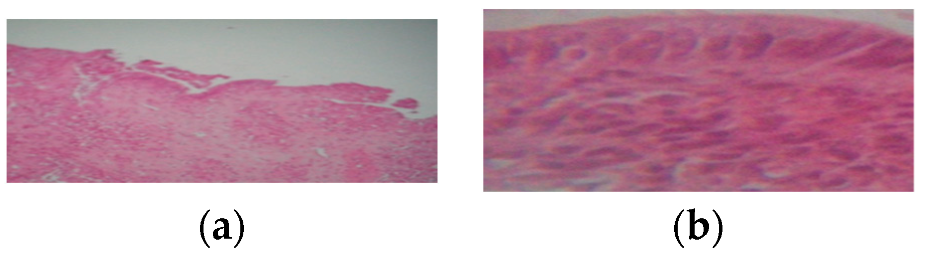
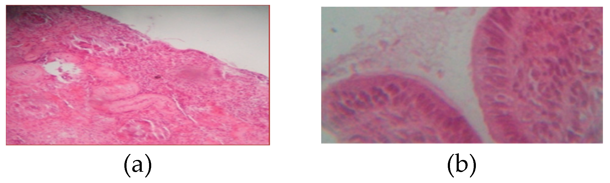
3.1. From 3rd Postpartum Day

Our results show that on the third day after parturition the epithelium of the uterine endometrium presents the following characteristics: Discontinuous, desquamated surface epithelium that is detached in places (Figure 1a). On Figure 1b we can observe a simple prismatic surface epithelium with cells with large nuclei.

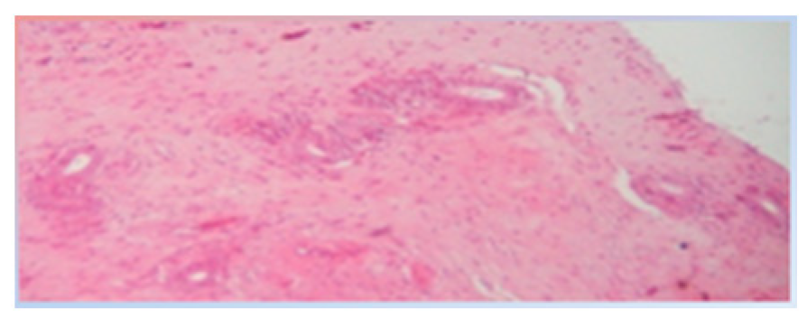
3.2. Between 5th and the 7th Day Postpartum

During the period between the fifth and the seventh day of the postpartum one observes a epithelium is more intact than that of the surface, and it is darker. The chorion is loose and characterized separately by less important vascularization. The number of uterine glands is a few or they are absent (Figure 2). They have small circular sizes and are scattered in the chorion.

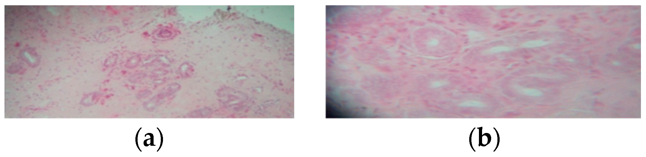
3.3. From 10th Postpartum Day

From the tenth day after parturition there is appearance of the uterine glands. The uterine glands are more numerous compared to the number of those at the previous stage. These glands are small in size, disseminated in the chorion, and their lumen can be expanded or reduced (Figure 3).

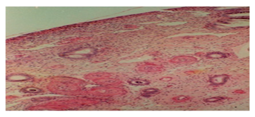
3.4. From 11th to 14th Postpartum Day

During the second week we observe: The surface epithelium is poorly regenerated and desquamated in a few places (Figure 4a). The uterine glands underwent an increase in number and size, they are circular or elongated in shape, they can be scattered in the lamina propria or organized in clusters, and they are surrounded by blood vessels (Figure 4b).

3.5. From 15th to 18th Postpartum Day

At the beginning of the second week the surface epithelium is not yet completely restored, and it is of the simple cylindrical type. Many uterine glands are disseminated in the chorion. Neovascularization is very important as is increases the number and size of glands (Figure 5).

3.6. From 19th to 21st Postpartum Day

At the end of the third week at magnification (10) (Figure 6a), the surface epithelium appears to be more intact, uniform, and continuous over its entire surface.
At the highest magnification (40) (Figure 6b), the epithelium appears to be simple and cylindrical. In light of the histological results of the endometrium, it appears that uterine involution in camels completes on the 21st postpartum day. These results agree with those in Ref. [7], which reports a duration of 40 days. We also used the method of transrectal palpation. Our results are in agreement with those declared by the authors of Refs. [4,8], who reported a duration from 15 to 28 days. The uterine lining is completely restored from the 18th postpartum day.
The process of uterine involution occurs through the association of endometrial regeneration phenomena and vascular phenomena.

4. Conclusions

It appears, in this brief and clear study that the restoration of the epithelium of the uterine endometrium completes at the end of the third week.

Author Contributions

Conceptualization, R.K., H.Z. and D.A.; methodology, B.M., A.S. and Y.R.; validation, N.D. and S.F.; writing—original draft preparation, R.K.; writing—review and editing, H.Z. and D.A.; project administration and interpretation of data, R.K.; the decision to publish the results, R.K., H.Z., A.S. and D.A. All authors have read and agreed to the published version of the manuscript.

Funding

This research received no external funding.

Institutional Review Board Statement

During this study, no invasive procedure was performed during the sampling.

Informed Consent Statement

Not applicable.

Data Availability Statement

Not applicable.

Acknowledgments

Our thanks go to the director of the experimental station at the university veterinary institute of Blida, the director of the high commission for the development of the steppe, and the director of the research center on agropastoralism for their help.

Conflicts of Interest

The authors declare no conflict of interest.

References

  1. Zarrouk, A.; Souilem, O.; Beckers, J. Actualités sur la reproduction chez la femelle dromadaire (Camelus dromedarius). Rev. Elev. Med. Vet. Pays Trop. 2003, 56, 95–102. (In French) [Google Scholar] [CrossRef] [Green Version]
  2. Skidmore, J.A. Reproductive physiology in female old world camelids. Anim. Reprod. Sci. 2011, 124, 148154. [Google Scholar] [CrossRef] [PubMed]
  3. Kelanemer, R.; Antoine-Moussiaux, N.; Moula, N.; Abu-Median, A.A.K.; Hanzen, C.H.; Kaidi, R. Effect of nutrition on reproductive performance during the peri-partum period of female camel (Camelus dromaderius) in Algeria. J. Anim. Vet. Adv. 2015, 14, 192–196. [Google Scholar]
  4. Vaughan, J.; Tibary, A. Reproduction in female South American camelids: A review and clinical observations. Small Rumin. Res. 2006, 61, 259–281. [Google Scholar] [CrossRef]
  5. Musa, B.; Makawi, S. Involution of the uterus and the first postpartum heat in the camel (Camelus dromedarius). In Proceedings of the Conference on Animal Production in Arid Zones, Damascus, Syria, 7–12 September 1985. [Google Scholar]
  6. Tibary, A.; Anouassi, A. Uterine infections in camelidae. Vet. Sci. Tomorrow 2001, 1, 1–12. [Google Scholar]
  7. El-Wishy, A. Functional morphology of the ovaries of the dromedary camel. In Proceedings of the First International Camel Conference, Dubai, United Arab Emirates, 2–6 February 1992. [Google Scholar]
  8. Derar, R.; Ali, A.; Al-Sobayil, F. The postpartum period in dromedary camels: Uterine involution, ovarian activity, hormonal changes, and response to GnRH treatment. Anim. Reprod. Sci. 2014, 151, 186–193. [Google Scholar] [CrossRef] [PubMed]
Figure 1. (a) Histological section of the endometrium on the third day postpartum (10). (b) Histological section of the endometrium on the third day postpartum (40).
Blsf 22 00021 g001
Figure 2. Histological section of the endometrium between the fifth and seventh day postpartum (10).
Blsf 22 00021 g002
Figure 3. Histological section of the endometrium on the tenth day postpartum (40).
Blsf 22 00021 g003
Figure 4. (a) Histological section of the endometrium between the eleventh and fourteenth day postpartum (10). (b) Histological section of the endometrium between the eleventh and fourteenth day postpartum (40).
Blsf 22 00021 g004
Figure 5. Histological section of the endometrium between the fifteenth and eighteenth day postpartum (40).
Blsf 22 00021 g005
Figure 6. (a) Histological section of the endometrium between the nineteenth and twenty-first days postpartum(10). (b) Histological section of the endometrium between the nineteenth and twenty-first days postpartum (40).
Blsf 22 00021 g006
Disclaimer/Publisher’s Note: The statements, opinions and data contained in all publications are solely those of the individual author(s) and contributor(s) and not of MDPI and/or the editor(s). MDPI and/or the editor(s) disclaim responsibility for any injury to people or property resulting from any ideas, methods, instructions or products referred to in the content.

