Review

Three-Dimensional Cell Culture Methods in Infectious Diseases and Vaccine Research

1
Department of Vaccine Technology, Hacettepe University Vaccine Institute, 06100 Ankara, Turkey
2
Department of Infectious Diseases and Clinical Microbiology, Faculty of Medicine, Hacettepe University, 06100 Ankara, Turkey
*
Author to whom correspondence should be addressed.
Future Pharmacol. 2023, 3(1), 48-60; https://doi.org/10.3390/futurepharmacol3010004
Submission received: 31 October 2022 / Revised: 23 December 2022 / Accepted: 4 January 2023 / Published: 6 January 2023
(This article belongs to the Special Issue Three-Dimensional In Vitro Cell Culture Models in Drug Discovery)

Abstract

:
Cells, the basic structures of all living organisms, reside in an extracellular matrix consisting of a complex three-dimensional architecture and interact with neighboring cells both mechanically and biochemically. Cell–cell and cell–extracellular matrix interactions form a three-dimensional network that maintains tissue specificity and homeostasis. Important biological processes in a cell cycle are regulated by principles organized by the microenvironment surrounding the cell. The conventional cell culture methods failed to mimic in vivo-like structural organization and are insufficient to examine features such as connectivity of cells, cellular morphology, viability, proliferation, differentiation, gene and protein expression, response to stimuli, and drug/vaccine metabolism. Three-dimensional cell culture studies are very important in terms of reducing the need for in vivo studies and creating an intermediate step. Three-dimensional cell culture methods have attracted attention in the literature in recent years, especially in examining the cellular distribution of organs in the presence of infectious diseases, elucidating the pathogenic mechanism of action of viruses, and examining virus–host interactions. This review highlights the use and importance of three-dimensional cell culture methods in the design and characterization of novel vaccine formulations and the pathogenesis of infectious diseases.

Graphical Abstract

1. Introduction

Cell-based assays provide a simple, rapid, and low-cost option to avoid large-scale and costly animal testing. Therefore, cell culture studies are an important step in the vaccine development process. Conventional cell culture studies are based on the development of a monolayer of adherent cells grown on flat, two-dimensional (2D) substrates such as polystyrene or glass. 2D cell culture studies play an important role in advancing viral biology, tissue morphogenesis, disease mechanisms, vaccine studies, large-scale protein production, tissue engineering, and regenerative medicine [1,2,3,4,5]. However, the need for new methods such as three-dimensional (3D) cell culture has increased, especially due to the inability to mimic in vivo conditions and provide adequate physiological compatibility.
In recent years, 3D cell culture studies have attracted great interest in vaccine development studies in terms of host-virus interaction, infection mechanisms, vaccine screening, and replication kinetics. Considering COVID-19, the 21st century pandemic, the virus–host interaction, and cellular entry of SARS-CoV-2 clarified by conventional cell culture studies [6,7]. Performing cell culture studies, which are important at every stage of vaccine development studies, in 3D models that can imitate the natural morphology of the virus and cells, provides many indisputable advantages.
Cell-based assays are the main tool used in vaccine development to evaluate the potential efficacy of a new antigen, adjuvant, delivery system, or vaccine formulation [8,9]. To obtain the most reliable results, the cell culture model used as the test platform should work similarly to in vivo models. There are several reasons for the differences between the 2D and 3D cell culture methods. One of the most important of these reasons is that the cell morphology changes according to the culture method. In 2D cell culture, cells are stretched in an unnatural state on a flat surface, while cells replicated in 3D on a biological or synthetic scaffold material maintain normal morphology. In addition, the expression level and membrane arrangement of cell surface receptors are quite different in 3D and 2D cell culture methods. This directly affects many parameters, especially virus–host interactions. Another important difference between these cell culture methods is that the gene expression levels of the cells vary according to the method used. Cells growing in 2D and monolayers are under stress, and therefore some expressed genes and proteins are altered as a result of this unnatural state [10,11,12]. These genes and proteins directly affect the efficacy and cellular response of the tested vaccine formulation. Moreover, 3D cell culture also has some advantages in vaccine development and virus–host interaction studies compared to the in vivo animal model. Although the results obtained with rodents are accepted, they cannot adequately mimic the virus-cell interaction and the pathophysiology of the disease due to the lack of human cells [13,14,15]. The purpose of this review is to provide an overview of the use of 3D cell culture techniques in vaccine development and pathogen-host interaction studies in infectious diseases.

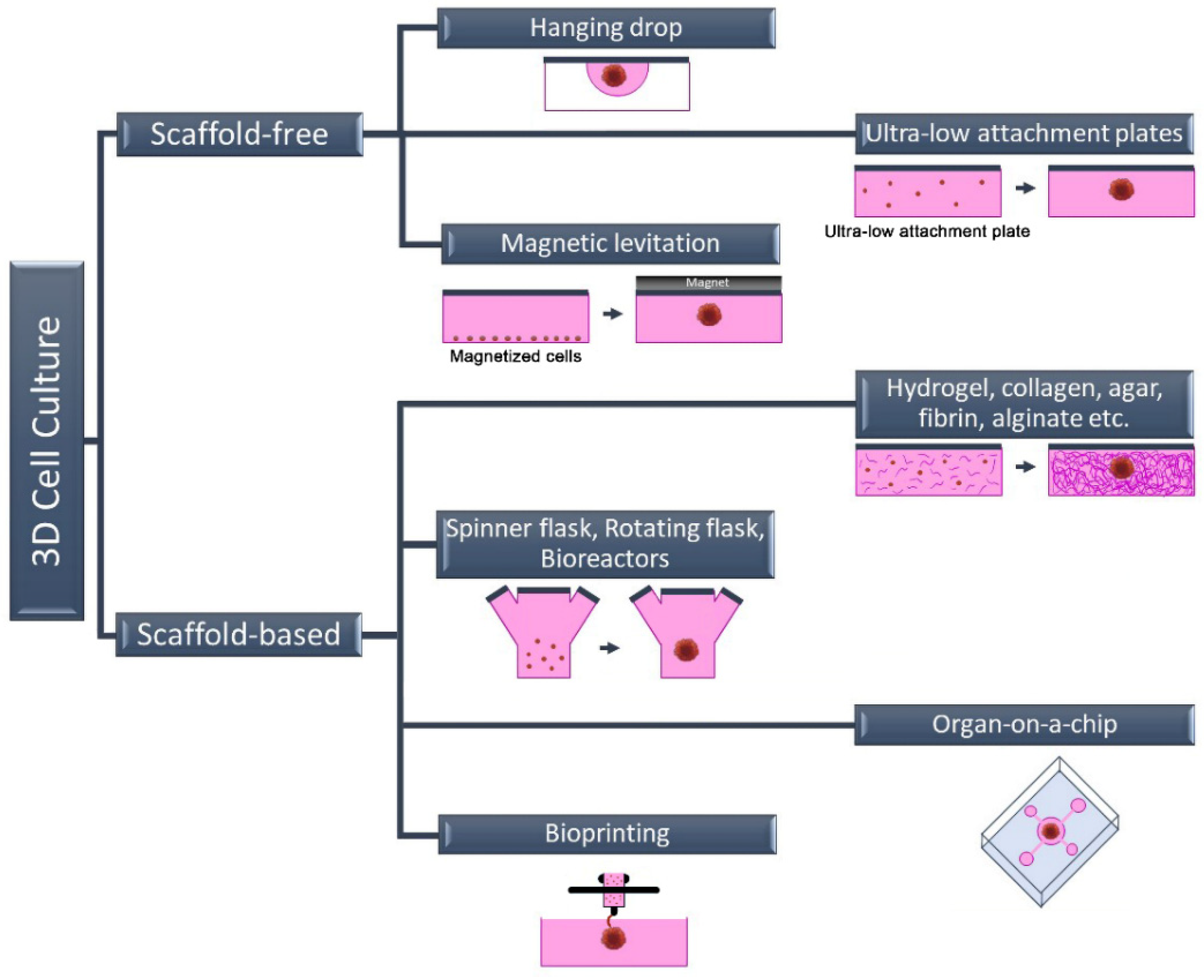
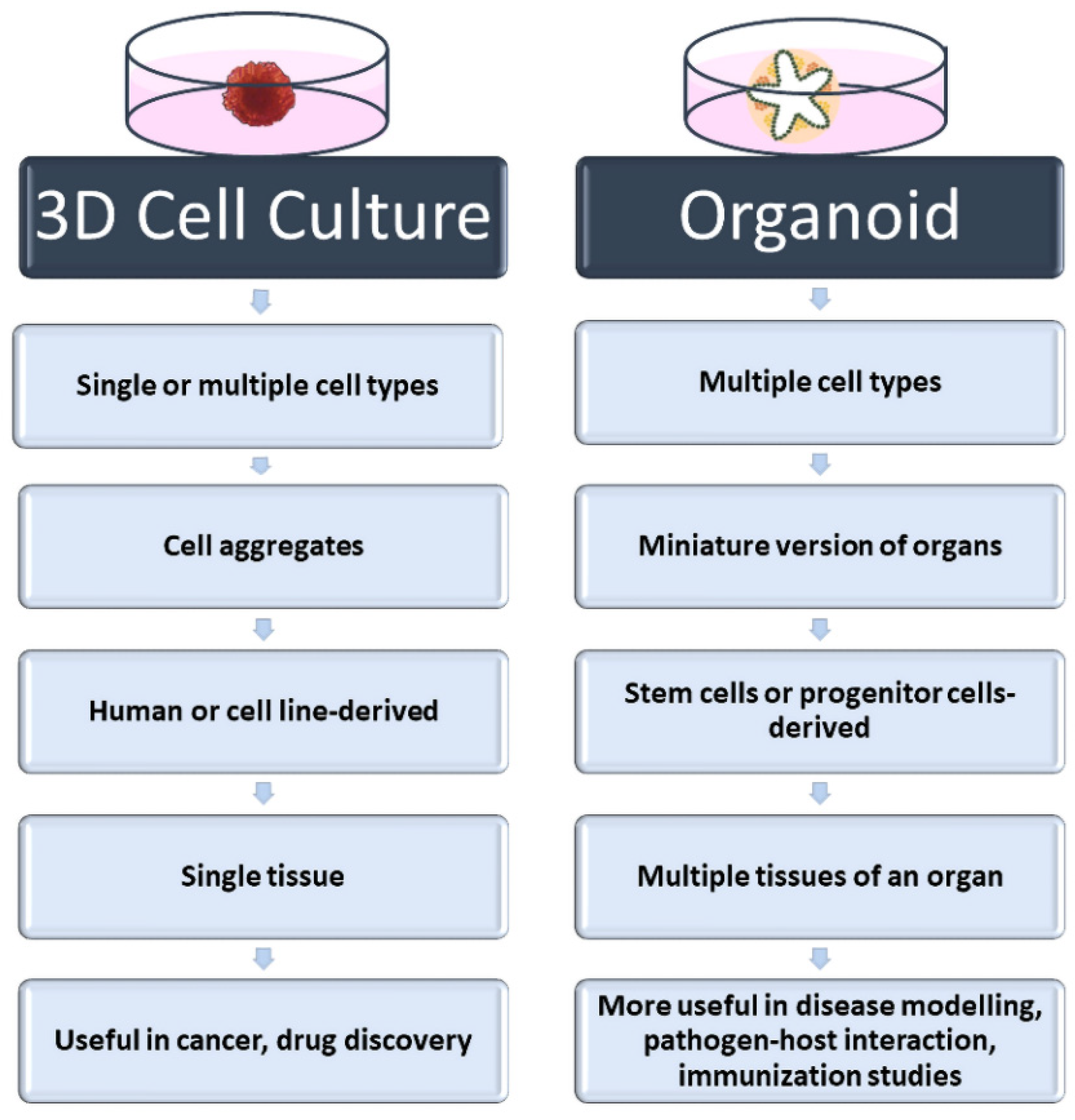
2. Three-Dimensional Cell Culture Methods

Cells, the building blocks of tissues and organs in the organism, reside in a complex 3D extracellular matrix (ECM) environment. This complex 3D architecture allows cells to interact both with each other and with the ECM. In this way, each cell acquires its own morphology and maintains the specificity and homeostasis of the tissue. Today, new methods have been developed that enable cells to grow in this complex 3D architecture in a laboratory environment. These methods are basically divided into two categories: scaffold-free methods and scaffold-based methods (Figure 1).

2.1. Scaffold-Free 3D Cell Culture Method

The most commonly used scaffold-free 3D cell culture techniques are cell suspension culture on non-adhesive or ultra-low attachment plates (liquid overlay), hanging drop, magnetic levitation, and microfluidic. Scaffold-free methods are generally fast and economical. This method is a “bottom-up” approach and is based on the fact that cells come together to form a spheroid structure. The formation of 3D spheroids depends entirely on the natural abilities of the cells because there is no material that can be used as a scaffold in these techniques. The hanging drop technique gravitationally collects cells in the form of suspended drops at a spherical air–liquid interface, thus facilitating the formation of a 3D cell structure without a scaffold [16]. Similar to the cell suspension method, the cell density of the suspension can be varied depending on the desired cell aggregate size. Spheroid formation, in contrast to these approaches, can be accomplished through external physical intervention in some scaffold-free techniques such as magnetic levitation or agitation bioreactor. The biggest shortcoming of scaffold-free 3D cell culture methods is the absence of an ECM component. Since there is no ECM in the environment, only cell–cell interaction occurs. Cell–ECM interaction is missing, unlike in their natural environment [17]. The wells of the plates can also be coated with various chemicals, such as poly (2-hydroxyethyl methacrylate), agar, agarose, or pol (N-p vinyl benzyl-D-lactone amide), to produce a non-attached surface. However, this incurs additional equipment and/or costs. These approaches might not be adequate because they lack the scaffold support necessary for the cell–matrix interaction needed for cell biology and proper functioning [18,19].

2.2. Scaffold-Based 3D Cell Culture Method

In scaffold-based 3D cell culture methods, ECM components are also added to the culture to provide extracellular components that mimic the biological environment. For this purpose, commercially available, ready-to-use scaffolds as well as suitable ECM components can be used. Unlike scaffold-free methods, the ECM is a complex and dynamic structure found between cells in these methods. In addition to providing structural support, ECM plays an active role in helping cells acquire tissue-specific properties. Although its content varies according to the characteristics of tissues and cells, ECM basically consists of two components: proteins (collagen, elastin, fibronectin, laminin, fibrillin, etc.) and proteoglycans (heparan sulfate, chondroitin sulfate, etc.) [20,21]. Each of these components varies according to the task undertaken by the cell, and they also change in different physiological and biochemical events such as proliferation, genetic changes, differentiation, attachment, and migration. Therefore, the ECM is defined as a dynamic structure. In scaffold-based 3D cell culture, natural polymers such as collagen, hydroxyapatite, agar, fibrin or alginate are used as scaffolds, as well as biodegradable synthetic polymers such as poly (ethylene glycol) and poly (lactide-co-glycolide) [20,21].
In addition to homologous 3D cell culture, heterologous 3D spheroids and organoids can be prepared by scaffold-based methods. Organoids called “Culturable Mini-Organs” are thought of as miniature versions of organs. On the other hand, spheroids can be prepared either homogeneously (from a single cell type) or heterogeneously (from different cell types) (Figure 2). Mostly, immortal cell lines are used for spheroids, while organoids are prepared from adult or embryonic stem cells. For this reason, organoids are complex structures that can better mimic organs [22,23]. On the contrary, spheroids are more economical and easy-to-prepare structures that can also be called “cell aggregates” or “organotypic culture” [24,25]. Although both approaches are used in vaccine and drug research and in in vitro disease modeling, spheroids are frequently preferred in tumor and drug development studies. Organoids are preferred in vaccine and pathogen interaction studies for assessing immune responses, especially in mimicking complex and multi-component organs such as the respiratory tract. Heterologous 3D models, in which more than one cell type is cultured together, also provide cell–cell interactions where growth factors and other biological factors can be exchanged [26,27]. The interactions of cells with each other and with the ECM are very important in terms of cell polarity. Cell polarity plays a direct role in the viral–host relationship as it affects the expression of the relevant receptor. For this reason, heterologous 3D cell culture studies attract attention as an effective in vitro study method in infection and virology studies.
Bioprinting, which is used to create a 3D cell culture model, is a very successful method of mimicking the complexity of biological structures and is a computer-based approach. Although it mostly has applications in tissue engineering studies, it also allows the production of 3D tissues by printing a solution consisting of one or more cell types and ECM, called bioink, on the desired surface layer by layer with a printer. In the bioprinting process, since the modeling and printing are carried out under computer control, the printing of the cells into the layers can be carried out in a very sensitive and controlled manner. Although it is a costly approach compared to other 3D cell culture models, it offers unique opportunities in artificial tissue production and artificial disease models. Another advantage of this method is that it allows the interaction of cells with each other and with the ECM, as well as the ability to diversify the printing pattern. The addition of bioactive components to the pattern, which can be changed by cells after printing, provides the cells with physiological conditions in the biological environment [28,29,30]. Furthermore, bioprinting offers the opportunity to make copies of tissues and even organs with different tissues in a biological environment. Since the first years of bioprinting studies, the production of artificial organs has been the most attractive field of biomedicine. However, nowadays, they are preferred not only in the production of artificial organs or tissues but also in the use of these organs in the investigation of the pathology of the disease and its relationship with the pathogen.
Bioreactors are another method used to prepare 3D cell cultures and organoid models. This method was originally designed to minimize the effect of gravity and allow cells to clump together in a liquid medium to form spheroids. Bioreactors can generally be classified into four groups: perfusion bioreactors, spinner flask bioreactors, rotating vessels, and mechanical force systems. In the spinner flask approach, one of the two most commonly used types, there is a constantly rotating impeller inside the bioreactor tank and it moves the liquid, allowing the cells to interact with each other. The second method was developed to eliminate the physical effect of the propeller on the cells. In this method, which is called a rotating vessel bioreactor, the tank containing the cells rotates. The most important advantage of bioreactors over other methods is that they provide the same physical conditions to all cells in a dynamic environment. With the continuous movement of the bioreactor tanks, nutrients can be delivered to the cells in many ways. The bioreactor approach is frequently used in studies of pathogen–host interactions. It allows the expression of cell connections and surface molecules that play a direct role in the entry of viruses into cells and thus infecting them. Bioreactors are also suitable for large-scale production when compared to other methods [31,32,33].
Organ-on-a-chip systems promise much more than current 3D cell culture studies, and they are seen as the future of these studies. Organ-on-a-chip technology, which has become more popular in the last 10 years, provides the desired artificial organ to mimic the biology of the disease [34,35]. Besides providing a 3D structure like other methods, it makes it possible to design a system with adjacent tissues. This approach allows for tissue and organ formation that more realistically mimics the biological structure in vitro. Especially thanks to the advances in nanotechnology, the diversity of applications has increased in the organ-on-chip approach. There are organ-on-a-chip models on the market that are designed separately for almost every organ and are commercially available. Moreover, there are organ-on-a-chip designs developed with sensors for imaging and biological/physiological changes designed in accordance with the experiments to be carried out. Virus–host interaction is one of the areas where organ-on-a-chip technology is used most frequently. Since this technology allows the formation of miniature tissues and organoids, studies on the examination of the interaction of pathogens with the host and the determination of the subsequent physiological changes with sensors are quite surprising and promising [36,37]. It is a very useful approach, especially in the elucidation of complex systems in which multiple biological factors play an active role, such as the development of resistance in infectious diseases and the evaluation of the immune response developing after vaccination.
As already mentioned, each of the different 3D cell culture methods has its own advantages and limitations. When all methods are compared, it can be said that organoids and organ-on-a-chip approaches are the best imitation techniques in terms of containing more than one tissue type and using stem cells. The lack of vascularization and blood vessels, interorgan communication, and immune system components prevent even organoids from fully mimicking in vivo conditions. As one of the most researched organoid models in the field of infectious illnesses, intestinal organoids include a variety of cell types from human tissues, enabling the investigation of heterocellular interactions. The major drawbacks are the absence of neural innervation, lumen content, and fluid flow, in addition to vascularization. One of the most popular techniques for the lung, another organ where pathogen–host interaction is frequently studied, is air–liquid interface (ALI) culture. The key advantage of ALI culture over other techniques is that it gives the cells an apical-basal configuration, enabling cell development towards a mucociliary phenotype. In this method, the apical surfaces of the cells are exposed to air, while the basal surfaces are in the liquid cell culture medium. Despite the fact that lung organoids better mimic the real functioning of the organ than the ALI method, they are still insufficient, particularly in terms of immune cell depletion and lack of vascularization. Furthermore, all 3D cell culture models developed for the lung are under ambient air pressure. Given the close correlation between respiratory rate and pressure in vivo, the models are insufficient to depict this relationship.

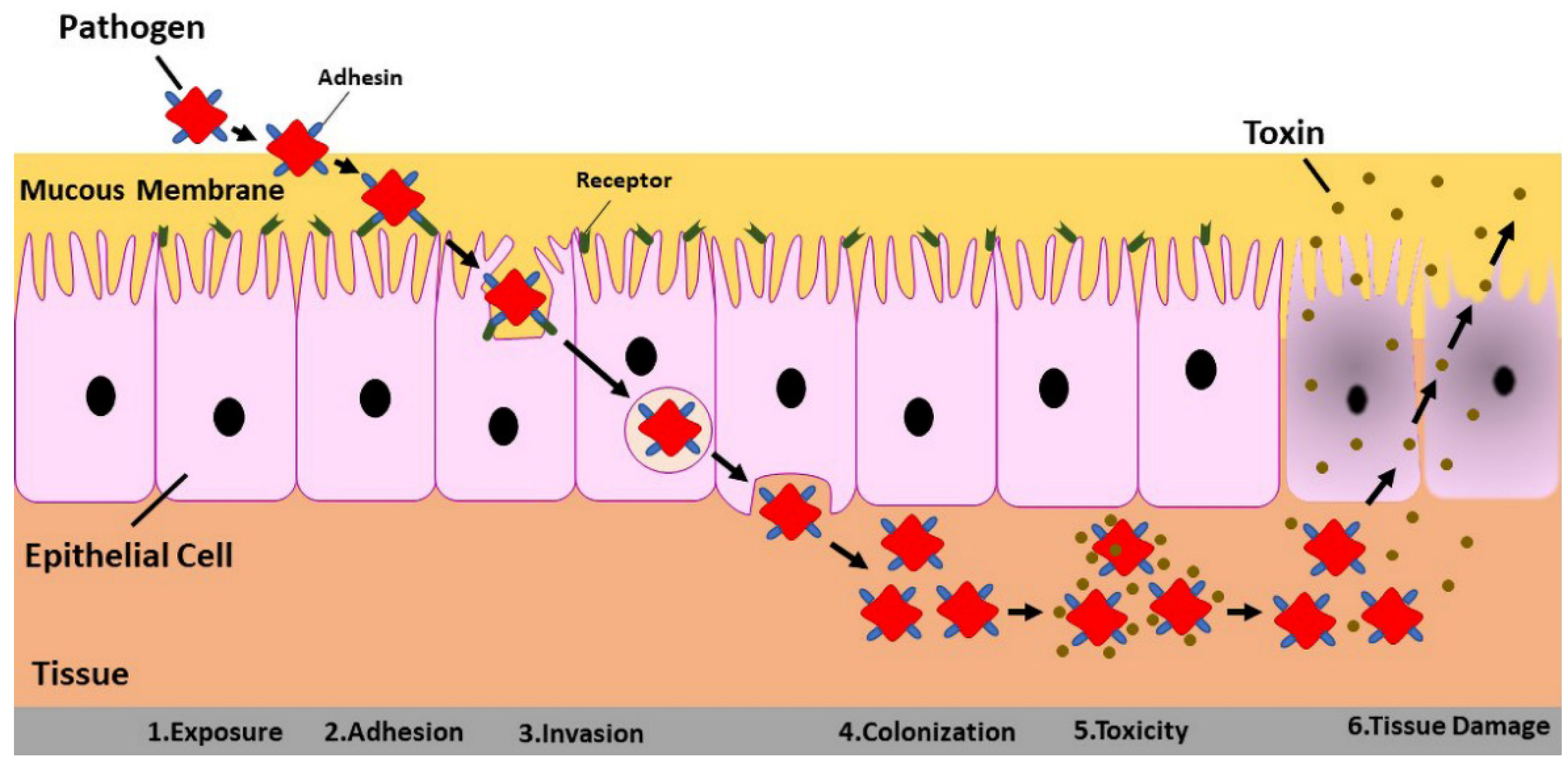
3. Three-Dimensional Cell Culture in Virus–Host Interaction

Cells can grow and multiply in an environment where their morphological development is supported and the biological factors necessary for their development are provided. A niche is a dynamic environment that varies with cell type. This dynamic and multi-component environment in cell culture is provided by the ECM. One of the most important features that distinguishes 3D cell culture from conventional cell culture is the availability of ECM. The biological structure can be better imitated by adding the components required by the cell to the medium according to the cell type. Mimicry of the organism in vitro also allows for more realistic investigation and understanding of complex and multifactorial processes such as pathogen–cell interaction. As seen in the recent SARS-CoV-2 pandemic, the first studies were to elucidate the structure and cellular interactions of the virus.
The spike proteins on the surface of the virus assume the primary role in the entry of the SARS-CoV-2 virus into the cell. Glycan-coated spike proteins play a role in the initiation of infection by penetrating the virus into the cell. The receptor-binding domain in the spike protein binds to a receptor called angiotensin-converting enzyme 2 (ACE2), which is expressed in type 2 alveolar epithelial cells in the human lungs [38]. The interaction of the pathogen with the cell and the understanding of its infecting pathways are modeled by molecular dynamics simulations. Vaccine studies for the unpredictable pandemic were initiated after elucidating the pathogen–host cell interaction. Therefore, it is very important to determine the pathogen–host cell interaction in vaccine and drug development, especially for infectious diseases. The most basic purpose of developing an effective vaccine is to mimic the pathogen and stimulate the immune response. Infection formation consists of many sequential events: pathogen exposure, adhesion, invasion, colonization, and cell damage [39] (Figure 3).
Developing an experimental design that can mimic the in vivo environment for each of these steps will yield more realistic results. Imitation of complex pathophysiological mechanisms in infectious diseases in vitro will be very beneficial in determining the stages of the disease and accelerating vaccine development. For this purpose, there are various 3D cell culture studies in the literature for different pathogens. In order to summarize the examples of different studies performed in this field in recent years, the literature samples are presented in Table 1.
In research on the pathogen–host interaction, a novel model is primarily required since microenvironmental elements cannot be provided in conventional cell culture. The dynamic microenvironment of tissues and organs contributes to the harmony of homeostasis. This microenvironment is superior in many ways, including immunology, due to its abundance of different types of cells with various activities. For example, secretions act as a barrier and provide defense against viruses. In vitro modeling of the microenvironment is unavoidable, particularly in organs like the lungs and intestines where mucosa and polarity directly influence pathogen interaction. The use of 3D cell culture and organoid models to study pathogen–host interactions has been received special attention, especially during the pandemic. Organoids were developed to examine the function, virulence, and pathogenicity of the SARS-CoV-2 virus in various organs such as the brain [47,48,49], intestinal [50,51,52], and respiratory tract [53,54,55]. Following SARS-CoV-2 infection, significant chemokine induction was shown in a lung organoid model established using human pluripotent stem cells, similar to that reported in COVID-19 patients. Additionally, hPSC-derived colonic organoids were developed for gastrointestinal symptoms typically seen in COVID-19 patients. It is interesting to note that ACE2 expression and its interactions with the virus were observed in the derived colonic organoids. In addition to pathogen interaction, inhibition of virus entry by drugs approved by the FDA for the treatment of SARS-CoV-2 was also determined [56]. A related study found that SARS-CoV-2 infection mimics IFN signaling in colonic epithelial cells, inducing ACE-2 expression. Based on the findings and research done in the human colon organoid model it was hypothesized that a therapeutic that is effective on the IFN-γ signaling pathway can limit or inhibit viral replication [50].
Studies with the Zika Virus (ZIKV) are among the first examples of the use of organoids in infectious diseases. Studies were carried out on the effect of the disease on the brain, viral tropism, and cell death on the brain organoids. Garceaz et al. used neural stem cells, neurospheres, and brain organoids to investigate the effects of ZIKV infection on neuronal differentiation and neural stem cell growth. Firstly, human-induced pluripotent stem cells were cultured in a suitable medium on a Matrigel-coated surface. The cells were then differentiated into neural stem cells using neurobasal medium and pluripotent stem cell neural induction medium before being exposed to ZIKV. After 24 h, viral RNA and the viral envelope protein were detected in neural stem cells. The effects of ZIKV on neural differentiation were investigated on spheroids prepared from neural stem cells. Spheroids were prepared by growing virus-infected or uninfected neural stem cells under 90 rpm rotation. It was reported that round neurospheres were obtained with uninfected neural stem cells, but the cells were separated while the spheroids were prepared from virus-infected cells. Furthermore, it was stated that the survival time of virus-infected spheroids decreased in cell culture conditions. The effect of ZIKV infection on neurogenesis was investigated in brain organoids derived from human-induced pluripotent stem cells. Differentiation into brain organoids was performed by the spin flask method under 40 rpm rotation. Organoids grown in neuronal differentiation media were subsequently embedded in Matrigel and grown for 35 days. When the growth rates of virus-infected or non-infected organoids were examined, it was observed that ZIKV infection reduced the growth rate of organoids by 40% [57]. Escherichia coli strain Nissle is used as a probiotic. It has also been reported that this strain protects mice from some enterohemorrhagic E. coli strains that cause infection in humans. Therefore, mouse models are not well-suited to studying E. coli infection. Pradhan et al. investigated the safety of the Nissle strain and the pathogenicity of enterohemorrhagic E. coli strains in human intestinal organoids. Human intestinal organoids were prepared by directed differentiation of the H1 line, one of the most commonly used human embryonic stem cell lines. Spheroids were obtained from a hospital pluripotent stem cell facility, embedded in Matrigel, and incubated in reconstituted intestinal medium. It was emphasized that human intestinal organoids were able to mimic the architecture and function of human small intestinal tissue thanks to their internal lumens and epithelial barriers surrounded by a single layer of differentiated epithelial cells (enterocytes, goblet cells, and enteroendocrine cells) surrounded by a diffuse mesenchymal layer. It was reported that the Nissle strain injected into the lumen of the human intestinal organoids can be recovered from the lumen after 3 days. Interestingly, it was observed that pathogenic strains (enterohemorrhagic E. coli and uropathogenic E. coli) were removed from the lumen by disrupting the epithelial barrier. In addition, it was determined that the effect of pathogenic strains decreased in organoids pre-incubated with the Nissle strain. As a result of the co-culture of the organoids with Nissle and pathogenic strains, it was observed that the pathogenic strains replicated while the Nissle strain did not replicate. Studies in stem cell-derived human intestinal organoids showed that the Nissle strain eliminates pathogenic strains through activation of host defenses [58].
Utilizing multiple pathogens simultaneously in a study is one of the biggest advantages of using 3D cell culture in pathogen–host interaction. This type of co-infection allows for natural and complex infection patterns to be mirrored, offering more realistic results. Human papillomavirus (HPV) and Chlamydia trachomatis virus and bacteria, respectively, are sexually transmitted infections and appear quite common. There is an increase in the coinfection of these two pathogens in patients with cervical and ovarian cancer, but the mechanism has not been clarified yet. Koster et al. developed organoids with ectocervical stem cells from cancer patients and healthy donors to investigate the mechanism of the increase in the incidence of coinfection. Human ectocervical organoids were prepared by embedding stem cells in Matrigel. The stem cells were isolated from biopsy samples obtained from standard surgical procedures. As a second approach, primary cells were cultured in a collagen-coated cell culture flask and then cultured in irradiated mouse fibroblasts (J2-3T3) for long-term culture prior to embedding in Matrigel. 3T3-J2 irradiated feeder cells are mitotically inactivated by radiation but retain metabolic activity. Organoids prepared from HPV-negative and E6E7-expressing HPV-positive stem cells were infected with C. trachomatis and used to illuminate the pathways of both single and coinfection. As a result of the study, they reported that the bacteria significantly inhibited the mismatch repair pathway at both the transcriptional and post-translational levels. It was also stated that this effect is more effective in HPV-induced organoids [59].

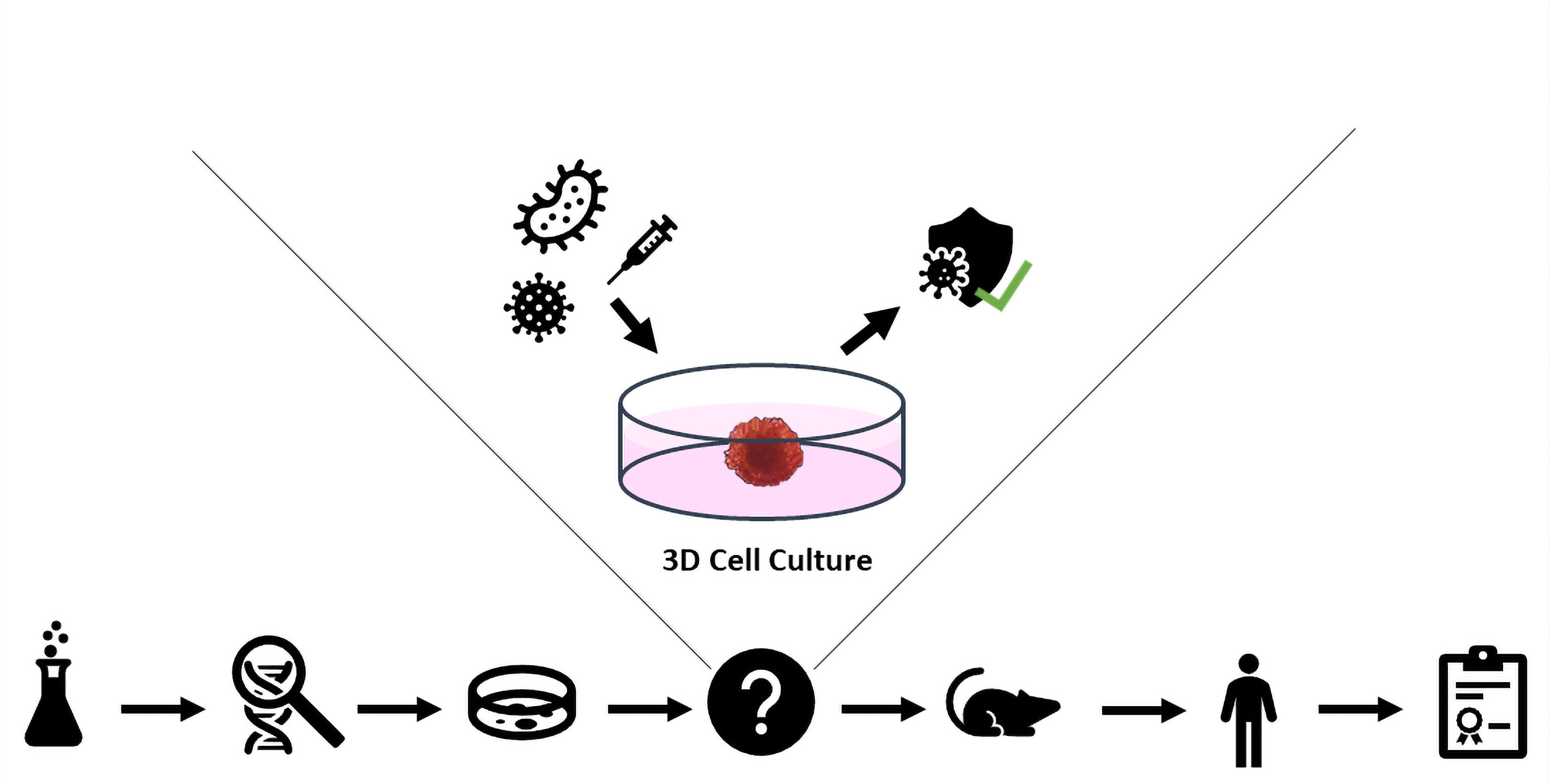
4. Three-Dimensional Cell Culture in Vaccine Development Studies: Future Direction

Animal models are still used in vaccine development studies as the only preclinical models demonstrating the vaccine’s efficacy. For a newly developed vaccine, choosing which animal species to utilize is crucial. The need for a novel preclinical approach is heightened by the inadequacy of the animals employed to mimic humans’ immune response systems and the ethical responsibility to reduce the use of animal experiments. The effectiveness of 3D cell culture studies is encouraging for the application of these techniques in vaccine research. 3D cell culture methods can be an effective tool for modeling the immune system in vitro. However, there has not yet been a 3D cell culture technique developed for vaccine research. The recent SARS-CoV-2 pandemic, which claimed many lives, demonstrated that next-generation technologies and delivery systems can be used in vaccine formulations. The addition of nanoparticulate delivery systems or new adjuvants to vaccine formulations is inevitable, but new in vitro methods involving immune system components are required for preclinical testing of these.
For centuries, vaccination has been the most trusted and secure way to protect people from infectious diseases. Thanks to the vaccine developed in 1796, smallpox was defeated and eradicated by the World Health Organization in 1980. An extensive and organized vaccination program was implemented globally as part of the 1967-launched struggle to eradicate smallpox [60,61]. Additionally, diphtheria, polio, and measles may all be prevented by immunization today. The first experiments in the fight against SARS-CoV-2, which was classified as a pandemic in 2019 and is currently active, were focused on creating vaccines and therapeutics. Safety is the essential feature of a prophylactic vaccine designed against an infectious disease. Therefore, a vaccine candidate must comply with regulatory health agency standards for safety, toxicity, and immunogenic response. Various techniques are used to evaluate the efficacy and toxicity of the vaccine, and clinical trials are initiated based on the preclinical results obtained. Unfortunately, the results of three-stage clinical trials often do not correspond to preclinical trial results obtained in vitro. According to reports, only 16.2% of vaccine development studies conducted in the United States between 2006 and 2015 received FDA approval [62]. The primary reason for this problem in vaccine development studies is the inadequate biological mimicking of the biological system and organism by the preclinical tests currently available and in use for the evaluation of immune response and toxicity. The development of vaccines is a challenging and drawn-out process that necessitates funding, effort, and time before it reaches the clinical stage. The need for novel preclinical studies is heightened by the fact that the majority of vaccine candidates generated through this drawn-out and expensive procedure failed in clinical trials [63,64,65].
Nowadays, the immunogenicity of developed vaccines is determined by in vivo studies. Although it varies according to the disease, studies using mostly mice may be insufficient to determine the true immunogenic properties of the vaccine. The different quantities and distribution of viral ligands in human cells are the most fundamental aspects of this. The viral infection cycle is directly impacted by this distinction. Additionally, because different species’ immune systems interact differently, mimicking human pathophysiology is insufficient. In addition to all of these, ethical issues exist [64,65].
Cell culture studies are one of the most crucial and fundamental phases in vaccine development as well as drug research. The cell culture method serves as the foundation for investigations prior to vaccine formulation development, as well as for analyzing the vaccine’s effectiveness and toxicity. The vaccine must resemble the pathogen causing the disease at the highest rate possible in order to generate a lengthy and effective immune response, which is the most crucial attribute required from the vaccine candidate. Hundreds of vaccine and therapeutic research investigations were launched shortly after the virus’s shape, pathogenicity, and genetic sequence were found in the SARS-CoV-2 pandemic. A pathogen’s structural and genetic properties, its interaction with the target organ and cells, and the course and pathogenicity of the disease it produces are all necessary for a successful immune response with a vaccine. However, current in vitro cell culture assays are based on conventional methods and investigate vaccination toxicity in monolayer cells planted on plastic plates. When it comes to measuring the immune response, conventional single-cell culture is inadequate. Vaccine immunogenicity is an extremely complicated process involving several cytokines, immunological components, and cells. Only by mimicking biological components in vitro can this process be measured in preclinical research. In recent years, 3D cell culture technologies have been developed to address these shortcomings, particularly in pathogen–host interactions. The in vitro imitation of the target organ allows for the study of the pathogen–cell interaction as well as the prediction of undesired side effects and/or insufficient immune responses in vaccines, thereby saving time and money and avoiding unnecessary in vivo research.
In vitro studies involving components of the immune system must be part of the preclinical studies used to assess the immunogenicity and potency of vaccine candidates. In addition to the gastrointestinal, respiratory, and genital-urinary systems, the mucous membrane covers many organs and cavities in the body, such as the conjunctiva and inner ear. The mucosa is frequently exposed to pathogens. To combat pathogens, a specialized immune system has evolved along mucosal surfaces. This local immune system, called the mucosal immune system, includes lymphoid tissue clusters (mucosal-associated lymphoid tissue) just like the lymph node and spleen [66,67]. The mucosa is home to roughly 75% of the total lymphocytes. Additionally, because of the microflora they create, non-pathogenic bacteria that coexist with their hosts offer defense against diseases. In addition to these, IgA is the most fundamental component of mucosal immunity. In the light of this knowledge, preclinical in vitro studies investigating the efficacy of the vaccine should be tested in a method that also includes immune system components such as macrophages, T cells, dendritic cells, and neutrophils [68]. Only a system with immune system cells will allow for meaningful investigation of infection development and vaccine immunogenicity in a cell culture medium. In this context, alternative techniques are utilized to include immune system cells in cell culture studies. The most common technique involves isolating peripheral blood mononuclear cells from healthy human blood. Cells can only survive for a few passages, making it impossible to use them in in vitro cell culture for an extended period. In the second method, commercially available cell lines like THP-1 (human monocytic cell line derived from an acute monocytic leukemia patient), are preferred. However, the presence of immune system cells in the study significantly limits repeatability and reproducibility, in addition to the already present constraints of conventional cell culture.
Although novel in vitro models are required for vaccine development studies, there have been very few studies in this field. Studies utilizing organoids and 3D cell culture are mostly focused on pathogen–host interactions and anticancer research. In a recent study, human tonsil organoids were used to assess the vaccines’ immunogenicity. Wagar et al. performed a surgical harvest of healthy tonsils from patients, which were then roughly mechanically divided into cells and maintained in optimal conditions. Transwell membranes were used to develop organoids, and complete media containing recombinant human B cell activating factor were used to promote the growth of organoids underneath the membranes. Tonsil organoids were used to evaluate the immune response to the live attenuated influenza vaccination, the measles, mumps, and rubella vaccine, the rabies vaccine, and the Ad5-vectored SARS-CoV-2 vaccine candidate. Tonsil organoids were used in this study to assess the effectiveness of several adjuvants, including alum and imiquimod. It was reported that follicular dendritic cells and follicular helper T cells interact when the tonsil organoids are stimulated by the influenza vaccine, aiding in the development of memory B cells. In particular, 14 days after the organoids received the vaccine treatment, it was revealed that a considerable increase in CD8+ T cell activation was seen compared to the control group. Increased levels of specific IgG and IgA also suggest that tonsil organoids may be a useful tool for assessing the immunogenicity of vaccines and predicting personalized responses [69].

5. Conclusions

Thanks to the many features and innovations they offer, three-dimensional cell culture studies continue to advance the field. The use of 3D cell culture techniques shows promise for assessing vaccines’ ability to elicit an immune response as well as for filling the preclinical research gap in the study of pathogen traits like virulence and intracellular entry. The long-term culture of immune cells and the availability of various ECM and cell culture media for various tissues on a single platform are two significant gaps that must be filled in addition to the successful work. The development of tissue-specific growth organs, spheroid and organoid approaches, commercially available kits for specific organs, and biological factors are all promising advancements in this field. Complex immunocompetent 3D in vitro models will enable more accurate results prior to in vivo studies and will serve as better models for vaccine screening and development studies. Day after day, new techniques are helping to close the gap between traditional cell culture and in vivo studies.

Author Contributions

Conceptualization, G.V. and S.U.; writing and original draft preparation, G.V.; review and editing, S.U. All authors have read and agreed to the published version of the manuscript.

Funding

This research received no external funding.

Institutional Review Board Statement

Not applicable.

Informed Consent Statement

Not applicable.

Conflicts of Interest

The authors declare no conflict of interest.

References

  1. Dolskiy, A.A.; Grishchenko, I.V.; Yudkin, D.V. Cell Cultures for Virology: Usability, Advantages, and Prospects. Int. J. Mol. Sci. 2020, 21, 7978. [Google Scholar] [CrossRef] [PubMed]
  2. Han, F.; Wang, J.; Ding, L.; Hu, Y.; Li, W.; Yuan, Z.; Guo, Q.; Zhu, C.; Yu, L.; Wang, H.; et al. Tissue Engineering and Regenerative Medicine: Achievements, Future, and Sustainability in Asia. Front. Bioeng. Biotechnol. 2020, 8, 83. [Google Scholar] [CrossRef] [Green Version]
  3. Hudu, S.A.; Alshrari, A.S.; Syahida, A.; Sekawi, Z. Cell Culture, Technology: Enhancing the Culture of Diagnosing Human Diseases. J. Clin. Diagn. Res. 2016, 10, DE01–DE05. [Google Scholar] [CrossRef] [PubMed]
  4. Verma, A.; Verma, M.; Singh, A. Animal tissue culture principles and applications. In Animal Biotechnology; Academic Press: Cambridge, MS, USA, 2020; pp. 269–293. [Google Scholar]
  5. O’Flaherty, R.; Bergin, A.; Flampouri, E.; Mota, L.M.; Obaidi, I.; Quigley, A.; Xie, Y.; Butler, M. Mammalian cell culture for production of recombinant proteins: A review of the critical steps in their biomanufacturing. Biotechnol. Adv. 2020, 43, 107552. [Google Scholar] [CrossRef]
  6. Ju, X.; Zhu, Y.; Wang, Y.; Li, J.; Zhang, J.; Gong, M.; Ren, W.; Li, S.; Zhong, J.; Zhang, L.; et al. A novel cell culture system modeling the SARS-CoV-2 life cycle. PLoS Pathog. 2021, 17, e1009439. [Google Scholar]
  7. Shang, J.; Wan, Y.; Luo, C.; Ye, G.; Geng, Q.; Auerbach, A.; Li, F. Cell entry mechanisms of SARS-CoV-2. Proc. Natl. Acad. Sci. USA 2020, 117, 11727–11734. [Google Scholar] [CrossRef]
  8. Graham, B.S. Advances in antiviral vaccine development. Immunol. Rev. 2013, 255, 230–242. [Google Scholar] [CrossRef]
  9. Josefsberg, J.O.; Buckland, B. Vaccine process technology. Biotechnol. Bioeng. 2012, 109, 1443–1460. [Google Scholar]
  10. Cacciamali, A.; Villa, R.; Dotti, S. 3D Cell Cultures: Evolution of an Ancient Tool for New Applications. Front. Physiol. 2022, 13, 836480. [Google Scholar] [CrossRef]
  11. Jensen, C.; Teng, Y. Is It Time to Start Transitioning From 2D to 3D Cell Culture? Front. Mol. Biosci. 2020, 7, 33. [Google Scholar] [CrossRef] [PubMed] [Green Version]
  12. Kapałczyńska, M.; Kolenda, T.; Przybyła, W.; Zajączkowska, M.; Teresiak, A.; Filas, V.; Ibbs, M.; Bliźniak, R.; Łuczewski, Ł.; Lamperska, K. 2D and 3D cell cultures - a comparison of different types of cancer cell cultures. Arch. Med. Sci. 2018, 14, 910–919. [Google Scholar] [CrossRef] [PubMed]
  13. Barrila, J.; Crabbé, A.; Yang, J.; Franco, K.; Nydam, S.D.; Forsyth, R.J.; Davis, R.R.; Gangaraju, S.; Ott, C.M.; Coyne, C.B.; et al. Modeling Host-Pathogen Interactions in the Context of the Microenvironment: Three-Dimensional Cell Culture Comes of Age. Infect. Immun. 2018, 86, e00282-18. [Google Scholar] [CrossRef] [PubMed]
  14. Häfner, S.J. Level up for culture models - How 3D cell culture models benefit SARS-CoV-2 research. Biomed. J. 2021, 44, 1–6. [Google Scholar] [CrossRef]
  15. Harb, A.; Fakhreddine, M.; Zaraket, H.; Saleh, F.A. Three-Dimensional Cell Culture Models to Study Respiratory Virus Infections Including COVID-19. Biomimetics 2021, 7, 3. [Google Scholar] [CrossRef]
  16. Hsiao, A.Y.; Tung, Y.C.; Qu, X.; Patel, L.R.; Pienta, K.J.; Takayama, S. 384 hanging drop arrays give excellent Z-factors and allow versatile formation of co-culture spheroids. Biotechnol. Bioeng. 2012, 109, 1293–1304. [Google Scholar] [CrossRef] [Green Version]
  17. Alghuwainem, A.; Alshareeda, A.T.; Alsowayan, B. Scaffold-Free 3-D Cell Sheet Technique Bridges the Gap between 2-D Cell Culture and Animal Models. Int. J. Mol. Sci. 2019, 20, 4926. [Google Scholar] [CrossRef] [Green Version]
  18. Foty, R. A simple hanging drop cell culture protocol for generation of 3D spheroids. J. Vis. Exp. 2011, 51, e2720. [Google Scholar] [CrossRef]
  19. Shri, M.; Agrawal, H.; Rani, P.; Singh, D.; Onteru, S.K. Hanging Drop, A Best Three-Dimensional (3D) Culture Method for Primary Buffalo and Sheep Hepatocytes. Sci. Rep. 2017, 7, 1203. [Google Scholar] [CrossRef] [Green Version]
  20. Hu, M.; Ling, Z.; Ren, X. Extracellular matrix dynamics: Tracking in biological systems and their implications. J. Biol. Eng. 2022, 16, 13. [Google Scholar] [CrossRef]
  21. Nicolas, J.; Magli, S.; Rabbachin, L.; Sampaolesi, S.; Nicotra, F.; Russo, L. 3D Extracellular Matrix Mimics: Fundamental Concepts and Role of Materials Chemistry to Influence Stem Cell Fate. Biomacromolecules 2020, 21, 1968–1994. [Google Scholar] [CrossRef] [PubMed]
  22. Koike, H.; Takebe, T. Generating Mini-Organs in Culture. Curr. Pathobiol. Rep. 2016, 4, 59–68. [Google Scholar] [CrossRef]
  23. Dutta, D.; Clevers, H. Organoid culture systems to study host-pathogen interactions. Curr. Opin. Immunol. 2017, 48, 15–22. [Google Scholar] [CrossRef] [PubMed]
  24. Gunti, S.; Hoke, A.T.K.; Vu, K.P.; London, N.R., Jr. Organoid and Spheroid Tumor Models: Techniques and Applications. Cancers 2021, 13, 874. [Google Scholar] [CrossRef] [PubMed]
  25. Zanoni, M.; Cortesi, M.; Zamagni, A.; Arienti, C.; Pignatta, S.; Tesei, A. Modeling neoplastic disease with spheroids and organoids. J. Hematol. Oncol. 2020, 13, 97. [Google Scholar] [CrossRef] [PubMed]
  26. Altmann, B.; Grün, C.; Nies, C.; Gottwald, E. Advanced 3D Cell Culture Techniques in Micro-Bioreactors, Part II: Systems and Applications. Processes 2021, 9, 21. [Google Scholar] [CrossRef]
  27. Sangeeta, B.; Ankita Jaywant, D.; Shafina, S.; Jyotirmoi, A.; Soumya, B. Two-Dimensional and Three-Dimensional Cell Culture and Their Applications, in Cell Culture; Zhan, X., Ed.; IntechOpen: Rijeka, Croatia, 2021. [Google Scholar]
  28. Dey, M.; Ozbolat, I.T. 3D bioprinting of cells, tissues and organs. Sci. Rep. 2020, 10, 14023. [Google Scholar] [CrossRef] [PubMed]
  29. Kabir, A.; Datta, P.; Oh, J.; Williams, A.; Ozbolat, V.; Unutmaz, D.; T Ozbolat, I. 3D Bioprinting for fabrication of tissue models of COVID-19 infection. Essays Biochem. 2021, 65, 503–518. [Google Scholar]
  30. Koban, R.; Lam, T.; Schwarz, F.; Kloke, L.; Bürge, S.; Ellerbrok, H.; Neumann, M. Simplified Bioprinting-Based 3D Cell Culture Infection Models for Virus Detection. Viruses 2020, 12, 1298. [Google Scholar] [CrossRef]
  31. Grün, C.; Altmann, B.; Gottwald, E. Advanced 3D Cell Culture Techniques in Micro-Bioreactors, Part I: A Systematic Analysis of the Literature Published between 2000 and 2020. Processes 2020, 8, 1656. [Google Scholar] [CrossRef]
  32. Kizilova, N.; Rokicki, J. 3D Bioreactors for Cell Culture: Fluid Dynamics Aspects. In Biomechanics in Medicine, Sport and Biology; Springer International Publishing: Cham, Switzerland, 2022. [Google Scholar]
  33. Yi, T.; Huang, S.; Liu, G.; Li, T.; Kang, Y.; Luo, Y.; Wu, J. Bioreactor Synergy with 3D Scaffolds: New Era for Stem Cells Culture. ACS Appl. Bio. Mater. 2018, 1, 193–209. [Google Scholar] [CrossRef] [PubMed]
  34. Ingber, D.E. Human organs-on-chips for disease modelling, drug development and personalized medicine. Nat. Rev. Genet. 2022, 23, 467–491. [Google Scholar] [CrossRef] [PubMed]
  35. Sun, W.; Luo, Z.; Lee, J.; Kim, H.-J.; Lee, K.; Tebon, P.; Feng, Y.; Dokmeci, M.R.; Sengupta, S.; Khademhosseini, A. Organ-on-a-Chip for Cancer and Immune Organs Modeling. Adv. Healthc. Mater. 2019, 8, 1801363. [Google Scholar] [CrossRef] [PubMed]
  36. Tang, H.; Abouleila, Y.; Si, L.; Ortega-Prieto, A.M.; Mummery, C.L.; Ingber, D.E.; Mashaghi, A. Human Organs-on-Chips for Virology. Trends Microbiol. 2020, 28, 934–946. [Google Scholar] [CrossRef]
  37. Wang, Y.; Wang, P.; Qin, J. Human Organoids and Organs-on-Chips for Addressing COVID-19 Challenges. Adv. Sci. 2022, 9, 2105187. [Google Scholar] [CrossRef]
  38. Li, G.; He, X.; Zhang, L.; Ran, Q.; Wang, J.; Xiong, A.; Wu, D.; Chen, F.; Sun, J.; Chang, C. Assessing ACE2 expression patterns in lung tissues in the pathogenesis of COVID-19. J. Autoimmun. 2020, 112, 102463. [Google Scholar]
  39. Galán, J.E. The cell biology of microbial infections: Coming of age. J. Cell Biol. 2002, 158, 387–388. [Google Scholar] [CrossRef] [Green Version]
  40. Geiser, J.; Boivin, G.; Huang, S.; Constant, S.; Kaiser, L.; Tapparel, C.; Essaidi-Laziosi, M. RSV and HMPV Infections in 3D Tissue Cultures: Mechanisms Involved in Virus–host and Virus-Virus Interactions. Viruses 2021, 13, 139. [Google Scholar] [CrossRef]
  41. Heo, I.; Dutta, D.; Schaefer, D.A.; Iakobachvili, N.; Artegiani, B.; Sachs, N.; Boonekamp, K.E.; Bowden, G.; Hendrickx, A.P.A.; Willems, R.J.L.; et al. Modelling Cryptosporidium infection in human small intestinal and lung organoids. Nat. Microbiol. 2018, 3, 814–823. [Google Scholar] [CrossRef]
  42. Berg, J.; Hiller, T.; Kissner, M.S.; Qazi, T.H.; Duda, G.N.; Hocke, A.C.; Hippenstiel, S.; Elomaa, L.; Weinhart, M.; Fahrenson, C.; et al. Optimization of cell-laden bioinks for 3D bioprinting and efficient infection with influenza A virus. Sci. Rep. 2018, 8, 13877. [Google Scholar] [CrossRef] [Green Version]
  43. Berg, J.; Weber, Z.; Fechler-Bitteti, M.; Hocke, A.C.; Hippenstiel, S.; Elomaa, L.; Weinhart, M.; Kurreck, J. Bioprinted Multi-Cell Type Lung Model for the Study of Viral Inhibitors. Viruses 2021, 13, 1590. [Google Scholar] [CrossRef] [PubMed]
  44. Nie, Y.Z.; Zheng, Y.W.; Miyakawa, K.; Murata, S.; Zhang, R.R.; Sekine, K.; Ueno, Y.; Takebe, T.; Wakita, T.; Ryo, A.; et al. Recapitulation of hepatitis B virus–host interactions in liver organoids from human induced pluripotent stem cells. EBioMedicine 2018, 35, 114–123. [Google Scholar] [CrossRef] [PubMed] [Green Version]
  45. Long, R.K.M.; Moriarty, K.P.; Cardoen, B.; Gao, G.; Vogl, A.W.; Jean, F.; Hamarneh, G.; Nabi, I.R. Super resolution microscopy and deep learning identify Zika virus reorganization of the endoplasmic reticulum. Sci. Rep. 2020, 10, 20937. [Google Scholar] [CrossRef] [PubMed]
  46. Eriksen, A.Z.; Møller, R.; Makovoz, B.; Uhl, S.A.; tenOever, B.R.; Blenkinsop, T.A. SARS-CoV-2 infects human adult donor eyes and hESC-derived ocular epithelium. Cell Stem Cell 2021, 28, 1205–1220. [Google Scholar] [CrossRef]
  47. Jacob, F.; Pather, S.R.; Huang, W.K.; Zhang, F.; Wong, S.Z.H.; Zhou, H.; Cubitt, B.; Fan, W.; Chen, C.Z.; Xu, M.; et al. Human Pluripotent Stem Cell-Derived Neural Cells and Brain Organoids Reveal SARS-CoV-2 Neurotropism Predominates in Choroid Plexus Epithelium. Cell Stem Cell 2020, 27, 937–950. [Google Scholar] [CrossRef]
  48. Ramani, A.; Müller, L.; Ostermann, P.N.; Gabriel, E.; Abida-Islam, P.; Müller-Schiffmann, A.; Mariappan, A.; Goureau, O.; Gruell, H.; Walker, A.; et al. SARS-CoV-2 targets neurons of 3D human brain organoids. EMBO J. 2020, 39, e106230. [Google Scholar] [CrossRef]
  49. Zhang, B.Z.; Chu, H.; Han, S.; Shuai, H.; Deng, J.; Hu, Y.F.; Gong, H.R.; Lee, A.C.; Zou, Z.; Yau, T.; et al. SARS-CoV-2 infects human neural progenitor cells and brain organoids. Cell Res. 2020, 30, 928–931. [Google Scholar] [CrossRef]
  50. Heuberger, J.; Trimpert, J.; Vladimirova, D.; Goosmann, C.; Lin, M.; Schmuck, R.; Mollenkopf, H.J.; Brinkmann, V.; Tacke, F.; Osterrieder, N.; et al. Epithelial response to IFN-γ promotes SARS-CoV-2 infection. EMBO Mol. Med. 2021, 13, e13191. [Google Scholar] [CrossRef]
  51. Jang, K.K.; Kaczmarek, M.E.; Dallari, S.; Chen, Y.H.; Tada, T.; Axelrad, J.; Landau, N.R.; Stapleford, K.A.; Cadwell, K. Variable susceptibility of intestinal organoid-derived monolayers to SARS-CoV-2 infection. PLoS Biol. 2022, 20, e3001592. [Google Scholar] [CrossRef]
  52. Zhou, J.; Li, C.; Liu, X.; Chiu, M.C.; Zhao, X.; Wang, D.; Wei, Y.; Lee, A.; Zhang, A.J.; Chu, H.; et al. Infection of bat and human intestinal organoids by SARS-CoV-2. Nat. Med. 2020, 26, 1077–1083. [Google Scholar] [CrossRef]
  53. Lamers, M.M.; van der Vaart, J.; Knoops, K.; Riesebosch, S.; Breugem, T.I.; Mykytyn, A.Z.; Beumer, J.; Schipper, D.; Bezstarosti, K.; Koopman, C.D.; et al. An organoid-derived bronchioalveolar model for SARS-CoV-2 infection of human alveolar type II-like cells. EMBO J. 2021, 40, e105912. [Google Scholar] [CrossRef] [PubMed]
  54. Milewska, A.; Kula-Pacurar, A.; Wadas, J.; Suder, A.; Szczepanski, A.; Dabrowska, A.; Owczarek, K.; Marcello, A.; Ochman, M.; Stacel, T.; et al. Replication of Severe Acute Respiratory Syndrome Coronavirus 2 in Human Respiratory Epithelium. J. Virol. 2020, 94, e00957-20. [Google Scholar] [CrossRef] [PubMed]
  55. Tran, B.M.; Grimley, S.L.; McAuley, J.L.; Hachani, A.; Earnest, L.; Wong, S.L.; Caly, L.; Druce, J.; Purcell, D.F.J.; Jackson, D.C.; et al. Air-Liquid-Interface Differentiated Human Nose Epithelium: A Robust Primary Tissue Culture Model of SARS-CoV-2 Infection. Int. J. Mol. Sci. 2022, 23, 835. [Google Scholar] [CrossRef]
  56. Han, Y.; Duan, X.; Yang, L.; Nilsson-Payant, B.E.; Wang, P.; Duan, F.; Tang, X.; Yaron, T.M.; Zhang, T.; Uhl, S.; et al. Identification of SARS-CoV-2 inhibitors using lung and colonic organoids. Nature 2021, 589, 270–275. [Google Scholar] [CrossRef] [PubMed]
  57. Garcez, P.P.; Loiola, E.C.; Madeiro da Costa, R.; Higa, L.M.; Trindade, P.; Delvecchio, R.; Nascimento, J.M.; Brindeiro, R.; Tanuri, A.; Rehen, S.K. Zika virus impairs growth in human neurospheres and brain organoids. Science 2016, 352, 816–818. [Google Scholar] [CrossRef] [PubMed]
  58. Pradhan, S.; Weiss, A.A. Probiotic Properties of Escherichia coli Nissle in Human Intestinal Organoids. mBio 2020, 11, e01470-20. [Google Scholar] [CrossRef] [PubMed]
  59. Koster, S.; Gurumurthy, R.K.; Kumar, N.; Prakash, P.G.; Dhanraj, J.; Bayer, S.; Berger, H.; Kurian, S.M.; Drabkina, M.; Mollenkopf, H.J.; et al. Modelling Chlamydia and HPV co-infection in patient-derived ectocervix organoids reveals distinct cellular reprogramming. Nat. Commun. 2022, 13, 1030. [Google Scholar] [CrossRef]
  60. Riedel, S. Edward Jenner and the History of Smallpox and Vaccination. In Baylor University Medical Center Proceedings; Taylor Francis Group: Abingdon, UK, 2005; Volume 18, pp. 21–25. [Google Scholar]
  61. Strassburg, M.A. The global eradication of smallpox. Am. J. Infect. Control. 1982, 10, 53–59. [Google Scholar] [CrossRef]
  62. Lawko, N.; Plaskasovitis, C.; Stokes, C.; Abelseth, L.; Fraser, I.; Sharma, R.; Kirsch, R.; Hasan, M.; Abelseth, E.; Willerth, S.M. 3D Tissue Models as an Effective Tool for Studying Viruses and Vaccine Development. Front. Mater. 2021, 8, 80. [Google Scholar] [CrossRef]
  63. Excler, J.-L.; Saville, M.; Berkley, S.; Kim, J.H. Vaccine development for emerging infectious diseases. Nat. Med. 2021, 27, 591–600. [Google Scholar] [CrossRef]
  64. Han, Y.; Yang, L.; Lacko, L.A.; Chen, S. Human organoid models to study SARS-CoV-2 infection. Nat. Methods 2022, 19, 418–428. [Google Scholar] [CrossRef] [PubMed]
  65. Hong, K.J.; Seo, S.H. Organoid as a culture system for viral vaccine strains. Clin. Exp. Vaccin. Res. 2018, 7, 145–148. [Google Scholar] [CrossRef] [PubMed]
  66. Holmgren, J.; Czerkinsky, C. Mucosal immunity and vaccines. Nat. Med. 2005, 11, S45–S53. [Google Scholar] [CrossRef] [PubMed]
  67. Li, Y.; Jin, L.; Chen, T. The Effects of Secretory IgA in the Mucosal Immune System. BioMed Res. Int. 2020, 2020, 2032057. [Google Scholar] [CrossRef] [Green Version]
  68. Kessie, D.K.; Rudel, T. Advanced human mucosal tissue models are needed to improve preclinical testing of vaccines. PLoS Biol. 2021, 19, e3001462. [Google Scholar] [CrossRef]
  69. Wagar, L.E.; Salahudeen, A.; Constantz, C.M.; Wendel, B.S.; Lyons, M.M.; Mallajosyula, V.; Jatt, L.P.; Adamska, J.Z.; Blum, L.K.; Gupta, N.; et al. Modeling human adaptive immune responses with tonsil organoids. Nat. Med. 2021, 27, 125–135. [Google Scholar] [CrossRef]
Figure 1. Schematic representation of three-dimensional cell culture methods.
Futurepharmacol 03 00004 g001
Figure 2. Main characteristics of spheroids, and organoids.
Futurepharmacol 03 00004 g002
Figure 3. Mechanisms of microbial infections.
Futurepharmacol 03 00004 g003
Table 1. Selected studies of three-dimensional cell culture and emphasized results.
Pathogen/Targeted OrganCell line(s)/3D MethodResultsRef
Respiratory Syncytial virus and human Metapneumovirus/ LungCiliated cells, Goblet cells, Basal Cells / Commercially available in vitro reconstituted 3D airway
epithelium.
HMPV was less pathogenic and more sensitive to IFNs than RSV.
RSV-infected epithelia were less receptive to HMPV in dual infections.
Including the innate immunological response of the epithelium in both single- and
dual-infection viral-host interactions.
[40]
Cryptosporidium parvum/ small intestinal and lungSmall intestinal: duodenal biopsy samples were embedded in
Matrigel.
Lung: bronchial airway tissue
resection from cancer patients
embedded in BME type 2.
Organoids used to demonstrate the complex life cycle of a parasite.
The oocysts produced in organoids are
infectious and comparable to those seen in
infected host animals.
[41]
Influenza A virus / lungHuman epithelial lung carcinoma cells containing bioink /
3D Bioprinting.
A549 cells are effectively infected by the virus in both 2D and 3D culture, but the clustered.
infection pattern of 3D culture is more similar to the normal biological state seen in human lung cells.
Antiviral IL-29 was released in 3D printed cells.
[42]
Influenza A virus/LungMonocytic THP-1 and A549 alveolar epithelial cells / Bioprinted human lung model.Immune response generated through
proinflammatory cytokines in organoids
treated with bacterial toxins.
[43]
Hepatitis B virus / liverHuman induced pluripotent stem cell,
HUVEC and BM-MSCs / 3D microwell plate.
Compared to conventionally grown cells, organoids demonstrated increased
sensitivity toward HBV infection.
Organoids with viral infections may acquire hepatic dysfunction.
[44]
Zika virus / brainhPSC-derived cerebral organoids / Cerebral Organoid Kit.The endoplasmic reticulum is specially reorganized by pathogen.[45]
SARS-CoV-2/ eyeWhole-eye organoid model from human embryonic stem cells onto Matrigel-coated dishes.Eye organoids and cadaver samples both showed comparable viral replication.[46]
BM-MSCs Bone Marrow Mesenchymal Stem Cells, HUVEC Human umbilical vein endothelial cell; HMPV human Metapneumovirus; RSV Respiratory Syncytial virus; HBV Hepatitis B virus; hPSC Human pluripotent stem cell.
Disclaimer/Publisher’s Note: The statements, opinions and data contained in all publications are solely those of the individual author(s) and contributor(s) and not of MDPI and/or the editor(s). MDPI and/or the editor(s) disclaim responsibility for any injury to people or property resulting from any ideas, methods, instructions or products referred to in the content.

