
Plant-Derived Metal Nanoparticles (PDMNPs): Synthesis, Characterization, and Oxidative Stress-Mediated Therapeutic Actions


Abstract
:1. Introduction
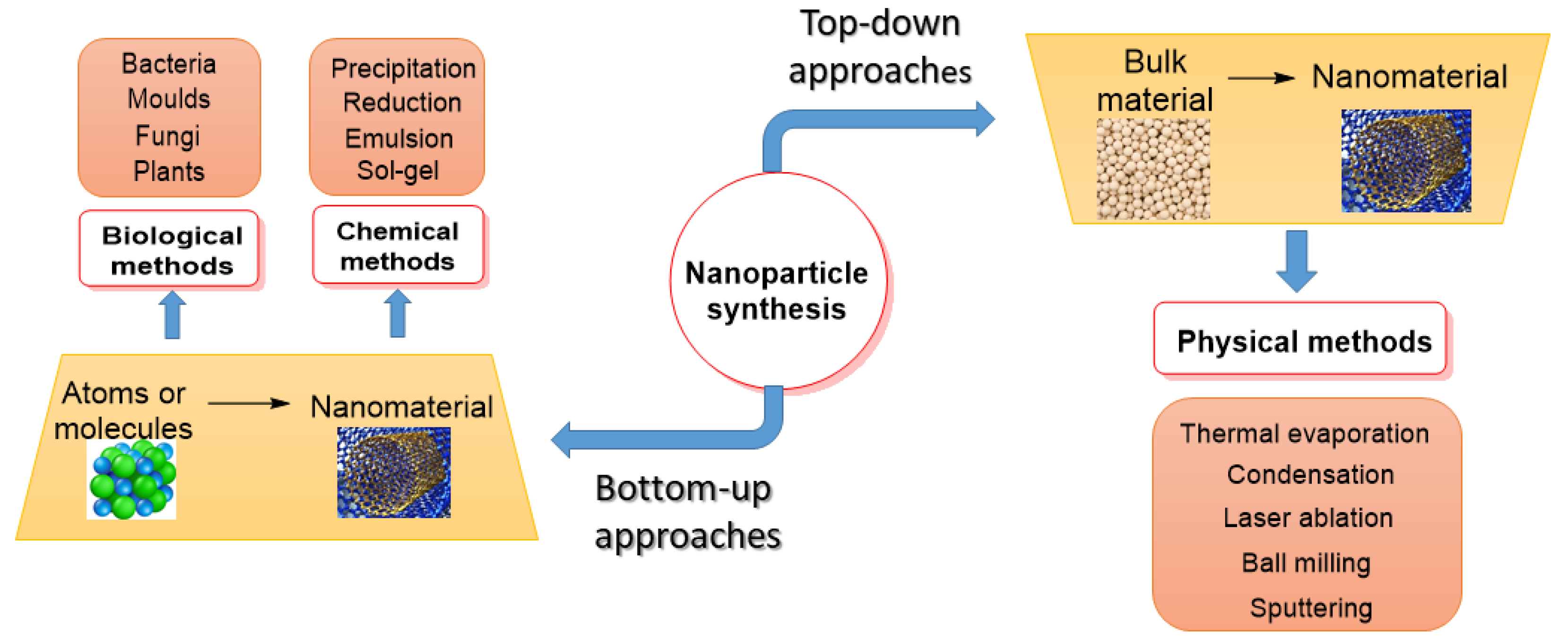
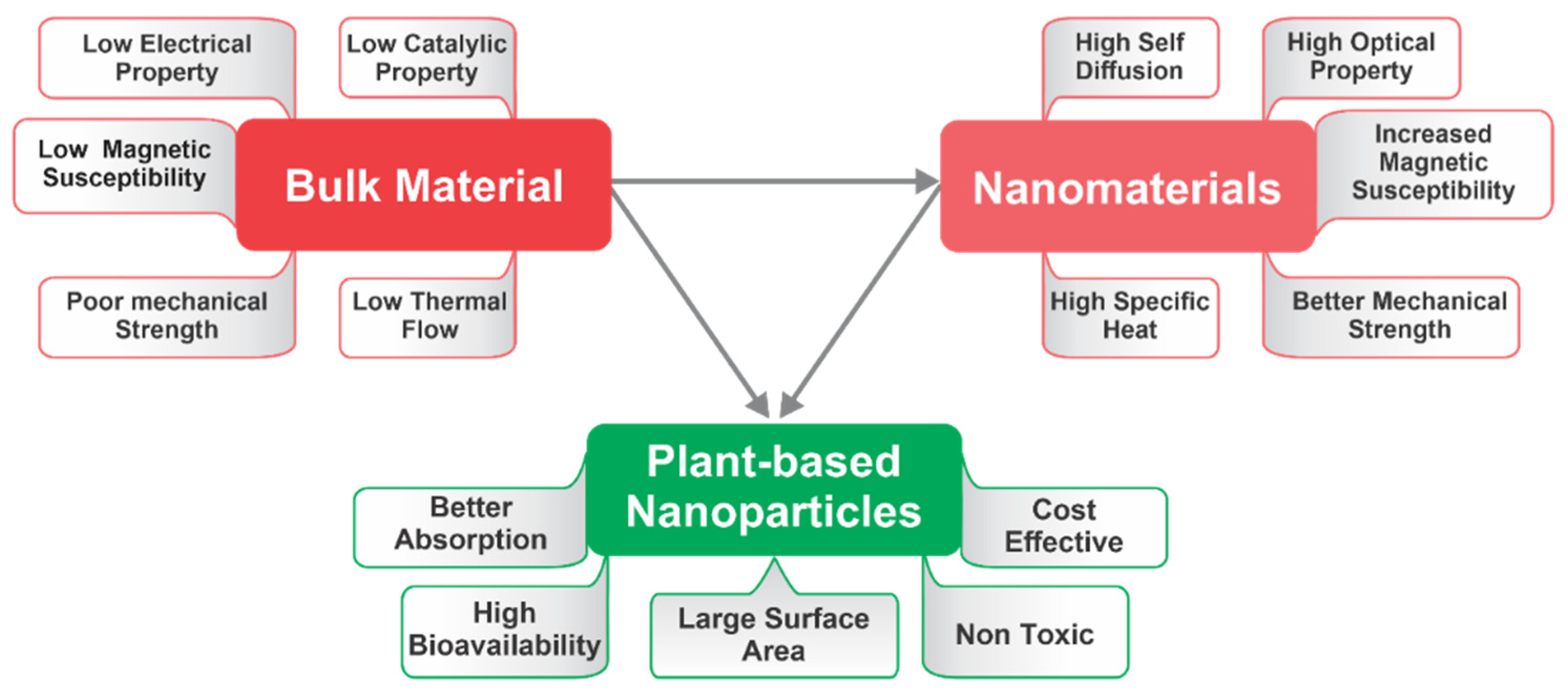
2. Overview of Nanoparticles
2.1. History and Development of Nanoparticles
2.2. Methodology
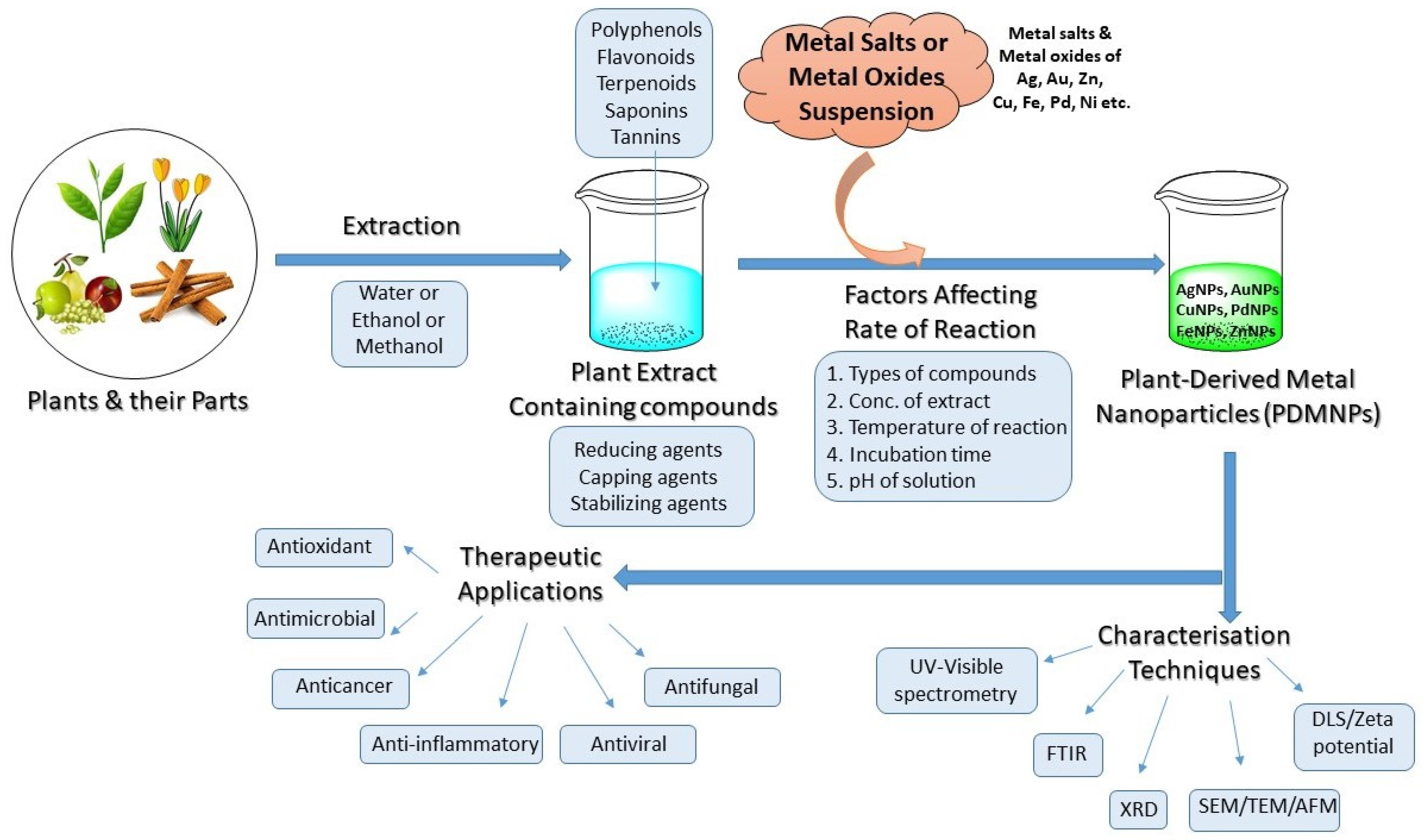
3. Synthesis of PDMNPs
3.1. General Synthesis
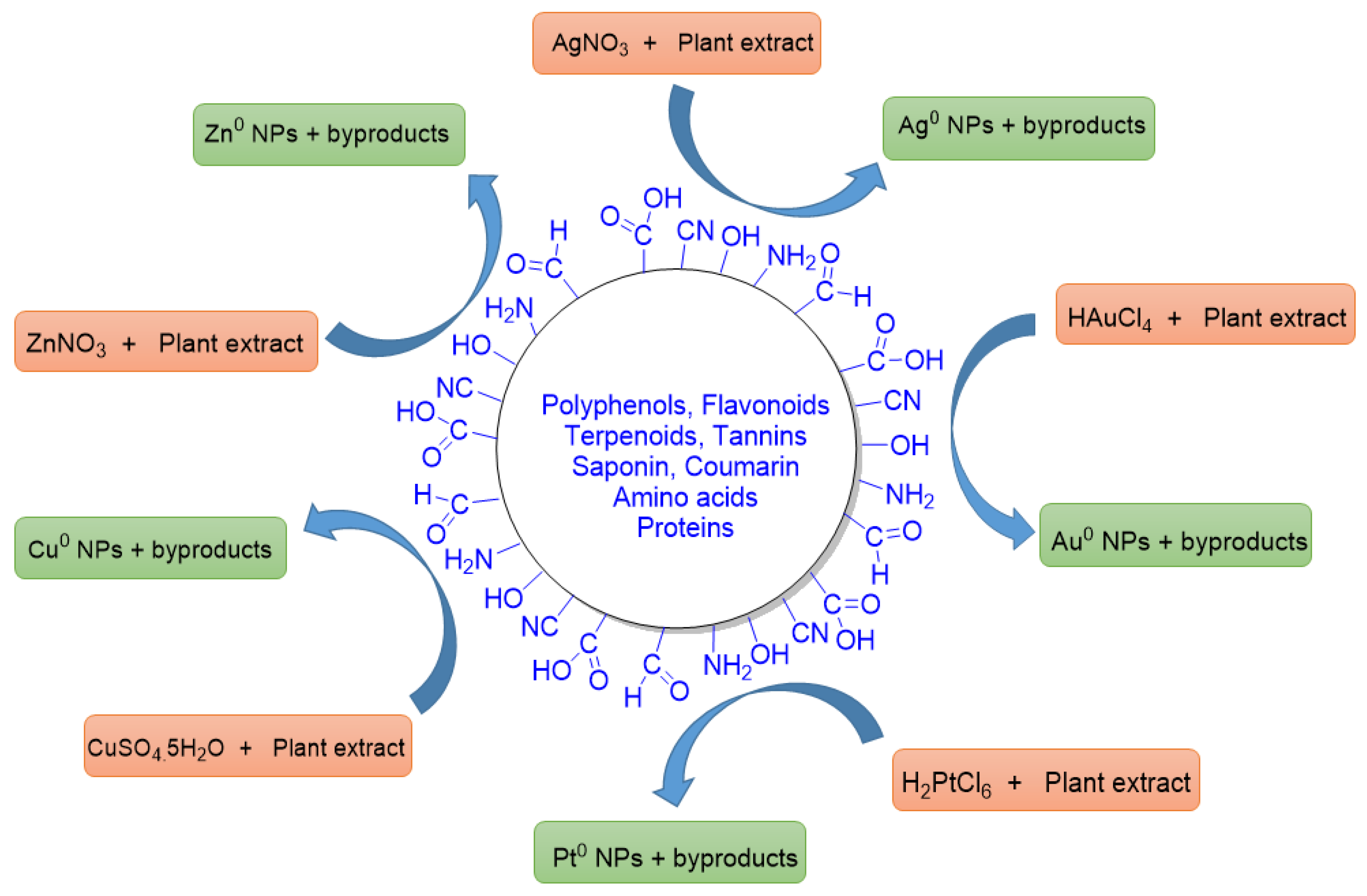
3.2. Plant Mediated Synthesis
4. Factors Affecting the Synthesis of PDMNPs
4.1. Effect of Extract Concentration
4.2. Effect of Time
4.3. Effect of pH
4.4. Effect of Temperature
4.5. Effect of Types of Bioactive Compounds
4.6. Effect of Agitation
5. Structural Characterization of PDMNPs
5.1. Ultra Violet-Visible Spectroscopy
5.2. X-ray Crystallography
5.3. Fourier-Transform Infrared Spectroscopy (FTIR)
5.4. Microscopic Techniques
5.5. Powder XRD Technique
5.6. Light Scattering Methods and Zeta-Potential (ζ)

| Name of Technique | Source of Detection | Study |
|---|---|---|
| UV-Visible absorption spectroscopy | Monochromatic wavelength | For identifying, characterizing, and studying nanomaterials in terms of size, shape, concentration, and agglomeration state |
| Fourier-Transform Infrared (FTIR) spectroscopy | Infrared radiations | Identifying the surface adsorption of functional groups on nanoparticles |
| Scanning Electron Microscopy (SEM) | Electron beam | Characterization of size (below 1.0 nm) and shape of nanoparticles |
| Transmission Electron Microscopy (TEM) | Electron beam | Measurement of nanoparticle’s size, distribution, and morphology |
| Atomic Force Microscopy (AFM) | Electron beam from the laser light | Evaluation of size and shape of nanoparticles in three dimensions. It also determines the surface morphology and elemental composition in a very quick time |
| Dynamic Light Scattering (DLS) | Scattered light from a laser | Studying the aggregation and colloidal state of nanoparticles in suspension |
| Zeta Potential Measurement | Free ions | Studying the physical stability in terms of surface charge of colloidal nanoparticles |
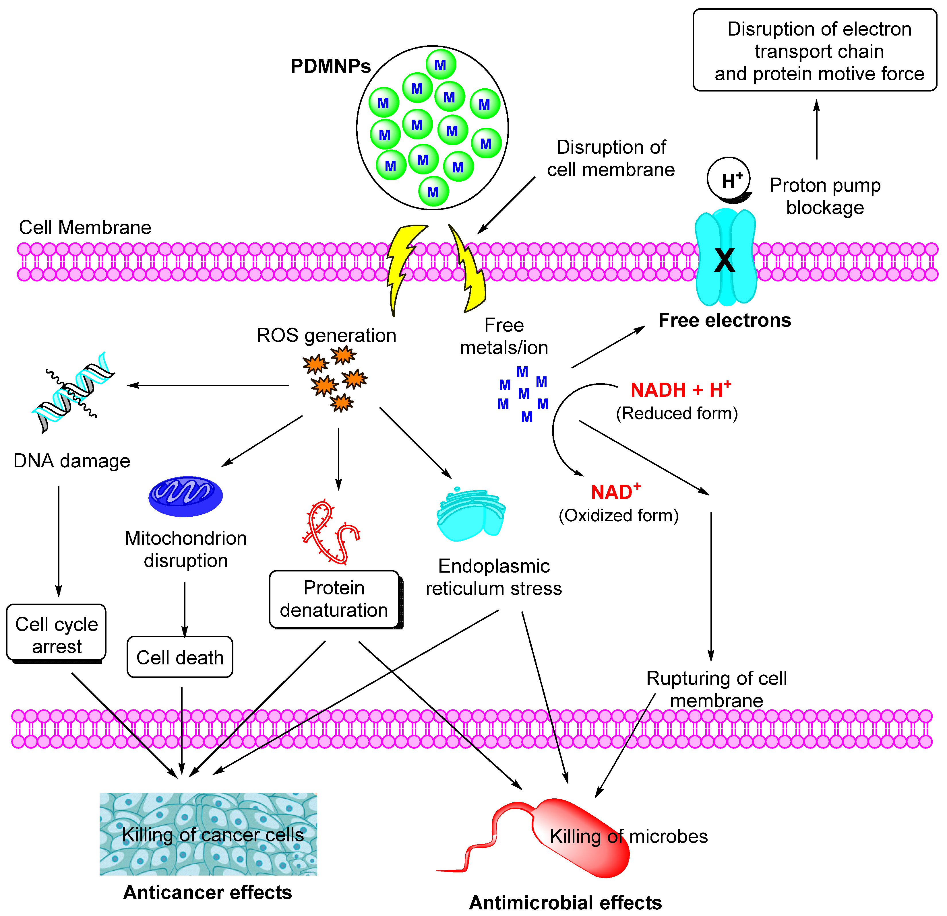
6. Assessment of Oxidative Stress-Mediated Therapeutic Actions
6.1. Antioxidant Action
6.2. Antimicrobial Action
6.3. Anticancer Action
| Plant Name (Part Used) | Extract | Nanoparticles | Findings | Ref. |
|---|---|---|---|---|
| Quercus robur, Eucalyptus globulus, Camellia sinensis, Thimus mastichina, Thimus vulgaris, Thuja occidentalis, Mentha sp., Rosmarinus officinalis, Laurus nobilis, Citrus limon (leaves) | Aqueous | Metal: Ag, Cu, Fe, Pd, Ni, Shape: Hexagonal, spherical, triangular, and rod shaped Size: 50 nm | Antioxidant activity: Evaluated by using DPPH, FRAP, FC, and cyclic voltammetry methods | [202] |
| Camellia sinensis, Ilex paraguariensis, Salvia officinalis, Tilia cordata, Levisticum officinale, Aegopodium podagraria, Urtica dioica, Capsicum baccatum, Viscum album (whole plants) | Methanol | Metal: Ag Shape: spherical, and rod-shaped Size: 100–200 nm | Antioxidant activity: Evaluated by DPPH, CUPRAC, and SNPAC assays | [203] |
| Phoenix Dactylifera (leaves) | Ethanol/water mixture (70%/30%) | Metal: Cu Shape: spherical and rhombohedral Size: 25–100 nm | Antioxidant activity: Evaluated by using DPPH, phosphomolybdenum, and ferric-reducing antioxidant power (FRAP) assays with IC50 of 0.39 mg/mL | [204] |
| Rosa floribunda charisma (flowers) | Phenyl ethyl alcohol | Metal: Mg Shape: Polyhedral Size: 35.25–55.14 nm | Antioxidant activity: Evaluated by using inhibition of superoxide (IC50 26.2 µg/mL), nitric oxide (IC50 52.9 µg/mL), hydroxyl radical (IC50 31.9 µg/mL), and xanthine oxidase (IC50 15.9 µg/mL) assays Antibacterial activity: Against Staphylococcus epidermidis (MIC of 15.63), Staphylococcus pyogenes (MIC of 7.81), and Pseudomonas aeruginosa (MIC of 31.25) µg/mL as well as with minimum biofilm inhibitory concentrations 1.95, 1.95, 7.81 µg/mL, respectively | [205] |
| Cestrum nocturnum (leaves) | Aqueous | Metal: Ag Shape: spherical Size: 20 nm | Antioxidant activity: Evaluated by DPPH (29.55% inhibition) hydrogen peroxide (45.41% inhibition), hydroxyl radical (20% inhibition), and superoxide radical scavenging (8% inhibition) methods Antibacterial activity: Against Citrobacter, Escherichia faecalis, Salmonella typhi, Escherichia coli, Proteus vulgaris, and Vibrio cholera with MIC values of 16, 4, 16, 8, 8, and 16 μg/mL | [206] |
| Plantago lanceolate (whole) | Aqueous | Metal: Ag Shape: spherical Size: 30 ± 4 nm | Antioxidant activity: Evaluated by using DPPH assay with IC50 369.5 ± 13.42 µg/mL Antibacterial activity: Against Agrobacterium tumefaciens, Proteus vulgaris, Staphylococcus aureus, and Escherichia coli with IC50 values 08.02 ± 0.68, 55.78 ± 1.01, 12.34 ± 1.35 and 11.68 ± 1.42 µg/mL, respectively | [207] |
| Flemingia wightiana (leaves) | Aqueous | Metal: Ag Shape: spherical Size: 20–40 nm | Antioxidant activity: Evaluated by using DPPH and H2O2 scavenging activity with IC50 values of 71.96 and 80.59 µg/mL respectively Anticancer activity: Against SKOV3 (inhibition 83.2%) and COLO205 (inhibition 75.9%) cancer cells | [208] |
| Artemisia absinthium, Humulus lupulus, and Thymus vulgaris (leaves) | Ethanol | Metal: Ag Shape: spherical and wedge Size: 42–48 nm | Antioxidant activity: Evaluated by using DPPH with 0.14 ± 0.00 inhibition for A. absinthium/AgNPs, 0.11 ± 0.00 inhibition for H. lupulus/AgNPs, and 0.14 ± 0.00 mmol/g for T. vulgaris/AgNPs while 0.55 ± 0.05; 0.86 ± 0.05 and 0.55 ± 0.05 mmol/g inhibitions in ABTS method for A. absinthium/AgNPs, H. lupulus/AgNPs, and T. vulgaris/AgNPs samples. Antibacterial activity: Evaluated with inhibition zones in the range 9.0 ± 0.1 to 20.4 ± 0.3 mm for all the samples | [209] |
| Sesamum indicum (oil cake) | Aqueous | Metal: Ag Shape: spherical Size: 6.6–14.8 nm | Anticancer activity: Against MCF-7 cell lines with 72.02% cell viability, 15.18% late apoptosis, and 1.20% necrosis at 2.5 μg/mL concentration of AgNPs whereas against with 56.97% cell viability, 31.19% late apoptosis, and 4.85% necrosis at 7.5 μg/mL concentration of AgNPs Antibacterial activity: With minimum inhibitory concentration (0.5 μg/mL) against Pseudomonas aeruginosa, Klebsiella pneumoniae, and Escherichia coli. | [210] |
| Dodonaea viscosa (leaves) | Aqueous, acetone, methanol, acetonitrile | Metal: Ag Shape: spherical and dendritic Size: 15, 18, 12, and 20 nm | Antibacterial activity: Against Streptococcus pyogenes with a zone of inhibition (20, 16, 13, 18 mm) for AgNPs synthesized by methanol, acetone, acetonitrile, and water extracts nanoparticles, respectively Anticancer activity: Against A549 NSCLC cell lines with IC50 values 14, 3, 80, and 4 μg/mL for methanol, acetone, acetonitrile, and water extract derived nanoparticles, respectively. | [211] |
| Mangifera indica (leaves) | Aqueous | Metal: Ag Shape: rod-shaped Size: 500–900 nm | Antioxidant activity: Evaluated by using DPPH with inhibition 83.7 ± 0.2%, 89.8 ± 0.3%, and 96.4 ± 0.3% for 0.1 w/v, 1 w/v, and 10% w/v concentration, respectively. Anticancer activity: Against MCF-7 and HCT-116 cell lines with cell growth 49.1 ± 0.5% and 58.2 ± 0.2%, respectively, at 10% w/v | [212] |
| Impatiens balsamina and Lantana camara (leaves) | Aqueous | Metal: Ag Shape: spherical Size: <24 nm | Antioxidant activity: Against Staphylococcus aureus and Escherichia coli with average ZOI values 13.8 and 8.9 mm for Impatiens balsamina and 15.8 and 15.4 mm Lantana camara, respectively | [213] |
| Jasminum auriculatum (leaves) | Aqueous | Metal: Au Shape: spherical Size: 8–37 nm | Anticancer activity: Against cervical cancer Antibacterial activity: Against Streptococcus pyogenes, Staphylococcus aureus, Escherichia coli, and Klebsiella pneumoniae Antifungal activity: Against Candida albicans, Aspergillus fumigatus, Lecanicillium lecanii, and Trichoderma viride | [214] |
| Alpinia nigra (fruits) | Aqueous | Metal: Ag Shape: spherical Size: 6 nm | Anticancer activity: Against human cervical cancer cell line (HeLa) with the IC50 value of 104 μg/mL Antibacterial activity: Against Streptococcus pyogenes, Staphylococcus aureus, Escherichia coli, and Klebsiella pneumoniae with ZOI values of 20, 9, 12, and 7 mm, respectively Antifungal activity: Candida Albicans, Aspergillus fumigatus, Lecanicillium lecanii, and Trichoderma viride with ZOI values of 4, 4, 5, and 5 mm, respectively | [215] |
| Lycium chinense (fruits) | Aqueous | Metal: Au, Ag Shape: spherical Size: 536 and 480 nm | Anticancer activity: Aignificant cytotoxicity to the human breast cancer MCF7 cell line for AgNPs and whereas no toxicity to non-diseased RAW264.7 (murine macrophage) cells, whereas no toxicity on both cell lines for AuNPs | [216] |
| Garcinia Indica (fruit) | Aqueous | Metal: Au, Ag Shape: spherical Size: 20–30 nm | Antioxidant activity: Evaluated by DPPH assay with 32–72% inhibition | [217] |
| Ziziphora clinopodioides (whole plant) | Aqueous | Metal: Ag Shape: spherical Size: 20–45 nm | Antibacterial activity: Against Staphylococcus aureus and Escherichia coli with MIC values at 200 and 400 ppm concentrations, respectively | [218] |
| Calophyllum tomentosum (leaves) | Aqueous | Metal: Ag Shape: spherical Size: NA | Antioxidant activity: Evaluated by using DPPH (90% inhibition), H2O2 scavenging (83.94%), nitric oxide scavenging power (78.4%), reducing power assays Antibacterial activity: Against Klebsiella aerogenes, Staphylococcus, auereus, Pseudomonas aeruginosa, and Escherichia coli with a maximum inhibition at 100 µg/mL concentration | [219] |
| Pistacia atlantica (leaves and fruit) | Aqueous | Metal: Au Shape: spherical Size: 40–50 nm | Antioxidant activity: Evaluated by using DPPH assay Anticancer activity: Against cervical cancer with a maximum inhibition of cells at 200 μg/mL concentration Antibacterial activity: Against Escherichia coli, Pseudomonas aeruginosa, Staphylococcus aureus, and Bacillus subtilis with MIC values 7.81, 3.9, 7.81, and 3.25 μg/mL | [220] |
| Allium rotundum L., Falcaria vulgaris Bernh. and Ferulago angulate Boiss. (leaves) | Aqueous | Metal: Ag Shape: spherical Size: 20.5 nm | Antimicrobial activity: Against Pseudomonas aeruginosa and Staphylococcus aureus | [221] |
| Carya illinoinensis (leaves) | Methanol | Metal: Ag Shape: spherical Size: 20 nm | Antibacterial activity: Against Saphylococcus aureus (MIC: 128), Listeria monocytogenes (MIC: 64), Escherichia coli (MIC: 16), Pseudomonas aeruginosa (MIC: 32) | [222] |
| Tasmannia lanceolata and Backhousia citriodora (leaves) | Aqueous | Metal: Au Shape: spherical Size: 7.10 ± 0.66 nm | Anticancer activity: Against liver cancer (HepG2), melanoma cancer (MM418C1), and breast cancer (MCF-7) cell lines. AuNPs showed 20% better inhibition of these cancer cell lines than plant extracts | [223] |
| Ougeinia oojeinensis (leaves) | Ethanol | Metal: Ag Shape: spherical Size: 5–100 nm | Antioxidant activity: Evaluated by DPPH assay with IC50 value 21.95 ± 1.02 Antibacterial activity: Against Escherichia coli, Bacillus cereus, Pseudomonas aeruginosa, and Staphylococcus aureus with ZOI values 14.58 ± 0.79, 16.55 ± 0.37, 16.25 ± 0.63, and 23.83 ± 0.44 mm, respectively. Antifungal activity: Aspergillus niger, Rhizopus oryzae, Mucor, azygosporus, and Penicillum chrysogenum with a zone of inhibition 20.17 ± 0.6, 16.50 ± 0.29, 20.25 ± 0.38, 21.82 ± 0.43 mm, respectively | [224] |
| Holoptelea integrifolia (leaves) | Aqueous | Metal: Ag Shape: spherical Size: 32–38 nm | Antioxidant activity: Evaluated by using DPPH, metal chelating, and nitric oxide assay with inhibition 51.49 ± 3.33, 41.18 ± 2.27, and 74.59 ± 3.08%, respectively Antibacterial activity: Against Escherichia coli and Salmonella typhimurium with MIC value ranges from 75 to 150 μg | [225] |
| Aesculus hippocastanum (leaves) | Aqueous | Metal: Ag Shape: spherical Size: 50 ± 5 nm | Antioxidant activity: Evaluated by using DPPH and superoxide radical scavenging assays with 54.72% and 62.9% inhibition at the highest concentration of 100 ppm. Antibacterial activity: Against Pseudomonas aeruginosa, Pseudomonas fluorescens, Staphylococcus aureus, Staphylococcus epidermidis, Listeria monocytogenes, Bacillus subtilis, Corynebacterium renale, Micrococcus luteus, Enterococcus faecalis, and Bacillus cereus with ZOI values as 20.0, 8.0, 8.0, 17.5 ± 2.12, 13 ± 0.00, 13 ± 0.00, 15 ± 2.64, 12 ± 0.00, 17 ± 0.00, 10.5 ± 1.41, 10.5 ± 1.41 mm diameter, respectively | [226] |
| Punica granatum L. (seeds) | Oil | Metal: Au Shape: elongated, and rectangular Size: 70 nm | Antioxidant activity: Evaluated by using DPPH scavenging (23.6 ± 1.5 to 62.5 ± 1.8%) and H2O2 scavenging (21.6 ± 1.3 to 62.8 ± 1.8%) at different concentrations Anticancer activity: Against lung and colon cancer with the cell viability ranging from 80.3 to 25% and 83.3 to 28.4.2%, respectively | [227] |
| Alternanthera bettzickiana (leaves) | Aqueous | Metal: Au Shape: spherical Size: 80–120 nm | Antibacterial activity: Bacillus subtilis, Staphylococcus aureus, Salmonella typhi, Pseudomonas aeruginosa, Micrococcus luteus, and Enterobacter aerogens Anticancer activity: Against human lung cancer | [228] |
| Green tea and black tea (leaves) | Aqueous | Metal: Au, Ag Shape: spherical Size: ∼10 nm for AuNPs, and ∼30 nm for AgNPs | Antibacterial activity: Against Bacillus subtilis, Staphylococcus aureus, Salmonella typhi, Pseudomonas aeroginosa, Micrococcus luteus, and Enterobacter aerogenes with ZOI values 14 ± 0.43, 19 ± 0.33, 17 ± 0.13, 28 ± 0.33, 30 ± 0.33, 24 ± 0.17 mm diameters for AgNPs while AuNPs show the ZOI values as 16 ± 0.88, 16 ± 0.44, 14 ± 0.58, and 22 ± 0.44 mm diameter against Salmonella typhi, Pseudomonas aeruginosa, Micrococcus luteus, and Enterobacter aerogenes, respectively | [229,230] |
| Phoenix dactylifera (root hairs) | Aqueous | Metal: Ag Shape: spherical Size: 15–40 nm | Antibacterial activity: Against Candida albicans and Escherichia coli with 20 and 22 mm ZOI, respectively Anticancer activity: Against MCF7 cell lines with IC50 values of 29.6 μg/mL | [231] |
| Sida cordifolia (whole plant) | Aqueous | Metal: Ag Shape: spherical Size: 3–6 nm | Antibacterial activity: Against Escherichia coli, Klebsiella pneumoniae, Bacillus subtilis, and Staphyloccocus aureus with ZOI values as 15.0 ± 0.63, 17.17 ± 0.75, 18.00 ± 0.63, and 19.50 ± 0.55 mm, respectively | [232] |
| Salvia spinose (whole plant) | Aqueous | Metal: Ag Shape: spherical Size: 450 nm | Antibacterial activity: Against Bacillus subtilis, Bacillus vallismortis, and Escherichia coli with zone of inhibition diameters of 15, 16, and 12 mm | [233] |
| Melaleuca alternifolia (leaves) | Aqueous | Metal: Ag Shape: spherical Size: 11.56 nm | Antimicrobial activity: Against Staphylococcus aureus, Staphylococcus epidermidis, Streptococcus pyogenes, Klebsiella pneumoniae, Pseudomonas aeruginosa, Trichophyton mentagrophytes, and Candida albicans with ZOI values ranges from 14.8 to 24.7 mm | [234] |
| Parkia speciosa (leaves) | Aqueous | Metal: Ag Shape: spherical Size: 31–35 nm | Antimicrobial activity: Against Escherichia coli, Staphylococcus aureus, Pseudomonas aeruginosa, and Bacillus subtilis with ZOI values ranging from 4.0 to 10 mm in diameter Antioxidant activity: Evaluated by DPPH assay with IC50 value of 15.26 μg/mL | [235] |
| Hygrophila spinosa (whole plant) | Aqueous | Metal: Au Shape: spherical Size: 68.44 ± 0.30 nm | Anticancer activity: Against MCF-7 and MDA-MB-231 (breast cancer), SKOV-3 (ovarian cancer) NCI/ADR (multi-drug resistant), and U-87 (glioblastoma, brain cancer) cell lines with significant percentage cell viability 43.78, 39.34, 21.45, 31.48, and 27.89%, respectively | [236] |
| Clerodendrum phlomidis (leaves) | Aqueous | Metal: Ag Shape: spherical Size: 23–42 nm | Antioxidant activity: Evaluated by using phosphomolybdate (910 AEAA), ferric reducing power (1.63 AU), superoxide radical scavenging (IC50 55.86 μg/mL), and DPPH (IC50 9.12 μg/mL) assays Anticancer activity: Against Ehrlich ascites carcinoma (EAC) and human colorectal adenocarcinoma (HT29) cell lines with 91.84% and 84.91% inhibition, respectively | [237] |
| Aconitum toxicum Rchb (rhizome) | Aqueous | Metal: Ag, Au Shape: spherical Size: 9–15 nm for AuNPs and 53–67 nm for AgNPs | Antioxidant activity: Evaluated by using DPPH assay with inhibition in between 78% and 84.32% at different concentrations | [238] |
| Musa acuminata colla (flowers) | Aqueous | Metal: Ag, Au Shape: spherical Size: 12.6–15.7 nm for AgNPs and 10.1–15.6 nm for AuNPs | Antibacterial activity: Against Enterococcus faecalis, Staphylococcus aureus, Klebsiella pneumoniae, Salmonella typhi, Escherechia coli, Proteus mirabilis, and Pseodomonas aeruginosa with ZOI values 13, 9, 10, 9, 12, 6, and 12 mm for AgNPs) and 11, 0, 10, 9, 7, 8, and 9 for AuNPs at 1000 µg concentration Anticancer activity: 50% cell viability at 55.0 µg/mL and 35 µg/mL concentrations for AuNPs, and AgNPs respectively | [239] |
| Allium cepa (cloves) | Aqueous | Metal: Ag Shape: spherical Size: 10–50 nm | Antibacterial activity: Against methicillin-resistant Staphyllococcus aureus and Pseudomonas aerigunosa with a maximum inhibition at 100 μg/mL concentration Anticancer activity: Against human breast cancer cells (MCF-7) with a maximum inhibition at 100 μg/mL concentration after 24 h | [240] |
| Solanum nigurum (leaves) | Aqueous | Metal: Au, Ag, Pd Shape: spherical Size: 3.46 nm for AgNPs, 9.39 nm for AuNPs, and 21.55 nm for PdNPs | Antibacterial activity: Against Escherichia coli with ZOI values 19.2 and 20 mm, 23 and 20 mm, and 18 and 19 mm for AuNPs, AgNPS, and PdNPs, respectively, at 5 and 10 mL concentration | [241] |
| Carthamus tinctorius L (flowers) | Aqueous | Metal: Ag Shape: spherical Size: 38 nm | Antibacterial activity: Against Escherichia coli with inhibition of 98% and 85% at 40 °C and 80 °C temperatures | [242] |
| Carpesium cernuum (whole plant) | Aqueous | Metal: Ag Shape: spherical Size: 13.0 ± 0.2 nm | Antioxidant activity: Evaluated by using DPPH assay with IC50 value 0.121 ± 0.005 mg/mL Anticancer activity: Against human lung cancer A549 and B16F10 cell lines with 44.5 and 36.0% cytotoxicity on A549 and B16F10 respectively at 100 µg/mL concentration | [243] |
| Ananas comosus (peels) | Aqueous | Metal: Fe Shape: spherical Size: 17.87 nm | Antifungal activity: Against Fusarium verticilliodes, Aspergillus flavus, and Alternaria alternate with inhibition zones ranging from 18.96 to 39.23 mm diameters | [244] |
| Aaronsohnia factorovskyi (whole plant) | Aqueous | Metal: Ag Shape: spherical Size: 104–140 nm | Antibacterial activity: Against Staphylococcus aureus, Bacillus subtilis, Pseudomonas aeruginosa, and Escherichia coli with an inhibition zone diameter of about 19.00 ± 2.94 mm Antifungal activity: Against Fusarium oxysporum, Fusarium solani, Helminthosporium rostratum, and Alternaria alternate with reduced the growth of fungal yarn to 1.5 mm. | [245] |
| Vernonia amygdalina (leaves) | Aqueous | Metal: CuO Shape: spherical Size: 19.68 nm | Antibacterial activity: Against Staphylococcus aureus, Escherichia coli, Pseudomonas aeruginosa, and Enterobacter aerogenes. The uppermost zone of inhibition of 15 mm was observed for E. aerogenes | [246] |
| Artemisia ciniformis (leaves) | Aqueous | Metal: Ag Shape: spherical Size: 4–14 nm | Anticancer activity: Against gastric cancer with the highest inhibition of cell proliferation at 100 μg/mL concentration | [247] |
| Cichorium intybus (leaves) | aqueous | Metal: Ag Shape: spherical Size: 17.17 nm | Anticancer activity: Against human breast cancer (MCF-7) with IC50 value 507.58 μg/mL after 24 h. | [248] |
| Rhynchosia suaveolens (leaves) | Aqueous | Metal: Ag Shape: spherical Size: 10–30 nm | Anticancer activity: Against DU145 and PC-3 (human prostate carcinoma cell lines), SKOV3 (human ovarian carcinoma), and A549 (human lung adenocarcinoma) with IC50 values of 4.35, 7.72, 4.2, and 24.7 μg/mL, respectively | [249] |
| Solanum lycopersicum (leaves) | Aqueous | Metal: FeO Shape: flower shaped Size: 483.8 nm | Anticancer activity: Against human lung cancer cell line A549 with IC50 value 69 ± 0.50 μg/mL | [250] |
| Ageratum conyzoides (leaves) | Aqueous | Metal: Ag Shape: spherical Size: 14–48 nm | Antioxidant activity: Evaluated by using DPPH and ABTS free radical scavenging assays with % inhibition 53.61 ± 0.01 to 89.82 ± 0.017 and 40.16 ± 0.13 to 81.1 ± 0.13 in 31.25 to 500 mg/mL concentrations, respectively. | [251] |
| Blumea eriantha DC (whole plant) | Ethanol | Metal: Ag, Fe Shape: spherical Size: 50 nm | Antioxidant activity: Evaluated by using DPPH (% inhibition 20.66 ± 0.90 for AgNPs and 17.25 ± 1.19 for FeNPs), ABTS (% inhibition 86.31 ± 0.21 for AgNPs, 74.94 ± 1.72 for FeNPs), H2O2 scavenging (% inhibition 92.14 ± 1.06 for AGNPs and, 57.00 ± 0.58 for FeNPs), and total antioxidant assay (% Inhibition 70.10 ± 0.53 for AgNPs and 56.14 ± 0.64% for FeNPs) Antibacterial activity: Against Staphylococcus aureus (ZOI 16.17 ± 2.08 for AgNPs and 13.06 ± 0.57 FeNPs), Bacillus subtilis (ZOI 14.12 ± 1.52 for AgNPs and 12.45 ± 0.52 for FeNPs), Bacillus cereus (ZOI 11.20 ± 1.15 for AgNPs and 10.12 ± 1.02 for FeNPs), and Escherichia coli (ZOI 15.24 ± 1.52 for AgNPs and 11.55 ± 1.18 for FeNPs) Anticancer activity: Against MCF-7 (human breast adenocarcinoma cell line) with % inhibition 15.45, 20.25, and 28.16 at the concentrations 25, 50, and 100 μg/mL, respectively, for AgNPs as well as 11.09, 17.81, and 22.25 at the concentration 25, 50, and 100 μg/mL respectively for FeNPs | [252] |
| Artemisia abrotanum (whole plant) | Aqueous | Metal: MgO Shape: spherical Size: 10 nm | Antioxidant activity: Evaluated by using DPPH assay with IC50 4.73 µg/mL | [253] |
| Cnici Benedictus (whole plant) | Aqueous | Metal: Au, CuO, and ZnO Shape: spherical Size: 13 nm for Au-CuONPs and 28 nm for CuO-ZnONPs | Antibacterial activity: Against Staphylococcus aureus (MIC 0.3125), Escherichia coli (MIC 0.625), Pseudomonas aeruginosa (MIC 2.5) Antifungal activity: Against Candida albicans (MIC 1.25). Anticancer activity: Against rat glioma C6 cells with IC50 0.907 and 4.91 for Au-CuONP and CuO-ZnONPs, respectively | [254] |
| Elephantopus scaber (leaves) | Aqueous | Metal: Ag Shape: spherical Size: 37.86 nm | Anticancer activity: Against human skin carcinoma cells on A375 and L929 cell lines with IC50 values 15.68 ± 0.15 μg/mL and 65.49 ± 0.40 μg/mL, respectively Antioxidant activity: Evaluated by using the DPPH method with an IC50 value of 6.629 µg/mL Antibacterial activity: Against Bacillus subtilis, Lactococcus lactis, Pseudomonas fluorescens, and Pseudomonas aeruginosa Antifungal activity: Against Aspergillus flavus and Aspergillus penicillioides with ZOI ranges from 12 to 24 mm for all the strains | [255] |
| Acanthospermum hispidum (leaves) | Aqueous | Metal: Ag Shape: quasi-spherical Size: 20–60 nm | Antibacterial activity: Against Pseudomonas aeruginosa, Streptococcus pyogenes, Staphylococcus aureus, and Escherichia coli with a zone of inhibition of 17–19 at 100 µg/mL concentration Antifungal activity: Against Candida albicans, Aspergillus niger, and Aspergillus clavatus with MIC 500, 250, and 500 MIC µg/mL, respectively Antimicrobacterial activity: Against Mycobacterium tuberculosis H37RV with MIC 100 MIC µg/mL | [256] |
| Leucaena leucocephala L. (leaves) | Aqueous | Metal: Ag Shape: quasi-spherical Size: 35–47 nm | Antibacterial activity: Against Pseudomonas aeruginosa, Streptococcus pyogenes, Staphylococcus aureus, Escherichia coli, Salmonella typhi, Bacillus subtilis with ZOI values of 16–19 at 100 µg/mL concentration Antimycobacterial activity: Against Mycobacterium tuberculosis with MIC of 125 μg/mL | [257] |
| Silybum marianum (whole plant) | Aqueous | Metal: Ag-ZnO, ZnO Shape: quasi-spherical Size: 31.2 nm for ZnO, and 35.3 nm for Ag–ZnO | Antibacterial activity: Against Staphylococcus epidermidis, Bacillus subtilis, Klebsiella pneumonia, Escherichia coli, and Pseudomonas aeruginosa with MIC values 250, 50, 150, 100, and 250 µg/mL for ZnONPs and 150, 50, 250, 150, and 150 µg/mL for Ag-ZnONPs, respectively Antifungal activity: Fusarium solani, Aspergillus flavus, Aspergillus fumigatus, and Aspergillus niger with MIC values 50, 250, 500, and 100 µg/mL for ZnONPs and 50, 150, 700, and 150 µg/mL for Ag-ZnONPs, respectively Antioxidant activity: Total antioxidant capacity (67.6 ± 1.44 for ZnONPs and 72.6 ± 1.32 for Ag-ZnONPs, at 1000 μg/mL concentration), total reducing power (72.4 ± 2.78 for Ag-ZnONPs and 68.1 ± 1.31 for ZnONPs at 1000 μg/mL concentration) and DPPH (67.22 ± 2.1 for Ag-ZnONPs and 56.31 ± 1.4 for ZnONPs at 1000 μg/mL concentration) | [258] |
| Nyctanthes arbor-tristis (flowers) | Aqueous | Metal: ZnO Shape: spherical Size: 12–32 nm | Antifungal activity: Alternaria alternata, Aspergillus niger, Botrytis cinerea, Fusarium oxysporum, and Penicillium expansum with MIC values 64, 16, 128, 64, and 128 µg/mL | [259] |
| Melia azedarach (leaves) | Aqueous | Metal: Ag Shape: spherical Size: 23 nm | Antifungal activity: Against Colletotrichum coccodes, Monilinia sp., and Pyricularia with growth inhibition of 18%, 33%, and 51%, respectively. | [260] |
| Kleinia grandiflora (leaves) | Aqueous | Metal: Ag Shape: spherical Size: 20–50 nm | Anticancer activity: Against Dalton’s lymphoma ascites (DLA) cell lines with 40% cytotoxicity at 10 µg/mL concentration and IC50 of 500 nM after 6 h of treatment Antimicrobial activity: Against Escherichia coli, Pseudomonas aeruginosa, Candida albicans, and Aspergillus niger with a zone of inhibition as 13 ± 0.7, 17 ± 1.02, 13 ± 0.32, and 15 ± 0.46, respectively | [261] |
| Medicago sativa L. | Aqueous | Metal: ZnO Shape: hexagonal Size: 14 nm | Antimicrobial activity: Against Staphylococcus epidermidis, Lactococcus lactis, and Lactobacillus casei with MIC values in the range of 0.58–9.31 μg/mL Antifungal activity: Candida albicans, and Saccharomyces cerevisiae with MIC values of 9.31 and 4.65 μg/mL | [262] |
| Tussilago farfara (flower buds) | Aqueous | Metal: Ag, Au Shape: spherical Size: 13.57 ± 3.26 nm for AgNPs and 18.20 ± 4.11 nm for AuNPs | Antibacterial activity: Against Escherichia coli, Enterococcus faecalis, Pseudomonas aeruginosa, and Staphylococcus aureus with MIC values ranging from 10 to 40 µg/mL for AuNPs and AgNPs samples Anticancer activity: Against PANC-1 cells with IC50 values 166.1 and 71.2 µM for AgNPs and AuNPs, respectively. Against AGS cell lines with IC50 values 338 and 77.9 µM for AgNPs and AuNPs, respectively. Against HT-29 cell lines with IC50 values 275.3 and 87 µM for AgNPs and AuNPs, respectively | [263] |
| Celastrus paniculatus (leaves) | Aqueous | Metal: Cu Shape: spherical Size: 2−10 nm | Antifungal activity: Against plant pathogenic fungi Fusarium oxysporum with showing 76.29 ± 1.52 maximum mycelial inhibition | [264] |
| Pechuel-loeschea leubnitziae (roots) | Methanol | Metal: Ag Shape: spherical Size: 100 nm | Anticancer activity: Against the U87 glioma cell line with an IC50 value in the range of 0.64–0.71 µg/mL | [265] |
| Centaurea pumilio L. (aerial parts) | Methanol | Metal: Ag Shape: spherical Size: 6 and 8 nm | Antimicrobial activity: Against Stapholococcus aureus, Streptococcus pyogenes, Pseudomonas aeruginosa, Escherichia coli, and Candida albicans with ZOI values 22, 17, 12, 12 mm and no value, respectively. Antioxidant activity: Through estimation of SOD with activity in a dose-dependent-manner on the 4th and 7th days and then decreasing on the 14th day | [266] |
| Artemisia turcomanica (leaves) | Aqueous | Metal: Ag Shape: spherical Size: 22 nm | Anticancer activity: Against AGS (Human Gastric Adenocarcinoma) and normal L-929 cell lines with the IC50 value 15.43 and 14.56 μg/mL | [267] |
| Hagenia abyssinica (Brace) JF. Gmel. (leaves) | Metal: Cu Shape: spherical, hexagonal, triangular, cylindrical, and irregularly shaped Size: 34.76 nm | Antibacterial activity: Against Escherichia coli, Pseudomonas aeruginosa, Staphylococcus aureus, and Bacillus subtilis with ZOI 12.7, 12.7, 14.7, and 14.2 mm, respectively | [268] | |
| Solanum nigrum (leaves) | Aqueous | Metal: CuO Shape: spherical Size: 32 and 25 nm | Antioxidant activity: Evaluated by using DPPH assay with 9–60% inhibition at different concentrations from ranges 15, 30, 60, 125, 250, and 500 µg/mL Antibacterial activity: Against Bacillus subtilis, Staphylococcus, saprohyticus, Escherichia coli, and Pseudomonas aeruginosa with ZOI 13 ± 0.1, 11 ± 0.2, 15 ± 0.4, and 12 ± 0.6 nm, respectively, at a concentration of 100 µg | [269] |
| Solanum nigrum (leaves) | Aqueous | Metal: ZnO Shape: spherical Size: 49 nm size | Antioxidant activity: Evaluated by using DPPH (21–94% inhibition) andH2O2 scavenging (12–95% inhibition) assays at 15–500 μg/mL concentrations with IC50 values 130.54 and 126.14 μg/mL, respectively. Antibacterial activity: Against Bacillus Subtilis, Staphylococcus saprohyticus, Escherichia coli, and Pseudomonas aeruginosa with ZOI 17, 15, 19, and 17 nm at 100 μg concentration | [270] |
| Polyalthia longifolia (leaves) | Aqueous | Metal: CuO Shape: quasi-spherical Size: 5–60 nm | Antibacterial activity: Against Pseudomonas aeruginosa, Staphylococcus aureus, Escherichia coli, and Staphyloccocus pyogenes with MIC values 100, 12.5, 25, and 125 μg/mL respectively Antifungal activity: Aspergillus niger, Epidermophyton floccosum, Aspergillus clavatus, and Candida albicans with MIC values 1000, 100, 1000, and 1000 μg/mL, respectively | [271] |
| Limonia acidissima (fruits) | Aqueous | Metal: MgO Shape: flake-like structure Size: 10–15 nm | Antibacterial activity: Against Escherichia coli, Klebsiella pneumoniae, Pseudomonas aeruginosa, and Staphylococcus aureus with MIC 0.25, 0.025, 0.25, and 0.025 μg/mL, respectively Antifungal activity: Against Alternaria alternate and Phomopsis azadirachtae with 91.48% and 95.33% inhibitions, respectively | [272] |
| Cardiospermum halicacabum (leaves) | Aqueous | Metal: ZnO Shape: hexagonal quartzite Size: 65, 62, 55, and 48 nm | Antibacterial activity: Staphylococcus aurius, Bacillus subtilis, Escherichia coli, and Pseudomonas aeruginosa with ZOI inhibition 20, 20, 21, and 19 mm at 0.6 mg concentration, respectively | [273] |
| Oedera genistifolia (leaves) | Aqueous | Metal: Ag Shape: spherical Size: 34.2 nm | Cytotoxicity: No cytotoxicity against HeLa cells Antibacterial activity: Against Listeria ivanovic, Streptococcus uberis, Staphylococcus aureus, Mycobacterium, smergatis, Enterobacter cloacae, and Vibrio sp with MIC values 1.0, 0.5, 0.5, 0.25, 0.5, and 0.25 mg/mL, respectively | [274] |
| Vernonia amygdalina (leaves) | Aqueous | Metal: Cu, Zn Shape: hexagonal wurtzite Size: 34 to 39 nm | Antibacterial activity: Against Staphylococcus aureus, Pseudomonas aeruginosa, and Escherichia coli with ZOI values 21, 24, and 25 mm diameter, respectively | [275] |
| Moringa oleifera (leaves) | Aqueous | Metal: ZnO Shape: quasi-spherical Size: 35–95 nm | Antifungal activity: Against Candida albicans, Aspergillus niger, Aspergillus clavatus, Trichophyton mentographytes, and Epidermophyton floccosum with MIC values 250, 250, 250, 100, and 100 µg/mL, respectively | [276] |
| Wedelia chinensis (leaves) | Aqueous | Metal: Ag Shape: spherical Size: 31.68 nm | Antioxidant activity: Evaluated by using DPPH and reducing power assay with a maximum inhibition at 200 μg/mL and at absorbance 0.81 ± 0.146 Antibacterial activity: Against Escherichia coli and Listeria monocytogenes with ZOI 25.4 and 21.7 mm, respectively Cytotoxic: Showed dose-dependent cytotoxicity against Hep G2 cells with an IC50 value of 25 μg/mL | [277] |
| Seripheidium quettense (leaves) | aqueous | Metal: Ag Shape: spherical Size: 49.96–54.36 nm | Antibacterial activity: Bacillus subtilis, Staphylococcus aureus, Staphylococcus epidermidis, Escherichia coli, Pseudomonas aeruginosa, and Kle sela pneumonia with MIC 33.3, -, 100, 11.1, 100, and 33.3 μg/mL, respectively Antifungal activity: Against Aspergillus fumigatus, Aspergillus flavus, Aspergillus niger, and Mucor spp., with ZOI 12 ± 0.33, 10 ± 0.41, 13.2 ± 0.72, and 11 ± 0.78 mm diameter, respectively Anticancer activity: Demonstrated cytotoxic effects against HepG2 cells with IC50 of 62.5 µg/mL | [278] |
| Albizia procera (leaves) | Aqueous | Metal: Ag Shape: spherical Size: 6.18 nm | Antibacterial activity: Escherichia coli and Staphylococcus aureus with ZOI 13.5 ± 3.1 and 18.5 ± 6.75 at 100 μg concentration dose | [279] |
| Cynara scolymus (Leaves) | Aqueous | Metal: ZnO Shape: spherical Size: 65.9 nm | Antiproliferative activity: Against human breast cancer cell line (MCF 7) and Vero cells with IC50 values of 65.31 μg/μL and 957.85 μg/μL, respectively Antimicrobial activity: Against Staphylococcus aureus, Escherichia coli, Pseudomonas aeruginosa, Candida albicans, and Candida tropicalis with MIC50 > 7, 25, >100, >100, and 0.35 µg/mL concentrations, respectively | [280] |
| Mangifera indica (leaves) | Aqueous | Metal: ZnO Shape: spherical and hexagonal quartzite Size: 45–60 nm | Antioxidant activity: Evaluated by DPPH assay with 20–90% inhibition at different concentrations Anticancer property: Evaluated cytotoxicity against lung cancer A549 cell lines with 85% cell viability at 1.0 μg/mL concentration | [281] |
| Cissus arnotiana (leaves) | Aqueous | Metal: Cu Shape: spherical Size: 60–90 nm | Antibacterial activity: Against Escherichia coli, Streptococcus sp., Rhizobium sp., and Klebsiella sp., with 22.20 ± 0.16, 20.23 ± 0.35, 16.33 ± 0.13, and 18.25 ± 0.12 at 75 μg mL concentration Antioxidant activity: Evaluated by using DPPH assay with % radical scavenging activity 18 ± 1, 21 ± 2, 20 ± 8, 19 ± 6, 18 ± 2 at 20, 40, 60, 80, and 100 μg/mL concentrations | [282] |
| Tridax. procumbens (leaves) | Aqueous | Metal: Ag Shape: spherical Size: 54.34 nm | Antioxidant activity: Evaluated by using DPPH free radical scavenging activity (60.96 ± 0.61% inhibition), metal chelating activity (53.24 ± 0.56% inhibition), and β-carotene linoleic (85.26 ± 0.16% inhibition) assays Antibacterial activity: against Pseudomonas aeruginosa, Serratia marcescens, Shigella fexneri, Salmonella typhi, Escherichia coli, Proteus mirabilis, Klebsiella pneumonia, Enterococcus faecalis, and Staphylococcus aureus with ZOI inhibition ranges from 11 ± 1.00 to 15.33 ± 0.58 mm diameter and MIC values from 11.43 to 102.8 µg/mL, respectively, for all the strains, respectively | [283] |
| Taraxacum officinale (leaves) | Aqueous | Metal: Ag Shape: spherical Size: 15 nm | Antioxidant activity: Evaluated by using DPPH radical scavenging ((IC50 = 56.1 μg/mL), ABTS radical scavenging (IC50 = 45.6 μg/mL), and nitric oxide (NO) scavenging (IC50 = 55.2 μg/mL) assays Anticancer activity: Against human liver carcinoma cells HepG2 cells with a maximum inhibition at 25 μg/mL Antimicrobial activity: Against Xanthomonas axonopodis pv. citri and Pseudomonas syringae with ZOI 17.2 ± 0.65, 20.2 ± 0.84, 22.0 ± 0.84 mm, and 15.4 ± 0.32, 17.2 ± 0.65, 19.5 ± 0.66 mm at 10, 20, and 30 µg/mL concentrations, respectively | [284] |
| Tragopogon collinus (whole plant) | Ethanol and methanol | Metal: Ag Shape: spherical Size: 7–18 nm | Antibacterial activity: Against Staphylococcus aureus with ZOI 2, 5, and 10 mm and Escherichia coli with ZOI 4, 7, and 8 mm at 6000, 7000, and 8000 µg/mL concentrations, respectively | [285] |
| Deverra tortuosa (aerial part) | Aqueous | Metal: ZnO Shape: hexagonal and Wurtzite structures Size: 9.26–31.18 nm | Anticancer activity: Against cancer cell lines namely human colon adenocarcinoma “Caco-2” with IC50 50.81 μg/mL and human lung adenocarcinoma “A549” with IC50 83.47 μg/mL, respectively | [286] |
| Pulicaria vulgaris Gaertn. (aerial part) | Aqueous | Metal: Ag Shape: spherical Size: 28.6 ± 9.0 nm | Antibacterial activity: Against Staphylococcus aureus, and Escherichia coli with MIC values ranging from 60 to 80 µg/mL and MBC values ranging from 80 to 100 µg/mL Antifungal activity: Against Candida albicans, and Candidia glabrata MIC values ranging from 40 to 60 µg/mL and MCF values ranging from 80 to 100 µg/mL Antioxidant activity: Evaluated by using DPPH radical scavenging assay with maximum scavenging activity at 120 µg/mL concentration | [287] |
| Rhododendron arboretum (leaves) | Aqueous | Metal: MgO Shape: spherical Size: NA | Antibacterial activity: Escherichia coli, Spectrococous mutans, and Proteus vulgaris with ZOI 36 mm, 32 mm, and 24 mm at 10 mg/L concentration, respectively | [288] |
| Aegle marmelos (leaves) | Aqueous | Metal: ZnFe2O4 Shape: spherical Size: NA | Antibacterial activity: Against Escherichia coli, Pseudomonas aeruginosa, Staphylococcus aureus, and Bacillus subtilis with ZOI 17, 25, 22, and 23 nm | [289] |
| Costus pictus D.(leaves) | Aqueous | Metal: MgO Shape: hexagonal Size: 50 nm | Antibacterial activity: Staphylococcus aureus, Bacillus subtilis, Escherichia coli, and Salmonella paratyphi with ZOI 5.5, 10.0, 12.5, and 15.0 mm Antifungal activity: Candidia albicans and Aspergillus niger with ZOI 10.5 and 12.5 mm Anticancer activity: Against DLA cell line with % inhibition 2 ± 0.152, 9 ± 0.025, 18 ± 0.174, 30 ± 0.035, and 52 ± 0.0.053 at 10, 20, 50, 100, and 200 µg/mL concentrations, respectively | [290] |
| Tropaeolum majus L. (leaves) | Aqueous | Metal: Ag Shape: spherical Size: 35–55 nm | Antibacterial activity: Against Staphylococcus aureus, Enterococcus faecalis, Escherichia coli, Salmonella typhi, and Pseudomonas aeruginosa with MIC values from 50 to 450 µg/mL Antifungal activity: Aspergillus niger, Candida albicans, Penicillium notatum, Trichoderma viridiae, and Mucor sp. with MIC values from 12 to 170 µg/mL Anticancer activity: Against MCF7 and VERO cells with 50% inhibition at 3–4 and 5–6 µg/mL concentrations, respectively. | [291] |
| Psidium guajava L. (leaves) | Aqueous | Metal: Ag Shape: spherical Size: 20–35, 25 nm, and 25–35 nm | Antioxidant activity: Evaluated by using DPPH (IC50 values of 52.53 ± 0.31 μg/mL) and ABTS (IC50 values of 55.10 ± 0.29 μg/mL) radical scavenging assays Antimicrobial activity: Saccharomyces cerevisiae, Aspergillus niger, Rhizopus, Alcaligenes faecalis, and Escherichia coli with a maximum inhibition at 100 μg/mL concentration. | [292] |
| Handelia trichophylla (flowers) | Aqueous | Metal: Ag Shape: spherical Size: 20–50 nm | Antibacterial activity: Against Staphylococcus aureus, Bacillus subtilis, Escherichia coli, and Pseudomonas aeruginosa with ZOI 10.2, 9.1, and 8.5 mm for negative control as well as 16.3, 11.3, 11.3, and 9.8 mm for positive control, respectively | [293] |
| Pimpinella anisum (seeds) | Methanol | Metal: Ag, Au Shape: spherical Size: ~15 nm | Antioxidant activity: Evaluated by using DPPH assay with IC50 45.53 and 191.58 µg/mL for AgNPs and AuNPs samples, respectively Antibacterial activity: Against Staphylococcus aureus and Escherichia coli with ZOI 11 and 13 mm for AgNPs while AuNPs were inactive against these strains Antifungal activity: Against Aspergillus flavus and Candida albicans with ZOI 10 and 11 mm for AgNPs while AuNPs were inactive against these strains | [294] |
| Gundelia tournefortii L. (leaves) | Aqueous | Metal: Au Shape: spherical Size: 40–45 nm | Antioxidant activity: Evaluated by using DPPH assay with IC50 194 μg/mL Antibacterial activity: Pseudomonas aeruginosa, Escherichia coli, Bacillus subtilis, Staphylococcus aureus, Salmonella typhimurium, and Streptococcus pneumonia with MIC values 2 ± 0, 4 ± 0, 2 ± 0, 2 ± 0, 2 ± 0, 4 ± 0 mg/mL, respectively Antifungal activity: Candida albicans, Candida glabrata, Candida krusei, and Candida guilliermondii with MIC values 4 ± 0, 4 ± 0, 2 ± 0, 2 ± 0 mg/mL, respectively | [295] |
| Plectranthus asirensis (aerial parts) | Ethanol | Metal: Ag Shape: spherical Size: 20 nm | Antioxidant activity: Evaluated by using DPPH assay with IC50 12.725 ± 0.326 and 14.541 ± 0.225 µg/mL values for luteolin and stigmasterol in extract, respectively | [296] |
| Harpagophytum procumbens (roots) | Aqueous | Metal: Ag Shape: spherical Size: 82 nm | Antioxidant activity: Evaluated by chemiluminescence method for short-lived free radicals (inhibition = 73%) and ABTS method for long-lived free radicals (inhibition = 18.3%) respectively | [297] |
| Hyacinthus orientalis L. and Dianthus caryophyllus L. (leaves) | Aqueous | Metal: Ag Shape: spherical Size: 61.45 and 89.6 nm | Antioxidant activity: Evaluated by chemiluminescence method with inhibition of free radicals ranging between 88.30 and 97.38% | [298] * |
| Salvia officinalis L. (leaves) | Aqueous | Metal: Ag Shape: spherical Size: 75 nm | Antioxidant activity: Evaluated by chemiluminescence method with 98.6% inhibition of free radicals | [299] * |
| Anthriscus cerefolium L. (aerial part) | Hydroalcoholic | Metal: Ag Shape: spherical Size: 10–15 nm | Antioxidant activity: Evaluated by DPPH radical scavenging and chemiluminescence method with significant percentage inhibition (55–78%) | [300] * |
7. Future Outlooks and Directions
- When produced through physical and chemical processes, nanoparticles are uniform and homogeneous. However, synthesized by biological methods, PDMNPs are variable in shape and size. Consequently, logical investigations need to be employed to ensure the uniformity of particles.
- The exact amount of reducing agents (bioactive compounds) in plant extracts is unknown if it is not standardized quantitatively. Therefore, plant extract should be standardized qualitatively and quantitatively in order to maintain the homogeneity of formed PDMNPs.
- PDMNPs are synthesized using metals, which may be toxic to the human body if consumed in large quantities. Most reports do not include the toxicity profile of synthesized PDMNPs, as well as biological studies. To address these issues of the precise mechanism, distribution, toxicity, and adverse effects, comprehensive pharmacokinetic studies are required extensively.
- The majority of PDMNP therapeutic applications and molecular mechanisms are based on ROS generated during biological actions. Despite these studies, the negative effects on normal cells/tissues lack a mode of action, which is one of the most pressing issues that must be addressed accurately.
- Extensive clinical or in vivo research is also required to develop PDMNPs in the appropriate dosage forms for the treatment of a variety of diseases.
- Despite ROS-mediated therapeutic actions, other modes of action of PDMNPs must be investigated further in order to be effective against other diseases.
8. Conclusions
Author Contributions
Funding
Institutional Review Board Statement
Informed Consent Statement
Data Availability Statement
Acknowledgments
Conflicts of Interest
Abbreviations
References
- Bayda, S.; Adeel, M.; Tuccinardi, T.; Cordani, M.; Rizzolio, F. The History of Nanoscience and Nanotechnology: From Chemical–Physical Applications to Nanomedicine. Molecules 2020, 25, 112. [Google Scholar] [CrossRef] [PubMed] [Green Version]
- Mittal, D.; Kaur, G.; Singh, P.; Yadav, K.; Ali, S.A. Nanoparticle-Based Sustainable Agriculture and Food Science: Recent Advances and Future Outlook. Front. Nanotechnol. 2020, 2, 579954. [Google Scholar] [CrossRef]
- Gatoo, M.A.; Naseem, S.; Arfat, M.Y.; Dar, A.M.; Qasim, K.; Zubair, S. Physicochemical Properties of Nanomaterials: Implication in Associated Toxic Manifestations. Biomed Res. Int. 2014, 2014, 498420. [Google Scholar] [CrossRef] [Green Version]
- Mansoori, G.; Fauzi, S.T. Nanotechnology—An Introduction for the Standards Community; ASTM International: West Conshohocken, PN, USA, 2005; Volume 2, pp. 1–22. [Google Scholar]
- Liu, Y.; Mai, S.; Li, N.; Yiu, C.K.; Mao, J.; Pashley, D.H.; Tay, F.R. Differences between top-down and bottom-up approaches in mineralizing thick, partially demineralized collagen scaffolds. Acta Biomater. 2011, 7, 1742–1751. [Google Scholar] [CrossRef] [PubMed] [Green Version]
- Khan, I.; Saeed, K.; Khan, I. Nanoparticles: Properties, applications and toxicities. Arab. J. Chem. 2019, 12, 908–931. [Google Scholar] [CrossRef]
- Bhardwaj, B.; Singh, P.; Kumar, A.; Kumar, S.; Budhwar, V. Eco-Friendly Greener Synthesis of Nanoparticles. Adv. Pharm. Bull. 2020, 10, 566–576. [Google Scholar] [CrossRef] [PubMed]
- Mohamad, N.A.N.; Arham, N.A.; Jai, J.; Hadi, A. Plant extract as reducing agent in synthesis of metallic nanoparticles: A review. Adv. Mater. Res. 2013, 832, 350–355. [Google Scholar] [CrossRef]
- Logozzi, M.; Di Raimo, R.; Mizzoni, D.; Fais, S. The Potentiality of Plant-Derived Nanovesicles in Human Health-A Comparison with Human Exosomes and Artificial Nanoparticles. Int. J. Mol. Sci. 2022, 23, 4919. [Google Scholar] [CrossRef]
- Roy, A. Plant Derived Silver Nanoparticles and their Therapeutic Applications. Curr. Pharm. Biotechnol. 2021, 22, 1834–1847. [Google Scholar] [CrossRef]
- Vippola, M.; Valkonen, M.; Sarlin, E.; Honkanen, M.; Huttunen, H. Insight to Nanoparticle Size Analysis—Novel and Convenient Image Analysis Method versus Conventional Techniques. Nanoscale Res. Lett. 2016, 11, 169. [Google Scholar] [CrossRef] [Green Version]
- Simpson, D.S.A.; Oliver, P.L. ROS Generation in Microglia: Understanding Oxidative Stress and Inflammation in Neurodegenerative Disease. Antioxidants 2020, 9, 743. [Google Scholar] [CrossRef] [PubMed]
- Dröge, W. Free radicals in the physiological control of cell function. Physiol. Rev. 2002, 82, 47–95. [Google Scholar] [CrossRef] [PubMed] [Green Version]
- Forrester, S.J.; Kikuchi, D.S.; Hernandes, M.S.; Xu, Q.; Griendling, K.K. Reactive Oxygen Species in Metabolic and Inflammatory Signaling. Circ Res. 2018, 122, 877–902. [Google Scholar] [CrossRef] [PubMed]
- Alzoghaibi, M.A. Concepts of oxidative stress and antioxidant defense in Crohn’s disease. World J. Gastroenterol. 2013, 19, 6540–6547. [Google Scholar] [CrossRef] [PubMed]
- Gilgun-Sherki, Y.; Melamed, E.; Offen, D. Oxidative stress induced-neurodegenerative diseases: The need for antioxidants that penetrate the blood brain barrier. Neuropharmacology 2001, 40, 959–975. [Google Scholar] [CrossRef] [PubMed]
- Rasheed, A.; Li, H.; Tahir, M.M.; Mahmood, A.; Nawaz, M.; Shah, A.N.; Aslam, M.T.; Negm, S.; Moustafa, M.; Hassan, M.U.; et al. The role of nanoparticles in plant biochemical, physiological, and molecular responses under drought stress: A review. Front. Plant Sci. 2022, 13, 976179. [Google Scholar] [CrossRef] [PubMed]
- Abdal Dayem, A.; Hossain, M.K.; Lee, S.B.; Kim, K.; Saha, S.K.; Yang, G.-M.; Choi, H.Y.; Cho, S.-G. The Role of Reactive Oxygen Species (ROS) in the Biological Activities of Metallic Nanoparticles. Int. J. Mol. Sci. 2017, 18, 120. [Google Scholar] [CrossRef] [Green Version]
- AshaRani, P.V.; Low, K.M.G.; Hande, M.P.; Valiyaveettil, S. Cytotoxicity and genotoxicity of silver nanoparticles in human cells. ACS Nano. 2009, 24, 279–290. [Google Scholar] [CrossRef]
- Carlson, C.; Hussain, S.M.; Schrand, A.M.; Braydich-Stolle, L.K.; Hess, K.L.J.R. Unique cellular interaction of silver nanoparticles: Size-dependent generation of reactive oxygen species. J. Phys. Chem. B. 2008, 112, 13608–13619. [Google Scholar] [CrossRef]
- Baig, N.; Kammakakam, I.; Falath, W. Nanomaterials: A review of synthesis methods, properties, recent progress, and challenges. Mater. Adv. 2021, 2, 1821–1871. [Google Scholar] [CrossRef]
- Nancharaiah, Y.V.; Mohan, S.V.; Lens, P.N.L. Biological and bioelectrochemical recovery of critical and scarce metals. Trends Biotechnol. 2016, 34, 137–155. [Google Scholar] [CrossRef]
- Jamkhande, P.G.; Ghule, N.W.; Bamer, A.H.; Kalaskar, M.G. Metal nanoparticles synthesis: An overview on methods of preparation, advantages and disadvantages, and applications. J. Drug. Deliv. Sci. Technol. 2019, 53, 101174. [Google Scholar] [CrossRef]
- Barhoum, A.; García-Betancourt, M.L.; Jeevanandam, J.; Hussien, E.A.; Mekkawy, S.A.; Mostafa, M.; Omran, M.M.; Abdalla, M.S.; Bechelany, M. Review on Natural, Incidental, Bioinspired, and Engineered Nanomaterials: History, Definitions, Classifications, Synthesis, Properties, Market, Toxicities, Risks, and Regulations. Nanomaterials 2022, 12, 177. [Google Scholar] [CrossRef]
- Jeevanandam, J.; Barhoum, A.; Chan, Y.S.; Dufresne, A.; Danquah, M.K. Review on nanoparticles and nanostructured materials: History, sources, toxicity and regulations. Beilstein J. Nanotechnol. 2018, 9, 1050–1074. [Google Scholar] [CrossRef] [PubMed] [Green Version]
- Shehzad, K.; Xu, Y.; Gaoc, C.; Duan, X. Three-dimensional macro-structures of two-dimensional nanomaterials. Chem. Soc. Rev. 2016, 45, 5541–5588. [Google Scholar] [CrossRef]
- Vollath, D. Nanomaterials: An Introduction to Synthesis, Properties and Applications, 2nd ed.; Wiley-VCH: Weinheim, Germany, 2013; ISBN 978-3-527-33379-0. [Google Scholar]
- Paras; Yadav, K.; Kumar, P.; Teja, D.R.; Chakraborty, S.; Chakraborty, M.; Mohapatra, S.S.; Sahoo, A.; Chou, M.M.C.; Liang, C.-T.; et al. A Review on Low-Dimensional Nanomaterials: Nanofabrication, Characterization and Applications. Nanomaterials 2023, 13, 160. [Google Scholar] [CrossRef] [PubMed]
- Kelly, K.L.; Coronado, E.; Zhao, L.L.; Schatz, G.C. The Optical Properties of Metal Nanoparticles: The Influence of Size, Shape, and Dielectric Environment. J. Phys. Chem. B 2003, 107, 668–677. [Google Scholar] [CrossRef]
- Yaqoob, A.A.; Ahmad, H.; Parveen, T.; Ahmad, A.; Oves, M.; Ismail, I.M.I.; Qari, H.A.; Umar, K.; Ibrahim, M.N.M. Recent Advances in Metal Decorated Nanomaterials and Their Various Biological Applications: A Review. Front. Chem. 2020, 8, 341. [Google Scholar] [CrossRef]
- Yaqoob, S.B.; Adnan, R.; Khan, R.M.R.; Rashid, R. Gold, Silver, and Palladium Nanoparticles: A Chemical Tool for Biomedical Applications. Front. Chem. 2020, 8, 376. [Google Scholar] [CrossRef]
- Chavali, M.S.; Nikolova, M.P. Metal oxide nanoparticles and their applications in nanotechnology. SN Appl. Sci. 2019, 1, 607. [Google Scholar] [CrossRef] [Green Version]
- Huynh, K.-H.; Pham, X.-H.; Kim, J.; Lee, S.H.; Chang, H.; Rho, W.-Y.; Jun, B.-H. Synthesis, Properties, and Biological Applications of Metallic Alloy Nanoparticles. Int. J. Mol. Sci. 2020, 21, 5174. [Google Scholar] [CrossRef] [PubMed]
- Nguyen, T.K.; Thanh, N.M.; Mahiddine, S. Mechanisms of Nucleation and Growth of Nanoparticles in Solution. Chem. Rev. 2014, 114, 7610–7630. [Google Scholar]
- Harish, V.; Ansari, M.M.; Tewari, D.; Gaur, M.; Yadav, A.B.; García-Betancourt, M.-L.; Abdel-Haleem, F.M.; Bechelany, M.; Barhoum, A. Nanoparticle and Nanostructure Synthesis and Controlled Growth Methods. Nanomaterials 2022, 12, 3226. [Google Scholar] [CrossRef] [PubMed]
- Ijaz, I.; Gilani, E.; Nazir, A.; Bukhari, A. Detail review on chemical, physical and green synthesis, classification, characterizations and applications of nanoparticles. Green Chem. Lett. Rev. 2020, 13, 223–245. [Google Scholar] [CrossRef]
- Thakkar, K.; Mhatre, S.S.; Parikh, R.Y. Biological synthesis of metallic nanoparticles. Nanomed. Nanomed.-Nanotechnol. 2010, 6, 257–262. [Google Scholar] [CrossRef] [PubMed]
- Dhand, C.; Dwivedi, N.; Loh, X.J.; Ying, A.N.J.; Verma, N.K.; Beuerman, R.W.; Lakshminarayanan, R.; Ramakrishna, S. Methods and strategies for the synthesis of diverse nanoparticles and their applications: A comprehensive overview. RSC Adv. 2015, 5, 105003–105037. [Google Scholar] [CrossRef]
- Abinaya, S.; Kavitha, H.P.; Prakash, M.; Muthukrishnaraj, A. Green synthesis of magnesium oxide nanoparticles and its applications: A review. Sustain. Chem. Pharm. 2021, 19, 100368. [Google Scholar] [CrossRef]
- Singh, J.; Dutta, T.; Kim, K.-H.; Rawat, M.; Samddar, P.; Kumar, P. Green’ synthesis of metals and their oxide nanoparticles: Applications for environmental remediation. J. Nanobiotechnol. 2018, 16, 84. [Google Scholar] [CrossRef]
- Viswanath, B.; Kim, S. Influence of nanotoxicity on human health and environment: The alternative strategies. Rev. Environ. Contam. Toxicol. 2015, 240, 77. [Google Scholar]
- Rastogi, A.; Tripathi, D.K.; Yadav, S.; Chauhan, D.K.; Živčák, M.; Ghorbanpour, M.; El-Sheery, N.I.; Bresticet, M. Application of silicon nanoparticles in agriculture. 3 Biotech 2019, 9, 90. [Google Scholar] [CrossRef] [Green Version]
- Abedini, A.; Daud, A.R.; Hamid, M.A.A.; Othman, N.K.; Saion, E. A review on radiation-induced nucleation and growth of colloidal metallic nanoparticles. Nanoscale Res. Lett. 2013, 8, 1–10. [Google Scholar] [CrossRef] [Green Version]
- Ullah, A.; Lim, S.I. Plant extract-based synthesis of metallic nanomaterials, their applications, and safety concerns. Biotechnol. Bioeng. 2022, 119, 2273–2304. [Google Scholar] [CrossRef] [PubMed]
- Zhang, D.; Ma, X.-L.; Gu, Y.; Huang, H.; Zhang, G.-W. Green Synthesis of Metallic Nanoparticles and Their Potential Applications to Treat Cancer. Front. Chem. 2020, 8, 799. [Google Scholar] [CrossRef] [PubMed]
- Moghaddam, A.B.; Namvar, F.; Moniri, M.; Tahir, P.M.; Azizi, S.; Mohamad, R. Nanoparticles Biosynthesized by Fungi and Yeast: A Review of Their Preparation, Properties, and Medical Applications. Molecules 2015, 20, 16540–16565. [Google Scholar] [CrossRef]
- Park, Y.; Hong, Y.N.; Weyers, A.; Kim, Y.S.; Linhardt, R.J. Polysaccharides and phytochemicals: A natural reservoir for the green synthesis of gold and silver nanoparticles. IET Nanobiotechnol. 2011, 5, 69–78. [Google Scholar] [CrossRef]
- Rónavári, A.; Igaz, N.; Adamecz, D.I.; Szerencsés, B.; Molnar, C.; Kónya, Z.; Pfeiffer, I.; Kiricsi, M. Green Silver and Gold Nanoparticles: Biological Synthesis Approaches and Potentials for Biomedical Applications. Molecules 2021, 26, 844. [Google Scholar] [CrossRef]
- Das, S.K.; Thatoi, H. Biotechnological Utilization of Mangrove Resources, 1st ed.; Academic Press: Cambridge, MA, USA; Elsevier: Amsterdam, The Netherlands, 2020; pp. 355–370. [Google Scholar]
- Adeyemi, J.O.; Oriola, A.O.; Onwudiwe, D.C.; Oyedeji, A.O. Plant Extracts Mediated Metal-Based Nanoparticles: Synthesis and Biological Applications. Biomolecules 2022, 12, 627. [Google Scholar] [CrossRef] [PubMed]
- Marslin, G.; Siram, K.; Maqbool, Q.; Selvakesavan, R.K.; Kruszka, D.; Kachlicki, P.; Franklin, F. Secondary Metabolites in the Green Synthesis of Metallic Nanoparticles. Materials 2018, 11, 940. [Google Scholar] [CrossRef] [Green Version]
- Xulu, J.H.; Ndongwe, T.; Ezealisiji, K.M.; Tembu, V.J.; Mncwangi, N.P.; Witika, B.A.; Siwe-Noundou, X. The Use of Medicinal Plant-Derived Metallic Nanoparticles in Theranostics. Pharmaceutics 2022, 14, 2437. [Google Scholar] [CrossRef]
- Gawande, M.B.; Goswami, A.; Felpi, F.X.; Asefa, T.; Huang, X.; Silva, R.; Zou, X.; Zboril, R.; Varma, R.S. Cu and Cu-Based Nanoparticles: Synthesis and Applications in Catalysis. Chem. Rev. 2016, 116, 3722–3811. [Google Scholar] [CrossRef] [Green Version]
- Rudramurthy, G.R.; Swamy, M.K.; Sinniah, U.R.; Ghasemzadeh, A. Nanoparticles: Alternatives against Drug-Resistant Pathogenic Microbes. Molecules 2016, 21, 836. [Google Scholar] [CrossRef]
- Khezerlou, A.; Alizadeh-Sani, M.; Azizi-Lalabadi, M.; Ehsani, A. Nanoparticles and their antimicrobial properties against pathogens including bacteria, fungi, parasites and viruses. Microb. Pathog. 2018, 123, 505–526. [Google Scholar] [CrossRef]
- Benassai, E.; Bubba, M.D.; Ancillotti, C.; Colzi, I.; Gonnelli, C.; Calisi, N.; Salvatici, M.C.; Casalone, E.; Ristori, S. Green and cost-effective synthesis of copper nanoparticles by extracts of non-edible and waste plant materials from Vaccinium species: Characterization and antimicrobial activity. Mater. Sci. Eng. C Mater. Biol. Appl. 2021, 119, 111453. [Google Scholar] [CrossRef]
- Naghdi, S.; Sajjadi, M.; Nasrollahzadeh, M.; Rhee, K.Y.; Sajadi, S.M.; Jaleh, B. Cuscuta refexa leaf extract mediated green synthesis of the Cu nanoparticles on graphene oxide/manganese dioxide nanocomposite and its cata lytic activity toward reduction of nitroarenes and organic dyes. J. Taiwan Inst. Chem. Eng. 2018, 86, 158–173. [Google Scholar] [CrossRef]
- Kumar, B.; Smita, K.; Cumbal, L.; Debut, A. Green approach for fabrication and applications of zinc oxide nanoparticles. Bioinorg. Chem. Appl. 2014, 2014, 523869. [Google Scholar] [CrossRef] [PubMed] [Green Version]
- Kesharwani, J.; Yoon, K.Y.; Hwang, J.; Rai, M. Phytofabrication of silver nanoparticles by leaf extract of Datura metel: Hypothetical mechanism involved in synthesis. J. Bionanoscience 2009, 3, 39–44. [Google Scholar] [CrossRef]
- Patra, J.K.; Baek, K.-H. Green Nanobiotechnology: Factors Affecting Synthesis and Characterization Techniques. J. Nanomater. 2014, 2014, 417305. [Google Scholar] [CrossRef] [Green Version]
- Pyrzynska, K.; Sentkowska, A. Biosynthesis of selenium nanoparticles using plant extracts. J. Nanostruct. Chem. 2022, 12, 467–480. [Google Scholar] [CrossRef]
- Gopinath, K.; Arumugam, G.S. Phytosynthesis of silver nanoparticles using Pterocarpus santalinus leaf extract and their antibacterial properties. J. Nanostruc. Chem. 2013, 3, 68. [Google Scholar] [CrossRef]
- Shaikh, W.A.; Chakraborty, S.; Owens, G.; Ul-Islam, R. A review of the phytochemical mediated synthesis of AgNP (silver nanoparticle): The wonder particle of the past decade. Appl. Nanosci. 2021, 11, 2625–2660. [Google Scholar] [CrossRef]
- Adeyemia, J.O.; Elemikec, E.E.; Onwudiwea, D.C.; Singh, M. Bio-inspired synthesis and cytotoxic evaluation of silver-gold bimetallic nanoparticles using Kei-Apple (Dovyalis caffra) fruits. Inorg. Chem. Comm. 2019, 109, 107569. [Google Scholar] [CrossRef]
- Jain, S.; Mehata, M.S. Medicinal Plant Leaf Extract and Pure Flavonoid Mediated Green Synthesis of Silver Nanoparticles and their Enhanced Antibacterial Property. Sci. Rep. 2017, 7, 15867. [Google Scholar] [CrossRef] [PubMed] [Green Version]
- Christopher, J.S.G.; Saswati, B.; Ezilrani, P.S. Optimization of Parameters for Biosynthesis of Silver Nanoparticles Using Leaf Extract of Aegle marmelose. Braz. Arch. Biol. Technol. 2015, 58, 702–710. [Google Scholar] [CrossRef] [Green Version]
- Moosa, A.A.; Ridha, A.M.; Al-Kaser, M. Process Parameters for Green Synthesis of Silver Nanoparticles using Leaves Extract of Aloe Vera Plant. Int. J. Multidiscip. Curr. Res. 2015, 3, 966–976. [Google Scholar]
- Gericke, M.; Pinches, A. Microbial production of gold nanoparticles. Gold Bull. 2006, 39, 22–28. [Google Scholar] [CrossRef] [Green Version]
- Sangaonkar, G.M.; Pawar, K.D. Garcinia indica mediated biogenic synthesis of silver nanoparticles with antibacterial and antioxidant activities. Colloids Surf. B 2018, 164, 210–217. [Google Scholar] [CrossRef] [PubMed]
- Krishnaprabha, M.; Pattabi, M. Synthesis of gold nanoparticles using garcinia indica fruit rind extract. Int. J. Nanosci. 2016, 15, 1660015. [Google Scholar] [CrossRef]
- Dwivedi, A.D.; Gopal, K. Biosynthesis of gold and silver nanoparticles using Chenopodium album leaf extract. Colloids Surf. A 2010, 369, 27–33. [Google Scholar] [CrossRef]
- Dubey, S.P.; Lahtinen, M.; Sillanpaa, M. Tansy fruit mediated greener synthesis of silver and gold nanoparticles. Process Biochem. 2010, 45, 1065–1071. [Google Scholar] [CrossRef]
- Fayaz, A.M.; Balaji, K.; Kalaichelvan, P.T.; Venkatesan, R. Fungal based synthesis of silver nanoparticles-an effect of temperature on the size of particles. Colloids Surf. B 2009, 74, 123–126. [Google Scholar] [CrossRef]
- Shaligram, N.S.; Bule, M.; Bhambure, R.; Singhal, R.S.; Singh, S.K.; Szakacs, G.; Pandey, A. Biosynthesis of silver nanoparticles using aqueous extract from the compactin producing fungal strain. Process Biochem. 2009, 44, 939–943. [Google Scholar] [CrossRef]
- Veerasamy, R.; Xin, T.Z.; Gunasagaran, S.; Xiang, T.F.W.; Yang, E.F.C.; Jeyakumar, N.; Dhanraj, S.A. Biosynthesis of silver nanoparticles using mangosteen leaf extract and evaluation of their antimicrobial activities. J. Saudi Chem. Soc. 2011, 15, 113–120. [Google Scholar] [CrossRef] [Green Version]
- Ghoreishi, S.M.; Behpour, M.; Khayatakashani, M. Green synthesis of silver and gold nanoparticles using Rosa damascena and its primary applications in electrochemistry. Phys. E 2011, 44, 97–104. [Google Scholar] [CrossRef]
- Gardea-Torresdey, J.L.; Tiemann, K.J.; Gamez, G.; Dokken, K.; Cano-Aguilera, I.; Furenlid, L.R.; Renner, M.W. Reduction and accumulation of gold(III) by Medicago sativa alfalfa biomass: X-ray absorption spectroscopy, pH and temperature dependence. Environ. Sci. Technol. 2000, 34, 4392–4396. [Google Scholar] [CrossRef] [PubMed] [Green Version]
- Begum, R.; Farooqi, Z.H.; Naseem, K.; Ali, F.; Batool, M.; Xiao, J.; Irfan, A. Applications of UV/Vis Spectroscopy in Characterization and Catalytic Activity of Noble Metal Nanoparticles Fabricated in Responsive Polymer Microgels: A Review. Crit. Rev. Anal. Chem. 2018, 48, 506–516. [Google Scholar] [CrossRef]
- Armendariz, V.; Herrera, I.; Peralta-Videa, J.R.; Jose-Yacaman, M.; Troiani, H.; Santiago, P.; Gardea-Torresdey, J.L. Size controlled gold nanopartilce formation by Avena sativa biomass: Use of plants in nanobiotechnology. J. Nanopart. Res. 2004, 6, 377–382. [Google Scholar] [CrossRef]
- Singh, A.K.; Srivastava, O.N. One-step Green synthesis of gold nanoparticles using black cardamom and effect of pH on its synthesis. Nanoscale Res. Lett. 2015, 10, 353. [Google Scholar] [CrossRef] [Green Version]
- Sathishkumar, M.; Sneha, K.; Won, S.W.; Cho, C.W.; Kim, S.; Yun, Y.S. Cinnamon zeylanicum bark extract and powder mediated green synthesis of nano-crystalline silver particles and its bactericidal activity. Colloids Surf. B 2009, 73, 332–338. [Google Scholar] [CrossRef]
- Vincent, J.; Lau, K.S.; Evyan, Y.C.-Y.; Chin, S.X.; Sillanpää, M.; Chia, C.H. Biogenic Synthesis of Copper-Based Nanomaterials Using Plant Extracts and Their Applications: Current and Future Directions. Nanomaterials 2022, 12, 3312. [Google Scholar] [CrossRef]
- Marciniak, L.; Nowak, M.; Trojanowska, A.; Tylkowski, B.; Jastrzab, R. The Effect of pH on the Size of Silver Nanoparticles Obtained in the Reduction Reaction with Citric and Malic Acids. Materials 2020, 13, 5444. [Google Scholar] [CrossRef]
- Bernabé-Antonio, A.; Martínez-Ceja, A.; Romero-Estrada, A.; Sánchez-Carranza, J.N.; Columba-Palomares, M.C.; Rodríguez-López, V.; Meza-Contreras, J.C.; Silva-Guzmán, J.A.; Gutiérrez-Hernández, J.M. Green Synthesis of Silver Nanoparticles Using Randia aculeata L. Cell Culture Extracts, Characterization, and Evaluation of Antibacterial and Antiproliferative Activity. Nanomaterials 2022, 12, 4184. [Google Scholar] [CrossRef]
- Chitra, K.; Annadurai, G. Antibacterial Activity of pH-Dependent Biosynthesized Silver Nanoparticles against Clinical Pathogen. BioMed Res. Int. 2014, 2014, 725165. [Google Scholar] [CrossRef] [Green Version]
- Suresh, A.P.; Kalarikkal, S.P.; Pullareddy, B.; Sundaram, G.M. Low pH-Based Method to Increase the Yield of Plant-Derived Nanoparticles from Fresh Ginger Rhizomes. ACS Omega 2021, 6, 17635–17641. [Google Scholar] [CrossRef] [PubMed]
- Khan, S.A.; Shahid, S.; Sajid, M.R.; Noreen, F.; Kanwal, S. Biogenic synthesis of CuO Nanoparticles and their biomedical applications: A current review. Int. J. Adv. Res. 2017, 5, 925–946. [Google Scholar] [CrossRef] [PubMed] [Green Version]
- Pereira, L.; Mehboob, F.; Stams, A.J.M.; Mota, M.M.; Rijnaarts, H.H.M.; Alves, M.M. Metallic nanoparticles: Microbial synthesis and unique properties for biotechnological applications, bioavailability and biotransformation. Crit. Rev. Biotechnol. 2013, 35, 114–128. [Google Scholar] [CrossRef] [PubMed]
- Rai, A.; Singh, A.; Ahmad, A.; Sastry, M. Role of halide ions and temperature on the morphology of biologically synthesized gold nanotriangles. Langmuir 2006, 22, 736–741. [Google Scholar] [CrossRef] [PubMed]
- Gericke, M.; Pinches, A. Biological synthesis of metal nanoparticles. Hydrometallurgy 2006, 83, 132–140. [Google Scholar] [CrossRef]
- Lengke, M.F.; Fleet, M.E.; Southam, G. Biosynthesis of silver nanoparticles by filamentous cyanobacteria from a silver (I) nitrate complex. Langmuir 2007, 23, 2691–2699. [Google Scholar] [CrossRef]
- Andreescu, D.; Eastman, C.; Balantrapu, K.; Goia, D.V.A. Simple route for manufacturing highly dispersed silver nanoparticles. J. Mater. Res. 2007, 22, 2488–2496. [Google Scholar] [CrossRef]
- Sathishkumar, M.; Krishnamurthy, S.; Yun, Y.S. Immobilization of silver nanoparticles synthesized using the Curcuma longa tuber powder extract on cotton cloth for bactericidal activity. Biores. Technol. 2010, 101, 7958–7965. [Google Scholar] [CrossRef]
- Kumari, M.; Mishra, A.; Pandey, S.; Singh, S.P.; Chaudhry, V.; Mudiam, M.K.R.; Shukla, S.; Kakkar, P.; Nautiyal, C.S. Physico-Chemical Condition Optimization during Biosynthesis lead to development of Improved and Catalytically Efficient Gold Nano Particles. Sci. Rep. 2016, 6, 27575. [Google Scholar] [CrossRef] [PubMed] [Green Version]
- Kobylinska, N.; Klymchuk, D.; Khaynakova, O.; Duplij, V.; Matvieieva, N. Morphology-Controlled Green Synthesis of Magnetic Nanoparticles Using Extracts of ‘Hairy’ Roots: Environmental Application and Toxicity Evaluation. Nanomaterials 2022, 12, 4231. [Google Scholar] [CrossRef] [PubMed]
- Mittal, J.; Batra, A.; Singh, A.; Sharma, M.M. Phytofabrication of nanoparticles through plant as nanofactories. Adv. Nat. Sci. Nanosci. Nanotechnol. 2014, 5, 043002. [Google Scholar] [CrossRef]
- Lu, Y.; Foo, L.Y. Polyphenolics of Salvia-A review. Phytochemistry 2002, 59, 117–140. [Google Scholar] [CrossRef]
- Shankar, S.S.; Rai, A.; Ahmad, A.; Sastry, M. Rapid synthesis of Au, Ag, and bimetallic Au core-Ag shell nanoparticles using Neem (Azadirachta indica) leaf broth. J. Colloid Interface Sci. 2004, 275, 496–502. [Google Scholar] [CrossRef]
- Prasad, R. Synthesis of Silver Nanoparticles in Photosynthetic Plants. J. Nanopart. 2014, 2014, 963961. [Google Scholar] [CrossRef] [Green Version]
- Saxena, A.; Tripathi, R.M.; Zafar, F.; Singh, P. Green synthesis of silver nanoparticles using aqueous solution of Ficus benghalensis leaf extract and characterization of their antibacterial activity. Mater. Lett. 2012, 67, 91–94. [Google Scholar] [CrossRef]
- Aziz, N.; Fatma, T.; Varma, A.; Prasad, R. Biogenic Synthesis of Silver Nanoparticles Using Scenedesmus abundans and Evaluation of Their Antibacterial Activity. J. Nanopart. 2014, 2014, 689419. [Google Scholar] [CrossRef] [Green Version]
- Al-Zahrani, S.; Astudillo-Calderón, S.; Pintos, B.; Pérez-Urria, E.; Manzanera, J.A.; Martín, L.; Gomez-Garay, A. Role of Synthetic Plant Extracts on the Production of Silver-Derived Nanoparticles. Plants 2021, 10, 1671. [Google Scholar] [CrossRef]
- Kutchan, T.M. A role for intra- and intercellular translocation in natural product biosynthesis. Curr. Opin. Plant Biol. 2005, 8, 292–300. [Google Scholar] [CrossRef]
- Hussain, K.; Saquib, M.; Khan, M.F. Techniques for Extraction, Isolation, and Standardization of Bio-Active Compounds from Medicinal Plants. Natural Bio-Active Compounds: Volume 2: Chemistry, Pharmacology and Health Care Practices; Swamy, M.K., Akhtar, M.S., Eds.; Springer: Singapore, 2019; pp. 179–200. ISBN 9789811372056. [Google Scholar]
- Teffane, M.; Boudries, H.; Bey, M.B.; Kadi, A.; Boukhalfa, F. Effect of Solvent Type, Extraction Temperature, Agitation Speed and Microwave Power on Phenolic Compound Extraction and Antioxidant Activity of Apricot Kernels (Prunus armeniaca L.). Curr. Bioact. Compd. 2022, 18, e260821191386. [Google Scholar] [CrossRef]
- Ahmed, S.; Ahmad, M.; Swami, B.L.; Ikram, S. A review on plants extract mediated synthesis of silver nanoparticles for antimicrobial applications: A green expertise. J. Adv. Res. 2016, 7, 17–28. [Google Scholar] [CrossRef] [Green Version]
- Hernández-Pinero, J.L.; Terrón-Rebolledo, M.; Foroughbakhch, R.; Moreno-Limón, S.; Melendrez, M.F.; Solís-Pomar, F.; Pérez-Tijerina, E. Effect of heating rate and plant species on the size and uniformity of silver nanoparticles synthesized using aromatic plant extracts. Appl. Nanosci. 2016, 6, 1183–1190. [Google Scholar] [CrossRef] [Green Version]
- Deen, G.R.; Hannan, F.A.; Henari, F.; Akhtar, S. Effects of Different Parts of the Okra Plant (Abelmoschus esculentus) on the Phytosynthesis of Silver Nanoparticles: Evaluation of Synthesis Conditions, Nonlinear Optical and Antibacterial Properties. Nanomaterials 2022, 12, 4174. [Google Scholar] [CrossRef]
- Aspé, E.; Fernández, K. The effect of different extraction techniques on extraction yield, total phenolic, and anti-radical capacity of extracts from Pinus radiata Bark. Ind. Crops Prod. 2011, 34, 838–844. [Google Scholar] [CrossRef]
- Hano, C.; Abbasi, B.H. Plant-Based Green Synthesis of Nanoparticles: Production, Characterization and Applications. Biomolecules 2022, 12, 31. [Google Scholar] [CrossRef] [PubMed]
- Jiang, J.; Oberdorster, G.; Biswas, P. Characterization of size, surface charge, and agglomeration state of nanoparticle dispersions for toxicological studies. J. Nanopart. Res. 2009, 11, 77–89. [Google Scholar] [CrossRef]
- Chen, X.; Jensen, L. Understanding the shape effect on the plasmonic response of small ligand coated nanoparticles. J. Opt. 2016, 18, 074009. [Google Scholar] [CrossRef]
- Soliman, M.K.Y.; Salem, S.S.; Abu-Elghait, M.; Azab, M.S. Biosynthesis of Silver and Gold Nanoparticles and Their Efficacy Towards Antibacterial, Antibiofilm, Cytotoxicity, and Antioxidant Activities. Appl. Biochem. Biotechnol. 2023, 195, 1158–1183. [Google Scholar] [CrossRef]
- Sun, S.; Murray, C.B.; Weller, D.; Folks, L.; Moser, A. Monodisperse FePt nanoparticles and ferromagnetic FePt nanocrystal superlattices. Science 2000, 287, 1989–1992. [Google Scholar] [CrossRef]
- Noah, N. Green Synth Characterizat Applicat Nanoparticles, 1st ed.; Elsevier: Amsterdam, The Netherlands, 2019; Volume 53, pp. 111–135. [Google Scholar]
- Jyoti, K.; Baunthiyal, M.; Singh, A. Characterization of silver nanoparticles synthesized using Urtica dioica Linn. leaves and their synergistic effects with antibiotics. J. Rad. Res. App. Sci. 2016, 9, 217–227. [Google Scholar] [CrossRef] [Green Version]
- Anandalakshmi, K.; Venugob, J.; Ramasamy, V. Characterization of silver nanoparticles by green synthesis method using Pedalium murex leaf extract and their antibacterial activity. App. Nanosci. 2016, 6, 399–408. [Google Scholar] [CrossRef] [Green Version]
- Krithiga, N.; Rajalakshmi, N.; Jayachitra, A. Green synthesis of silver nanoparticles using leaf extracts of Clitoria ternatea and Solanum nigrum and study of its antibacterial effect against common nosocomial pathogens. J. Nanosci. 2015, 2015, 928204. [Google Scholar] [CrossRef] [Green Version]
- Mallikarjuna, K.; Nasif, O.; Ali, A.S.; Chinni, S.V.; Reddy, L.V.; Reddy, M.R.V.; Sreeramanan, S. Phytogenic Synthesis of Pd-Ag/rGO Nanostructures Using Stevia Leaf Extract for Photocatalytic H2 Production and Antibacterial Studies. Biomolecules 2021, 11, 190. [Google Scholar] [CrossRef] [PubMed]
- Khan, S.A.; Shahid, S.; Shahid, B.; Fatima, U.; Abbasi, S.A. Green Synthesis of MnO Nanoparticles Using Abutilon indicum Leaf Extract for Biological, Photocatalytic, and Adsorption Activities. Biomolecules 2020, 10, 785. [Google Scholar] [CrossRef] [PubMed]
- Baudot, C.; Tan, C.M.; Kong, J.C. FTIR spectroscopy as a tool for nano-material characterization. Infrared Phys. Technol. 2010, 53, 434–438. [Google Scholar] [CrossRef]
- Kiefer, J.; Grabow, J.; Kurland, H.-D.; Muller, F.A. Characterization of nanoparticles by solvent infrared spectroscopy. Anal. Chem. 2015, 87, 12313–12317. [Google Scholar] [CrossRef] [Green Version]
- Chithrani, B.D.; Ghazani, A.A.; Chan, W.C.W. Determining the size and shape dependence of gold nanoparticle uptake into mamelian cells. Nano Lett. 2006, 6, 662–668. [Google Scholar] [CrossRef]
- Ansari, M.A.; Murali, M.; Prasad, D.; Alzohairy, M.A.; Almatroudi, A.; Alomary, M.N.; Udayashankar, A.C.; Singh, S.B.; Asiri, S.M.M.; Ashwini, B.S.; et al. Cinnamomum verum Bark Extract Mediated Green Synthesis of ZnO Nanoparticles and Their Antibacterial Potentiality. Biomolecules 2020, 10, 336. [Google Scholar] [CrossRef] [Green Version]
- Cherian, T.; Ali, K.; Saquib, Q.; Faisal, M.; Wahab, R.; Musarrat, J. Cymbopogon Citratus Functionalized Green Synthesis of CuO-Nanoparticles: Novel Prospects as Antibacterial and Antibiofilm Agents. Biomolecules 2020, 10, 169. [Google Scholar] [CrossRef] [Green Version]
- Jain, N.; Jain, P.; Rajput, D.; Patil, U.K. Green synthesized plant-based silver nanoparticles: Therapeutic prospective for anticancer and antiviral activity. Micro Nano Syst. Lett. 2021, 9, 5. [Google Scholar] [CrossRef]
- Rautela, A.; Rani, J.; Das, M.D. Green synthesis of silver nanoparticles from Tectona grandis seeds extract: Characterization and mechanism of antimicrobial action on different microorganisms. J. Ana. Sci. Tech. 2019, 10, 1–10. [Google Scholar] [CrossRef] [Green Version]
- Utsunomiya, S.; Ewing, R.C. Application of high-angle annular dark field scanning transmission electron microscopy, scanning transmission electron microscopy-energy dispersive X-ray spectrometry, and energy-filtered transmission electron microscopy to the characterization of nanoparticles in the environment. Environ. Sci. Technol. 2003, 37, 786–791. [Google Scholar] [PubMed]
- Shaik, M.R.; Khan, M.; Kuniyil, M.; Al-Warthan, A.; Alkhathlan, H.Z.; Siddiqui, M.R.H.; Shaik, J.P.; Ahamed, A.; Mahmood, A.; Khan, M.; et al. Plant-Extract-Assisted Green Synthesis of Silver Nanoparticles Using Origanum vulgare L. Extract and Their Microbicidal Activities. Sustainability 2018, 10, 913. [Google Scholar] [CrossRef] [Green Version]
- Gardea-Torresdey, J.L.; Gomez, E.; Peralta-Videa, J.R.; Parsons, J.G.; Troiani, H.E.; Jose-Yacaman, M. Alfalfa sprouts: A natural source for the synthesis of silver nanoparticles. Langmuir 2003, 19, 1357–1361. [Google Scholar] [CrossRef]
- Sivalingam, D.; Karthikeyan, S.; Arumugam, P. Biosynthesis of silver nanoparticles from Glycyrrhiza glabra root extract. Arch. App. Sci. Res. 2012, 4, 178–187. [Google Scholar]
- Joudeh, N.; Linke, D. Nanoparticle classification, physicochemical properties, characterization, and applications: A comprehensive review for biologists. J. Nanobiotechnol. 2022, 20, 262. [Google Scholar] [CrossRef]
- Srirangam, G.M.; Rao, K.P. Synthesis and characterization of silver nanoparticles from the leaf extract of Malachra capitata (l). Ras. J. Chem. 2017, 10, 46–53. [Google Scholar]
- Alexander, V.; Vorontsov, O.; Sergei, V. Tsybulya. Influence of Nanoparticles Size on XRD Patterns for Small Monodisperse Nanoparticles of CuO and TiO2 Anatase. Ind. Eng. Chem. Res. 2018, 57, 2526–2536. [Google Scholar]
- Tan, Y.; Sun, D.; Yu, H.; Yang, B.; Shi, Y.G.; Chen, Z.; Cai, Q.; Wu, Z. Crystallization mechanism analysis of noncrystalline Ni–P nanoparticles through XRD, HRTEM and XAFS. CrystEngComm 2014, 16, 9657–9668. [Google Scholar] [CrossRef]
- Gardea-Torresdey, J.; Rodriguez, E.; Parsons, J.G.; Peralta Videa, J.R.; Meitzner, G.; Cruz-Jimenez, G. Use of ICP and XAS to determine the enhancement of gold phytoextraction by Chilopsis linearis using thiocyanate as a complexing agent. Anal. Bioanal. Chem. 2005, 382, 347–352. [Google Scholar] [CrossRef] [PubMed]
- Velavan, S.; Amargeetha, A. X-ray Diffraction (XRD) and Energy Dispersive Spectroscopy (EDS) Analysis of Silver Nanoparticles Synthesized from Erythrina Indica Flowers. Nanosci Technol. 2018, 5, 1–5. [Google Scholar]
- Meva, F.E.; Ntoumba, A.A.; Kedi, P.B.E.; Tchoumbi, E.; Schmitz, A.; Schmolke, L.; Klopotowski, M.; Moll, B.; Kökcam-Demir, Ü.; Mpondo, E.A.M.; et al. Silver and palladium nanoparticles produced using a plant extract as reducing agent, stabilized with an ionic liquid: Sizing by X-ray powder diffraction and dynamic light scattering. J. Mater. Res. Technol. 2019, 8, 1991–2000. [Google Scholar] [CrossRef]
- Bayat, M.; Zargar, M.; Astarkhanova, T.; Pakina, E.; Ladan, S.; Lyashk, M.; Shkurkin, S.I. Facile Biogenic Synthesis and Characterization of Seven Metal-Based Nanoparticles Conjugated with Phytochemical Bioactives Using Fragaria ananassa Leaf Extract. Molecules 2021, 26, 3025. [Google Scholar] [CrossRef] [PubMed]
- Scimeca, M.; Bischetti, S.; Lamsira, H.K.; Bonfiglio, R.; Bonanno, E. Energy Dispersive X-ray (EDX) microanalysis: A powerful tool in biomedical research and diagnosis. Eur. J. Histochem. 2018, 62, 2841. [Google Scholar] [CrossRef]
- Carvalho, P.M.; Felício, M.R.; Santos, N.C.; Gonçalves, S.; Domingues, M.M. Application of Light Scattering Techniques to Nanoparticle Characterization and Development. Front. Chem. 2018, 6, 237. [Google Scholar] [CrossRef]
- Rasmussen, M.K.; Pedersen, J.N.; Marie, R. Size and surface charge characterization of nanoparticles with a salt gradient. Nat. Commun. 2020, 11, 2337. [Google Scholar] [CrossRef]
- Gurunathan, S.; Qasim, M.; Park, C.; Yoo, H.; Kim, I.D.J.H.; Hong, K. Cytotoxic potential and molecular pathway analysis of silver nanoparticles in human colon cancer cells HCT116. Int. J. Mol. Sci. 2018, 19, 2269. [Google Scholar] [CrossRef] [Green Version]
- Dikshit, P.K.; Kumar, J.; Das, A.K.; Sadhu, S.; Sharma, S.; Singh, S.; Gupta, P.K.; Kim, B.S. Green Synthesis of Metallic Nanoparticles: Applications and Limitations. Catalysts 2021, 11, 902. [Google Scholar] [CrossRef]
- Tantra, R.; Schulze, P.; Quincey, P. Effect of nanoparticle concentration on zeta-potential measurement results and reproducibility. Particuology 2010, 8, 279–285. [Google Scholar] [CrossRef]
- Mourdikoudis, S.; Pallares, R.M.; Thanh, N.T.K. Characterization techniques for nanoparticles: Comparison and complementarity upon studying nanoparticle properties. Nanoscale 2018, 10, 12871–12934. [Google Scholar] [CrossRef] [PubMed] [Green Version]
- Phaniendra, A.; Jestadi, D.; Periyasamy, L. Free Radicals: Properties, Sources, Targets, and Their Im-plication in Various Diseases. Indian J. Clin. Biochem. 2015, 30, 11–26. [Google Scholar] [CrossRef] [PubMed] [Green Version]
- Khezerlou, A.; Akhlaghi, A.P.; Alizadeh, A.M.; Dehghan, P.; Malekie, P. Alarming impact of the excessive use of tert-butylhydroquinone in food products: A narrative review. Toxicol. Rep. 2022, 9, 1066–1075. [Google Scholar] [CrossRef] [PubMed]
- Galib, B.M.; Mashru, M.; Jagtap, C.; Patgiri, B.J.; Prajapati, P.K. Therapeutic potentials of metals in ancient India: A review through Charaka Samhita. J. Ayurveda. Integr. Med. 2011, 2, 55–63. [Google Scholar] [CrossRef] [PubMed] [Green Version]
- Bedlovičová, Z.; Strapáč, I.; Baláž, M.; Salayová, A. A Brief Overview on Antioxidant Activity Determi-nation of Silver Nanoparticles. Molecules 2020, 25, 3191. [Google Scholar] [CrossRef] [PubMed]
- El-Shafey, A.M. Green synthesis of metal and metal oxide nanoparticles from plant leaf extracts and their applications: A review. Green Process. Synth. 2020, 9, 304–339. [Google Scholar] [CrossRef]
- Kumar, M.; Ahmad, A.; Rawat, P.; Khan, M.F.; Rasheed, N.; Gupta, P.; Sathiamoorthy, B.; Bhatia, G.; Palit, G.; Maurya, R. Antioxidant flavonoid glycosides from Evolvulus alsinoides. Fitoterapia 2010, 81, 234–242. [Google Scholar] [CrossRef]
- Akinmoladun, A.C.; Khan, M.F.; Sarkar, J.; Farombi, E.O.; Maurya, R. Distinct radical scavenging and antiproliferative properties of Spondias mombin and antioxidant activity-guided isolation of quercetin-3-O-β-D-glucopyranoside and undec-1. Afr. J. Pharm. Pharmacol. 2015, 9, 506–513. [Google Scholar]
- Ansari, J.A.; Khan, A.R.; Ahmad, M.K.; Fatima, N.; Rastogi, N.; Khan, M.F.; Khan, H.J.; Mahdi, A.A. Phytochemicals, Antioxidant and Antiproliferative, Studies of Some Medicinal Plants from Indian Sub-continent. Br. J. Pharm. Res. 2016, 11, 1–11. [Google Scholar] [CrossRef]
- Ahamad, T.; Khan, M.A.; Ansari, W.A.; Khan, M.F. Antioxidant and Anticancer Activities of Selected Indian Medicinal Plant Viz., Artocarpus Lakoocha, Kigelia Pinnata, and Amaranthus Viridis. Era’s J. Med. Res. 2020, 7, 1–6. [Google Scholar] [CrossRef]
- Ahamad, T.; Negi, D.S.; Khan, M.F. Phytochemical analysis, total phenolic content, antioxidant and antidiabetic activity of Sansevieria cylindrica leaves extract. J. Nat. Prod. Resour. 2017, 3, 134–136. [Google Scholar]
- Nelson, B.; Johnson, M.; Walker, M.; Riley, K.R.; Sims, C.M. Antioxidant cerium oxide nanoparticles in biology and medicine. Antioxidants 2016, 5, 15. [Google Scholar] [CrossRef] [PubMed] [Green Version]
- Ahamad, T.; Ansari, W.A.; Negi, D.S.; Khan, M.F. Non-Enzymatic Natural Reactive Oxygen Scavengers (Ros): A Review on Structures and Mode of Action. Era’s J. Med. Res. 2019, 6, 103–112. [Google Scholar] [CrossRef]
- Apak, R.; Ozyurek, M.; Guclu, K.; Capanoglu, E. Antioxidant Activity/Capacity Measurement. Reactive Oxygen and Nitrogen Species (ROS/RNS) Scavenging Assays, Oxidative Stress Biomarkers, and Chromatographic/Chemometric Assays. J. Agric. Food Chem. 2016, 64, 1046–1070. [Google Scholar] [CrossRef] [PubMed]
- Hirayama, O.; Takagi, M.; Hukumoto, K.; Katoh, S. Evaluation of antioxidant activity by chemiluminescence. Anal. Biochem. 1997, 247, 237–241. [Google Scholar] [CrossRef] [PubMed]
- Christodouleas, D.; Papadopoulos, K.; Calokerinos, A.C. Determination of Total Antioxidant Activity of Edible Oils as well as Their Aqueous and Organic Extracts by Chemiluminescence. Food Anal. Methods 2011, 4, 475–484. [Google Scholar] [CrossRef]
- Bunaciu, A.A.; Danet, A.F.; Fleschin, S.; Aboul-Enein, H.Y. Recent Applications for in Vitro Antioxidant Activity Assay. Crit. Rev. Anal. Chem. 2016, 46, 389–399. [Google Scholar] [CrossRef] [PubMed]
- Popa, C.; Farcasanu, I.; Jipa, S.; Zaharescu, T.; Danet, A. Chemiluminescence Determination of the Total Antioxidant Capacity of Rosemary Extract. Rev. Chim. 2012, 63, 715–719. [Google Scholar]
- Popa, C.-V.; Cristea, N.-I.; Farcasanu, I.-C.; Danet, A.F. Total Antioxidant Capacity of Some Fruit Seeds Extracts. Rev. Chim. 2013, 64, 1377–1380. [Google Scholar]
- Popa, C.-V.; Lungu, L.; Savoiu, M.; Bradu, C.; Dinoiu, V.; Danet, A.F. Total antioxidant activity and phenols and flavonoids content of several plant extracts. Int. J. Food Prop. 2012, 15, 691–701. [Google Scholar] [CrossRef]
- Popa, C.; Danet, A.; Jipa, S.; Zaharescu, T. Determination of Total Antioxidant Activity of Wines Using a Flow Injection Method with Chemiluminescence Detection. Rev. Chim. 2010, 61, 11–16. [Google Scholar]
- Popa, C.V.; Danet, A.F.; Jipa, S.; Zaharescu, T. Determination of Total Antioxidant Capacity of Some Fruit Juices and Noncarbonated Soft Drinks by a FIA-CL Method. Rev. Chim. 2012, 63, 978–983. [Google Scholar]
- Iranifam, M.; Al Lawati, H.A.J. Monitoring the antioxidant capacity in honey and fruit juices using a microfluidic device with a NaHCO3-H2O2-Co2+ chemiluminescence reaction. Food Chem. 2019, 297, 124930. [Google Scholar] [CrossRef] [PubMed]
- Fair, R.J.; Tor, Y. Antibiotics and Bacterial Resistance in the 21st Century. Perspect. Medicin. Chem. 2014, 6, 25–64. [Google Scholar] [CrossRef] [Green Version]
- Verma, N.; Kumar, N. Synthesis and biomedical applications of copper oxide nanoparticles: An expanding horizon. ACS Biomater Sci. Eng. 2019, 5, 1170–1188. [Google Scholar] [CrossRef]
- Mba, I.E.; Nweze, E.I. Nanoparticles as therapeutic options for treating multidrug-resistant bacteria: Research progress, challenges, and prospects. World J. Microbiol. Biotechnol. 2021, 37, 108. [Google Scholar] [CrossRef]
- Eltarahony, M.; Zaki, S.; Elkady, M.; Abd-El-Haleem, D. Biosynthesis, characterization of some combined nanoparticles, and its biocide potency against a broad spectrum of pathogens. J. Nanomater. 2018, 2018, 5263814. [Google Scholar] [CrossRef] [Green Version]
- Shaikh, S.; Nazam, N.; Rizvi, S.M.D.; Ahmad, K.; Baig, M.H.; Lee, E.J.; Choi, I. Mechanistic Insights into the Antimicrobial Actions of Metallic Nanoparticles and Their Implications for Multidrug Resistance. Int. J. Mol. Sci. 2019, 20, 2468. [Google Scholar] [CrossRef] [Green Version]
- Kumar, H.; Bhardwaj, K.; Kuča, K.; Kalia, A.; Nepovimova, E.; Verma, R.; Kumar, D. Flower-based green synthesis of metallic nanoparticles: Applications beyond fra-grance. Nanomaterials 2020, 10, 766. [Google Scholar] [CrossRef] [Green Version]
- Rajeshkumar, S.; Bharath, L.V. Mechanism of plant-mediated synthesis of silver nanoparticles—A review on biomole-cules involved, characterisation and antibacterial activity. Chem. Biol. Interact. 2017, 273, 219–227. [Google Scholar] [CrossRef]
- Sánchez-López, E.; Gomes, D.; Esteruelas, G.; Bonilla, L.; Lopez-Machado, A.L.; Galindo, R.; Cano, A.; Espina, M.; Ettcheto, M.; Camins, A.; et al. Metal-Based Nanoparticles as Antimicrobial Agents: An Overview. Nanomaterials 2020, 10, 292. [Google Scholar] [CrossRef] [Green Version]
- Khalil, M.M.H.; Ismail, E.H.; El-Baghdady, K.Z.; Mohamed, D. Green synthesis of silver nanoparticles using olive leaf ex-tract and its antibacterial activity. Arab. J. Chem. 2014, 7, 1131–1139. [Google Scholar] [CrossRef] [Green Version]
- Franci, G.; Falanga, A.; Galdiero, S.; Luciana Palomba, L.; Rai, M.; Morelli, G.; Galdiero, M. Silver nanoparticles as potential antibacterial agents. Molecules 2015, 20, 8856–8874. [Google Scholar] [CrossRef] [PubMed] [Green Version]
- Mukunthan, K.; Elumalai, E.; Patel, T.N.; Murty, V.R. Catharanthus roseus: A natural source for the synthesis of silver na-noparticles. Asian Pac. J. Trop. Biomed. 2011, 1, 270–274. [Google Scholar] [CrossRef] [PubMed] [Green Version]
- Dehnavi, A.S.; Raisi, A.; Aroujalian, A. Control size and stability of colloidal silver nanoparticles with antibacterial activity prepared by a green synthesis method. Synth. React. Inorg. Met. Nano-Metal Chem. 2013, 43, 543–551. [Google Scholar] [CrossRef]
- Tiwari, V.; Mishra, N.; Gadani, K.; Solanki, P.S.; Shah, N.A.; Tiwari, M. Mechanism of Anti-bacterial Activity of Zinc Oxide Nanoparticle Against Carbapenem-Resistant Acinetobacter baumannii. Front. Microbiol. 2018, 9, 1218. [Google Scholar] [CrossRef] [Green Version]
- Bogdanovi’c, U.; Lazi’c, V.; Vodnik, V.; Budimir, M.; Markovi’c, Z.; Dimitrijevi’c, S. Copper nanoparticles with high antimicrobial activity. Mater. Lett. 2014, 128, 75–78. [Google Scholar] [CrossRef]
- Li, Y.; Zhang, W.; Niu, J.; Chen, Y. Mechanism of photogenerated reactive oxygen species and correlation with the antibacterial properties of engineered metal-oxide nanoparticles. ACS Nano 2012, 6, 5164–5173. [Google Scholar] [CrossRef]
- Lipovsky, A.; Nitzan, Y.; Gedanken, A.; Lubart, R. Antifungal activity of ZnO nanoparticles-the role of ROS mediated cell injury. Nanotechnology 2011, 22, 105101. [Google Scholar] [CrossRef]
- Romero, M.; Cantón, E.; Pemán, J.; Gobernado, M. Antifúngicos inhibidores de la síntesis del glucano. Rev. Esp. Quimioter. 2005, 18, 281–299. [Google Scholar]
- Kumari, M.; Giri, V.P.; Pandey, S.; Kumar, M.; Katiyar, R.; Nautiyal, C.S.; Mishra, R. An insight into the mecha nism of antifungal activity of biogenic nanoparticles than their chemical counterparts. Pestic. Biochem. Physiol. 2019, 157, 45–52. [Google Scholar] [CrossRef]
- Sun, Q.; Li, J.; Le, T. Zinc Oxide nanoparticle as a novel class of antifungal agents: Current advances and future perspectives. J. Agric. Food Chem. 2018, 66, 11209–11220. [Google Scholar] [CrossRef]
- Ferlay, J.; Colombet, M.; Soerjomataram, I.; Parkin, D.M.; Piñeros, M.; Znaor, A.; Bray, F. Cancer statistics for the year 2020: An overview. Int. J. Cancer 2021, 149, 778–789. [Google Scholar] [CrossRef]
- Sung, H.; Ferlay, J.; Siegel, R.L.; Laversanne, M.; Soerjomataram, I.; Jemal, A.; Bray, F. Global Cancer Statistics 2020: GLOBOCAN Estimates of Incidence and Mortality Worldwide for 36 Cancers in 185 Coun-tries. CA Cancer J. Clin. 2021, 71, 209–249. [Google Scholar] [CrossRef] [PubMed]
- Falzone, L.; Salomone, S.; Libra, M. Evolution of Cancer Pharmacological Treatments at the Turn of the Third Millennium. Front. Pharmacol. 2018, 9, 1300. [Google Scholar] [CrossRef] [PubMed] [Green Version]
- Andra, S.; Balu, S.K.; Jeevanandham, J.; Muthalagu, M.; Vidyavathy, M.; San Chan, Y.; Danquah, M.K. Phytosynthesized metal oxide nanoparticles for pharmaceutical applications. Naunyn-Schmiedeberg’s Arch. Pharmacol. 2019, 392, 755–771. [Google Scholar] [CrossRef] [PubMed]
- Toy, R.; Bauer, L.; Hoimes, C.; Ghaghada, K.B.; Karathanasis, E. Targeted Nanotechnology for Cancer Imaging. Adv. Drug Deliv. Rev. 2014, 30, 79–97. [Google Scholar] [CrossRef] [Green Version]
- Khan, M.A.; Ahamad, T.; Saquib, M.; Hussain, M.K.; Khan, M.F. Unmodified household coffee maker assisted extraction and purification of anticancer agents from Dillenia indica fruits. Nat. Prod. Res. 2021, 35, 984–987. [Google Scholar] [CrossRef]
- Mishra, D.P.; Khan, M.A.; Yadav, D.K.; Rawat, A.K.; Singh, R.K.; Ahamad, T.; Hussain, M.K.; Saquib, M.; Khan, M.F. Monoterpene Indole Alkaloids from Anthocephalus cadamba Fruits Exhibiting Anticancer Activity in Human Lung Cancer Cell Line H1299. ChemistrySelect 2018, 3, 8468–8472. [Google Scholar] [CrossRef]
- Verma, V.; Sharma, V.; Singh, V.; Kumar, R.; Khan, M.F.; Singh, A.K.; Sharma, R.; Arya, K.R.; Maikhuri, J.P.; Dalela, D.; et al. Labda-8(17),12,14-trien-19-oic Acid Contained in Fruits of Cupressus sempervirens Suppresses Benign Prostatic Hyperplasia in Rat and In Vitro Human Models Through Inhibition of Androgen and STAT-3 Signaling. Phytother. Res. 2014, 28, 1196–1203. [Google Scholar] [CrossRef]
- Ovais, M.; Khalil, A.T.; Raza, A.; Khan, M.A.; Ahmad, I.; Islam, N.U.; Saravanan, M.; Ubaid, M.F.; Ali, M.; Shinwari, Z.K. Green synthesis of silver nanoparticles via plant extracts: Beginning a new era in cancer theranostics. Nanomedicine 2016, 12, 3157–3177. [Google Scholar] [CrossRef] [PubMed]
- Ovais, M.; Raza, A.; Naz, S.; Islam, N.U.; Khalil, A.T.; Ali, S.; Khan, M.A.; Shinwari, Z.K. Current state and prospects of the phytosynthesized colloidal gold nanoparticles and their applications in cancer theranostics. Appl. Microbiol. Biotechnol. 2017, 101, 3551–3565. [Google Scholar] [CrossRef] [PubMed]
- Kumari, R.; Saini, A.K.; Kumar, A.; Saini, R.V. Apoptosis induction in lung and prostate cancer cells through silver nano-particles synthesized from Pinus roxburghii bioactive fraction. J. Bio. Inorg. Chem. 2020, 25, 23–37. [Google Scholar] [CrossRef] [PubMed]
- Sahay, G.; Alakhova, D.Y.; Kabanov, A.V. Endocytosis of nanomedicines. J. Control. Release 2010, 145, 182–195. [Google Scholar] [CrossRef] [PubMed] [Green Version]
- Patil, M.P.; Kim, G.-D. Eco-friendly approach for nanoparticles synthesis and mechanism behind antibacterial activity of silver and anticancer activity of gold nanoparticles. Appl. Microbiol. Biotechnol. 2017, 101, 79–92. [Google Scholar] [CrossRef] [PubMed]
- Kim, Y.-J.; Perumalsamy, H.; Castro-Aceituno, V.; Kim, D.; Markus, J.; Lee, S.; Kim, S.; Liu, Y.; Yang, D.C. Photoluminescent and self-assembled hyaluronic acid-zinc ox-ide-ginsenoside Rh2 nanoparticles and their potential caspase-9 apoptotic mechanism towards cancer cell lines. Int. J. Nano-Med. 2019, 14, 8195–8208. [Google Scholar] [CrossRef] [PubMed] [Green Version]
- Martínez-Cabanas, M.; López-García, M.; Rodríguez-Barro, P.; Vilariño, T.; Lodeiro, P.; Herrero, R.; Barriada, J.L.; de Vicente, M.E.S. Antioxidant Capacity Assessment of Plant Extracts for Green Synthesis of Nanoparticles. Nanomaterials 2021, 11, 1679. [Google Scholar] [CrossRef] [PubMed]
- Flieger, J.; Franus, W.; Panek, R.; Szymańska-Chargot, M.; Flieger, W.; Flieger, M.; Kołodziej, P. Green Synthesis of Silver Nanoparticles Using Natural Extracts with Proven Antioxidant Activity. Molecules 2021, 26, 4986. [Google Scholar] [CrossRef]
- Djamila, B.; Eddine, L.S.; Abderrhmane, B.; Nassiba, A.; Barhoum, A. In vitro antioxidant activities of copper mixed oxide (CuO/Cu2O) nanoparticles produced from the leaves of Phoenix dactylifera L. Biomass Conv. Bioref. 2022; in press. [Google Scholar] [CrossRef]
- Younis, I.Y.; El-Hawary, S.; Eldahshan, S.O.A.; Abdel-Aziz, M.M.; Ali, Z.Y. Green synthesis of magnesium nanoparticles mediated from Rosa floribunda charisma extract and its antioxidant, antiaging and antibiofilm activities. Sci. Rep. 2011, 11, 16868. [Google Scholar] [CrossRef]
- Keshari, A.K.; Srivastava, R.; Singh, P.; Yadav, V.B.; Nath, G. Antioxidant and antibacterial activity of silver nanoparticles synthesized by Cestrum nocturnum. J. Ayurveda Integr. Med. 2020, 11, 37–44. [Google Scholar] [CrossRef]
- Shah, M.Z.; Guan, Z.H.; Din, A.U.; Ali, A.; Rehman, A.U.; Jan, K.; Faisal, S.; Saud, S.; Adnan, M.; Wahid, F.; et al. Synthesis of silver nanoparticles using Plantago lanceolata extract and assessing their antibacterial and antioxidant activities. Sci. Rep. 2021, 11, 20754. [Google Scholar] [CrossRef]
- Reddy, N.V.; Satyanarayana, B.M.; Sivasankar, S.; Pragathi, D.; Subbaiah, K.V.; Vijaya, T. Eco-friendly synthesis of silver nanoparticles using leaf extract of Flemingia wightiana: Spectral characterization, antioxidant and anticancer activity studies. SN Appl. Sci. 2020, 2, 884. [Google Scholar] [CrossRef] [Green Version]
- Balciunaitiene, A.; Viskelis, P.; Viskelis, J.; Streimikyte, P.; Liaudanskas, M.; Bartkiene, E.; Zavistanaviciute, P.; Zokaityte, E.; Starkute, V.; Ruzauskas, M.; et al. Green Synthesis of Silver Nanoparticles Using Extract of Artemisia absinthium L., Humulus lupulus L. and Thymus vulgaris L., Physico-Chemical Characterization, Antimicrobial and Antioxidant Activity. Processes 2021, 9, 1304. [Google Scholar] [CrossRef]
- Alfuraydi, A.A.; Devanesan, S.; Al-Ansari, M.; AlSalhi, M.S.; Ranjitsingh, A.J. Eco-friendly green synthesis of silver nanoparticles from the sesame oil cake and its potential anticancer and antimicrobial activities. J. Photochem. Photobiol. B Biol. 2019, 192, 83–89. [Google Scholar] [CrossRef] [PubMed]
- Anandan, M.; Poorani, G.; Boomi, P.; Kumar, V.K.; Anand, K.; Chuturgoon, A.A.; Prabu, G.H. Green synthesis of anisotropic silver nanoparticles from the aqueous leaf extract of Dodonaea viscosa with their antibacterial and anticancer activities. Process Biochem. 2019, 80, 80–88. [Google Scholar] [CrossRef]
- Narayanan, A.V.; Jacob, R.; Paulson, J.M.; Kumar, D.B.; Narayana, C.R. Mango leaf extract synthesized silver nanorods exert anticancer activity on breast cancer and colorectal carcinoma cells. J. Drug Deliv. Sci. Technol. 2018, 44, 8–12. [Google Scholar]
- Aritonang, H.F.; Koleangan, H.; Wuntu, A.D. Synthesis of silver nanoparticles using aqueous extract of medicinal plants’ Impatiens balsamina and Lantana camara fresh leaves and analysis of antimicrobial activity. Int. J. Microbiol. 2019, 2019, 8642303. [Google Scholar] [CrossRef] [Green Version]
- Balasubramanian, S.; Kala, S.M.J.; Pushparaj, T.L. Biogenic synthesis of gold nanoparticles using Jasminum auriculatum leaf extract and their catalytic, antimicrobial and anticancer activities. J. Drug Deliv. Sci. Technol. 2020, 57, 101620. [Google Scholar] [CrossRef]
- Baruah, D.; Yadav, R.N.S.; Yadav, A.; Das, A.M. Alpinia nigra fruits mediated synthesis of silver nanoparticles and their antimicrobial and photocatalytic activities. J. Photochem. Photobiol. B Biol. 2019, 201, 111649. [Google Scholar] [CrossRef]
- Chokkalingam, M.; Singh, P.; Huo, Y.; Soshnikova, V.; Ahn, S.; Kang, J.; Yang, D.C. Facile synthesis of Au and Ag nanoparticles using fruit extract of Lycium chinense and their anticancer activity. J. Drug Deliv. Sci. Technol. 2019, 49, 308–315. [Google Scholar] [CrossRef]
- Desai, M.P.; Sangaokar, G.M.; Pawar, K.D. Kokum fruit mediated biogenic gold nanoparticles with photoluminescent, photocatalytic and antioxidant activities. Process Biochem. 2018, 70, 188–197. [Google Scholar] [CrossRef]
- Esmaile, F.; Koohestani, H.; Abdollah-Pour, H. Characterization and antibacterial activity of silver nanoparticles green synthesized using Ziziphora clinopodioides extract. Environ. Nanotechnol. Monit. Manag. 2020, 14, 100303. [Google Scholar] [CrossRef]
- Govindappa, M.; Hemashekhar, B.; Arthikala, M.-K.; Ravishankar, R.V.; Ramachandra, Y.L. Characterization, antibacterial, antioxidant, antidiabetic, anti-inflammatory and antityrosinase activity of green synthesized silver nanoparticles using Calophyllum tomentosum leaves extract. Results Phys. 2018, 9, 400–408. [Google Scholar] [CrossRef]
- Hamelian, M.; Hemmati, S.; Varmira, K.; Veisi, H. Green synthesis, antibacterial, antioxidant and cytotoxic effect of gold nanoparticles using Pistacia Atlantica extract. J. Taiwan Inst. Chem. Eng. 2018, 93, 21–30. [Google Scholar] [CrossRef]
- Hekmati, M.; Hasanirad, S.; Khaledi, A.; Esmaeili, D. Green synthesis of silver nanoparticles using extracts of Allium rotundum l, Falcaria vulgaris Bernh, and Ferulago angulate Boiss, and their antimicrobial effects in vitro. Gene Rep. 2020, 19, 100589. [Google Scholar] [CrossRef]
- Dalir, J.B.S.; Djahaniani, H.; Nabati, F.; Hekmati, M. Characterization and the evaluation of antimicrobial activities of silver nanoparticles biosynthesized from Carya illinoinensis leaf extract. Heliyon 2020, 6, e03624. [Google Scholar] [CrossRef]
- Khandanlou, R.; Murthy, V.; Wang, H. Gold nanoparticle-assisted enhancement in bioactive properties of Australian native plant extracts, Tasmannia lanceolata and Backhousia citriodora. Mater. Sci. Eng. C 2020, 112, 110922. [Google Scholar] [CrossRef]
- Kumar, D.; Arora, S.; Abdullah; Danish, M. Plant based synthesis of silver nanoparticles from Ougeinia oojeinensis leaves extract and their membrane stabilizing, antioxidant and antimicrobial activities. Mater. Today Proc. 2019, 17, 313–320. [Google Scholar] [CrossRef]
- Kumar, V.; Singh, S.; Srivastava, B.; Bhadouria, R.; Singh, R. Green synthesis of silver nanoparticles using leaf extract of Holoptelea integrifolia and preliminary investigation of its antioxidant, anti-inflammatory, antidiabetic and antibacterial activities. J. Environ. Chem. Eng. 2019, 7, 103094. [Google Scholar] [CrossRef]
- Küp, F.Ö.; Çoşkunçay, S.; Duman, F. Biosynthesis of silver nanoparticles using leaf extract of Aesculus hippocastanum (horse chestnut): Evaluation of their antibacterial, antioxidant and drug release system activities. Mater. Sci. Eng. C 2020, 107, 110207. [Google Scholar] [CrossRef]
- Lydia, D.E.; Khusro, A.; Immanuel, P.; Esmail, G.A.; Al-Dhabi, N.A.; Arasu, M.V. Photoactivated synthesis and characterization of gold nanoparticles from Punica granatum L. seed oil: An assessment on antioxidant and anticancer properties for functional yoghurt nutraceuticals. J. Photochem. Photobiol. B Biol. 2020, 206, 111868. [Google Scholar] [CrossRef]
- Nagalingam, M.; Kalpana, V.N.; Devi Rajeswari, V.; Panneerselvam, A. Biosynthesis, characterization, and evaluation of bioactivities of leaf extract-mediated biocompatible gold nanoparticles from Alternanthera bettzickiana. Biotechnol. Rep. 2018, 19, e00268. [Google Scholar]
- Onitsuka, S.; Hamada, T.; Okamura, H. Preparation of antimicrobial gold and silver nanoparticles from tealeaf extracts. Colloids Surf. B 2019, 173, 242–248. [Google Scholar] [CrossRef]
- Rajawat, S.; Malik, M.M. Anticancer activity of green silver nanoparticles against He-La cervical cancer cell lines. Mater. Today Proc. 2019, 18, 841–847. [Google Scholar]
- Oves, M.; Aslam, M.; Rauf, M.A.; Qayyum, S.; Qari, H.A.; Khan, M.S.; Ismail, I.M.I. Antimicrobial and anticancer activities of silver nanoparticles synthesized from the root hair extract of Phoenix dactylifera. Mater. Sci. Eng. C 2018, 89, 429–443. [Google Scholar] [CrossRef]
- Pallela, P.N.V.K.; Ummey, S.; Ruddaraju, L.K.; Pammi, S.V.N.; Yoon, S.-G. Ultra-small, mono dispersed green synthesized silver nanoparticles using aqueous extract of Sida cordifolia plant and investigation of antibacterial activity. Microb. Pathog. 2018, 124, 63–69. [Google Scholar] [CrossRef]
- Pirtarighat, S.; Ghannadnia, M.; Baghshahi, S. Green synthesis of silver nanoparticles using the plant extract of Salvia spinosa grown in vitro and their antibacterial activity assessment. J. Nanostruct. Chem. 2019, 9, 1–9. [Google Scholar] [CrossRef] [Green Version]
- Ramadan, M.A.; Shawkey, A.E.; Rabeh, M.A.; Abdellatif, A.O. Promising antimicrobial activities of oil and silver nanoparticles obtained from Melaleuca alternifolia leaves against selected skin-infecting pathogens. J. Nat. Med. 2019, 20, 100289. [Google Scholar] [CrossRef]
- Ravichandran, V.; Vasanthi, S.; Shalini, S.; Shah, S.A.A.; Tripathy, M.; Paliwal, N. Green synthesis, characterization, antibacterial, antioxidant and photocatalytic activity of Parkia speciosa leaves extract mediated silver nanoparticles. Results Phys. 2019, 15, 102565. [Google Scholar] [CrossRef]
- Satpathy, S.; Patra, A.; Ahirwar, B.; Hussain, M.D. Process optimization for green synthesis of gold nanoparticles mediated by extract of Hygrophila spinosa T. Anders and their biological applications. Physica. E Low Dimens. Syst. Nanostruct. 2020, 121, 113830. [Google Scholar] [CrossRef]
- Sriranjani, R.; Srinithya, B.; Vellingiri, V.; Brindha, P.; Anthony, S.P.; Sivasubramanian, A.; Muthuraman, M.S. Silver nanoparticle synthesis using Clerodendrum phlomidis leaf extract and preliminary investigation of its antioxidant and anticancer activities. J. Mol. Liq. 2016, 220, 926–930. [Google Scholar] [CrossRef]
- Sutan, N.A.; Manolescu, D.S.; Fierascu, I.; Neblea, A.M.; Sutan, C.; Ducu, C.; Fierascu, R.C. Phytosynthesis of gold and silver nanoparticles enhance in vitro antioxidant and mitostimulatory activity of Aconitum toxicum Reichenb. rhizomes alcoholic extracts. Mater. Sci. Eng. C 2018, 93, 746–758. [Google Scholar] [CrossRef]
- Valsalam, S.; Agastian, P.; Esmail, G.A.; Ghilan, A.-K.M.; Al-Dhabi, N.A.; Arasu, M.V. Biosynthesis of silver and gold nanoparticles using Musa acuminata colla flower and its pharmaceutical activity against bacteria and anticancer efficacy. J. Photochem. Photobiol. B Biol. 2019, 201, 111670. [Google Scholar] [CrossRef]
- Vijayakumar, S.; Malaikozhundan, B.; Saravanakumar, K.; Durán-Lara, E.F.; Wang, M.-H.; Vaseeharan, B. Garlic clove extract assisted silver nanoparticle—Antibacterial, antibiofilm, anitihelminthic, anti-inflammatory, anticancer and ecotoxicity assessment. J. Photochem. Photobiol. B Biol. 2019, 198, 111558. [Google Scholar] [CrossRef]
- Vijilvani, C.; Bindhu, M.R.; Frincy, F.C.; AlSalhi, M.S.; Sabitha, S.; Saravanakumar, K.; Atif, M. Antimicrobial and catalytic activities of biosynthesized gold, silver and palladium nanoparticles from Solanum nigurum leaves. J. Photochem. Photobiol. B Biol. 2020, 202, 111713. [Google Scholar] [CrossRef]
- Barani, H.; Aboutorabi, S.N.; Nasiriboroumand, M.; Mohammadi, P.; Sheibani, H. Biosynthesis of silver nanoparticles using saflower flower: Structural characterization, and its antibacterial activity on applied wool fabric. J. Inorg. Organomet. Polym. Mater. 2018, 28, 2525–2532. [Google Scholar]
- Ahn, E.-Y.; Jin, H.; Park, Y. Green synthesis and biological activities of silver nanoparticles prepared by Carpesium cernuum extract. Arch. Pharm. Res. 2019, 42, 926–934. [Google Scholar] [CrossRef]
- Akpomie, K.G.; Ghosh, S.; Gryzenhout, M.; Conradie, J. Ananas comosus peel-mediated green synthesized magnetite nanoparticles and their antifungal activity against four flamentous fungal strains. Biomass Convers. Biorefnery, 2021; in press. [Google Scholar] [CrossRef]
- Al-Otibi, F.; Al-Ahaidib, R.A.; Alharbi, R.I.; Al-Otaibi, R.M.; Albasher, G. Antimicrobial potential of biosynthesized silver nanoparticles by Aaronsohnia factorovskyi extract. Molecules 2020, 26, 130. [Google Scholar] [CrossRef]
- Murthy, A.H.C.; Zeleke, T.D.; Tan, K.B.; Ghotekar, S.; Alam, M.W.; Balachandran, R.; Chan, K.-Y.; Sanaulla, P.F.; Kumar, M.R.A.; Ravikum, C.R. Enhanced multifunctionality of CuO nanoparticles synthesized using aqueous leaf extract of Vernonia amygdalina plant. Results Chem. 2021, 3, 100141. [Google Scholar] [CrossRef]
- Aslany, S.; Tafvizi, F.; Naseh, V. Characterization and evaluation of cytotoxic and apoptotic effects of green synthesis of silver nanoparticles using Artemisia Ciniformis on human gastric adenocarcinoma. Mater. Today Commun. 2020, 24, 101011. [Google Scholar] [CrossRef]
- Behboodi, S.; Baghbani-Arani, F.; Abdalan, S.; Sadat, S.S.A. Green engineered biomolecule-capped silver nanoparticles fabricated from cichorium intybus extract: In vitro assessment on apoptosis properties toward human breast cancer (MCF-7) Cells. Biol. Trace Elem. Res. 2019, 187, 392–402. [Google Scholar] [CrossRef] [PubMed]
- Bethu, M.S.; Netala, V.R.; Domdi, L.; Vijaya, T.; Janapala, V.R. Potential anticancer activity of biogenic silver nanoparticles using leaf extract of Rhynchosia suaveolens: An insight into the mechanism. Artif Cells. Nanomed. Biotechnol. 2018, 46, 104–114. [Google Scholar] [CrossRef] [PubMed] [Green Version]
- Bharathi, D.; Preethi, S.; Abarna, K.; Nithyasri, M.; Kishore, P.; Deepika, K. Bio-inspired synthesis of fower shaped iron oxide nanoparticles (FeONPs) using phytochemicals of Solanum lycopersicum leaf extract for biomedical applications. Biocatal. Agric. Biotechnol. 2020, 27, 101698. [Google Scholar] [CrossRef]
- Chandraker, S.K.; Lal, M.; Shukla, R. DNA-binding, antioxidant, H2O2 sensing and photocatalytic properties of biogenic silver nanoparticles using Ageratum conyzoides L. leaf extract. RSC Adv. 2019, 9, 23408–23417. [Google Scholar] [CrossRef] [Green Version]
- Chavan, R.R.; Bhinge, S.D.; Bhutkar, M.A.; Randive, D. Characterization, antioxidant, antimicrobial and cytotoxic activities of green synthesized silver and iron nanoparticles using alcoholic Blumea eriantha DC plant extract. Mater. Today Commun. 2020, 24, 101320. [Google Scholar] [CrossRef]
- Dobrucka, R. Synthesis of MgO nanoparticles using artemisia abrotanum herba extract and their antioxidant and photocatalytic properties. Iran J. Sci. Technol. Trans A Sci. 2018, 42, 547–555. [Google Scholar] [CrossRef] [Green Version]
- Dobrucka, R.; Kaczmarek, M.; Łagiedo, M.; Kielan, A.; Dlugaszewska, J. Evaluation of biologically synthesized Au-CuO and CuO-ZnO nanoparticles against glioma cells and microorganisms. Saudi Pharm. J. 2019, 27, 373–383. [Google Scholar] [CrossRef]
- Francis, S.; Joseph, S.; Koshy, E.P.; Mathew, B. Microwave assisted green synthesis of silver nanoparticles using leaf extract of elephantopus scaber and its environmental and biological applications. Artif. Cells Nanomed. Biotechnol. 2018, 46, 795–804. [Google Scholar] [CrossRef] [Green Version]
- Ghotekar, S.; Pansambal, S.; Pawar, S.P.; Pagar, T.; Oza, R.; Bangale, S. Biological activities of biogenically synthesized fluorescent silver nanoparticles using Acanthospermum hispidum leaves extract. SN Appl. Sci. 2019, 1, 1342. [Google Scholar] [CrossRef] [Green Version]
- Ghotekar, S.; Savale, A.; Pansambal, S. Phytofabrication of fluorescent silver nanoparticles from Leucaena leucocephala L. leaves and their biological activities. J. Water Environ. Nanotechnol. 2018, 3, 95–105. [Google Scholar]
- Hameed, S.; Khalil, A.T.; Ali, M.; Numan, M.; Khamlich, S.; Shinwari, Z.K.; Maaza, M. Greener synthesis of ZnO and Ag–ZnO nanoparticles using Silybum marianum for diverse biomedical applications. Nanomedicine 2019, 14, 655–673. [Google Scholar] [CrossRef]
- Jamdagni, P.; Khatri, P.; Rana, J.S. Green synthesis of zinc oxide nanoparticles using fower extract of Nyctanthes arbor-tristis and their antifungal activity. J. King Saud Univ. Sci. 2018, 30, 168–175. [Google Scholar] [CrossRef] [Green Version]
- Jebril, S.; Jenana, K.B.R.; Dridi, C. Green synthesis of silver nanoparticles using Melia azedarach leaf extract and their antifungal activities: In vitro and in vivo. Mater. Chem. Phys. 2020, 248, 122898. [Google Scholar] [CrossRef]
- Kanagamani, K.; Muthukrishnan, P.; Shankar, K.; Kathiresan, A.; Barabadi, H.; Saravanan, M. Antimicrobial, cytotoxicity and photocatalytic degradation of Norfoxacin using Kleinia grandifora mediated silver nanoparticles. J. Clust. Sci. 2019, 30, 1415–1424. [Google Scholar] [CrossRef]
- Król, A.; Railean-Plugaru, V.; Pomastowski, P.; Buszewski, B. Phytochemical investigation of Medicago sativa L. extract and its potential as a safe source for the synthesis of ZnO nanoparticles: The proposed mechanism of formation and antimicrobial activity. Phytochem. Lett. 2019, 31, 170–180. [Google Scholar] [CrossRef]
- Lee, Y.J.; Song, K.; Cha, S.-H.; Cho, S.; Kim, Y.S.; Park, Y. Sesquiterpenoids from Tussilago farfara fower bud extract for the eco-friendly synthesis of silver and gold nanoparticles possessing antibacterial and anticancer activities. Nanomaterials 2019, 9, 819. [Google Scholar] [CrossRef] [Green Version]
- Mali, S.C.; Dhaka, A.; Githala, C.K.; Trivedi, R. Green synthesis of copper nanoparticles using Celastrus paniculatus Willd. leaf extract and their photocatalytic and antifungal properties. Biotechnol. Rep. 2020, 27, e00518. [Google Scholar] [CrossRef]
- Mofolo, M.J.; Kadhila, P.; Chinsembu, K.C.; Mashele, S.; Sekhoacha, M. Green synthesis of silver nanoparticles from extracts of Pechuel-loeschea leubnitziae: Their anti-proliferative activity against the U87 cell line. Inorg. Nano-Metal Chem. 2020, 50, 949–955. [Google Scholar] [CrossRef]
- Mostafa, E.; Fayed, M.A.A.; Radwan, R.A.; Bakr, R.O. Centaurea pumilio L. extract and nanoparticles: A candidate for healthy skin. Colloids Surf. B Biointerfaces 2019, 182, 110350. [Google Scholar] [CrossRef]
- Mousavi, B.; Tafvizi, F.; Zaker, B.S. Green synthesis of silver nanoparticles using Artemisia turcomanica leaf extract and the study of anti-cancer effect and apoptosis induction on gastric cancer cell line (AGS). Artif. Cells Nanomed. Biotechnol. 2018, 46, 499–510. [Google Scholar] [CrossRef] [Green Version]
- Murthy, H.C.A.; Desalegn, T.; Kassa, M.; Abebe, B.; Assefa, T. Synthesis of green copper nanoparticles using Medicinal Plant Hagenia abyssinica (Brace) JF. Gmel. leaf extract: Antimicrobial properties. J. Nanomater. 2020, 2020, 3924081. [Google Scholar] [CrossRef]
- Muthuvel, A.; Jothibas, M.; Manoharan, C. Synthesis of copper oxide nanoparticles by chemical and biogenic methods: Photocatalytic degradation and in vitro antioxidant activity. Nanotechnol. Environ. Eng. 2020, 5, 14. [Google Scholar] [CrossRef]
- Muthuvel, A.; Jothibas, M.; Manoharan, C. Effect of chemically synthesis compared to biosynthesized ZnO-NPs using Solanum nigrum leaf extract and their photocatalytic, antibacterial and invitro antioxidant activity. J. Environ. Chem. Eng. 2020, 8, 103705. [Google Scholar] [CrossRef]
- Nagore, P.; Ghotekar, S.; Mane, K.; Ghoti, A. Structural properties and antimicrobial activities of Polyalthia longifolia leaf extract-mediated CuO nanoparticles. Bionanoscience 2021, 11, 579–589. [Google Scholar] [CrossRef]
- Nijalingappa, T.B.; Veeraiah, M.K.; Basavaraj, R.B.; Darshand, G.P.; Sharma, S.C.; Nagabhushana, H. Antimicrobial properties of green synthesis of MgO micro architectures via Limonia acidissima fruit extract. Biocatal. Agric. Biotechnol. 2019, 18, 100991. [Google Scholar] [CrossRef]
- Nithya, K.; Kalyanasundharam, S. Effect of chemically synthesis compared to biosynthesized ZnO nanoparticles using aqueous extract of C. halicacabum and their antibacterial activity. OpenNano 2019, 4, 100024. [Google Scholar] [CrossRef]
- Okaiyeto, K.; Ojemaye, M.O.; Hoppe, H.; Mabinya, L.V.; Okoh, A.I. Phytofabrication of silver/silver chloride nanoparticles using aqueous leaf extract of Oedera genistifolia: Characterization and antibacterial potential. Molecules 2019, 24, 4382. [Google Scholar] [CrossRef] [Green Version]
- Okeke, I.S.; Agwu, K.K.; Ubachukwu, A.A.; Maaza, M.; Ezema, F.I. Impact of Cu doping on ZnO nanoparticles phytochemically synthesized for improved antibacterial and photocatalytic activities. J. Nanoparticle Res. 2020, 22, 272. [Google Scholar] [CrossRef]
- Pagar, K.; Ghotekar, S.; Pagar, T.; Nikam, A.; Pansambal, S.; Oza, R.; Sanap, D.; Dabhane, H. Antifungal activity of biosynthesized CuO nanoparticles using leaves extract of Moringa oleifera and their structural characterizations. Asian J. Nanosci. Mater. 2020, 3, 15–23. [Google Scholar]
- Paul, D.M.; Rebecca, L.J.; Veluswamy, P.; Das, J. Exploration of Wedelia chinensis leaf-assisted silver nanoparticles for antioxidant, antibacterial and in vitro cytotoxic applications. J. Food Drug. Anal. 2018, 26, 917–925. [Google Scholar] [CrossRef] [Green Version]
- Nasar, M.Q.; Zohra, T.; Khalil, A.T.; Saqib, S.; Ayaz, M.; Ahmad, A.; Shinwari, Z.K. Seripheidium quettense mediated green synthesis of biogenic silver nanoparticles and their theranostic applications. Green Chem. Lett. Rev. 2019, 12, 310–322. [Google Scholar] [CrossRef] [Green Version]
- Rafique, M.; Sadaf, I.; Tahir, M.B.; Rafique, M.S.; Nabi, G.; Iqbal, T.; Sughra, K. Novel and facile synthesis of silver nanoparticles using Albizia procera leaf extract for dye degradation and antibacterial applications. Mater. Sci. Eng. C 2019, 99, 1313–1324. [Google Scholar] [CrossRef] [PubMed]
- Rajapriya, M.; Sharmili, S.A.; Baskar, R.; Balaji, R.; Alharbi, N.S.; Kadaikunnan, S.; Khaled, J.M.; Alanzi, K.F.; Vaseeharan, B. Synthesis and characterization of zinc oxide nanoparticles using cynara scolymus leaves: Enhanced hemolytic, antimicrobial, antiproliferative, and photocatalytic activity. J. Clust. Sci. 2020, 31, 791–801. [Google Scholar] [CrossRef]
- Rajeshkumara, S.; Venkat, K.S.; Ramaiah, A.; Agarwal, H.; Lakshmi, T.; Roopan, S.M. Biosynthesis of zinc oxide nanoparticles using Mangifera indica leaves and evaluation of their antioxidant and cytotoxic properties in lung cancer (A549) cells. Enzyme Microb. Technol. 2018, 117, 91–95. [Google Scholar] [CrossRef] [PubMed]
- Kumar, R.S.; Menon, S.; Kumar, V.; Tambuwala, M.M.; Bakshi, H.A.; Mehta, M.; Satija, S.; Gupta, G.; Chellappan, D.K.; Thangavelu, K.; et al. Antibacterial and antioxidant potential of biosynthesized copper nanoparticles mediated through Cissus arnotiana plant extract. J. Photochem. Photobiol. B Biol. 2019, 197, 111531. [Google Scholar]
- Rani, R.; Sharma, D.; Chaturvedi, M.; Yadav, J.P. Green synthesis of silver nanoparticles using Tridax procumbens: Their characterization, antioxidant and antibacterial activity against MDR and reference bacterial strains. Chem. Pap. 2020, 74, 1817–1830. [Google Scholar] [CrossRef]
- Saratale, R.G.; Benelli, G.; Kumar, G.; Kim, D.S.; Saratale, G.D. Bio-fabrication of silver nanoparticles using the leaf extract of an ancient herbal medicine, dandelion (Taraxacum ofcinale), evaluation of their antioxidant, anticancer potential, and antimicrobial activity against phytopathogens. Environ. Sci. Pollut. Res. 2018, 25, 10392–10406. [Google Scholar] [CrossRef]
- Seifpour, R.; Nozar, M.; Pishkar, L. Green synthesis of silver nanoparticles using tragopogon collinus leaf extract and study of their antibacterial effects. J. Inorg. Organomet. Polym. Mater. 2020, 30, 2926–2936. [Google Scholar] [CrossRef]
- Selim, Y.A.; Azb, M.A.; Ragab, I.; El-Azim, A.M. Green synthesis of zinc oxide nanoparticles using aqueous extract of deverra tortuosa and their cytotoxic activities. Sci. Rep. 2020, 10, 3445. [Google Scholar] [CrossRef] [Green Version]
- Sharif-Rad, M.; Pohl, P. Synthesis of biogenic silver nanoparticles (AgCl-NPs) using a pulicaria vulgaris gaertn. aerial part extract and their application as antibacterial, antifungal and antioxidant agents. Nanomaterials 2020, 10, 638. [Google Scholar] [CrossRef] [Green Version]
- Singh, A.; Joshi, N.C.; Ramola, M. Magnesium oxide Nanoparticles (MgONPs): Green synthesis, characterizations and antimicrobial activity. Res. J. Pharm. Technol. 2019, 12, 4644. [Google Scholar] [CrossRef]
- Sriramulu, M.; Shukla, D.; Sumathi, S. Aegle marmelos leaves extract mediated synthesis of zinc ferrite: Antibacterial activity and drug delivery. Mater. Res. Express 2018, 5, 115404. [Google Scholar] [CrossRef]
- Suresh, J.; Pradheesh, G.; Alexramani, V.; Sundrarajan, M.; Hong, S.I. Green synthesis and characterization of hexagonal shaped MgO nanoparticles using insulin plant (Costus pictus D. Don) leave extract and its antimicrobial as well as anticancer activity. Adv. Powder. Technol. 2018, 29, 1685–1694. [Google Scholar] [CrossRef]
- Valsalam, S.; Agastian, P.; Arasu, M.V.; Al-Dhabi, N.A.; Ghilan, A.K.M.; Kaviyarasu, K.; Ravindran, B.; Chang, S.W.; Arokiyaraj, S. Rapid biosynthesis and characterization of silver nanoparticles from the leaf extract of Tropaeolum majus L. and its enhanced in-vitro antibacterial, antifungal, antioxidant and anticancer properties. J. Photochem. Photobiol. B Biol. 2019, 191, 65–74. [Google Scholar] [CrossRef] [PubMed]
- Wang, L.; Wu, Y.; Xie, J.; Wu, S.; Wu, Z. Characterization, antioxidant and antimicrobial activities of green synthesized silver nanoparticles from Psidium guajava L. leaf aqueous extracts. Mater. Sci. Eng. C 2018, 86, 1–8. [Google Scholar] [CrossRef] [PubMed]
- Yazdi, M.E.T.; Amiri, M.S.; Hosseini, H.A.; Reza, K.O.; Mosawee, H.; Pakravanan, K.; Darroudi, M. Plant-based synthesis of silver nanoparticles in Handelia trichophylla and their biological activities. Bull. Mater. Sci. 2019, 42, 155. [Google Scholar] [CrossRef] [Green Version]
- Zayed, M.F.; Mahfoze, R.A.; El-kousy, S.M.; Al-Ashkar, E.A. In-vitro antioxidant and antimicrobial activities of metal nanoparticles biosynthesized using optimized Pimpinella anisum extract. Colloids Surf. A Physicochem. Eng. Asp. 2020, 585, 124167. [Google Scholar] [CrossRef]
- Han, S.; Ahmeda, A.; Jalalvand, A.R.; Lu, W.; Zangeneh, M.M.; Zangeneh, A. Application of silver nanoparticles containing Gundelia tournefortii L. leaf aqueous extract in the treatment of microbial diseases and cutaneous wound healing. Appl. Organomet. Chem. 2022, 36, e6311. [Google Scholar] [CrossRef]
- Amina, M.; Alarfaj, N.A.; El-Tohamy, M.F.; Al-Musayeib, N.M.; Oraby, H.F. Sequential injection-chemiluminescence evaluation of stigmasterol glucoside and luteolin via green synthesis of silver nanoparticles using biomass of Plectranthus asirensis. Green Chem Lett Rev. 2018, 11, 523–533. [Google Scholar] [CrossRef] [Green Version]
- Besliu, D.-V.; Meghea, A. Optimization of green synthesis of silver nanoparticles by using Harpagophytum procumbens root extract. UPB Sci. Bull 2021, 83, 19–28. [Google Scholar]
- Bunghez, R.; Patrascu, M.E.B.; Badea, N.; Doncea, S.M.; Popescu, A.; Ion, R.M. Antioxidant silver nanoparticles green synthesized using ornamental plants. J. Optoelectron. Adv. Mater. 2012, 14, 1016–1022. [Google Scholar]
- Barbinta-Patrascu, M.E.; Bunghez, I.-R.; Iordache, S.M.; Badea, N.; Fierascu, R.-C.; Ion, R.M. Antioxidant properties of biohybrids based on liposomes and Sage silver nanoparticles. J. Nanosci. Nanotechnol. 2013, 13, 2051–2060. [Google Scholar] [CrossRef] [PubMed]
- Ortana, A.; Fierascub, I.; Ungureanuc, C.; Fierascub, R.C.; Avramescud, S.M.; Dumitrescuc, O.; Dinu-Pirvue, C.E. Innovative phytosynthesized silver nanoarchitectures with enhanced antifungal and antioxidant properties. Appl. Surf. Sci. 2015, 358, 540–548. [Google Scholar] [CrossRef]


Disclaimer/Publisher’s Note: The statements, opinions and data contained in all publications are solely those of the individual author(s) and contributor(s) and not of MDPI and/or the editor(s). MDPI and/or the editor(s) disclaim responsibility for any injury to people or property resulting from any ideas, methods, instructions or products referred to in the content. |
© 2023 by the authors. Licensee MDPI, Basel, Switzerland. This article is an open access article distributed under the terms and conditions of the Creative Commons Attribution (CC BY) license (https://creativecommons.org/licenses/by/4.0/).
Share and Cite
Khan, M.F.; Khan, M.A. Plant-Derived Metal Nanoparticles (PDMNPs): Synthesis, Characterization, and Oxidative Stress-Mediated Therapeutic Actions. Future Pharmacol. 2023, 3, 252-295. https://doi.org/10.3390/futurepharmacol3010018
Khan MF, Khan MA. Plant-Derived Metal Nanoparticles (PDMNPs): Synthesis, Characterization, and Oxidative Stress-Mediated Therapeutic Actions. Future Pharmacology. 2023; 3(1):252-295. https://doi.org/10.3390/futurepharmacol3010018
Chicago/Turabian StyleKhan, Mohammad Faheem, and Mohd Aamish Khan. 2023. "Plant-Derived Metal Nanoparticles (PDMNPs): Synthesis, Characterization, and Oxidative Stress-Mediated Therapeutic Actions" Future Pharmacology 3, no. 1: 252-295. https://doi.org/10.3390/futurepharmacol3010018